the small bowel and functional dyspepsia
TRANSCRIPT

From the DEPARTMENT OF MEDICINE Karolinska Institutet, Stockholm, Sweden
THE SMALL BOWEL AND FUNCTIONAL DYSPEPSIA
PEPTIDE HORMONES AND NEUROTRANSMITTERS
Anne-Barbara Witte
Stockholm 2013

The frontcover illustration and drawings in the main text were created by the author Anne-Barbara Witte. The backcover photograph was taken by Yan Liljegren. All previously published papers were reproduced with permission from the publisher. Published by Karolinska Institutet. Printed by Larserics Digital Print AB. © Anne-Barbara Witte, 2013 ISBN 978-91-7549-289-6

Institutionen för Medicin Solna
The small bowel and functional dyspepsia Peptide hormones and neurotransmitters
AKADEMISK AVHANDLING som för avläggande av medicine doktorsexamen vid Karolinska Institutet offentligen försvaras i Rehabsalen, Karolinska Universitetssjukhuset Solna, Hus S2, Plan 1
Tisdagen den 10 december 2013, kl. 13.00
av Anne-‐Barbara Witte Legitimerad Läkare
Huvudhandledare: Docent Peter Thelin Schmidt Karolinska Institutet Institutionen för Medicin Solna Bihandledare: Professor Lars Agréus Karolinska Institutet Centrum för Allmänmedicin Huddinge
Fakultetsopponent: Docent Klas Sjöberg Lunds Universitet Institutionen för Kliniska Vetenskaper Malmö Betygsnämnd: Docent Staffan Eriksson Karolinska Institutet Institutionen för Fysiologi och Farmakologi Docent Mikael Alvarsson Karolinska Institutet Institutionen för Molekylär Medicin och Kirurgi Docent Riadh Sadik Göteborgs Universitet Institutionen för Medicin
Stockholm 2013

ABSTRACT Functional dyspepsia (FD) is believed to be caused by pathophysiological changes in the upper gut. Gastro-intestinal motility, epithelial transport and signalling is associated with the metabolism of nutrients and the complex regulation of hunger and satiety. Glucagon-like peptide 1 (GLP-1) and peptide YY (PYY) are considered “hot targets”. Both are anorexigenic, can induce nausea, and are involved in neuronal and hormonal feedback. Epithelial transport and signalling are partly controlled by the action of the neurotransmitter serotonin (5-HT). 5-HT forms the “link” between luminal stimulation and the enteric nervous system. We aimed at investigating if GLP-1, PYY and 5-HT are involved in the pathogenesis of FD. In study I and II healthy subjects were given a radiolabelled omelette during intravenous infusion of saline, PYY1-36, or PYY3-36 (study I) and saline or the GLP-1 receptor antagonist Exendin(9-39)amide (study II) in a single-blinded, randomized design. Gastric emptying (scintigraphy), appetite ratings (VAS), and plasma concentrations of insulin, glucose, GLP-1, PYY and glucagon were studied. In study III FD patients and controls consumed two liquid meals, first a fixed amount and then until maximal satiety. Gastric emptying (paracetamol absorption test) and plasma concentrations of GLP-1, glucose and insulin were assessed as well as appetite ratings and dyspeptic symptoms. In study IV duodenal mucosal biopsies from FD patients and controls were studied for the number of 5-HT-containing cells (immunohistochemistry) and the expression of different 5-HT receptors by means of PCR. Biopsies were also mounted in Ussing chambers for evaluation of basal and 5-HT-stimulated short-circuit current. In study V duodenal biopsies from non-patients with FD and controls from a population based upper endoscopy study were evaluated immunohistochemically for Chromogranin A (CGA) as endocrine cell marker and 5-HT. Individuals with FD were further divided into epigastric pain syndrome (EPS) and postprandial distress syndrome (PDS). PYY3-36 and PYY1-36 inhibits gastric emptying (PYY3-36 most effectively), and decreased the postprandial rise in insulin. PYY3-36 induced nausea and decreased prospective consumption. GLP-1 was involved in regulation of postprandial gastric motility, in insulin and glucose levels, and restrained glucagon secretion. Gastric emptying was not affected and we conclude that GLP-1 has a role as incretin hormone independent of gastric emptying. FD patients had normal postprandial glucose and GLP-1 concentrations. The FD-EPS subgroup had higher postprandial insulin levels compared to controls. Exogenous 5-HT induced lower short-circuit current and higher electrical resistance in FD. FD patients had higher gene expression of HTR3E and SERT and lower expression of HTR7 and TPH1. The number of 5-HT containing cells in duodenal mucosa was similar in FD patients and controls, and adults with FD had less endocrine cells and a normal number of 5-HT containing cells compared to controls. Endocrine cells was significantly decreased in the duodenal bulb in EPS but not PDS. Our results provide new evidence that altered endocrine secretion in the small bowel is part of the disease mechanism in FD, with PYY and GLP-1 as key candidates. GLP-1 specifically contributes to the development of nausea. Furthermore, FD patients have abnormal 5-HT stimulated electrolyte secretion in the duodenum with possible involvement of the 5-HT receptors 3E and 7.

LIST OF PUBLICATIONS
This thesis is based on the following papers that are referred to in the text by their Roman numbers:
I. Differential effect of PYY1-36 and PYY3-36 on gastric emptying in man. Witte AB, Grybäck P, Holst JJ, Hilsted L, Hellström PM, Jacobsson H, Schmidt PT. Regulatory Peptides 2009; 158(1-3): 57-62.
II. Involvement of endogenous glucagon-like peptide-1 in regulation of gastric motility and pancreatic endocrine secretion. Witte AB, Grybäck P, Jacobsson H, Näslund E, Hellström PM, Holst JJ, Hilsted L, Schmidt PT. Scandinavian Journal of Gastroenterology 2011; 46(4): 428-35.
III. Glucose homeostasis and GLP-1 in Functional Dyspepsia – relation to dyspeptic symptoms and satiety measures. Witte AB, Hilsted L, Holst JJ, Schmidt PT. Manuscript.
IV. Duodenal epithelial transport in functional dyspepsia: Role of serotonin. Witte AB, D'Amato M, Poulsen SS, Laurent A, Knuhtsen S, Bindslev N, Hansen MB, Schmidt PT. World Journal of Gastrointestinal Pathophysiology 2013; 4(2): 28-36.
V. Decreased number of duodenal endocrine cells with unaltered serotonin containing cells in Functional Dyspepsia. Witte AB, Walker MM, Aro P, Ronkainen J, Marrazzo V, Talley NJ, Agréus L, Schmidt PT. Manuscript.

TABLE OF CONTENTS
1 Background .................................................................................................................. 7
1.1 Introduction ...................................................................................................... 7 1.2 Digestion .......................................................................................................... 9
1.2.1 Cephalic phase and the role of the stomach .................................... 9 1.2.2 Gastric motility ................................................................................ 9 1.2.3 Small bowel physiology ................................................................ 10
1.3 Glucose metabolism ...................................................................................... 12 1.3.1 Pancreatic function ........................................................................ 12
1.4 Epithelial sensing and the serotonin neurotransmitter .................................. 13 1.5 Functional dyspepsia ..................................................................................... 15
1.5.1 Definition and epidemiology ........................................................ 15 1.5.2 Disease mechanisms ...................................................................... 16
2 Aims ........................................................................................................................... 18 3 Methods ..................................................................................................................... 19
3.1 Duodenal cell physiology .............................................................................. 19 3.2 Duodenal epithelial function ......................................................................... 22 3.3 The endocrine system .................................................................................... 25 3.4 Gastric emptying ............................................................................................ 28 3.5 Helicobacter pylori testing ............................................................................ 29 3.6 Psychometric measurements ......................................................................... 31 3.7 Study populations .......................................................................................... 31 3.8 Statistical evaluation ...................................................................................... 32 3.9 Ethical considerations .................................................................................... 34
4 Results ....................................................................................................................... 35 4.1 Role of GLP-1 and PYY in gastric motility .................................................. 35 4.2 Effect of GLP-1 and PYY on endocrine pancreatic functions and glucose
control ............................................................................................................ 36 4.3 Dyspeptic symptoms and satiety measures – Role of GLP-1 and PYY ....... 37 4.4 Serotonin and epithelial function in functional dyspepsia ............................ 40
5 Discussion .................................................................................................................. 44 5.1 The role of GLP-1 and PYY in gastric motility ............................................ 44 5.2 Effect of GLP-1 and PYY on endocrine pancreatic functions and glucose
control ............................................................................................................ 45 5.3 Dyspeptic symptoms and satiety measures – The role of GLP-1 and PYY . 46 5.4 Serotonin and epithelial function in functional dyspepsia ............................ 47
6 Conclusions ............................................................................................................... 50 7 Populärvetenskaplig sammanfattning ....................................................................... 52 8 Acknowledgements ................................................................................................... 54 9 References ................................................................................................................. 56 10 Appendix (Questionnaires) ....................................................................................... 61

LIST OF ABBREVIATIONS
ANOVA Analysis of variance ASQ Abdominal symptom questionnaire AUC Area under the curve CCK Cholecystokinin CGA Chromogranin A Cl- Chloride ions Ct Cycle threshold D1 Duodenal bulb D2 Second (descending) part of the duodenum DAB Diaminobenzidine DDP-4 Dipeptidyl peptidase-4 DNA Deoxyribonucleic acid EC Enterochromaffin (cell) ELISA Enzyme linked immunosorbent assay EPS Epigastric pain syndrome Ex(9-39) Exendin(9-39)amide (truncated) FD Functional dyspepsia GI Gastrointestinal GIP Gastric inhibitory peptide GLP-1 Glucagon-like peptide-1 GSRS Gastrointestinal symptom rating scale HCO3
- Bicarbonate ions HP Helicobacter pylori HPF High power field 5-HT 5-Hydroxytryptamine; serotonin HTR 5-Hydroxytryptamine receptor IBS Irritable bowel syndrome IHC Immunohistochemistry MUAS Modified Ussing air suction (chamber) PCR Polymerase chain reaction PDS Postprandial distress syndrome PPI Proton pump inhibitor PYY Peptide YY RIA Radioimmunoassay Ret120 Retention at 120 min RNA Ribonucleic acid RT-PCR Reverse transcriptase- polymerase chain reaction SCC Short-circuit current SEM Standard error of the mean SERT Serotonin plasma membrane transport protein SLC6A4 Official gene symbol for SERT Tc Technetium T50 Half-emptying time TPH Tryptophan hydroxylase VAS Visual analogue scale

7
1 BACKGROUND 1.1 INTRODUCTION “The digestive canal represents a tube passing through the entire organism and communicating with the external world, i.e. as it were the external surface of the body, but turned inwards and thus hidden in the organism.” This quotation from Ivan P. Pavlov (1849-1936) captures a number of important aspects of gastrointestinal (GI) physiology: Firstly, the intestinal canal consists of a series of hollow organs, highly specialised and separated from each other by sphincters. They are accompanied by some accessory glands, which provide secretions to the “tube lumen”. This thesis focuses on the small bowel, where the pancreas is an accessory gland. The pathophysiology of the stomach is also included, as its functions are closely connected. The stomach acts as storage organ and also initiates digestion by shredding and semi-liquefying ingested food. During the gastric emptying process, the mass is pressed through the pyloric canal into the duodenum, where nutrient absorption takes place and metabolic processes are initiated, assisted by the action of the pancreatic gland. The distal small bowel continues the digestive process, re-absorbs fluids and regulates gastric and duodenal activity via feedback mechanisms. The following figure illustrates the upper digestive system including a macroscopic view of the duodenal wall.

8
Secondly, similar to the external area of our body, the GI lumen is subject to intense luminal stress. Intact mucosal function is ensured by a complex defence system. Communication with the external world as well as inter-intestinal communication and signals to the brain are facilitated by approximately 100 million neurons, which form the enteric nervous system (ENS). Apart from this “mini-brain”, GI function is regulated by hormonal communication to such an extent that the GI system can be described as the largest endocrine organ in the human body. A deeper understanding of this gut-brain-energy axis will hopefully lead to the development of treatment options for some of the more complex functional GI disorders, namely functional dyspepsia (FD) and the irritable bowel syndrome (IBS), but also for nausea, eating disorders and diabetic gastroparesis, for which no specific treatment exists at present. Thirdly, “the organ is hidden” which is especially true of the small bowel, as it is not easily accessed by endoscopic techniques (except for the proximal duodenum). This has contributed to the focus on stomach pathology in previous FD research as well as the paucity of studies on small bowel dysfunction. Most gut functions are autonomic and thus “hidden” from consciousness. Ivan Pavlov was awarded the Nobel Prize for Physiology or Medicine in 1904 “in recognition of his work on the physiology of digestion, through which knowledge on vital aspects of the subject has been transformed and enlarged”.

9
1.2 DIGESTION 1.2.1 Cephalic phase and the role of the stomach The stomach is divided into four sections, as illustrated on the previous page. The epithelium consists of a single cell layer covered by protecting mucus and invaginated by gastric pits where glands are situated that contain mucus producing cells in the isthmus (“neck”) and endocrine cells in the base. Different exocrine and endocrine cell types are strategically placed within the different sections. Mucus secreting cells are, for example, situated in the cardia, fundus and pylorus, whilst acid secreting cells are found in the corpus. Digestion starts with the cephalic phase, first described by I. Pavlov. During this phase, which is initiated by the sight, smell, taste and thought of food, an estimated 50% of gastro-intestinal secretion and a distinct change in motility pattern, mediated by vagal nerve activity, take place in order to prepare the system to receive food. In the stomach, receptive relaxation of the fundus begins during this phase, which is further described below. When food enters the stomach through the lower esophageal sphincter, the digestive process starts with activation of proteases (mainly pepsinogen, secreted by “chief cells” in the fundus) by the presence of hydrochloric acid. This acid is also part of the defence system against microbial invasion. Solid particles are churned into small pieces by muscular action in the corpus, as will be described below. Gastric acid components are secreted by parietal (oxyntic) cells in the corpus. These cells secrete hydrogen and hydronium ions through an active, energy-dependent pump system, which works against a concentration gradient. They are stimulated and inhibited by different mediators, including the hormones gastrin (acid stimulator, secreted when intra-gastric pH rises), gastric inhibitory peptide (GIP) and secretin (acid inhibitors, secreted from duodenal cells when gastric acid leaks into the duodenal lumen), and somatostatin (acid inhibitor, secreted from endocrine cells in the pylorus in response to low pH). Proton pump inhibitors (PPI) in drug form are used for the treatment of gastroesophageal reflux disease and gastric ulcer disease and are partially effective in dyspeptic patients, although their role in “true” FD is questionable. 1.2.2 Gastric motility Gastric accommodation starts with receptive relaxation of the fundus, which begins during the cephalic phase and is triggered by mechanoreceptors in the oropharynx. A vagally mediated adaptive modulation of smooth muscle contractions in the proximal stomach leads to an increase in volume without increase of pressure. The process continues when food reaches the stomach until a threshold is reached, after which active dilatation of the fundus is necessary to further increase the volume. This phenomenon is called gastric accommodation and can be identified by an increase in intraluminal pressure. A similar reaction can be initiated by duodenal distension, as demonstrated by barostat studies. Accommodation does not take place to the same extent after vagotomy. The exact mechanisms are unclear and even if the vagus nerve

10
has a modulatory function, the enteric nervous system (ENS) is generally believed to be the primary regulator. Watery liquids pass through the stomach without delay and are emptied in an exponential pattern. Carbohydrate and fat rich liquids leave the stomach more slowly and a solid meal does not leave the stomach until it is reduced in size to less than 2 mm. This is achieved by mid-gastric corpus contractions together with the decomposing action of gastric acid and peptidases, followed by a process of repeated propulsion, grinding and retro-pulsion. The time that solid components remain in the stomach is known as the lag phase. A normal sized solid-liquid mixed meal has a lag-phase of approximately 60 minutes. Gastric emptying of solids occurs when the pulverised and liquefied material is propelled in small portions through the pylorus to the duodenum. Gastric emptying slows down as a result of inhibited antral motor activity, increased fundic relaxation, pyloric sphincter contractions and altered intestinal motility. Both in the proximal and the distal duodenum, receptors that sense pH, calorie amount, osmolarity and different nutrient types can, with the help of hormonal and neural mediators, activate feedback mechanisms that modulate the gastric emptying rate. In the duodenum, cholecystokinin (CCK) is the most important mediator and secretion is associated with a reduction in the desire to eat and a delay in gastric emptying. The “ileal brake” (see below) is another mechanism that results in inhibition of upper GI motility. Between meals, a different pattern of smooth muscle contraction originates in the stomach and continues through the small intestine to regularly “sweep out” luminal contents, called the migrating motor complex. Basal gastric tone is maintained through a balance between cholinergic (excitatory) and non-cholinergic (inhibitory, predominantly vagal) activity. 1.2.3 Small bowel physiology Entry of gastric acid into the duodenum induces a neutralising process, in which the key player is secretin (produced in duodenal cells) that stimulates bicarbonate (HCO3
-) and fluid secretion from pancreas. In addition to pancreatic HCO3
-, the proximal duodenum also secretes HCO3
- through a permeable epithelium that is held together by low-resistance tight junctions. In response to food in the lumen, hormones are released from endocrine cells in the duodenum and jejunum. These hormones have several effects including regulation of gastric empting and eating behaviour, the latter by reducing appetite and enhancing the sense of satiety and fullness. Satiety can be achieved, at least in part, by a reduction in gastric emptying. Exact hormone action mechanisms remain unclear, but secretion is associated with a modulation of vagal activity and effects on brain stem function, both via the vagal nerve system and hormones that are transported by the blood stream to stimulate receptors in the area postrema and hypothalamus. Features and function of glucagon-like peptide 1 (GLP-1) and peptide YY (PYY) are described below.

11
Furthermore, food carries a high antigen load, and a food intake-inhibiting role of cytokines produced in small bowel mucosa has been hypothesised. Moreover, some inflammatory mediators are known to modulate ENS function. Nutrient digestion and absorption mainly take place in the jejunum. Pancreatic enzymes catalyse the chemical digestion of nutrients upon CCK stimulation, after which they enter epithelial cells by diffusion. In order to increase the absorptive area, the duodenum is folded into the plicae circulares, villi and microvilli. Glucagon-like peptide 1 GLP-1 is a cleavage product from pro-glucagon. The precursor is produced in the pancreas and in specialized endocrine cells in the ileum and caecum mucosa (L-cells). In contrast to pancreatic tissue, where pro-glucagon is mainly cleaved into glucagon (and glycentin related pancreatic polypeptide), intestinal cells produce glycentin, GLP-1 and GLP-2. Secretion of GLP-1 is highly stimulated by luminal carbohydrates and lipids, as well as by some neuropeptides (e.g. Substance P). GLP-1 itself strongly induces a number of biological responses, which is a reason why the hormone has gained increased attention in different branches of GI research [1]. GLP-1 is probably best known for enhancing pancreatic insulin secretion and suppressing glucagon in a glucose dependent manner (only effective when glucose concentrations are high). Furthermore, GLP-1 appears to restore pancreatic glucose sensitivity and to stimulate proliferation of insulin-secreting cells. GLP-1 analogues as treatment for Type II Diabetes have already entered the Swedish market. In the GI tract, GLP-1 secretion leads to inhibition of gastric acid secretion and affects gastric motility, the latter indicating that GLP-1 participates in the “ileal brake”. More specifically, GLP-1 alters the motility pattern by relaxing the muscular layers in the antrum and duodenum at the same time as the pyloric tone increases. Vagal afferent and efferent nerve signalling is part of the process. Another GLP-1 mediated effect is the reduction of food intake, which can be seen in subjects who receive intravenous GLP-1 infusions at physiological doses. The GLP-1 receptor is highly GLP-1 selective with only low affinity for glucagon and GIP. The G-protein coupled receptor is a member of the glucagon receptor family and a membrane protein, which possesses seven membrane-spanning domains, organised in a ligand specific three-dimensional structure. In the inactive state, a G-protein is coupled to the intracellular end of the helix, which detaches upon receptor stimulation and induces intracellular responses via second messenger actions. Peptide YY PYY is co-localised with GLP-1 in endocrine cells, its structure is closely related to pancreatic polypeptide and, although more common in the large bowel, it is present throughout the entire GI canal. PYY3-36 is the most abundant of two biologically active isoforms (full-length PYY1-36 and N-terminally truncated PYY3-36) and best known for a pronounced anorectic effect [2, 3]. Nausea and a sense of fullness are the

12
dose-limiting side effects of the peptide. PYY1-36 has previously been shown to slow gastric emptying and inhibit intestinal fluid as well as electrolyte secretion [4, 5]. PYY is therefore considered an “ileal and colonic brake” mediator. Both isoforms seem to participate in modulation of insulin secretion. After subcutaneous administration of peptide analogues, the postprandial serum level of insulin increases both in the case of PYY1-36 and PYY3-36 [6]. PPY acts through neuropeptide Y receptors, which are G-protein coupled receptors that respond to PYY isoforms, pancreatic polypeptide and neuropeptide Y. There are five mammalian neuropeptide Y receptors, designated Y1 to Y5. PYY3-36 selectively works through the Y2 receptor, while PYY1-36 targets the Y1, Y2 and Y5 receptors [7]. Ileal brake The ileal brake can be described as a feedback mechanism that optimises absorption of luminal nutrients and is initiated when nutrients reach the distal parts of the small bowel. The process includes delayed gastric emptying and GI transit, relaxation of gastric and duodenal motility, decreased number of contractions in jejunal motility as well as reduced pancreatic enzyme, biliary and gastric acid secretion. The process is accompanied by a decrease in the sensation of hunger, less food intake and increased satiety. It has been suggested that the satiety increasing effect is a consequence of delayed gastric emptying. Activation of neural afferents as well as hormone release from intestinal epithelial cells has been shown to mediate the described effects, but the exact mechanisms are unclear. Apart from GLP-1 and PYY, which are considered “key candidate mediators”, also neurotensin, oxyntomodulin and various other neurotransmitters have been suggested to be involved. 1.3 GLUCOSE METABOLISM 1.3.1 Pancreatic function The pancreas is a multi-function gland with both endocrine and exocrine roles. The pancreatic fluid secreted to the duodenum is produced in acinar cells, which are connected to ducts and can be stimulated directly by hormones produced in adjacent endocrine cells, or by ENS signals. Four different endocrine cells, primarily secreting glucagon, insulin, somatostatin and pancreatic polypeptide, are localised close to blood vessels in the so-called islets of Langerhans. Their primary physiological role is the regulation of blood glucose levels and glucose metabolism, although they all seem to be involved in digestion and eating behaviour control. Pancreatic polypeptide, for example, has been shown to stimulate gastric acid secretion, inhibit exocrine pancreatic secretion, and slow gastric emptying and reduce food intake via vagal nerve action [8-11]. The primary regulators of glucose homeostasis are glucagon and insulin. Glucose itself is believed to play a role in eating behaviour, because food intake is preceded by a decrease in serum glucose concentrations. High glucose levels after nutrient absorption

13
trigger insulin secretion, and insulin has been hypothesised to be involved in appetite regulation. Insulin Insulin producing cells constitute 65-80% of all endocrine cells in the pancreas. The hormone is derived from precursor pre-pro-insulin by two cleaving steps and structured as a dimer of two protein chains linked together by disulphide bonds. This three dimensional conformation is very similar among species, and any vertebrate-derived insulin is likely to be biologically active in humans. Insulin is stored in secretory vesicles and rapidly released in the presence of glucose in serum by a glucose-transporter coupled process. Independent of glucose levels, secretion can also be stimulated by CCK, GLP-1, GIP and vagus nerve action. Insulin allows glucose to be taken up by the liver, skeletal muscle and adipose tissue, which are primary sites of glucose metabolism and storage. In diabetes, insulin secretion is impaired or target cells are insulin resistant. Glucagon Glucagon is released from pancreatic cells during hypoglycemia and acts primarily on glucagon receptors in liver cells, in which glucose is stored in form of glycogen. Activation of the glucagon receptor starts glycogenolysis and gluconeogenesis, which lead to a rapid rise in serum glucose levels. Glucagon injections are used in emergency situations to correct hypoglycemia. Insulin inhibits the release and degradation of glucagon. 1.4 EPITHELIAL SENSING & THE SEROTONIN NEUROTRANSMITTER Epithelial secretion in the small bowel is regulated by sensory neurons via intrinsic afferent neurons (local afferents) and extrinsic afferent neurons (vagal and spinal pathways), which are part of the ENS. These neurons are activated upon stimulation of strategically positioned entero-endocrine and mast cells in the mucosa, which respond to osmolarity changes, different composition of nutrients, mechanical distortion, drugs or bacterial products, etc. Distribution and content of the entero-endocrine cells differ between species and various parts of the GI tract. The duodenum has a high content of enterochromaffin (EC) cells, which have both an endocrine and neuro-endocrine function, as well as being the main site of serotonin (5-hydroxytryptamine, 5-HT) biosynthesis. The luminal stimulus for the release of 5-HT is mainly mechanical. The pressure that a food bolus exerts on the intestinal wall can activate EC cells either directly or indirectly via submucosal mechanosensors. 5-HT plasma levels are further raised in response to intraluminal pH changes, the presence of bile acid or luminal glucose. Alterations in the 5-HT related intestinal glucose sensing system and in EC cell function have been observed in diabetic mice [12]. An altered EC cell number has also been described in FD [13, 14].

14
Following secretion, 5-HT exerts its effect on 5-HT receptors (HTR), of which fourteen have been characterised to date. The following are expressed in the GI canal: HTR1A, 1B, 1P, 2A, 2B, 3, 4 and 7. With the exception of HTR3, all are G-protein coupled receptors. HTR3 is a ligand-gated ion channel. Excitatory ENS receptors include HTR1P, 3 and 4, while the only inhibitory receptor is HTR1A. HTR3 and 4 can also be epithelial, i.e. non-neuronal, in nature. HTR2A is also an epithelial receptor, which can be directly activated by luminal stimuli. The figure below illustrates the organisation of the ENS with localisation of various 5-HT receptors in the intestinal wall. Information from afferent sensory neurons is passed to the submucosal and myenteric plexus of the ENS by a number of different pathways. Several pharmacological agents targeting 5-HT receptors have been developed. HTR3 antagonists relieve nausea and can be symptom relieving in FD symptoms. HTR4 agonists affect intestinal motility while HTR2C agonists are used as weight-loss drugs.
Exogenous 5-HT strongly stimulates epithelial secretion of electrolytes and fluids. In-vitro, the effect is highest when 5-HT is added to the serosal as opposed to the mucosal side of an epithelial sheet [15]. In-vitro studies have further demonstrated that secretion in the duodenum is electrogenic and probably composed of chloride ions (Cl-) and HCO3
- [16]. In the jejunum and ileum, Cl- secretion and sodium ion absorption account for most of the 5-HT stimulated ion transport [17]. Tryptophan hydroxylase (TPH) is the rate-limiting enzyme in the biosynthesis of 5-HT. A tryptophan-amino acid free diet slows gastric emptying in healthy subjects and alters visceral pain perception. Increased transcription of isoform TPH1 has been described throughout the gut in female patients with isolated FD [18].

15
The serotonin plasma membrane transport protein (SERT) transports the highly charged 5-HT molecule over the lipid-bilayers of cell membranes, thereby regulating 5-HT bioavailability. Pharmacological agents that inhibit SERT function also increase 5-HT serum levels. SERT malfunction has been found to result in abdominal pain and diarrhoea [19]. A relationship between SERT gene alterations and FD has been suggested [20, 21]. 1.5 FUNCTIONAL DYSPEPSIA 1.5.1 Definition and epidemiology FD is characterised by gastro-duodenal symptoms that cannot be explained by organic, systemic or metabolic disease. The main symptoms include epigastric pain, epigastric burning, bothersome postprandial bloating and early satiety. Descriptive definitions of individual symptoms have been presented by the Rome consensus committee, which also defined the diagnostic criteria that constitute the international standard in both research and clinical practice today [22]. Other common symptoms, although not stated among the Rome III diagnostic criteria, include nausea, vomiting, belching and weight loss. There are two FD sub-groups, defined by the dominant symptom profile: Postprandial Distress Syndrome (PDS), characterised by postprandial bloating and early satiety and Epigastric Pain Syndrome (EPS), characterised by epigastric pain or burning. Since almost all human beings can develop occasional dyspeptic symptoms, especially after acute intestinal infection, it is difficult to collect FD epidemiological data. Furthermore, there is a significant overlap with IBS and other functional intestinal disorders. Symptoms can vary over time to such a degree that the diagnosis may have to be changed. The prevalence of dyspepsia is 11-29%, depending on the definition [23, 24]. It is unclear whether or not there is a gender difference, although more women than men seek medical care for dyspeptic symptoms [25]. In the primary care setting, dyspepsia is common and rarely severe, and these patients not always have any investigation. The natural course of FD is not well known. In a Swedish study 13% of patients with un-investigated dyspepsia are symptom-free after 1 year and 17% after 7 years [26]. The subdivision into EPS and PDS was evaluated in the large population-based epidemiological Kalixanda study, data from which were used in Study V. Data were collected by means of a comprehensive questionnaire on different gastro-duodenal symptoms and upper endoscopic biopsies. Dyspeptic symptoms were found in 20% of the participants, who were representative of the Swedish national average. The prevalence of PDS was 12.2% and EPS 5.2%, with a minor overlap of 1.7% [27].

16
Epidemiological studies have indicated a correlation between FD, psychosocial factors and psychiatric disorders, especially anxiety and depression [27, 28]. So far, there is little evidence of a pathophysiological role in these disorders, but it is known that patients with psychological co-morbidity are more prone to seek health care [29]. Stress and emotional factors can also increase symptoms. Recent results from functional brain imaging studies suggest alterations in the interaction of gut-brain signals with signals from other brain related systems, including affective and cognitive circuits [30]. 1.5.2 Disease mechanisms The mechanisms behind FD are unclear. Experts generally believe that symptoms are due to a combination of causes. The term FD could also comprise a number of separate diseases. Neither Helicobacter pylori (HP) nor altered secretion of gastric acid seems to be of significance in FD, whereas duodenal hypersensitivity to acid might be of importance [31]. In clinical practice HP and acid secretion are nevertheless important, because of the overlap between FD and acid and/or HP-related organic disease. Pathophysiological changes described include delayed emptying of the stomach, which is seen in 20-40% of FD patients [32-34], and impaired accommodation of the stomach, reported in about one third of patients. The significance of these findings is unclear. It has been suggested that early satiety is a consequence of impaired accommodation [35]. Postprandial fullness and vomiting are more common in connection with delayed emptying of solids [33]. Visceral hypersensitivity is another pathophysiological finding, but is difficult to measure. In a Belgian study, 34% of FD patients had higher sensitivity to distension of the stomach, as measured by the barostat technique [36]. In the same way as IBS, FD can start after an acute intestinal infection. According to a large Belgian study [37] 17% of FD patients have an acute, presumed postinfectious onset of their symptoms, together with a high record of weight loss, early satiety and nausea, as well as impaired accommodation.
Recent studies show an interaction of mast cells, eosinophils and ECs [38-40]. Both eosinophils and mast cells are prevalent in duodenal mucosa and the former usually activate the latter during immune reactions. In IBS mast cell action is associated with the activation of enteric nerves [41]. Mast cells are also a significant source of 5-HT, which simulates receptors in the epithelium and on local, vagal and spinal nerve endings. A Chinese study recently revealed a higher level of ECs and 5-HT secretion in gastric mucosa in FD patients [14]. Eosinophils have been found to be present and de-granulated in FD, where they are related to dyspeptic symptoms and altered gastric

17
motility [42, 43]. In immune activation in the mucosa, mast cells and eosinophils also interact with T-lymphocytes [44]. The number of intraepithelial lymphocytes was studied in the duodenum of FD patients, but no difference was observed compared to healthy controls [43].
In addition to 5-HT, numerous other mediators are secreted by the intestinal wall in response to meal intake and transported by the circulatory system to target cells, where they initiate actions that regulate both motor and sensory functions. Upper gut mediators that might be of importance in FD pathophysiology include gastrin, which is secreted by gastric fundus cells, CCK and leptin, which are mainly secreted by the duodenal epithelium, as well as PYY and GLP-1, which are secreted by cells in the distal small bowel. Ghrelin is another candidate mediator, released during a fasting state, which has been shown to effectively accelerate gastric emptying and to be elevated in plasma in dysmotility-like FD. Ghrelin receptor agonists have been proposed as a novel treatment for FD and diabetic gastroparesis [45]. Gastrin stimulates gastric acid secretion and is involved in gastric emptying. Elevated plasma levels have been found in FD patients [46], but were not associated with symptoms. CCK acts on receptors situated in the gastric mucosa (CCK2), vagal nerve terminals (CCK1) and the brain (CCK1), and is highly involved in gastric motility mechanisms and eating behaviour [47]. Several studies have supported a role of CCK in FD. Intravenous administration of the CCK analogue CCK-8 can induce dyspeptic symptoms [48]. Leptin stimulates mucin secretion, reduces appetite and activates vagal afferents. Elevated leptin plasma levels have been observed in dysmotility-like FD [49]. Both PYY and GLP-1 affect food intake and can induce nausea. They are believed to be part of the ileal brake and, in the case of GLP-1, vagal nerve stimulation takes place [50]. Furthermore, GLP-1 is an incretin hormone and thus involved in glucose metabolism. PYY inhibits fluid and electrolyte secretion. The role of PYY and GLP-1 in the pathogenesis of FD has not yet been evaluated. Several of the entero-hormones discussed above are involved in vagal signalling, and it has been suggested that neuro-hormonal signalling between the ENS and CNS is impaired in FD [51]. It can be hypothesised that there is a correlation between a dysfunctional gut-brain-axis and psychological co-morbidity. New techniques, e.g. functional brain imaging, can facilitate further investigation of the gut-brain-axis. Aspects that merit greater attention include primary ENS changes, such as local inflammation or impaired intrinsic signalling, where 5-HT seems to be a key target [40, 52].

18
2 AIMS The general aim was to increase knowledge of small bowel function and its role in the pathophysiology of Functional Dyspepsia (FD) by studying the effect of hormones and neurotransmitters on gastric motility, duodenal epithelial function and pancreatic secretion and relating the findings to symptoms and metabolic control in patients. The specific aims were: Study I Does intravenous administration of PYY1-36 and PYY3-36 affect the gastric emptying rate or short-term metabolic control (glucose, insulin and GLP-1 levels) in healthy subjects? Do exogenous PYY1-36 and PYY3-36 affect appetite or satiety parameters? Study II What is the role of endogenous GLP-1 in intra-gastric distribution and gastric emptying of a solid meal in healthy subjects? To what extent does endogenous GLP-1 affect postprandial insulin, glucagon, glucose and PYY levels? Does endogenous GLP-1 influence appetite or satiety parameters? Study III Do FD patients have a different post-prandial secretion of glucose, insulin or GLP-1 compared to healthy controls? Is there a correlation between dyspeptic symptoms and GLP-1 secretion? Study IV Is 5-HT-induced duodenal epithelial ion transport altered in FD patients? Do these patients have a different expression of 5-HT receptors or number of 5-HT expressing cells in the duodenum? Study V Does the number of duodenal endocrine cells and specifically the relative content of 5-HT-containing endocrine cells differ between individuals with FD and controls? Is there a relationship to FD sub-types?

19
3 METHODS 3.1 DUODENAL CELL PHYSIOLOGY
Histological techniques Histological techniques, which involve the use of a microscope to study tissue samples, have played a central role in academic research since the 19th century. In 1906, the Nobel Prize in Medicine was awarded to histologists and medical doctors Camillo Golgi (1843-1926) and Santiago Ramon y Cajal (1852-1934). Histopathology, which examines manifestations of disease, is still an important tool in clinical practice. The tradition, feasibility and applicability of the technique means that material and expert help are usually easily accessible, standard protocols exist, results can be compared with other studies and the technique can be applied for different purposes. In immunohistochemistry (IHC) for example, peptides are stained with very high specificity, which can provide the researcher with an idea not only of the localization of the peptide in the cell or tissue complex, but also of the extent of peptide formation. In studies IV and V, fresh biopsies were fixated in formaldehyde, embedded in paraffin after 24 hours and later cut into 10 µm (study IV) and 3 µm (study V) sections. In histopathological evaluation, a standard combination of two stains is usually applied; the principal stain visualises the structure of interest, while the counterstain makes the principal stain more visible and also serves as a control. In study V, we stained with haematoxylin and eosin. The latter is an acidic dye, which binds to basic parts of the cell, such as the cytoplasm, while haematoxylin is a basic dye that can be found in acidic parts, such as the nucleus with its high concentration of nucleic acids (DNA and RNA). In study IV, we stained with haematoxylin and periodic acid-Schiff. Periodic acid reacts with certain carbohydrate macromolecules in a cell and forms aldehydes, which, in reaction with the Schiff reagent, produce a purple colour. Carbohydrate rich parts of the cell include connective tissue and mucus. Haematoxylin was used as a counterstain. When we evaluated a biopsy for possible pathology, we recorded the overall appearance, specifically the architecture of the villi, and the presence of possible acute and chronic inflammation (in form of inflammatory cells). We also assessed mucosal damage, especially edge damage, which results from squeezing the biopsy during endoscopy or mounting, and rated it with a severity score from 0-3. Immunohistochemistry We used immunostaining to detect 5-HT in studies IV and V and chromogranin A (CGA) as marker for endocrine cells in study V. Immunostaining is the process by which an antibody is directed against an antigen of interest, forming an antibody-antigen complex that can be highly specific. A “label” or “tag” then identifies the complex. The labelling molecule might be a fluorophore, which is detected using a

20
fluorescence microscope, or the horseradish peroxidase enzyme, which induces a distinct dark colour change due to a chemical reaction. Study IV: Sections of 5 µM were deparaffinised and incubated with anti-5-HT (the primary layer) for 18 hours. The immunoreactions were visualised by means of biotinylated rabbit anti-mouse immunoglobulin (a universal antibody that recognises immunoglobulin G chains) as the second layer, followed by streptavidin-peroxidase complex as the third. Streptavidin binds very strongly to biotin in the biotinylated antibody, and peroxidase is the labelling molecule, which in this case induced a colour change by oxidising subsequently added diaminobenzidine (DAB). The process is illustrated in the figure below. Sections were counterstained with haematoxylin. A limitation of this method is the fact that endogenous biotin can lead to significant background staining.
Study V: 3 µM sections underwent automated IHC with a Leica Bond Max system. This system works on the same principle as conventional IHC, starting with deparaffinisation of the sections, followed by incubation with either anti-5-HT or anti-CGA and finally the application of an advanced dextran polymer technique to tag the antibody-antigen complex. In this technique, complexes are bound to a polymer backbone, to which different enzymes and antibodies (provided by the manufacturer) are conjugated. Any non-specific background staining is thus significantly reduced. Procedures were standardised according to local protocols. Haematoxylin was used as counterstain. 5-HT stained cells can be quantified in different ways, e.g. as the number of stained cells per one hundred epithelial cells, the number of stained cells per high power field (HPF) or the number of stained cells per mm2. The total stained area per HPF or staining intensity might also be of interest. In study IV, the microscope was fitted with a camera and cells were counted from photomicrographs using the Image-Pro Plus 6.0 program, which makes it possible to determine both the number of stained cells and the stained area per HPF in an objective manner. We also determined the total stained area to obtain an idea of secretion

21
capacity independent of the number of 5-HT secreting cells. A field magnified 10 times was suitable for covering the biopsy area. In study V, stained cells were counted directly in the light microscope. In order to facilitate comparison with other studies, we decided to determine the number of cells per mm2. A field magnified 40 times was adequate for covering the sample area. Photographs were taken separately using a microscope equipped with a camera lens as well as Image J software in order to facilitate examination of the same regions for the two different stains and be able to compare as well as re-evaluate certain areas. In future studies, double-staining that co-localises CGA and another antigen of interest (5-HT, PYY, GLP-1) might be an alternative. Polymerase chain reaction One of the most commonly applied techniques in molecular biology is the Polymerase chain reaction (PCR), which was developed by Kary Mullis (1944-1983) for which he was awarded the Nobel Prize in Chemistry in 1993 (shared with Michael Smith). The concept of PCR lies in the amplification of a few copies of a DNA sequence to thousands of copies, which can be detected and quantified. Its name is derived from DNA polymerase, the DNA replicating enzyme, which selectively replicates the target region with the help of primers (short DNA fragments containing sequences that are complementary to the target sequence). The method itself is based on cycles of repeated heating for DNA melting and cooling to allow enzymatic reaction and can roughly be divided into the following stages:
1. At every cycle, the amount of product is doubled (assuming 100% reaction efficiency), which leads to exponential amplification.
2. The reaction slows down as reagents are consumed and the enzyme DNA polymerase activity reduces.
3. No more production occurs. To check whether the anticipated DNA fragment was generated, agarose gel electrophoresis is employed for size separation of the products, which are compared with DNA fragments of known size.
Over the years, traditional PCR has advanced from detection at the end-point of the reaction to detection while the reaction is in progress, which enables more accurate DNA and RNA quantification and does not require post-PCR methods. There is usually some confusion about the term real-time PCR, which refers to a quantitative PCR technique that uses fluorescent dyes to detect the reaction product (or even a sequence-specific DNA probe) at the end of each cycle, “in real time”. It is only during the exponential phase of the PCR reaction that it is possible to extrapolate backwards to determine the amount of starting template. The abbreviation of Real-time PCR is qPCR and not, as often believed, RT-PCR, which refers to the technique of reverse transcriptase PCR. RT-PCR qualitatively detects gene expression through the creation of complementary DNA transcripts from RNA. The two techniques can be combined to provide quantification of RNA. The combined assay, abbreviated as qRT-PCR or RT-qPCR, is currently the most powerful, sensitive

22
and quantitative assay for detection of RNA levels in human tissue and used in the clinical setting to identify diseases by their expression patterns as well as to perform expression analysis of single or multiple genes. In accordance with our hypothesis, we were interested in analysing the tissue for 5-HT receptor genes, the SERT gene SLC6A4 and the TPH1 gene, thus used qRT-PCR to perform expression analysis. In study IV, total RNA was extracted from duodenal biopsies and cDNA synthesised from it using commercially available kits. The quantitative RT-PCR process was automatised and performed separately for each gene; we employed an ABI Prism 7500 Sequence Detection System in combination with specific TaqMan Gene Expression Assays according to the manufacturer’s instructions. The TaqMan is a probe that generates a fluorescent signal. In order to compare samples, a signal threshold is determined, which is the point during the exponential phase where all samples can be compared. The threshold is defined as the point at which the generated signal exceeds any background fluorescence. The fractional number of PCR cycles required for the level of fluoresence to exceed the threshold is defined as the cycle threshold (Ct). Ct values are directly proportional to the amount of starting template, and mRNA expression levels can be calculated from these values. The following figure shows a re-construction of the Ct-plot from our study, including the actual Ct-values.
3.2 DUODENAL EPITHELIAL FUNCTION
Ussing chamber An Ussing chamber consists of two halves, between which a layer of epithelial tissue is placed. The polarised epithelium has the mucosal and the serosal side exposed to each half of the assembled chamber. Ion transport, which occurs across the epithelium, produces a potential difference (the voltage difference), which can be measured using two agar-bridges close to the tissue. Two current electrodes placed at a distance from the tissue are then used to cancel out the voltage difference. With no external, transmural difference in ion concentrations, the amount of current needed to eliminate a potential difference is defined as short-circuit current (SCC) and is the exact measure of an active net ion transport across the epithelium. The Danish scientist Hans H. Ussing (1911-2000) invented this technique in 1951 to study transport mechanisms across frog

23
skin. In study IV we used a modified version of his chamber system to study 5-HT stimulated epithelial transport. There have been few Ussing chamber studies on human GI tissue because of the limited availability of adequately sized tissue samples. Several miniaturised Ussing chamber systems have been developed, but increased edge damage, often as a result of squeezing of tissue, reduced tissue area due to employment of tissue glue, has probably limited their use. Niels Bindslev and his research group at the Panum Institute in Copenhagen designed a chamber that utilises air suction for mounting biopsies, known as the modified Ussing air suction (MUAS) chamber. Endoscopic biopsies are fixed in a central hole through two sandwiched discs by steady suction applied at one edge of the hole, leaving 5 mm2 of the biopsy exposed. The assembled discs with the mounted biopsy are inserted between two ordinary half-chambers. 10 mL Krebs-Ringer-HCO3
--buffer solution aerated with a CO2 containing gas in either half-chamber, maintains the pH and provides the cells with water and essential inorganic ions. Circulation is provided by gas-lift, and the temperature is maintained at 37 °C by water jackets. An automated voltage-clamp apparatus, adjusted to be sensitive to small signals, measures SCC and slope conductance from four units every 2 seconds when the clamp is on. Tips of agar bridges are inserted into the half-chambers for measurement of potential differences and platinum electrodes for current injection. In study IV, 10 mm d-glucose was added to the serosal side solution to provide the necessary “fuel” for transport and tissue viability and 10 mm d-sorbitol to the mucosal side solution for osmotic balance. Both sides were gassed with 95% O2 and 5% CO2.
Suction
37 C water
95 % O2 5 % CO2
Drugs Drugs
Bicarbonate Ringer
I
V
Ringer-Agar Bridge
Ag-AgCl Electrode
Tissue

24
The Bindslev group has developed a number of protocols for both absorptive and secretory processes, which can be used when designing control experiments or evaluating the viability and transport capacity of mounted tissue. Absorptive processes can be studied by applying different sugars to the mucosal bathing solution in increasing doses, followed by a single dose of an inhibitor. Secretory processes can be investigated by adding stimulating substances, such as 5-HT, to the serosal bathing solution. The dose-response relationship between increased concentrations of 5-HT in the solution and SCC can be analysed by non-linear fitting of parameters in the Michaelis-Menten or Hill equation. In study IV, after corrections for the solution resistance between the tips of the agar-bridges, each experiment began with documentation of baseline values for SCC, conductance and potential difference, which correspond to basal active ion transport in the biopsy. 5-HT was then applied in cumulative doses with 5-min intervals between applications. The resulting concentration in the bathing solution ranged from 3 to 243 µmol/L. D-Glucose, 5 mM, was later added to the mucosal side to test for tissue viability.
The Ussing chamber method can also be used to determine which 5-HT receptor subtypes are functionally involved in the process, as described by Engelmann et al. [16]. In that particular study, different 5-HT receptor antagonists were added after a single 5-HT dose in the plateau phase of the 5-HT response. Suitable chemicals are commercially available for several of the 5-HT receptors that are relevant in the context of functional intestinal disease, e.g. ketanserin (HTR2 antagonist) and ondansetron (HTR3 antagonist).
The “real” set-up

25
3.3 THE ENDOCRINE SYSTEM
Manipulating hormone action Hormones are mediators released from endocrine tissue into the bloodstream where they are transported to target tissue and generate a physiological response, either by interaction with specific receptors on the surface of their target cell or by binding an intracellular receptor in the cell cytoplasm. Intravenous infusion can ensure the full bioavailability of any endocrine substance over time and is therefore commonly used to study their effect. Infused quantities are calculated to correspond with either physiological or pharmacological plasma levels, which are normally determined based on previous studies. Receptor inhibitors can be used to further examine the nature and extent of the physiological action. In study I, we compared the effects of PYY1-36 with the truncated form PYY3-36 on gastric motility, hormone secretion and satiety mechanisms. Both forms are physiologically active in the human body, but their specific roles are still unclear. Subjects received intravenous infusions of either 0.8 pmol kg-1 min-1 synthetic human PYY1-36 or PYY3-36 dissolved in 0.9 % saline solution containing 0.1 % albumin, or a saline solution containing albumin alone, via an indwelling catheter placed in one antecubital vein. Infusions were started simultaneously with intake of a radiolabelled omelette (that was consumed within 10 min) and continued for 180 min. Total PYY levels were measured in serum at the same time as scintigraphic measurements (to study gastric motility) and the basal PYY level, maximal concentration and its stability were documented. Circulating GLP-1 in the blood stream is rapidly inactivated by the enzyme dipeptidyl peptidase-4 (DDP-4). A common way to study the endogenous actions of GLP-1 is by means of DDP-4 inhibitors, which block GLP-1 degradation, thereby enhancing its endogenous action. However, DDP-4 degrades not only GLP-1 but also the incretin hormone GIP, while plasma concentrations for both hormones are increased. We chose to study the role of endogenous GLP-1 using a specific competitive antagonist of the GLP-1 receptor, called Exendin(9-39)amide, Ex(9-39). Ex(9-39) is a truncated form of the non-mammalian exendin-4 peptide, which shares some sequence homology with GLP-1 and has a similar affinity to the GLP-1 receptor, and a sufficient dose can block receptor activity. In study II, subjects received an intravenous infusion of either 300 pmol�kg-1min-1
Ex(9-39) dissolved in a 0.9 % saline solution containing 0.1 % albumin or a saline solution only containing albumin. Infusions were started 50 min before food intake and continued for a total of 240 min. Gastric motility, hormone secretion and satiety mechanisms were studied.

26
Radioimmunoassay The Radioimmunoassay (RIA) is a method that allows very precise measurements of hormone levels in blood samples. This revolutionary technique was developed by Rosalyn Yalow (1921-2011) and Solomon Aaron Berson (1918-1972) and in 1977 Dr. Yalow received the Nobel Prize in Medicine specifically for the insulin assay. In the RIA a tracer, which is a radioactive labelled antigen (corresponding to the peptide that we intend to measure), is added to a solution with a known amount of antibody for that antigen. The solution is mixed with a sample of patient plasma, which contains an unknown amount of the antigen. The unlabelled (or "cold") antigen from the plasma will compete with the radiolabelled (or "hot") antigen in binding the antibody. The more plasma added, the more the “cold” antigen will replace the “hot” antigen. The ratio of antibody-bound radiolabelled antigen to free radiolabelled antigen will thus decrease. Antibody-bound antigen can then be removed from the solution and the remaining free radiolabelled antigen measured with a gamma counter. The process is illustrated in next page’s figure. In order to calculate the exact amount of antigen in patient plasma, the experiment is repeated with known standards. Amounts can then be derived from a comparing binding curve. Cross-reactivity with other antigens, the sensitivity of the assay (the limit of detection) and the intra-assay coefficient of variation are factors to consider. The latter describes the degree to which results differ between duplicates (the variation within the same data set).

27
Other immunoassays Since the development of the RIA, many advances in automation and sensitivity of immunoassays have been made. Technologies frequently used today include the Fluorescence Polarization Immunoassay and Microparticle Enzyme Immunoassay. The Microparticle assay is suitable for larger molecules (e.g. cancer markers or thyroid hormones), whereas the Fluorescence assay is utilised for measurement of smaller toxicology substances such as therapeutic drugs. In study III we used a fluorescence polarisation immunoassay kit to measure serum paracetamol concentrations. In these assays antigen is typically bound to a fluorescent label and competes with unlabelled antigen from the sample. A measure of the number of larger antibody-antigen complexes as opposed to smaller antigen-fluorescein molecules can be obtained by means of the ability of slower-moving, larger particles to polarise light.
In studies II and III we used the Abbot IMx insulin assay, a micro-particle enzyme immunoassay, to quantify insulin concentrations in serum samples. This competitive assay utilises the isolation of antibody/antigen complexes on a solid phase surface of small beads called micro-particles. It has high specificity, sensitivity and no cross-activity with pro-insulin. The clinical relevance of pro-insulin has not yet been established. When discussing enzyme assays, the well-known enzyme linked immunosorbent assay (ELISA) should be mentioned. Antibody or antigen is labelled with an enzyme, which coats a solid phase material, normally a microtiter plate. The enzyme converts a substrate to a product with a resulting signal, e.g. a colour change, which can be measured. We used an ELISA to detect HP presence in studies II and III. Other enzymatic methods In studies I, II and III we quantified glucose with the Roche glucose-assay on a Modular P instrument. This assay is a glucose specific enzyme assay, which means that it measures enzymatic activity. All enzyme assays measure either the consumption of the substrate or production of the product over time. The Roche assay is based on the reduction of NAD+ by means of a two-step reaction.

28
The product can be spectrophotometrically detected due to an increase in light absorbance of the assay solution at 340 nm. If the light in a spectrophotometric assay is in the visible region, one can actually see a change in the colour of the assay, so-called colorimetric assays. Other methods for measuring product generation during enzymatic reactions are based on fluorescence, chemiluminescence or radioactivity. 3.4 GASTRIC EMPTYING
The gastric emptying process can be studied with a range of different methods. Scintigraphy is the gold standard; other techniques include aspiration of stomach content [53], ultrasonography [54], magnetic resonance imaging [55], impedance measurements [56], breath tests [57] and absorption tests with pharmacological tracers [58]. We used scintigraphy in studies I and II. In study III, we chose the paracetamol absorption test for reasons of feasibility - it is easy to use, well tolerated and the tracer (paracetamol) can be measured in plasma samples. Scintigraphy In scintigraphy, the radiation emitted by radiolabelled tracers within the body (e.g. after intake of a labelled meal) is followed by a Gamma camera. This camera consists of at least one detector, which absorbs and counts gamma photons and is connected to a computer that converts the information into two-dimensional images. The most commonly used tracer is technetium-99m (99mTc), which has a relatively long half-life and can be incorporated in a variety of molecules. In studies I and II the meal consisted of a 310-kcal omelette with 99mTc -labelled macro-aggregated albumin. In order to imitate a normal meal situation, a non-labelled 70-kcal carbohydrate soft drink was imbibed as well. A dual head gamma camera was used, which obtained both anterior and posterior acquisitions every 5 min during the first 50 min, thereafter every 10 min for 70 min and finally one acquisition at 180 min. Geometric mean values of the acquisitions in a linear fit model were used to determine the linear emptying rate. The mid gastric band separating the antrum and fundus, indicated by an arrow in next page’s figure, could be identified in all cases and emptying parameters were studied separately for the antrum and fundus in study II. Apart from the emptying rate, the lag phase, defined as the time from meal termination until 10% of the radioactivity had left the stomach, was studied, as was half-emptying time (T50), defined as the time from meal termination until 50% of the radioactivity had left the stomach as well as retention at 120 min (Ret120) defined as the percentage of radioactivity remaining at 120 min.

29
Paracetamol emptying test Tracer methods for the study of gastric emptying rely on intestinal absorption of a tracer marker. Paracetamol is mainly absorbed in the duodenum, and serum paracetamol has been shown to correlate with the emptying of liquids [59, 60]. A gastric emptying profile can be generated after conversion of serum levels to cumulative values, reflecting the total absorption of paracetamol for 180 min after the meal.
In study III, we added commercially available effervescent paracetamol tablets to a nutrient drink in the single meal experiment. Paracetamol in serum samples was analysed by fluorescence polarization immunoassay technology. Disadvantages with paracetamol are varying pharmacokinetics between individuals and that the grinding function of the antrum is not tested due to the fact that the test is only applicable with liquid intake. 3.5 HELICOBACTER PYLORI TESTING
Various methods have been developed for HP diagnosis, including histological examination, culturing, the rapid urease test, urea breath tests, serology, PCR and faecal antigen testing. Endoscopic tests are preferable for primary diagnosis in patients 50 years (depending on the cancer incidence) and older in the clinical setting, while for younger patients alternative strategies are available [61, 62].

30
Rapid urease test In study III, results from rapid urease testing from endoscopic examination were available for nine FD patients. Urease is an enzyme produced by HP and not normally found in the human stomach. Urease converts urea to ammonia, resulting in a pH change that is detected by the test. Serology All remaining subjects in study III were examined serologically for the presence of HP. The commercially available ELISA Pyloriset EIA-G III kit was used to detect both IgG and IgA antibodies in serum, see chapter 1.3.3.3 for the ELISA concept. A disadvantage with serology is that the antibody concentration drops very slowly after eradication therapy and positive subjects might not have had active infection at the time of the study. Microscopic evaluation In study IV, one antrum and one fundus biopsy from each subject were stained with Giemsa dye, which colours human and bacterial cells purple and pink respectively. This allowed HP to be diagnosed in a light microscope. Giemsa is not HP specific and HP morphology has to be evaluated in order to increase specificity. As HP “likes to hide” beneath the mucus layer and in the crypts, the stain must be examined with relatively high power in order to definitively identify the organism. PPI or eradication treatment can lead to inhibition of urease activity and redistribution of the organism to the fundus. For this reason we examined biopsies from both antrum and fundus and the participants were considered positive if bacteria were present in either.
Helicobacter+pylori+hiding+in+a+gastric+pit,++Giemsa+stained.+Courtesy+of+Steen+Seier+Poulsen.++

31
3.6 PSYCHOMETRIC MEASUREMENTS
Dyspeptic symptoms cannot be directly measured. Different rating scales have been applied for quantification and statistical analysis of subjective measures. The visual analogue scale (VAS) is a continuous line between two endpoints (usually 100 mm in length) on which the human research subject marks his/her level of agreement. The score is determined by measuring the distance to the mark in millimetres. In contrast to discrete scales, continuous (analogue) scales capture the idea that symptoms are a type of continuous or ordinal data. As the assessments are clearly subjective, the scales are probably most valuable in situations when following a change within the same individual. In order to avoid overestimating a VAS when comparing groups, one can instead use methods based on the ranking of scores rather than their exact values. Our questionnaires included a number of ranking scores where the respondent rates his/her agreement on a verbal rating scale, which consists of a series of descriptive words (e.g. no pain, mild pain, moderate pain, severe pain). 3.7 STUDY POPULATIONS
Characterisation of patients Patient material was included in studies III, IV and V. At the time of the data analysis the golden rule was characterisation of FD patients according to the Rome III criteria, which were applied in studies III and IV for patient selection. The material employed in study V was from the Kalixanda study [63], which was designed shortly before the Rome II criteria were published. In that study, the abdominal symptom questionnaire (ASQ) was used to characterise patients and adjusted to fit the Rome II criteria. It was possible to sub-classify patients from study V into PDS and EPS (despite the fact that these diagnoses are not described in Rome II) as the dominating symptoms were obvious. Further inclusion criteria were aged 17-70 years, normal BMI, absence of over-consumption of alcohol and no regular medication use including PPI. Both the Rome II and III criteria require endoscopic evaluation in order to exclude structural disease. We therefore recruited subjects for studies III and IV from patients referred for an endoscopy by their GP or a physician at the hospital and in whom a diagnosis of FD was most likely (patient records were available in most cases and used to select potential candidates). It should be noted that referral for an endoscopy is usually preceded by a certain severity in symptoms. In contrast, the subjects in study V (the Kalixanda study) were sampled from the entire population of two neighbouring communities, Kalix and Haparanda, in Northern Sweden, a total of 21,610 inhabitants aged from 18-80 years. The age, gender and disease distribution of this population is representative of the Swedish national average [63]. A sample of 1,001 adults (1,000 with biopsies) aged between 20 and 80 years were invited for upper GI endoscopy. In studies III and IV, the gastrointestinal symptom rating scale (GSRS), which is a validated instrument comprising 15 items for the assessment of GI symptoms in IBS

32
and peptic ulcer disease was used to exclude co-morbidity (Appendix). Furthermore, non-validated Swedish and Danish versions of the dyspepsia questionnaire developed by Jan Tack and colleagues [36, 64] were used to confirm diagnosis and sub-classify patients into PDS and EPS respectively (Appendix). In study V, the ASQ was used. It has been validated in Swedish as well as Finnish and found to be reliable and reproducible [63, 65]. Questions focused on the presence or absence of abdominal symptoms from the lower and upper gut as well as on dyspeptic symptoms over the preceding 3 months (Appendix). All questionnaires were self-administered. Recruitment of healthy subjects Healthy volunteers recruited by advertisement participated in studies I, II, III and IV. They had to be free of previous or present diseases, not on any form of medication and have no dyspeptic symptoms. In order to detect significant differences and based on our experience from previous studies, we aimed to include nine healthy subjects in crossover experiments (studies I and II), twenty healthy controls in study III and ten healthy controls in study IV, with a 1:2 ratio between healthy subjects and FD patients in the latter two studies. Healthy controls in studies III and IV were asked to answer the same questionnaires as the patients and absence of symptoms could generally be confirmed. Endoscopic sampling Endoscopic biopsies were examined in studies IV and V. In study IV, biopsies were obtained from the duodenum at the border between the duodenal bulb (D1) and the descending duodenum (D2) using standard biopsy forceps. In study V, biopsies were obtained from both D1 and D2. Endoscopists were unaware of the subjects’ symptoms before and during endoscopy. 3.8 STATISTICAL EVALUATION
Data description We investigated complex and multi-factorial pathophysiological mechanisms in a heterogeneous disease, which required the use of miscellaneous experimental approaches and resulted in both categorical and numerical data, the latter mainly continuous. Whenever possible, we calculated mean ± standard error of the mean (SEM) to sum up the individual measurements. In study III, we completed data description with area under the curve (AUC) for both serum concentrations and VAS measurements, to include the factor of time and get an impression of the total process. In MUAS chamber experiments in study IV, increasing doses of 5-HT as stimulator were used and we calculated dose-response relationships in order to consolidate causality and thus reliability of the measurements. More specifically, the relationship between the concentration of 5-HT and induced SCC was investigated using a linear model, with treatment and log-transformed concentration as fixed effects.

33
Validation of results As stated above, the aim of statistical analysis is to use the newly gained information to make inferences about a population of interest (e.g. FD patients). We approached the matter statistically by starting out with a number of hypothesises about FD disease, designing studies to test those, and using P-values to indicate the strength of findings. P-values create an artificial dichotomy between significant and non-significant results, which is a simple and descriptive way of validation. Since our measurements were generally normally distributed, and observed differences could occur in two directions (e.g. patients might show both increased or decreased gastric motility compared to controls), the choice of statistical cut-off was a two-tailed P-value < 0.05. One should keep in mind that a statistical significant difference does not mean the result is clinically significant, but must be further evaluated based on the purpose of the study and clinical experience. The fact that we used small sample sizes increases the risk of type II errors. Our results should be regarded hypothesis generating and substantiating, but not proving. Study I and II were small, randomized controlled trials with a single-blinded cross-over design on separate days, and at least one week apart to ensure wash-out of administered substances. There are several advantages to a cross-over design. Each subject serves as his/her own control, which minimizes confounding factors. The design is statistical efficient and requires fewer subjects than non-cross-over designs or other repeated measures designs. Study III-V were case-control studies, in which data from FD patients was compared to data from healthy controls. The choice of an appropriate control population is fundamental for correct interpretation of the results. In study III and IV healthy controls were recruited during the same time period and in the same area as the patients. Patients were randomly selected from incoming upper endoscopy referrals during a certain time period at Karolinska and Bispebjerg hospital respectively. The question of whether or not one has a representative sample is a typical problem in statistical evaluation. We characterized our patients well, which partly eliminates this problem. Furthermore, in study V data from a population study was used, including even non-health care seeking individuals. The design of study V is best described as a nested case-control study, since we only used selected portions of the material in our experiments. Data comparison In study I and II, experiments were repeated in the same individuals, so data was compared as paired observations. Changes in appetite scores were calculated as the difference before and three hours after meal intake, and compared between two groups (saline versus Ex(9-39); study II) using the Wilcoxon rank-sum test and between three groups (PYY1-36 versus PYY3-36 and saline; study I) using the Friedman's test followed by Dunn's test (study I).

34
Evaluation of the emptying curve and plasma patterns of hormones was performed with repeated measures analysis of variance (ANOVA) followed by the Bonferroni test (study I and II). The ANOVA tests whether or not the means of several groups are equal and can be looked upon as a t-test that has been generalized to more than two groups. The Dunn’s and Bonferroni are so called post-hoc tests, which have been developed to point out the groups between which detected differences are significant. We had no paired samples in the remaining studies, and therefore used the commonly applied unpaired t-tests to compare mean values of two groups in the majority of cases:
• AUC values between patients and controls (study III) • Basal VAS scores between patients and controls (study III) • Basal SCC and conductance between patients and controls (study IV) • Stimulated SCC before and after application of glucose or 5-HT to the MUAS
chamber (study IV) • Numbers of stained cells between patients and controls (study IV and V)
To compare mRNA levels between patients and controls we chose instead to use the Wilcoxon rank-sum test (in the manuscript referred to as the Mann-Whitney U-test). For analysis of three independent groups one-way ANOVA followed by the Bonferroni test was applied, as indicated in comparison of EPS and PDS versus controls with regard to hormone levels (study III) and number of stained cells (study V). In study III we also aimed at determining the degree of correlation between different symptoms and basal values as well as AUC of serum GLP-1, and applied the Pearson’s correlation method in that purpose. This method reflects the degree of linear relationship between to independent variables. The resulting figure ranges from +1 to -1 and a correlation of +1 means that there is a perfect positive linear relationship. We used Prism 5.0 for Windows and SAS version 8.2 for all statistical analysis. 3.9 ETHICAL CONSIDERATIONS
All studies in this thesis were performed in accordance with the Declaration of Helsinki. Ethical approval was obtained from the regional ethics committees in Stockholm (Studies I, II, III and IV), Copenhagen (Study IV) and Umeå (Study V) as well as the radiation protection committee of the Karolinska University Hospital (Study I and II). Plasma was stored in a registered bank for biological samples and destroyed after analysis. All subjects provided written informed consent prior to participation. Endoscopy and insertion of an intravenous catheter led to a certain degree of discomfort for the subject. Many FD patients experienced their usual symptoms after food intake. We designed our studies in a way that ensured interpretability of the results and considered that their significance for clinical practice and future research outweighed the minor discomfort for the patients involved.
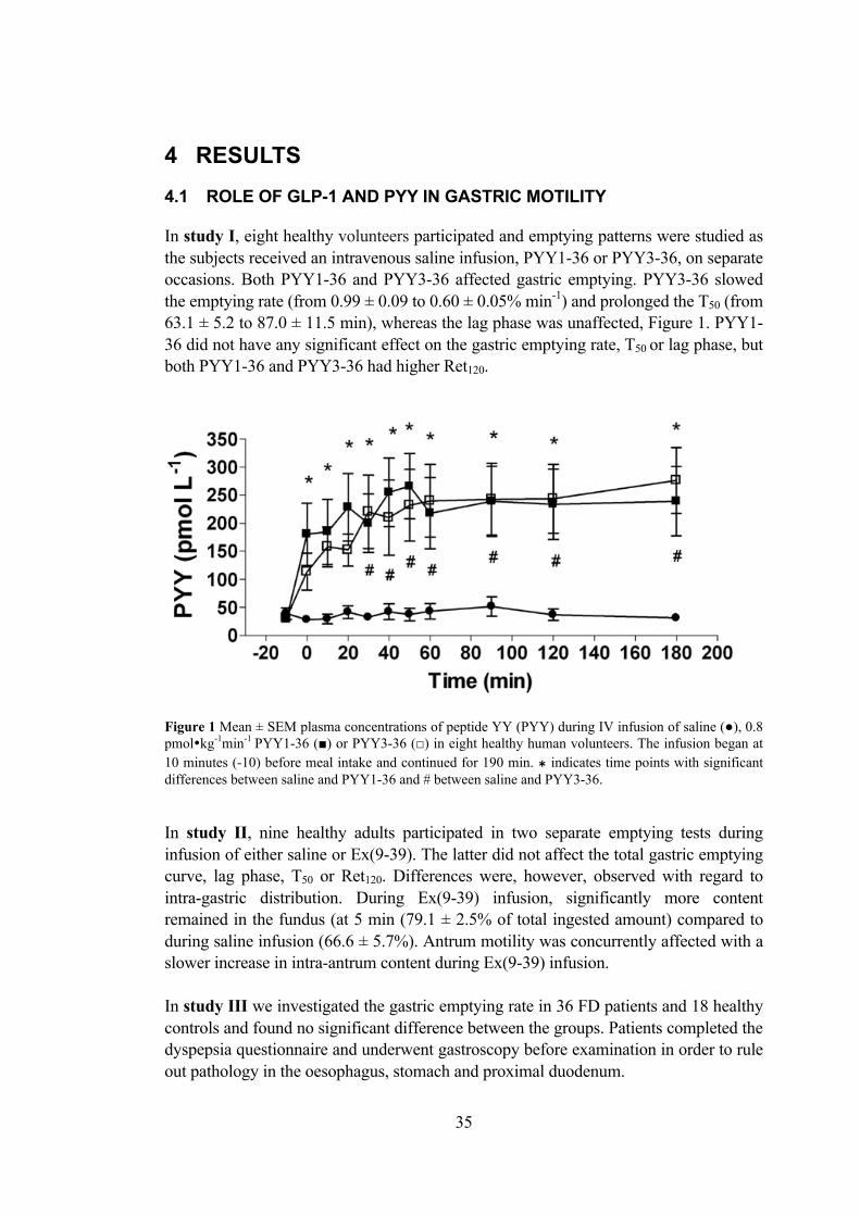
35
4 RESULTS 4.1 ROLE OF GLP-1 AND PYY IN GASTRIC MOTILITY
In study I, eight healthy volunteers participated and emptying patterns were studied as the subjects received an intravenous saline infusion, PYY1-36 or PYY3-36, on separate occasions. Both PYY1-36 and PYY3-36 affected gastric emptying. PYY3-36 slowed the emptying rate (from 0.99 ± 0.09 to 0.60 ± 0.05% min-1) and prolonged the T50 (from 63.1 ± 5.2 to 87.0 ± 11.5 min), whereas the lag phase was unaffected, Figure 1. PYY1-36 did not have any significant effect on the gastric emptying rate, T50 or lag phase, but both PYY1-36 and PYY3-36 had higher Ret120.
Figure 1 Mean ± SEM plasma concentrations of peptide YY (PYY) during IV infusion of saline (●), 0.8 pmol�kg-1min-1 PYY1-36 (■) or PYY3-36 (□) in eight healthy human volunteers. The infusion began at 10 minutes (-10) before meal intake and continued for 190 min. ⁎ indicates time points with significant differences between saline and PYY1-36 and # between saline and PYY3-36. In study II, nine healthy adults participated in two separate emptying tests during infusion of either saline or Ex(9-39). The latter did not affect the total gastric emptying curve, lag phase, T50 or Ret120. Differences were, however, observed with regard to intra-gastric distribution. During Ex(9-39) infusion, significantly more content remained in the fundus (at 5 min (79.1 ± 2.5% of total ingested amount) compared to during saline infusion (66.6 ± 5.7%). Antrum motility was concurrently affected with a slower increase in intra-antrum content during Ex(9-39) infusion. In study III we investigated the gastric emptying rate in 36 FD patients and 18 healthy controls and found no significant difference between the groups. Patients completed the dyspepsia questionnaire and underwent gastroscopy before examination in order to rule out pathology in the oesophagus, stomach and proximal duodenum.

36
4.2 EFFECT OF GLP-1 AND PYY ON ENDOCRINE PANCREATIC
FUNCTIONS AND GLUCOSE CONTROL
Insulin secretion In study I, insulin levels increased from pre-meal levels of 75.7 ± 9.7 to a maximum of 170.5 ± 8.1 pmol�L-1 30 min after food intake. Compared to saline, both PYY1-36 and PYY3-36 significantly moderated the postprandial rise in insulin. Despite the fact that the AUC for the whole period was not affected, the AUC30–60 for insulin was decreased by PYY3-36, but not by PYY1-36 (AUC30–60 9.3 ± 4.9 nmol�min-1 for saline, 7.4 ± 3.8 for PYY1-36 and 6.8 ± 3.4 for PYY3-36). In study II, Ex(9-39) did not affect preprandial insulin levels, but the postprandial rise in insulin was significantly enhanced, Figure 2.
Figure 2 Plasma levels of glucose, insulin and glucagon. Plasma concentrations of glucose, insulin and glucagon during IV infusion of saline (☐) or 300 pmol�kg-1 min-1 Ex(9-39)amide (●) in nine healthy human volunteers. “Meal” indicates the time it took to consume a 310-kcal solid meal. Mean ± SEM * indicates significant differences with P < 0.05.

37
In study III, we found a difference in insulin secretion between FD patients and healthy controls after the single meal. The AUC for insulin was significantly higher in FD patients (P = 0.030). When patient groups were divided into EPS and PDS, the difference in the AUC for insulin remained significant for EPS (P = 0.047). There was no such difference during the satiety test (P = 0.22). Glucose control In study I, glucose increased as expected during saline infusion from pre-meal levels of 5.1 ± 0.2 to a maximum of 6.1 ± 0.4 mmol�L-1 after food intake. Neither PYY1-36 nor PYY3-36 had any significant effects on that rise or on the glucose level AUC. In study II, preprandial glucose levels were not affected by Ex(9-39), but the postprandial rise was significantly enhanced, Figure 2. We found that during Ex(9-39) infusion, plasma glucagon levels were markedly increased at all times, including before the meal (after 40 min of Ex(9-39) infusion the glucagon level was 15.1 ± 0.7 compared to 5.4 ± 1.4 pmol�L-1 during saline infusion. In study III, FD patients and healthy controls had similar postprandial glucose patterns. Changes in GLP-1 concentrations In study I, the GLP-1 basal level was 14.0 ± 1.6 pmol�L-1 and neither the meal nor the infusion of the two forms of PYY had any significant effect on it. In study II, basal and postprandial GLP-1 levels during saline infusion were similar to those measured in study I. During Ex(9-39) infusion however, a statistically significant rise in postprandial GLP-1 concentrations was observed. In study III, basal levels of GLP-1 were similar to those reported in studies I and II. In contrast to previous studies, there was a clear postprandial rise, both after the single meal and during continuous food intake. There was no difference in the AUC of GLP-1 levels between FD patients and healthy controls. 4.3 DYSPEPTIC SYMPTOMS AND SATIETY MEASURES – ROLE OF
GLP-1 AND PYY
Nausea In study I, three of the first four subjects spontaneously reported nausea on one of the study occasions, which was subsequently identified as that of the PYY3-36 infusion. In the following subjects, nausea was rated using a 100 mm VAS 10 min before the meal, as well as 10, 30, 60, 120 and 180 min after it. Nausea was documented in the same way in studies II and III.

38
Compared to VAS ratings 10 min before the infusion, VAS for nausea increased significantly during PYY3-36 infusion in the healthy subjects (maximum at 120 min with VAS score of 50.3 ± 15.9 mm). Neither PYY1-36 nor Ex(9-39) infusions induced nausea in any subject, Figure 3.
Figure 3 VAS rating for nausea after intake of a solid meal during IV infusion of saline (●), 0.8 pmol�kg-
1 min-1 PYY1-36 (■) or PYY3-36 (□) in four healthy human volunteers; P < 0.05 for PYY3-36 vs. saline. Compared to healthy controls, FD patients experienced nausea to a significantly higher degree after meal intake (Study III, P < 0.0001) as well as before meal intake (P = 0.0025, time -10 min) and during the satiety test (continuous food intake, P = 0.027, AUC). Correlations between GLP-1 and symptoms were studied for basal levels (10 minutes before meal intake) and for the AUC. The AUC of GLP-1 correlated with that of nausea in both groups after the single meal (P = 0.015 for controls and 0.041 for FD), but not when analysed for continuous meal intake (satiety test). Hunger, desire to eat, prospective consumption and satiety In studies I, II and III, hunger, satiety, desire to eat and prospective consumption were rated in the same way as nausea (see above). Neither PYY1-36 nor PYY3-36 had significant effects on the desire to eat, hunger and satiety ratings. The prospective consumption rating decreased significantly on infusion of PYY3-36 but not PYY1-36, Figure 4 (the change in VAS score between -10 and 180 min was 9.5 ± 13.2 mm for PYY1-36, 39.5±7.7 mm for PYY3-36 and 4.1±12.5 for saline). Compared to saline, Ex(9-39) infusion did not affect ratings in any way. FD patients reported significantly higher prospective consumption scores after single meal intake in study III (P < 0.0001 for differences in the AUC). Ten minutes before meal intake, patients had a significantly higher level of satiety (P = 0.028). There was no significant correlation between GLP-1 levels and appetite ratings in study III, except for the AUC GLP-1 versus desire to eat for FD patients in the satiety test.

39
Figure 4 VAS rating 100 mm (actual values) for desire to eat, hunger, satiety and prospective consumption, performed after intake of a solid meal during IV infusion of saline (●), 0.8 pmol�kg-1min-1 PYY1-36 (■) or PYY3-36 (□) in eight healthy human volunteers. In study III, subjects also underwent the satiety drinking test. FD patients reported satiety after a mean time of 41 minutes (range 13-102), which corresponds to a 917 kcal intake. Healthy controls felt sated at 51 minutes (range 27-93, calorie intake 1,158 kcal), which was not significantly different from FD patients. Sensation of fullness, bloating and pain In study III, fullness, bloating and pain were also rated. Major differences were observed regarding the AUC after single meal intake, with significantly higher scores in patients (P = 0.0097 for differences in AUC fullness, P = 0.011 for AUC bloating, P = 0.0072 for AUC pain). Ten minutes before meal intake, patients had a significantly higher level of fullness (p = 0.019) and pain (P = 0.042), but not bloating. During the satiety drinking test, patients had significantly higher scores for bloating (P = 0.010) and pain (P = 0.015), but not fullness. Correlations between GLP-1 and symptoms were studied for basal values (at -10 min) and the AUC, Table 1. There was a significant correlation between baseline GLP-1 values and the sensation of fullness before the single meal in the control group (P = 0.0096). In controls, the AUC of GLP-1 correlated with that of pain (P = 0.015).

40
4.4 SEROTONIN AND EPITHELIAL FUNCTION IN FUNCTIONAL DYSPEPSIA
Immunohistochemistry In study IV, we invited healthy volunteers and patients with dyspeptic symptoms referred for gastroscopy at Bispebjerg Hospital (Copenhagen, Denmark) who fulfilled the ROME III criteria to participate. The dyspepsia questionnaire and GSRS were used to characterize subjects and differentiate “pure” FD from co-morbidity with IBS and reflux disease. Duodenal biopsies from 15 FD patients and 18 healthy controls were included in the histological studies. Gastric biopsies from the same subjects were assessed histologically for the presence of HP and one of the FD patients was found to be positive. Biopsies varied with regard to depth. All contained an intact surface epithelium and lamina propria. Several included the lamina muscularis mucosa, while others contained part of the sub-mucosal layer with Brunner’s glands. Epithelial damage ranged from 0-3 in both groups (mean score 0.7 in FD and 1.1 in healthy controls) with no significant difference (P = 0.179). The cell architecture was normal in both FD patients and healthy controls. 5-HT stained cells were found in the epithelium and Brunner’s glands, Figure 5. Only the epithelium was examined, as not all biopsies contained sub-mucosal tissue. The mean number of cells per HPF was 34.4 ± 8.4 in FD patients and 30.4 ± 3.70 in healthy controls with no significant difference between the groups (P = 0.647).
Figure 5 Paraformaldehyde-fixed duodenal tissue from a patient with functional dyspepsia. The tissue was immunohistochemically stained using a polyclonal antibody against serotonin (5-HT). There are many 5-HT immunoreactive cells in the surface epithelium, both in the crypts and in the villi. The biopsy has an intact surface epithelium with no signs of mechanical damage (magnification ×220).

41
In study V, we examined slides from the Kalixanda study, in which a random sample of an adult Swedish population (n = 1001) completed the ASQ and underwent upper endoscopy. Biopsies from several sections were taken including D1 and D2. We had access to a randomly selected portion of the material and identified the following groups: 24 FD patients and 12 healthy controls from D1 as well as 16 FD patients and 15 controls from D2. FD patients and healthy controls exhibited normal architecture in the D1 and D2 regions. Slides were examined to establish the number of CGA and 5-HT stained cells, and the same HPF were evaluated for both substances. The number of cells was then expressed as cells per mm2. Single stained cells were found in the epithelium, more specifically in the Lieberkühn crypts. There was no significant difference in the number of 5-HT stained cells (D1 P = 0.77, D2 P = 0.70). The number of CGA positive cells was significantly lower in FD patients both in D1 (P = 0.010) and D2 (P = 0.038), Figure 6.
Con
trol
bul
b
FD b
ulb
Con
trol
sec
ond
part
FD s
econ
d pa
rt
0
100
200 * *
CG
A p
ositi
ve c
ells
/ m
m2
Con
trol
bul
b
FD b
ulb
Con
trol
sec
ond
part
FD s
econ
d pa
rt
0
25
505-
HT
posi
tive
cells
/ m
m2
Figure 6 The number of Chromogranin A (CGA) and serotonin (5-HT) positive cells in the duodenal bulb and second part of the duodenum per mm2, respectively. Comparison between FD and controls. Based on the Rome III criteria and results from the ASQ, patients were further divided into EPS and PDS. The number of CGA but not 5-HT stained cells was found to be significantly decreased in D1 in EPS (CGA P = 0.028, 5-HT P = 0.94). There was no such difference in D2 (CGA P = 0.083, 5-HT P = 0.54).

42
Ussing chamber experiments In study IV, duodenal biopsies from 15 FD patients and 18 healthy controls were mounted in MUAS chambers. Mean basal SCC was 19.8 ± 3.0 µA�cm-2 for the FD patients and 21.4 ± 3.7 µA�cm-2 for the healthy controls with no significant difference between groups (P = 0.749). The addition of cumulative concentrations of 5-HT in steps of a factor of three from 3 to 243 µmol•L-1 on the serosal side induced a dose dependent SCC rise in both FD patients and healthy controls, Figure 7. The 5-HT-induced rise in SCC was significantly lower in FD patients (P < 0.001 for the overall difference). Glucose control values after 5-HT stimulation yielded a mean magnitude of 12.5 ± 2.0 µA�cm-2 for FD patients and 12.1 ± 2.5 µA�cm-2 for healthy controls (P = 0.906).
Figure 7 Dose-response curve of serotonin (5-HT)-induced short-circuit current (SCC). Addition of cumulative concentrations of 5-HT in steps of a factor of three from 3 to 243 µmol/L on the serosal side of duodenal biopsies mounted in a MUAS chamber resulted in an increased SCC (µA/cm2) in both FD patients (■; n = 8) and healthy controls (●; n = 9), with significantly lower values in the FD group (mean ± SE). Basal conductance, in millisiemens per square cm (mS�cm-2), was significantly lower in FD patients compared to healthy controls (42.4 ± 4.7 mS�cm-2 and 62.4 ± 4.5 mS�cm-2 respectively, P = 0.005). Histological examination revealed that the surface epithelium and entire lamina propria were intact in all samples after mounting and the experimental period.

43
Polymerase chain reaction In study IV, gene expression levels for 5-HT receptors 1A, 1B, 2A, 2B, 3A, 3B, 3C, 3D, 3E, 4 and 7 were quantified. Expression levels were undetectable for three receptor genes, HTR1A, HTR3B and HTR3D. Expression of HTR1B, HTR2A, HTR2B, HTR3A and HTR3C was very low. However, the receptor genes HTR3E, HTR4 and HTR7 were strongly expressed. Expression of HTR3E was significantly higher (P = 0.008) and HTR7 significantly lower in FD patients (P = 0.027) compared to healthy controls. There was no significant difference in expression of HTR4 between the two groups. SLC6A4 and the TPH1 gene were also quantified and found to be strongly expressed in all duodenal biopsies. There was a significant difference in expression level between FD patients and healthy controls, with FD patients exhibiting higher expression of SLC64 (P = 0.033) and lower expression of the TPH1 gene (P = 0.031). The number of biopsies studied for each gene was 8-10 for FD patients and 13-16 for healthy controls.

44
5 DISCUSSION 5.1 THE ROLE OF GLP-1 AND PYY IN GASTRIC MOTILITY
In animal studies, both PYY isoforms have been found to slow down the gastric emptying rate and T50, with PYY3-36 being ten times more effective in rats. In healthy humans, our results confirmed an inhibiting role of PYY3-36 in all gastric emptying-related parameters. PYY1-36 increased Ret120, but did not significantly change the other parameters. This is probably due to lack of power. While a sample of nine subjects is large enough to detect differences, type II errors (false negative outcomes) can still occur. A larger meal or higher dose of PYY1-36 could have led to increased power. The fact that PYY3-36 seems to be more effective might be a consequence of the different receptor binding profiles. The Y1 receptor mediated action in vagal parts of the brainstem in rats, leading to increased gastric motility, while the Y2 receptor mediated inhibition of motility [66]. PYY3-36, which is Y2 receptor specific, might thus exert its motility inhibiting action centrally through vagal efferent mechanisms, but this aspect remains to be elucidated. PYY1-36 has similar affinity to the Y2 receptor as PYY3-36 but in the physiological situation this form is not as prevalent in plasma. In addition, PYY1-36 targets other PYY receptors, and a motility inhibiting effect via Y2 receptors might be counteracted by activation of the Y1 receptor [67].
We could not confirm that endogenous GLP-1 plays a role in the gastric emptying process, but instead observed differences in the motility pattern in various gastric regions, suggesting that endogenous GLP-1 has an alternative regulating role in the intra-gastric distribution of food. When the action of endogenous GLP-1 is inhibited during Ex(9-39) infusion, food remains in the fundus for a longer time before being transported to the antrum. Based on this finding, one would expect blood GLP-1 levels to result in a more rapid transport of food to the antrum and consequently an increased emptying rate. However, in previous studies both exogenous and endogenous GLP-1 have been observed to inhibit the overall emptying rate [68-71], and the question is whether our results might represent a type I error. The calorie content of our meal was smaller and the food composition differed from similar studies. A larger meal could result in increased endogenous GLP-1 secretion, even during Ex(9-39) infusion. In our study, circulating GLP-1 concentrations were actually higher during Ex(9-39) infusion than during saline infusion. This means that even if the GLP-1 receptor is inhibited, the GLP-1 hormone could potentially mediate the effects of other mechanisms, as well as counteracting those induced by Ex(9-39). Higher GLP-1 concentrations during Ex(9-39) infusion might be explained by autocrine feedback. Schirra et al. published a study performed in healthy humans, where nutrients were infused into the duodenum with and without simultaneous intravenous Ex(9-39) infusion [72]. Gastric motility was assessed by means of barostat and manometry (a technique for measuring intraluminal pressure). Duodenal nutrients stimulated

45
relaxation of the upper stomach as well as inhibition of antrum activity. Antrum motility returned during Ex(9-39) inhibition, which supports the assumption that antrum inhibition is mediated by GLP-1. Inhibition of antrum activity has recently been observed after intra-duodenal infusion of protein [73], which also resulted in increased circulating GLP-1 levels. If the smooth muscles in the antrum wall contract during Ex(9-39) infusion, antral reception of food is hampered, which can explain why a meal remains in the upper part of the stomach for a longer period. In the context of gastric motility, it should be taken into account that liquid meal components were used in all our emptying studies. Theoretically, acceleration of liquid emptying during Ex(9-39) infusion could lead to redistribution of the solid meal components towards the upper stomach. Liquid components reaching the small bowel would also result in higher glucose and insulin levels. Hyperinsulinemia has been shown to delay gastric emptying independently of glucose [74]. Post-prandial levels were higher during Ex(9-39) infusion in our study, which could have had an inhibitory effect on gastric emptying that outweighed the potentially accelerating effect of Ex(9-39). We also found high glucagon levels during Ex(9-39) infusion, glucagon being another known inhibitor of the gastric emptying rate. The relative importance of liquid components should be further investigated in future experiments using dual-label scintigraphy. In general, liquid meals do not trigger gastric motility to the same extent as solid ones, which might explain why we did not observe any difference in gastric emptying patterns between healthy controls and FD patients in study III, in which intra-gastric motility changes were not assessed.
Both PYY3-36 and GLP-1 can induce nausea, which has been hypothesized to result from reduced gastric emptying. One could also hypothesize that nausea per se causes inhibition of gastric motility. 5.2 EFFECT OF GLP-1 AND PYY ON ENDOCRINE PANCREATIC
FUNCTIONS AND GLUCOSE CONTROL
PYY did not affect glucose levels in our study, but we observed a small inhibition in post-prandial insulin secretion, with both isoforms. This is in line with a previous human study by Sloth et al., who reported a higher post-prandial insulin response in the presence of PYY1-36 and PYY3-36 [75]. These researchers observed increased post-prandial glucose levels in the case of PYY3-36, which we were unable to confirm. In conclusion, there is no strong evidence that PYY has a role in the short-term control of glucose homeostasis. PYY3-36 might be linked to the development of insulin resistance. High levels of PYY3-36 during fasting have recently been found to be associated with insulin resistance independent of body mass [76]. Exogenous GLP-1 is known to stimulate insulin and inhibit glucagon secretion. Analogues are used in clinical practice for the treatment of diabetes. In contrast to

46
traditional treatment options, GLP-1 analogues have a lower risk of hypoglycaemia. The physiology behind GLP-1 related glucose homeostasis is not yet fully investigated. The GLP-1 inhibitor Ex(9-39) has been found to suppress the postprandial rise in insulin concentrations during application of a hyperglycaemic clamp [77, 78]. The clamp provides an opportunity to separate hormone-induced changes from those initiated by gastric emptying. The effect of endogenous GLP-1 on postprandial insulin levels is believed to be independent of gastric emptying [77], which is in line with our findings. Despite the fact that we did not detect any effect on the gastric emptying rate, endogenous GLP-1 leads to enhanced post-prandial insulin levels. We also found higher glucose levels, thus it could be assumed that the insulin increase is a consequence of greater glucose concentrations. Likewise, studies by Deane et al. and Edwards et al. identified increased glucose and insulin levels during Ex(9-39) infusion after meal intake [68, 79]. In contrast to changes in glucose and insulin concentrations, which only occurred after luminal food stimulation, we found glucagon levels to be constantly suppressed by Ex(9-39), even during fasting. Our finding that endogenous GLP-1 concentrations are sufficient to inhibit glucagon secretion in a consistent manner is in line with previous results [80]. Glucagon is known to be strongly involved in short-term control of glucose homeostasis and prevention of hyperglycaemia. Post-prandial glucose levels should naturally be higher during constant glucagon suppression. As pointed out already, the effect is meal dependent; glucagon suppression does not affect fasting levels of glucose or insulin. 5.3 DYSPEPTIC SYMPTOMS AND SATIETY MEASURES – THE ROLE OF
GLP-1 AND PYY
Satiety measures PYY1-36 did not have any effect on ratings for the desire to eat, hunger and satiety, which is in line with earlier studies. There is thus little evidence to suggest that PYY1-36 plays a role in satiety mechanisms. The only satiety measure affected by PYY3-36 in our study was prospective consumption. A recent study by Sloth et al. [75] using the same infusion doses, reported decreased ratings of perceived ability to eat during PYY3-36 infusion. Degen . found a reduced hunger score before meal intake [3], but not during or directly after the meal. Batterham et al. reported decreased hunger scores and food intake after a two hour infusion before meal intake [2]. PYY3-36 definitely seems to be involved in regulating food intake, but a longer infusion period before a meal is necessary to detect significant effects on hunger scores. Exogenous GLP-1 has been observed to increase satiety and decrease food intake in humans [81]. We did not find evidence of such a role for endogenous GLP-1 in our

47
study. Owing to the relatively small number of subjects, a minor effect on satiety parameters may have been overlooked. One could also hypothesize that the satiety increasing effect of exogenous GLP-1 is associated with inhibition of gastric emptying, as we did not observe a difference in the total gastric emptying rate caused by endogenous GLP-1. Further studies are needed to elucidate whether or not endogenous GLP-1 is involved in the complex physiology of satiety. Nausea In line with earlier studies [3, 75], we observed that PYY3-36 is capable of inducing nausea, which does not, however, seem to be food-related. Sloth et al. and Degen et al. used the same dose as in our study, but without a concomitant meal and found that a significant proportion of their subjects had to discontinue the PYY3-36 infusion because of side effects. Exogenous GLP-1 frequently causes nausea and vomiting [82, 83]. In study III, we measured GLP-1 levels pre- and post-prandially in FD patients, of whom a substantial number suffered from frequent nausea, and compared the results with those of healthy controls. As expected, the FD patients had significantly higher nausea ratings after meal intake compared to controls, although the AUC of GLP-1 concentrations was similar in both groups after the meal. In the correlation study, the AUC of GLP-1 correlated with that of nausea in both groups. In conclusion, GLP-1 should be regarded a candidate mediator in the pathogenesis of nausea, although its role in the pathogenesis of FD is unclear. Other dyspeptic symptoms In study III, FD patients presented with high levels of dyspeptic symptoms even at baseline. It is not possible to conclude whether these symptoms were present just before the meal (and might be induced by expectance of the meal and meal-related symptoms) or whether they were unrelated to food intake. In controls, there was a significant correlation between baseline GLP-1 values and the sensation of fullness before the single meal as well as between the AUC of GLP-1 and that of pain. Such an association was not found in patients. The number of subjects was relatively high, which makes a type II error unlikely. Patients were also carefully selected and there was no co-morbidity. Hence, our results do not support an association between altered GLP-1 secretion and FD symptom development, except in the case of nausea. 5.4 SEROTONIN AND EPITHELIAL FUNCTION IN FUNCTIONAL
DYSPEPSIA
Our results strongly support a 5-HT receptor mediated pathology in FD. Possible mechanisms include impaired HTR3 and HTR7 receptor function in duodenal epithelial transport or signalling.

48
Duodenal transport mechanisms were studied in the MUAS chamber, a sensitive system easily influenced by external factors. The SCC values in our studies were generally lower than those previously described by Engelmann et al. [16], which might be explained by the different settings in which the studies were conducted. The Engelmann study included different types of dyspeptic patient not covered by the Rome III criteria and had no control group, which may also have affected mean SCC measurements. We found lower SCC values in FD patients compared to controls when biopsies were stimulated with 5-HT. This can be interpreted as a decreased movement of negative ions to the duodenal lumen (which, in the duodenum, are most likely HCO3
- ions), increased movement of positive ions to the lumen (e.g. potassium secretion), decreased hydrogen absorption (from gastric acid) or less absorption of positively charged particles (e.g. potassium ions from gastric acid). Since HCO3
- secretion is an important part of the duodenal defence system, impaired secretion in FD patients is the most plausible explanation. One way to further elucidate this aspect would be to repeat the study, and include in-chamber pH measurements and/or single dose 5-HT stimulation with the subsequent addition of a HCO3
- transport inhibitor, e.g. acetazolamide, combined inhibition of Cl- secretion by for instance bumethanide (which blocks the co-transport of sodium, potassium and Cl- ions). HTR3 is known to be prevalent in duodenal mucosa [84], which is supported by our results. We also found higher expression of the HTR3E sub-type in FD patients compared to controls. HTR3E dysfunction in a subgroup of IBS has been suggested [85]. As the receptor is a gated cation-selective channel, a specific role in HCO3
- secretion as part of the altered epithelial transport is plausible, since HCO3
- secretion is both dependent on transport of sodium and potassium. One possible way of further elucidating receptor function in duodenal mucosa is to stimulate biopsies with a single dose of 5-HT followed by a specific receptor antagonist. Furthermore, an aspect that should be taken into account is the fact that HTR3E does not function alone, as shown by patch clamp studies [86] and thus functional studies on HTR3E should include other receptor sub-types (at least HTR3C, D, E). However, to date there are no specific ligands for the different sub-types. MUAS chamber studies should be performed using a general HTR3 antagonist, e.g. ondansetron, which has been in clinical use for many years for the treatment of nausea. A recommendation for future studies is to specifically target FD patients in whom nausea is the main symptom. HTR7 is a 5-HT receptor about which very little is known, although it has been described as being highly expressed in the stomach and ileum [87]. Receptor activation affects intestinal motility in dogs and guinea pigs [88, 89]. We found a high HTR7 expression in our duodenal samples, but lower expression in FD patients. Future studies on the involvement of HTR7 in duodenal epithelial transport would be facilitated by the development of a specific ligand, which does not exist at present. There is also a difference in SERT and TPH1 expression in duodenal mucosa that is not associated with the number of 5-HT secreting cells, which we investigated immunohistochemically in two independent studies (Studies IV and V). TPH1

49
expression was lower in patients; a similar relationship has been observed in IBS [90]. Decreased 5-HT bioavailability might thus be part of the FD mechanism. Gene expression of the 5-HT transporter is high in the duodenal biopsies and FD patients had increased levels compared to controls. If 5-HT bioavailability is actually decreased in patients, higher expression of the transporter protein could be a compensatory mechanism. The total number of endocrine cells is lower in FD. Since the amount of 5-HT is not altered, the involvement of other neuroendocrine mediators appears reasonable. Although PYY and GLP-1 are possible candidates, the number of cells containing GLP-1 and PYY in the duodenal mucosa is low (unpublished results). CGA is also found in gastrin and CCK cells. A decrease in CCK producing, but not 5-HT producing, cells could explain our results. Co-staining would be a way to further clarify this issue. Differences were specifically found for EPS, which suggests a connection between the number of endocrine cells and pain. It has been suggested that EPS and PDS are two different diseases. Recent results from Vanheel et al. indicate that epigastric pain seems to originate from the duodenal tract and that early satiety is mainly a stomach-related symptom [40]. The sample size in study V was rather small, but the patients were well characterized and did not suffer from IBS comorbidity. Our results are further strengthened by the fact that FD subjects were sampled in a population based manner and represented the average citizen rather than a patient seeking healthcare (who according to earlier studies suffers from psychological co-morbidity to a higher degree) or a patient in need of specialist care (who usually exhibits more severe symptomatology). Based on the results of this thesis, recommended future studies should include:
-‐ The effects of exogenous GLP-1 on gastric emptying and insulin levels in doses that are too low to induce nausea.
-‐ The number of GLP-1 and PYY expressing cells throughout the intestinal tract in EPS and PDS patients.
-‐ Functional studies of HTR3 receptors in duodenal ion transport in FD patients, especially those with nausea as the main symptom.
-‐ mRNA expression of TPH1 and SERT in PDS compared to EPS and controls. -‐ Co-staining for CCK, 5-HT and CGA in the duodenal mucosa in EPS, PDS
and healthy controls. -‐ Quantification of mucosal 5-HT staining cells in relation to mast cells,
eosinophils and intra-epithelial lymphocytes in post-infectious FD compared to other FD patients and healthy controls.

50
6 CONCLUSIONS
Study I This work contributes data to support the following conclusions: PYY3-36 and PYY1-36 both inhibit gastric emptying, PYY3-36 being the most effective. Neither PYY3-36 nor PYY1-36 influence glucose or GLP-1 concentrations, but both reduce the postprandial rise in insulin. PYY3-36 induces nausea in some subjects and reduces prospective consumption. The prolonged emptying process might be due to nausea. Decreased prospective consumption could be a result of reduced emptying as well as nausea. These issues are still to be investigated. Study II Endogenous GLP-1 does not affect the gastric emptying rate but seems to be involved in the regulation of gastric motility in relation to food intake and also in the regulation of post-prandial insulin and glucose levels. Furthermore, endogenous GLP-1 seems to tonically restrain glucagon secretion. Our results support a role of endogenous GLP-1 as an incretin hormone that is independent of gastric emptying. However, accelerating effects of GLP-1 receptor inhibition might have been missed due to enhanced insulin and glucagon levels. Both insulin and glucagon have been shown to inhibit gastric emptying in previous studies. We did not detect a role of endogenous GLP-1 in sensations of hunger, satiety, desire to eat, prospective consumption or nausea. Study III FD patients have higher postprandial insulin levels compared to healthy controls. When EPS and PDS are studied separately, increased insulin secretion is found with EPS, but not PDS. FD patients have normal postprandial glucose and GLP-1 concentrations. Higher satiety, nausea and pain scores in FD patients cannot be explained by delayed or accelerated gastric emptying. Nausea might be associated with postprandial GLP-1 secretion mechanisms. Study IV Duodenal ion transport in response to exogenous 5-HT is abnormal in FD patients, in whom exogenous 5-HT also induces higher epithelial electrical resistance. Based on previous results it is plausible that the resistance reflects lower HCO3
- and Cl- conductivity. 5-HT induces a dose dependent rise in SCC in both FD patients and healthy controls, but the rise is lower in the former. This can be a result of impaired bicarbonate secretion. Genes for the HTR3E, HTR4 and HTR7 5-HT receptors are highly expressed in human duodenal mucosa. The SERT gene (responsible for 5-HT transport over cell walls) and TPH1 (produces 5-HT in cells) are also strongly expressed. FD patients have higher gene expression of HTR3E and SERT and lower gene expression of HTR7 and TPH1 in their duodenal mucosal cells. An altered amount of HTR3E and SERT might be involved in the mechanisms behind impaired duodenal ion transport in response to exogenous 5-HT in FD patients. The number of duodenal epithelial cells containing 5-HT is similar in FD patients and healthy controls.

51
Study V Adult non-consulting FD subjects have less endocrine cells in the duodenum, as indicated by CGA staining, but a normal number of cells containing 5-HT. When EPS and PDS are studied separately, the number of endocrine cells is significantly lower in the in EPS but not in PDS. As 5-HT expressing cells are not altered, the involvement of other neuroendocrine substances, such as PYY and GLP-1, seems likely.

52
7 POPULÄRVETENSKAPLIG SAMMANFATTNING
”I 10 års tid har jag nu haft jätteont i övre magen, vissa dagar mer än andra. Min egen bedömning är magkatarr och jag har självbehandlat med alla receptfria läkemedel och dieter som finns, men inget hjälper, det blir bara värre. När jag vänder mig till läkare, diagnostiserar de Funktionell Dyspepsi – det betyder att de inte vet vad det är.” Detta inlägg är taget från ett patientforum på nätet. Författaren till inlägget har rätt. Vi vet i dagsläget inte vad Funktionell Dyspepsi (FD) är. FD är ett begrepp som omfattar symptom som troligen har sitt ursprung i magsäck och tunntarm men som inte förklaras av hittills känd organisk sjukdom, vilket innebär synliga vävnadsförändringar i magsäck eller tunntarm, infektion eller metabol sjukdom (exempelvis diabetes). För formell diagnos krävs minst sex månaders besvär med smärta eller brännande känsla i övre delen av magen, besvärande uppkördhet efter måltid eller tidig mättnadskänsla, eventuellt i kombination med andra övre magbesvär, och en normal gastroskopi. Magens fysiologi är komplicerad och man behöver titta på flera faktorer samtidigt för att få en bättre förståelse för uppkomsten av sjukdomar. Vi har undersökt hur olika signalämnen som utsöndras i tunntarmens slemhinna påverkar rörelsemönstret i magsäcken, sockerbalansen i blodet och upplevelsen av hunger, mättnad, illamående och dyspeptiska symtom. Vi har fokuserat på hormonerna (signalämnen som vandrar med blodet), PYY1-36, PYY3-36 och GLP-1 och nervsignalämnet serotonin. Vi har förutom studier kring magsäckstömning och blodsockerbalans räknat antalet celler som utsöndrar dessa ämnen i tolvfingertarmen. Vi har studerat transportmekanismer över tunntarmens slemhinna och hur dessa påverkas av signalämnet serotonin. Under projektets gång har vi specifikt letat efter och försökt förstå avvikelser hos FD patienter, både patienter som sökt vårdcentral och individer som inte känt något behov av att söka sjukvård. När vi gav PYY3-36 and PYY1-36 intravenöst (direkt i blodet) till friska försökspersoner, samtidigt som de fick inta en fast måltid, förlängdes magsäckstömningen och insulinnivån i blodet höjdes. Mättnadskänslan påverkades inte men däremot kunde PYY3-36 inducera illamående och försökspersonerna fick mindre lust att äta. Eftersom många FD patienter lider av illamående och oftast äter för lite, skulle det vara värdefullt att närmare kartlägga vilken roll PYY3-36 har i utvecklingen av dessa symptom. Vi undersökte GLP-1 närmare genom att hämma dess receptor (mottagare). Man har tidigare gjort försök där GLP-1 gavs direkt i blodet, men det finns inte många studier som fokuserat på funktionen av kroppseget GLP-1 i samband med måltid. Våra resultat visade att kroppseget GLP-1 effektivt och oberoende av matintag hämmar en substans som heter glukagon och som är mycket viktig i blodsockerregleringen. Det framkom även att kroppseget GLP-1 modulerar insulinnivåer i blodet efter måltid och kan ändra magsäckens rörelsemönster utan att hämma tömningshastigheten. Mättnadskänslan

53
påverkades inte och försökspersonerna blev inte illamående av den GLP-1 hämmande substansen (vilket de oftast blir av GLP-1 när det ges intravenöst). Vi undersökte GLP-1 närmare i FD patienter, genom att mäta GLP-1, insulin och glukosnivåer i blodet efter att patienterna (och friska kontroller) hade fått dricka näringslösning med en markör för mätning av magsäckstömning. FD patienter hade högre insulinnivåer efter måltid jämfört med kontrollpersonerna, samtidigt som blodsockernivåerna var normala. Detta gällde framförallt patienter med smärta som framträdande symptom. Patienterna uppgav i högre grad än kontrollpersonerna mättnadskänsla, illamående och smärta efter matintag, vilket vi inte kunde förklara med ändringar i magsäckens tömningshastighet. Att insulinnivåerna är annorlunda hos patienter med FD är nytt och bör undersökas närmare i framtida studier. Våra resultat talar även för att ett rörelsemönster i magsäckens olika delar kan vara viktigare i mekanism bakom FD än tömningshastigheten. GLP-1, liksom PYY3-36, verkar ha en funktion i uppkomsten av illamående. Det sker en livlig transport av olika ämnen mellan tarmen och blodet via tarmslemhinnan. När vi stimulerade slemhinnan med serotonin, kunde vi se att transporten för laddade ämnen är annorlunda hos FD patienter, sannolikt på grund av ökad bikarbonatutsöndring (ett ämne som neutraliserar och skyddar från frätande magsyra). I samma vävnad kunde vi kartlägga ändringar i genuttryck hos några av serotoninets olika receptorer. Vid utveckling av framtida läkemedel är det en fördel att veta vilka receptorer som är inblandade. Med hjälp av läkemedel som effektivt och specifikt hämmar eller aktiverar en av “nyckelreceptorerna” skulle man målinriktat kunna påverka de mekanismer som ger upphov till symptom. Baserat på våra resultat rekommenderas fler studier kring rollen hos serotoninreceptorerna 3E och 7. FD patienter hade mindre antal celler som kan utsöndra signalämnen av olika slag i tolvfingertarmens slemhinna. Detta gällde specifikt patienter med smärta som framträdande symptom. När vi enbart färgade celler som innehöll serotonin, var antalet celler lika hos patienter och friska. FD patienter verkar alltså ha ett mindre antal celler som innehåller andra signalsubstanser än serotonin i tunntarmens slemhinna. Att färga dessa celler för PYY3-36 och GLP-1 blir därför ett intressant framtida forskningsprojekt .

54
8 ACKNOWLEDGEMENTS
Family first I would like to thank my boyfriend Yan Hui and his wonderful son Oskar Yan-Song, for their sincere care and understanding, my parents Christina and Martin, which I think are the wisest people on earth, and my sisters Julia-Caroline and Maria-Henrike, for their optimism and support. I would further like to express my admiration to Matthias for his ability to enjoy life, when life is threatened by disease. And even if I do not name further members, I am glad to be part of a large and diverse family; my horizon has been widened by your existence. Essential leadership I am most grateful to main supervisor Associate Professor Peter Thelin Schmidt. Peter is a very good listener, a skillful scientist and a realist, who provided me with the necessary degree of freedom and passion. I could not have wished for better guidance. My gratitude further goes to Professor Lars Agréus as second supervisor. I have just started to walk towards new discoveries with him, and I feel inspired. I would like to thank my mentors Professor Suad Efendic and Doctor Peter Gustavsson for their listening ears and valuable advice. My former supervisors, Professor Bo Rydqvist and Professor Per Hellström, have been a source of knowledge and inspiration, thank you. Bosse, I still dream of patch clamping! I would like to mention my first academic tutor Associate Professor Elias Arnér, who supervised me at the Nobel Institute of Biochemistry. He taught me important basics of scientific research. I had the opportunity to assist the following students in their medical degree projects: Anne Marie Møller, Johan Widenberg and Maria-Henrike Witte. Thank you for your enthusiasm. Teamwork towards achievement The presented studies have been performed at several locations, including St Mary’s Hospital (Imperial College) in London, Bispebjerg Hospital, Panum Instituttet and Rigshospitalet in Copenhagen, Gastrocentrum Karolinska University Hospital, the Karolinska Institutet Dep. Medicine Solna and Dep. Molecular Medicine Huddinge, and the Centre of Family Medicine.
55
The following persons were of special importance for data sampling and analysis: Research nurses Kicki and Eva who took significant part in data sampling (Study III). The staff at Bispebjerg Endoscopic Unit for their help in obtaining biopsies (Study IV). Nicolas Kaltoft and Cristina Tilotta for introducing me to the Ussing chamber system (Study IV). Agneta Laurent for performing the PCR experiments (Study IV). Tom Storskrubb, Pertti Aro and Jukka Ronkainen for pursuing the Kalixanda study and providing me with samples (Study V). Vincenzo Marazzo for staining the slides in Study V. I further had the opportunity to collaborate with the following highly skilled scientists: Professors Marjorie Walker, Nicolas Talley, Lars Agréus, Jens Juul Holst, Per Hellström, Erik Näslund and Hans Jacobsson. Associate Professors Mauro D’Amato and Per Grybäck. PhDs Linda Hilsted, Niels Bindslev, Steen Poulsen, Svend Knuhtsen, and Mark Hansen. I admire those scientists, because they are involved in high quality research, teaching and clinical work, at the same time as they always find time to listen, give constructive feedback or even socialize. Big hug to fellow PhD student Johan Granlund – good luck with your book! It has not always been easy to combine a clinical career with research studies, and I would like to thank former and present employers for their support: Þórdís J. Hrafnkelsdóttir (Reykjavík University Hospital), Annica Bergqvist (Gastrocentrum, Karolinska University Hospital), Olle Lindström (Emergency Dep., Karolinska University Hospital), Susanne Blid (Capio Hagsätra Primary Care) and Birgit Ekholm (Ersta Psychiatric Clinic). Friends and colleagues (fellow doctors, nurses, health assistants, secretaries, cleaning personnel) - with your warmth and practical skills, you have all facilitated the pursuit of my studies. I will not name you individually - if you feel addressed, you are addressed, and you should be proud!

56
9 REFERENCES 1. Holst, J.J., Glucagon and glucagon-like peptides 1 and 2. Results Probl Cell Differ,
2010. 50: p. 121-35. 2. Batterham, R.L., et al., Gut hormone PYY(3-36) physiologically inhibits food intake.
Nature, 2002. 418(6898): p. 650-4. 3. Degen, L., et al., Effect of peptide YY3-36 on food intake in humans. Gastroenterology,
2005. 129(5): p. 1430-6. 4. Bilchik, A.J., et al., Peptide YY is a physiological regulator of water and electrolyte
absorption in the canine small bowel in vivo. Gastroenterology, 1993. 105(5): p. 1441-8.
5. Eto, B., et al., Comparison of the antisecretory effect of endogenous forms of peptide YY on fed and fasted rat jejunum. Peptides, 1997. 18(8): p. 1249-55.
6. Sloth, B., et al., Effect of subcutaneous injections of PYY1-36 and PYY3-36 on appetite, ad libitum energy intake, and plasma free fatty acid concentration in obese males. Am J Physiol Endocrinol Metab, 2007. 293(2): p. E604-9.
7. Holzer, P., F. Reichmann, and A. Farzi, Neuropeptide Y, peptide YY and pancreatic polypeptide in the gut-brain axis. Neuropeptides, 2012. 46(6): p. 261-74.
8. McTigue, D.M., N.K. Edwards, and R.C. Rogers, Pancreatic polypeptide in dorsal vagal complex stimulates gastric acid secretion and motility in rats. Am J Physiol, 1993. 265(6 Pt 1): p. G1169-76.
9. DeMar, A.R., R. Lake, and A.S. Fink, The effect of pancreatic polypeptide and peptide YY on pancreatic blood flow and pancreatic exocrine secretion in the anesthetized dog. Pancreas, 1991. 6(1): p. 9-14.
10. Schmidt, P.T., et al., A role for pancreatic polypeptide in the regulation of gastric emptying and short-term metabolic control. J Clin Endocrinol Metab, 2005. 90(9): p. 5241-6.
11. Kojima, S., et al., A role for pancreatic polypeptide in feeding and body weight regulation. Peptides, 2007. 28(2): p. 459-63.
12. Glisic, R., et al., Serotonin-producing enterochromaffin (EC) cells of gastrointestinal mucosa in dexamethasone-treated rats. Regul Pept, 2006. 136(1-3): p. 30-9.
13. Kulig, G., et al., [Evaluation of the number of enterochromaffin cells in gastric mucosa in subjects with functional dyspepsia]. Pol Merkur Lekarski, 2009. 26(155): p. 370-2.
14. Li, X., et al., The study on the role of inflammatory cells and mediators in post-infectious functional dyspepsia. Scand J Gastroenterol, 2010. 45(5): p. 573-81.
15. Tuo, B.G. and J.I. Isenberg, Effect of 5-hydroxytryptamine on duodenal mucosal bicarbonate secretion in mice. Gastroenterology, 2003. 125(3): p. 805-14.
16. Engelmann, B.E., et al., Functional characterization of serotonin receptor subtypes in human duodenal secretion. Basic Clin Pharmacol Toxicol, 2006. 98(2): p. 142-9.
17. Hansen, M.B., et al., Serotonin-induced short-circuit current in pig jejunum. Zentralbl Veterinarmed A, 1994. 41(2): p. 110-20.
18. Foxx-Orenstein A, C.M., Gershon MD, Linden DR, Mawe GM, Lewis JT, Jensen KL, Talley NJ, Szurszewski JH, Zinsmeister A, Alterations in intestinal serotonin

57
expression in dyspepsia and irritable bowel syndrome. Gastroenterology, 2007. 132: p. A72.
19. Kumar, S., et al., Serotonin transporter gene (SLC6A4) polymorphism in patients with irritable bowel syndrome and healthy controls. J Gastrointestin Liver Dis, 2012. 21(1): p. 31-8.
20. Toyoshima, F., et al., Serotonin transporter gene polymorphism may be associated with functional dyspepsia in a Japanese population. BMC Med Genet, 2011. 12: p. 88.
21. Park, C.S. and J.H. Uhm, Polymorphisms of the Serotonin Transporter Gene and G-Protein beta3 Subunit Gene in Korean Children with Irritable Bowel Syndrome and Functional Dyspepsia. Gut Liver, 2012. 6(2): p. 223-8.
22. Drossman DA, C.E., Delvaux M, Spuller RC, Talley NJ, Thompson WG, Whitehead WE (Eds) Rome III. The Functional Gastrointestinal Disorders. 3rd edition. 2006, Virginia, USA: Degnon Associates, Inc., McLean.
23. Agréus, L., et al., Irritable bowel syndrome and dyspepsia in the general population: overlap and lack of stability over time. Gastroenterology, 1995. 109(3): p. 671-80.
24. Mahadeva, S. and K.L. Goh, Epidemiology of functional dyspepsia: a global perspective. World J Gastroenterol, 2006. 12(17): p. 2661-6.
25. Flier, S.N. and S. Rose, Is functional dyspepsia of particular concern in women? A review of gender differences in epidemiology, pathophysiologic mechanisms, clinical presentation, and management. Am J Gastroenterol, 2006. 101(12 Suppl): p. S644-53.
26. Agréus, L., et al., Natural history of gastroesophageal reflux disease and functional abdominal disorders: a population-based study. Am J Gastroenterol, 2001. 96(10): p. 2905-14.
27. Aro, P., et al., Anxiety is associated with uninvestigated and functional dyspepsia (Rome III criteria) in a Swedish population-based study. Gastroenterology, 2009. 137(1): p. 94-100.
28. Agréus, L., Natural history of dyspepsia. Gut, 2002. 50 Suppl 4: p. iv2-9. 29. Van Oudenhove, L. and Q. Aziz, The role of psychosocial factors and psychiatric
disorders in functional dyspepsia. Nat Rev Gastroenterol Hepatol, 2013. 10(3): p. 158-67.
30. Mayer, E.A., Gut feelings: the emerging biology of gut-brain communication. Nat Rev Neurosci, 2011. 12(8): p. 453-66.
31. Oustamanolakis, P. and J. Tack, Dyspepsia: organic versus functional. J Clin Gastroenterol, 2012. 46(3): p. 175-90.
32. Quartero, A.O., et al., Disturbed solid-phase gastric emptying in functional dyspepsia: a meta-analysis. Dig Dis Sci, 1998. 43(9): p. 2028-33.
33. Sarnelli, G., et al., Symptoms associated with impaired gastric emptying of solids and liquids in functional dyspepsia. Am J Gastroenterol, 2003. 98(4): p. 783-8.
34. Stanghellini, V., et al., Risk indicators of delayed gastric emptying of solids in patients with functional dyspepsia. Gastroenterology, 1996. 110(4): p. 1036-42.
35. Karamanolis, G., et al., Association of the predominant symptom with clinical characteristics and pathophysiological mechanisms in functional dyspepsia. Gastroenterology, 2006. 130(2): p. 296-303.
36. Tack, J., et al., Symptoms associated with hypersensitivity to gastric distention in functional dyspepsia. Gastroenterology, 2001. 121(3): p. 526-35.

58
37. Tack, J., et al., Clinical and pathophysiological characteristics of acute-onset functional dyspepsia. Gastroenterology, 2002. 122(7): p. 1738-47.
38. Walker, M.M., et al., The role of eosinophils and mast cells in intestinal functional disease. Curr Gastroenterol Rep, 2011. 13(4): p. 323-30.
39. Dizdar, V., et al., Relative importance of abnormalities of CCK and 5-HT (serotonin) in Giardia-induced post-infectious irritable bowel syndrome and functional dyspepsia. Aliment Pharmacol Ther, 2010. 31(8): p. 883-91.
40. Vanheel, H., et al., Impaired duodenal mucosal integrity and low-grade inflammation in functional dyspepsia. Gut, 2013.
41. Barbara, G., et al., Mast cell-dependent excitation of visceral-nociceptive sensory neurons in irritable bowel syndrome. Gastroenterology, 2007. 132(1): p. 26-37.
42. Talley, N.J., et al., Non-ulcer dyspepsia and duodenal eosinophilia: an adult endoscopic population-based case-control study. Clin Gastroenterol Hepatol, 2007. 5(10): p. 1175-83.
43. Walker, M.M., et al., Duodenal mastocytosis, eosinophilia and intraepithelial lymphocytosis as possible disease markers in the irritable bowel syndrome and functional dyspepsia. Aliment Pharmacol Ther, 2009. 29(7): p. 765-73.
44. Moqbel, R. and J.J. Coughlin, Differential secretion of cytokines. Sci STKE, 2006. 2006(338): p. pe26.
45. Strasser, F., Clinical application of ghrelin. Curr Pharm Des, 2012. 18(31): p. 4800-12. 46. Walecka-Kapica, E., et al., [Gastrin secretion in patients with functional dyspepsia].
Pol Merkur Lekarski, 2009. 26(155): p. 362-5. 47. Berna, M.J. and R.T. Jensen, Role of CCK/gastrin receptors in
gastrointestinal/metabolic diseases and results of human studies using gastrin/CCK receptor agonists/antagonists in these diseases. Curr Top Med Chem, 2007. 7(12): p. 1211-31.
48. Chua, A.S. and P.W. Keeling, Cholecystokinin hyperresponsiveness in functional dyspepsia. World J Gastroenterol, 2006. 12(17): p. 2688-93.
49. Lankarani, K.B., et al., Serum leptin level in patients with functional dyspepsia. Dig Liver Dis, 2004. 36(11): p. 717-21.
50. Dockray, G.J., Enteroendocrine cell signalling via the vagus nerve. Curr Opin Pharmacol, 2013.
51. Sanger, G.J. and D.H. Alpers, Development of drugs for gastrointestinal motor disorders: translating science to clinical need. Neurogastroenterol Motil, 2008. 20(3): p. 177-84.
52. Witte, A.B., et al., Duodenal epithelial transport in functional dyspepsia: Role of serotonin. World J Gastrointest Pathophysiol, 2013. 4(2): p. 28-36.
53. Beckers, E.J., J.B. Leiper, and J. Davidson, Comparison of aspiration and scintigraphic techniques for the measurement of gastric emptying rates of liquids in humans. Gut, 1992. 33(1): p. 115-7.
54. Bolondi, L., et al., Measurement of gastric emptying time by real-time ultrasonography. Gastroenterology, 1985. 89(4): p. 752-9.
55. Schwizer, W., H. Maecke, and M. Fried, Measurement of gastric emptying by magnetic resonance imaging in humans. Gastroenterology, 1992. 103(2): p. 369-76.
56. McClelland, G.R. and J.A. Sutton, Epigastric impedance: a non-invasive method for the assessment of gastric emptying and motility. Gut, 1985. 26(6): p. 607-14.

59
57. Braden, B., Methods and functions: Breath tests. Best Pract Res Clin Gastroenterol, 2009. 23(3): p. 337-52.
58. Willems, M., A.O. Quartero, and M.E. Numans, How useful is paracetamol absorption as a marker of gastric emptying? A systematic literature study. Dig Dis Sci, 2001. 46(10): p. 2256-62.
59. Heading, R.C., et al., The dependence of paracetamol absorption on the rate of gastric emptying. Br J Pharmacol, 1973. 47(2): p. 415-21.
60. Clements, J.A., et al., A physiologically-based pharmacokinetic model for absorption of oral paracetamol in man [proceedings]. J Pharm Pharmacol, 1978. 30 Suppl: p. 60P.
61. SBU rapport, Ont i magen, Statens beredning för medicinsk utvärdering, 2000. 62. SBU rapport, Dyspepsi och reflux, Statens beredning för medicinsk utvärdering, 2007. 63. Aro, P., et al., Valid symptom reporting at upper endoscopy in a random sample of the
Swedish adult general population: the Kalixanda study. Scand J Gastroenterol, 2004. 39(12): p. 1280-8.
64. Tack, J., et al., Role of impaired gastric accommodation to a meal in functional dyspepsia. Gastroenterology, 1998. 115(6): p. 1346-52.
65. Agréus, L., et al., Reproducibility and validity of a postal questionnaire. The abdominal symptom study. Scand J Prim Health Care, 1993. 11(4): p. 252-62.
66. Chen, C.H., R.L. Stephens, Jr., and R.C. Rogers, PYY and NPY: control of gastric motility via action on Y1 and Y2 receptors in the DVC. Neurogastroenterol Motil, 1997. 9(2): p. 109-16.
67. Keire, D.A., et al., Primary structures of PYY, [Pro(34)]PYY, and PYY-(3-36) confer different conformations and receptor selectivity. Am J Physiol Gastrointest Liver Physiol, 2000. 279(1): p. G126-31.
68. Deane, A.M., et al., Endogenous glucagon-like peptide-1 slows gastric emptying in healthy subjects, attenuating postprandial glycemia. J Clin Endocrinol Metab, 2010. 95(1): p. 215-21.
69. Meier, J.J., et al., Normalization of glucose concentrations and deceleration of gastric emptying after solid meals during intravenous glucagon-like peptide 1 in patients with type 2 diabetes. J Clin Endocrinol Metab, 2003. 88(6): p. 2719-25.
70. Schirra, J., et al., Effects of glucagon-like peptide-1(7-36)amide on antro-pyloro-duodenal motility in the interdigestive state and with duodenal lipid perfusion in humans. Gut, 2000. 46(5): p. 622-31.
71. Schirra, J., et al., Effects of glucagon-like peptide-1(7-36)amide on motility and sensation of the proximal stomach in humans. Gut, 2002. 50(3): p. 341-8.
72. Schirra, J., et al., GLP-1 regulates gastroduodenal motility involving cholinergic pathways. Neurogastroenterol Motil, 2009. 21(6): p. 609-18, e21-2.
73. Ryan, A.T., et al., Intraduodenal protein modulates antropyloroduodenal motility, hormone release, glycemia, appetite, and energy intake in lean men. Am J Clin Nutr, 2012. 96(3): p. 474-82.
74. Kong, M.F., et al., Effect of euglycaemic hyperinsulinaemia on gastric emptying and gastrointestinal hormone responses in normal subjects. Diabetologia, 1998. 41(4): p. 474-81.

60
75. Sloth, B., et al., Effects of PYY1-36 and PYY3-36 on appetite, energy intake, energy expenditure, glucose and fat metabolism in obese and lean subjects. Am J Physiol Endocrinol Metab, 2007. 292(4): p. E1062-8.
76. Ukkola, O.H., et al., High serum fasting peptide YY (3-36) is associated with obesity-associated insulin resistance and type 2 diabetes. Regul Pept, 2011. 170(1-3): p. 38-42.
77. Salehi, M., T.P. Vahl, and D.A. D'Alessio, Regulation of islet hormone release and gastric emptying by endogenous glucagon-like peptide 1 after glucose ingestion. J Clin Endocrinol Metab, 2008. 93(12): p. 4909-16.
78. Schirra, J., et al., Endogenous glucagon-like peptide 1 controls endocrine pancreatic secretion and antro-pyloro-duodenal motility in humans. Gut, 2006. 55(2): p. 243-51.
79. Edwards, C.M., et al., Glucagon-like peptide 1 has a physiological role in the control of postprandial glucose in humans: studies with the antagonist exendin 9-39. Diabetes, 1999. 48(1): p. 86-93.
80. Schirra, J., et al., Exendin(9-39)amide is an antagonist of glucagon-like peptide-1(7-36)amide in humans. J Clin Invest, 1998. 101(7): p. 1421-30.
81. Verdich, C., et al., A meta-analysis of the effect of glucagon-like peptide-1 (7-36) amide on ad libitum energy intake in humans. J Clin Endocrinol Metab, 2001. 86(9): p. 4382-9.
82. Ritzel, R., et al., Pharmacokinetic, insulinotropic, and glucagonostatic properties of GLP-1 [7-36 amide] after subcutaneous injection in healthy volunteers. Dose-response-relationships. Diabetologia, 1995. 38(6): p. 720-5.
83. Nauck, M.A., et al., Effects of subcutaneous glucagon-like peptide 1 (GLP-1 [7-36 amide]) in patients with NIDDM. Diabetologia, 1996. 39(12): p. 1546-53.
84. van Lelyveld, N., et al., Regional differences in expression of TPH-1, SERT, 5-HT(3) and 5-HT(4) receptors in the human stomach and duodenum. Neurogastroenterol Motil, 2007. 19(5): p. 342-8.
85. Kapeller, J., et al., First evidence for an association of a functional variant in the microRNA-510 target site of the serotonin receptor-type 3E gene with diarrhea predominant irritable bowel syndrome. Hum Mol Genet, 2008. 17(19): p. 2967-77.
86. Holbrook, J.D., et al., Characterisation of 5-HT3C, 5-HT3D and 5-HT3E receptor subunits: evolution, distribution and function. J Neurochem, 2009. 108(2): p. 384-96.
87. Bard, J.A., et al., Cloning of a novel human serotonin receptor (5-HT7) positively linked to adenylate cyclase. J Biol Chem, 1993. 268(31): p. 23422-6.
88. Janssen, P., et al., Characterization of 5-HT7-receptor-mediated gastric relaxation in conscious dogs. Am J Physiol Gastrointest Liver Physiol, 2005. 289(1): p. G108-15.
89. Tonini, M., et al., 5-HT7 receptors modulate peristalsis and accommodation in the guinea pig ileum. Gastroenterology, 2005. 129(5): p. 1557-66.
90. Coates, M.D., et al., Molecular defects in mucosal serotonin content and decreased serotonin reuptake transporter in ulcerative colitis and irritable bowel syndrome. Gastroenterology, 2004. 126(7): p. 1657-64.