the sodium iodide symporter (nis): regulation and approaches to
TRANSCRIPT
Pharmacology & Therapeutics 135 (2012) 355–370
Contents lists available at SciVerse ScienceDirect
Pharmacology & Therapeutics
j ourna l homepage: www.e lsev ie r .com/ locate /pharmthera
Associate editor: J. Turgeon
The sodium iodide symporter (NIS): Regulation and approaches to targeting forcancer therapeutics
Takahiko Kogai ⁎, Gregory A. Brent ⁎⁎Molecular Endocrinology Laboratory, VA Greater Los Angeles Healthcare System, Departments of Medicine and Physiology, David Geffen School of Medicine at UCLA, Los Angeles,CA 90073, United States
Abbreviations: APL, acute promyelocytic leukemia; Bdexamethasone; DR, direct repeat; DUOX, dual oxidase; EGy, gray;HDAC, histone deacetylase; IGF, insulin-like growmurinemammary tumor virus-polyoma virusmiddle T anof tumor; PI3K, phosphatidylinositol 3-kinase; PKA, proteRAR, retinoic acid receptor; RTK, receptor tyrosine kinase;tRA, All-trans RA; TSH, thyroid-stimulating hormone; TSH
⁎ Correspondence to: T. Kogai, Molecular EndocrinolCA 90073, United States. Fax: 310 268 4035.⁎⁎ Correspondence to:G.A. Brent,Molecular Endocrinolog
United States. Fax: 310 268 4818.E-mail addresses: [email protected] (T. Kogai), gbrent
0163-7258/$ – see front matter. Published by Elsevier Idoi:10.1016/j.pharmthera.2012.06.007
a b s t r a c t
a r t i c l e i n f oKeywords:
Sodium iodide symporterThyroid cancerBreast cancerTranscriptional regulationPosttranslational regulationExpression of the sodium iodide symporter (NIS) is required for efficient iodide uptake in thyroid and lactat-ing breast. Since most differentiated thyroid cancer expresses NIS, β-emitting radioactive iodide is routinelyutilized to target remnant thyroid cancer and metastasis after total thyroidectomy. Stimulation of NIS expres-sion by high levels of thyroid-stimulating hormone is necessary to achieve radioiodide uptake into thyroidcancer that is sufficient for therapy. The majority of breast cancer also expresses NIS, but at a low level insuf-ficient for radioiodine therapy. Retinoic acid is a potent NIS inducer in some breast cancer cells. NIS is alsomodestly expressed in some non-thyroidal tissues, including salivary glands, lacrimal glands and stomach.Selective induction of iodide uptake is required to target tumors with radioiodide. Iodide uptake in mamma-lian cells is dependent on the level of NIS gene expression, but also successful translocation of NIS to the cellmembrane and correct insertion. The regulatory mechanisms of NIS expression and membrane insertion areregulated by signal transduction pathways that differ by tissue. Differential regulation of NIS confers selectiveinduction of functional NIS in thyroid cancer cells, as well as some breast cancer cells, leading to moreefficient radioiodide therapy for thyroid cancer and a new strategy for breast cancer therapy. The potentialfor systemic radioiodide treatment of a range of other cancers, that do not express endogenous NIS, hasbeen demonstrated in models with tumor-selective introduction of exogenous NIS.
Published by Elsevier Inc.
Contents
1. Introduction . . . . . . . . . . . . . . . . . . . . . . . . . . . . . . . . . . . . . . . . . . . . . . 3562. Physiology of iodide metabolism and sodium iodide symporter . . . . . . . . . . . . . . . . . . . . . . 3563. Radioiodide therapy in thyroid cancer treatment . . . . . . . . . . . . . . . . . . . . . . . . . . . . . 3574. Transcriptional regulation of sodium iodide symporter in thyroid . . . . . . . . . . . . . . . . . . . . . . 3575. Potential application of radioiodide therapy to non-thyroidal cancers . . . . . . . . . . . . . . . . . . . 3576. Regulation of sodium iodide symporter by retinoic acid in breast cancer cells . . . . . . . . . . . . . . . . 3587. Differential regulation of sodium iodide symporter expression in thyroid cells and breast cancer cells . . . . 3598. Posttranslational regulation of sodium iodide symporter . . . . . . . . . . . . . . . . . . . . . . . . . 3599. Conclusion . . . . . . . . . . . . . . . . . . . . . . . . . . . . . . . . . . . . . . . . . . . . . . 360
360361365365366
-ZIP, basic-leucine zipper; cAMP, cyclic AMP; CBZ, carbamazepine; CRE, cAMP-response element; CREM, CRE-modulator; Dex,C50, 50% effective concentration; ER, estrogen receptor; ERK, extracellular signal-regulated kinase; GR, glucocorticoid receptor;th factor;MAPK,mitogen-activated protein kinase;MEK,MAP/ERKkinase;MKK,MAPK kinase;MMI,methimazole;MMTV-PyVT,tigen; NIS, sodium iodide symporter; NUE,NIS upstream enhancer; PBF, PTTG1-binding factor; %ID/g, % of injected dose per gramin kinase-A; PPAR, peroxisome proliferator-activated receptor; PTTG1, pituitary tumor-transforming gene-1; RA, retinoic acid;RXR, retinoid-X receptor; Tg, thyroglobulin; TGF, transforming growth factor; TLR, Toll-like receptor; TPO, thyroid peroxidase;R, TSH Receptor.ogy Laboratory, Building 114, Room 229, VA Greater Los Angeles Healthcare System, 11301Wilshire Boulevard, Los Angeles,
y Laboratory, Building114, Room230, VAGreater LosAngelesHealthcare System, 11301Wilshire Boulevard, LosAngeles, CA90073,
@ucla.edu (G.A. Brent).
nc.
356 T. Kogai, G.A. Brent / Pharmacology & Therapeutics 135 (2012) 355–370
Acknowledgment . . . . . . . . . . . . . . . . . . . . . . . . . . . . . . . . . . . . . . . . . . . . 360366
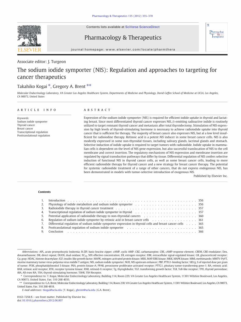
References . . . . . . . . . . . . . . . . . . . . . . . . . . . . . . . . . . . . . . . . . . . . . . . 360367Fig. 1. Schematic representation of iodide transport in the thyroid gland. The thyroidgland consist of follicles with one layer of epithelial cells surrounding the lumen. Iodide(I−) in circulation is transported into the lumen via basolateral NIS and apical pendrin.The activity of NIS requires the Na+-gradient maintained by Na+-K+ ATPase. Iodide inthe lumen is organified with Tg by TPO in the presence of H2O2 produced mainly byDUOX2. The iodinated tyrosine residues are used for synthesis of thyroid hormones,triiodothyronine (T3) or thyroxine (T4).
1. Introduction
Sodium iodide symporter (NIS, or SLC5A5, solute carrier family 5,member 5) (Dai et al., 1996; Smanik et al., 1997) is expressed at thehighest level in the thyroid and lactating breast (Dohan et al.,2003). Since NIS confers highly efficient iodide accumulation incells, its expression in cancer cells allows for the diagnostic andtherapeutic application of radioactive substrates of NIS, such as iodide(123I, 124I, and 131I) and pertechnetate (99mTcO4
−). Amajority (68–86%) ofthyroid cancer retains functional NIS expression (Castro et al., 2001;Wapnir et al., 2003). β-emitting radioiodide-131 (131I) is, therefore, rou-tinely used for ablation of remnant tumors after total thyroidectomy. Inthyroid cancer, the native NIS expression and radioiodide uptake are re-duced. Stimulation of NIS expression by increasing the serum levels ofthyroid-stimulating hormone (TSH), is required, prior to 131I administra-tion. Most differentiated thyroid cancer responds to these high levels ofserum TSH with an increase in NIS expression and iodide uptake(Schlumberger, 1998). The elevation of serum TSH can be achieved eitherby withdrawal of thyroid hormone supplement after thyroidectomy oradministration of recombinant TSH (thyrogen) (Ladenson et al., 1997).
The majority of breast cancer (70–80%) also expresses NIS(Tazebay et al., 2000; Wapnir et al., 2003), although iodide uptake isusually reduced or absent (Moon et al., 2001; Wapnir et al., 2004).Enhancement of the endogenous NIS expression in breast cancerhas been proposed as an approach that would allow 131I therapy(Boelaert & Franklyn, 2003). NIS, however, is expressed in the thyroidgland and other sites, such as stomach and salivary glands (Dohan etal., 2003), so selective induction of NIS in the target cancer is required.
The efficacy of 131I to destroy target tumors is dependent on thetissue-selective NIS gene induction, but also the effective transloca-tion of NIS protein to the cell membrane and correct membrane inser-tion. 131I retention in the target tumors, and the biological half-life of131I in the body, also influence treatment efficacy. Normal thyroid tis-sue incorporates the trapped iodide into thyroglobulin (Tg), referredto as organification, resulting in longer iodide retention. Iodide inmost thyroid cancer, as well as breast cancer, however, is not effi-ciently incorporated into proteins and hence more easily dischargedfrom cancer tissues (Schlumberger et al., 2007).
In this review, we will describe recent findings of pathways andagents that stimulate endogenous NIS gene expression, as well as in-tracellular NIS translocation, in thyroid cells and breast cancer cells.Dissection of signal transduction pathways for NIS regulation confersnovel potential targets to increase the efficacy of radioiodide therapyand expand its application to radioiodide-refractory thyroid cancer,as well as breast cancer and other NIS-expressing tumors.
2. Physiology of iodide metabolism and sodium iodide symporter
The thyroid must trap ~60 μg iodide/day from the bloodstream toproduce adequate thyroid hormone. The thyroid contains 70–90% ofthe iodide in the body (9–10 mg) (Riggs, 1952), and this iodide accu-mulation is dependent on NIS (Dai et al., 1996), expressed on thebasolateral membrane of thyroid follicular cells (Fig. 1). NIS is a glyco-sylated protein with 13 trans-membrane domains, transporting 2Na+ and one I−, dependent on the Na+ gradient maintained byNa+/K+ ATPase (Dohan et al., 2003). NIS activity produces the iodideconcentration gradient from blood to NIS-expressing cells, up to30-fold. Iodide taken up into the thyroid follicular cell by NIS, is re-leased to the lumen via pendrin, oxidized by thyroid peroxidase(TPO) with hydrogen peroxide (H2O2) produced mainly by dual
oxidase-2 (DUOX2), and binds to tyrosine residues of Tg accumulatedin the lumen (Fig. 1). The process of iodide incorporation into Tg istermed “organification”. The iodized tyrosine residues are then usedfor thyroid hormone synthesis. The transport of iodide into andthrough the thyroid gland is tightly regulated by TSH from the pituitarygland (Dohan et al., 2003; Kogai et al., 2006; Pesce et al., 2012). TSHstimulates NIS transcription (Kogai et al., 1997; Saito et al., 1997; Kogaiet al., 2000a), prolongs NIS protein half-life, and stimulates translocationof NIS into the cell membrane (Riedel et al., 2001), maximizing iodideuptake in thyroid cells.
Infants need ~90 μg/day of iodide to produce thyroid hormone, es-sential for normal brain development. Lactating mammary glands effi-ciently accumulates iodide so that breast milk contains 150–180 μg/Liodide (Semba & Delange, 2001). NIS is expressed on the basolateralmembrane of lactating mammary alveolar cells (Cho et al., 2000), andaccumulates iodide from the bloodstream intomilk. Expression of breastNIS is induced by oxytocin secreted from the posterior pituitary, and thisaction is enhanced by the elevated levels of serum prolactin and estro-gen present in the postnatal period (Cho et al., 2000; Tazebay et al.,2000).
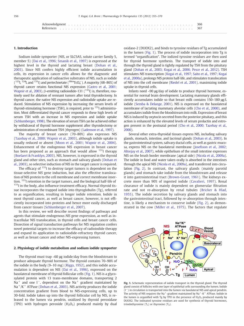
Several other extra-thyroidal tissues express NIS, including salivaryglands, stomach, intestine, and lacrimal glands (Dohan et al., 2003). Inthe gastrointestinal system, salivary ductal cells, as well as gastricmuco-sa, express NIS on the basolateral membrane (Josefsson et al., 2002;Altorjay et al., 2007), while epithelium of the small intestine expressesNIS on the brush border membrane (apical side) (Nicola et al., 2009a).The iodide in food and water taken orally is absorbed in the intestinesthrough the apical NIS (Nicola et al., 2009a), and transferred into circu-lation (Fig. 2). In contrast, the salivary glands (mainly parotidglands) and stomach take iodide from the bloodstream and releaseit into gastrointestinal tract (Brown-Grant, 1961). The kidneys ex-crete more than 90% of ingested iodide (Cavalieri, 1997). Renalclearance of iodide is mainly dependent on glomerular filtrationrate and not re-absorption by renal tubules (Bricker & Hlad,1955). The iodide secretion by salivary glands and stomach intothe gastrointestinal tract, followed by re-absorption through intes-tine, is likely a mechanism to conserve iodide (Fig. 2), as demon-strated in the cow (Miller et al., 1975). The factors that regulate
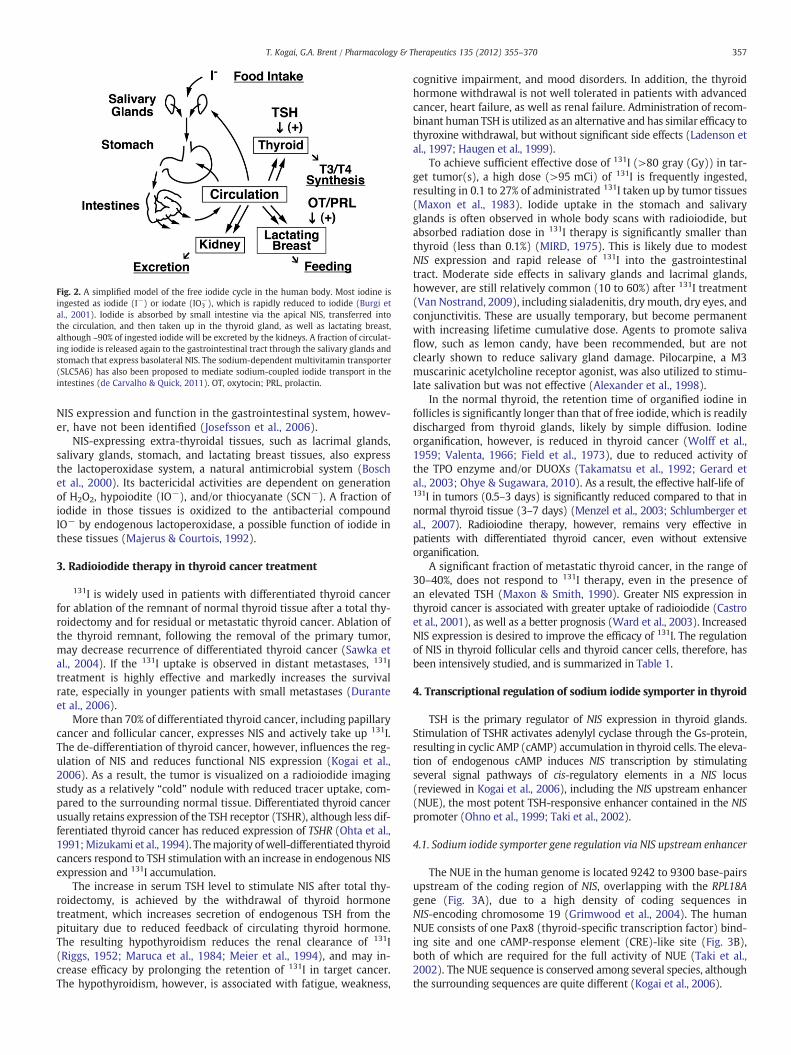
Fig. 2. A simplified model of the free iodide cycle in the human body. Most iodine isingested as iodide (I−) or iodate (IO3
−), which is rapidly reduced to iodide (Burgi etal., 2001). Iodide is absorbed by small intestine via the apical NIS, transferred intothe circulation, and then taken up in the thyroid gland, as well as lactating breast,although ~90% of ingested iodide will be excreted by the kidneys. A fraction of circulat-ing iodide is released again to the gastrointestinal tract through the salivary glands andstomach that express basolateral NIS. The sodium-dependent multivitamin transporter(SLC5A6) has also been proposed to mediate sodium-coupled iodide transport in theintestines (de Carvalho & Quick, 2011). OT, oxytocin; PRL, prolactin.
357T. Kogai, G.A. Brent / Pharmacology & Therapeutics 135 (2012) 355–370
NIS expression and function in the gastrointestinal system, howev-er, have not been identified (Josefsson et al., 2006).
NIS-expressing extra-thyroidal tissues, such as lacrimal glands,salivary glands, stomach, and lactating breast tissues, also expressthe lactoperoxidase system, a natural antimicrobial system (Boschet al., 2000). Its bactericidal activities are dependent on generationof H2O2, hypoiodite (IO−), and/or thiocyanate (SCN−). A fraction ofiodide in those tissues is oxidized to the antibacterial compoundIO− by endogenous lactoperoxidase, a possible function of iodide inthese tissues (Majerus & Courtois, 1992).
3. Radioiodide therapy in thyroid cancer treatment
131I is widely used in patients with differentiated thyroid cancerfor ablation of the remnant of normal thyroid tissue after a total thy-roidectomy and for residual or metastatic thyroid cancer. Ablation ofthe thyroid remnant, following the removal of the primary tumor,may decrease recurrence of differentiated thyroid cancer (Sawka etal., 2004). If the 131I uptake is observed in distant metastases, 131Itreatment is highly effective and markedly increases the survivalrate, especially in younger patients with small metastases (Duranteet al., 2006).
More than 70% of differentiated thyroid cancer, including papillarycancer and follicular cancer, expresses NIS and actively take up 131I.The de-differentiation of thyroid cancer, however, influences the reg-ulation of NIS and reduces functional NIS expression (Kogai et al.,2006). As a result, the tumor is visualized on a radioiodide imagingstudy as a relatively “cold” nodule with reduced tracer uptake, com-pared to the surrounding normal tissue. Differentiated thyroid cancerusually retains expression of the TSH receptor (TSHR), although less dif-ferentiated thyroid cancer has reduced expression of TSHR (Ohta et al.,1991;Mizukami et al., 1994). Themajority ofwell-differentiated thyroidcancers respond to TSH stimulation with an increase in endogenous NISexpression and 131I accumulation.
The increase in serum TSH level to stimulate NIS after total thy-roidectomy, is achieved by the withdrawal of thyroid hormonetreatment, which increases secretion of endogenous TSH from thepituitary due to reduced feedback of circulating thyroid hormone.The resulting hypothyroidism reduces the renal clearance of 131I(Riggs, 1952; Maruca et al., 1984; Meier et al., 1994), and may in-crease efficacy by prolonging the retention of 131I in target cancer.The hypothyroidism, however, is associated with fatigue, weakness,
cognitive impairment, and mood disorders. In addition, the thyroidhormone withdrawal is not well tolerated in patients with advancedcancer, heart failure, as well as renal failure. Administration of recom-binant human TSH is utilized as an alternative and has similar efficacy tothyroxine withdrawal, but without significant side effects (Ladenson etal., 1997; Haugen et al., 1999).
To achieve sufficient effective dose of 131I (>80 gray (Gy)) in tar-get tumor(s), a high dose (>95 mCi) of 131I is frequently ingested,resulting in 0.1 to 27% of administrated 131I taken up by tumor tissues(Maxon et al., 1983). Iodide uptake in the stomach and salivaryglands is often observed in whole body scans with radioiodide, butabsorbed radiation dose in 131I therapy is significantly smaller thanthyroid (less than 0.1%) (MIRD, 1975). This is likely due to modestNIS expression and rapid release of 131I into the gastrointestinaltract. Moderate side effects in salivary glands and lacrimal glands,however, are still relatively common (10 to 60%) after 131I treatment(Van Nostrand, 2009), including sialadenitis, dry mouth, dry eyes, andconjunctivitis. These are usually temporary, but become permanentwith increasing lifetime cumulative dose. Agents to promote salivaflow, such as lemon candy, have been recommended, but are notclearly shown to reduce salivary gland damage. Pilocarpine, a M3muscarinic acetylcholine receptor agonist, was also utilized to stimu-late salivation but was not effective (Alexander et al., 1998).
In the normal thyroid, the retention time of organified iodine infollicles is significantly longer than that of free iodide, which is readilydischarged from thyroid glands, likely by simple diffusion. Iodineorganification, however, is reduced in thyroid cancer (Wolff et al.,1959; Valenta, 1966; Field et al., 1973), due to reduced activity ofthe TPO enzyme and/or DUOXs (Takamatsu et al., 1992; Gerard etal., 2003; Ohye & Sugawara, 2010). As a result, the effective half-life of131I in tumors (0.5–3 days) is significantly reduced compared to that innormal thyroid tissue (3–7 days) (Menzel et al., 2003; Schlumberger etal., 2007). Radioiodine therapy, however, remains very effective inpatients with differentiated thyroid cancer, even without extensiveorganification.
A significant fraction of metastatic thyroid cancer, in the range of30–40%, does not respond to 131I therapy, even in the presence ofan elevated TSH (Maxon & Smith, 1990). Greater NIS expression inthyroid cancer is associated with greater uptake of radioiodide (Castroet al., 2001), as well as a better prognosis (Ward et al., 2003). IncreasedNIS expression is desired to improve the efficacy of 131I. The regulationof NIS in thyroid follicular cells and thyroid cancer cells, therefore, hasbeen intensively studied, and is summarized in Table 1.
4. Transcriptional regulation of sodium iodide symporter in thyroid
TSH is the primary regulator of NIS expression in thyroid glands.Stimulation of TSHR activates adenylyl cyclase through the Gs-protein,resulting in cyclic AMP (cAMP) accumulation in thyroid cells. The eleva-tion of endogenous cAMP induces NIS transcription by stimulatingseveral signal pathways of cis-regulatory elements in a NIS locus(reviewed in Kogai et al., 2006), including the NIS upstream enhancer(NUE), the most potent TSH-responsive enhancer contained in the NISpromoter (Ohno et al., 1999; Taki et al., 2002).
4.1. Sodium iodide symporter gene regulation via NIS upstream enhancer
The NUE in the human genome is located 9242 to 9300 base-pairsupstream of the coding region of NIS, overlapping with the RPL18Agene (Fig. 3A), due to a high density of coding sequences inNIS-encoding chromosome 19 (Grimwood et al., 2004). The humanNUE consists of one Pax8 (thyroid-specific transcription factor) bind-ing site and one cAMP-response element (CRE)-like site (Fig. 3B),both of which are required for the full activity of NUE (Taki et al.,2002). The NUE sequence is conserved among several species, althoughthe surrounding sequences are quite different (Kogai et al., 2006).
Table 1NIS stimulators in thyroid cells in vitro.
Agent Mechanism of actiona Cell lineb NISmRNA
NISprotein
I− uptakec References
TSH TSHR agonist FRTL5 Up Up 10–15 (Kogai et al., 1997)TSH TSHR agonist PCCL3 Up ~30 (Trapasso et al., 1999)TSH TSHR agonist Primary human thyroid Up Up ~13 (Saito et al., 1997;
Kogai et al., 2000a)Adenosine ADRA1 agonist FRTL5 Up Up ~7.5 (Harii et al., 1999)TSH/LPSd TLR4 agonist FRTL5, PCCL3 Up ~2.0 (Nicola et al., 2009b)TSH/L-NAMEd NOS inhibitor FRTL5 Up ~1.3 (Fozzatti et al., 2007)TSH/KT5823d cGK inhibitor FRTL5 ~2.2 (Fozzatti et al., 2007)TSH/LY294002d PI3K inhibitor FRTL5, PCCL3 Up Up ~3.0 (Kogai et al., 2008b)TSH/rapamycind mTOR inhibitor PCCL3 Up ~2.5 (de Souza et al., 2010)TSH/resveratrold Sirtuin activator FRTL5 Up ~3.0 (Sebai et al., 2010)LY294002 PI3K inhibitor NIS-BHP2-7 (TPC1) Up ~3.5 (Kogai et al., 2008b)Akti-1/2 AKT inhibitor NIS-BHP2-7 (TPC1) Up (Kogai et al., 2008b)Depsipeptide HDACi FTC133, SW1736 Up ~10 (Kitazono et al., 2001)Trichostatin A HDACi BHP18-21v (TPC1) Up Up ~3.0 (Furuya et al., 2004)SAHA HDACi K1, FTC133, C643 Up (Hou et al., 2010)
KAT18, OCUT1SAHA/perifosinee HDACi/AKTi K1, C643 Up (Hou et al., 2010)SAHA/RDEA119e HDACi/MEKi K1, C643 Up (Hou et al., 2010)SAHA/RDEA119/perifosinef HDACi/MEKi/AKTi C643, FTC133 Up (Hou et al., 2010)SAHA/RDEA119/perifosine/TSH HDACi/MEKi/AKTi/TSHR K1, C643, KAT18 Up 4–8 (Hou et al., 2010)Sunitinib/forskolin RTKi/AC agonist TPC1 (BHP) up (Fenton et al., 2010)Troglitazone PPAR agonist FTC133, TPC1 Up (Park et al., 2005)tRA RAR agonist FTC133 Up (Schmutzler et al., 1997)Lovastatin HMGR inhibitor CGTH Up ~3 (Frohlich et al., 2009)
a Abbreviations: ADRA1, adrenergic receptor α 1; NOS, nitric oxide synthase; cGK, cGMP-dependent protein kinase; mTOR, mammalian target of rapamycin; HMGR, HMG-CoAreductase; HDACi, HDAC inhibitor; AKTi, AKT inhibitor; MEKi, MEK inhibitor; RTKi, RTK inhibitor.
b Origins of FRTL5 and PCCL3 are rat thyroid glands; BHP2-7, BHP18-21, K1, TPC1, papillary thyroid cancer; CGTH, FTC133, follicular thyroid cancer; C643, KAT18, OCUT1,undifferentiated (anaplastic) thyroid cancer.
c Approximate fold-induction over the group without treatment is shown. Since specific activity of radioiodide in each study varies, values may not be compared among studies.d Enhances the TSH-induced NIS expression.e Enhances the SAHA-induced NIS expression.f Enhances the effects of double combination treatments.
Fig. 3. Regulation of the NUE in thyroid cells. A. Map of the human chromosome 19paround the NIS gene locus. The “A” in the translation start site (ATG) of NIS is referred toas+1.B. TSHR signaling pathways to NUE. NIS expression in thyroid cells is predominantlyregulated by the TSHR signaling to NUE. Gain-of-function studies of themolecules, indicat-ed by red color, have demonstrated stimulation of the NUE activity. The consensussequences of cis-elements of PAX8, CRE, and USF1 are indicated along with the sequenceof human NUE. *, Stimulatory effects have been reported with rat NUE, which containsan additional Pax8 element and anNFκB element (Nicola et al., 2010). AC, adenylyl cyclase;Ref-1, apurinic apyrimidinic endonuclease redox effector factor-1.
358 T. Kogai, G.A. Brent / Pharmacology & Therapeutics 135 (2012) 355–370
cAMP stimulates the NUE through both protein kinase-A (PKA)-dependent and -independent pathways in thyroid cells (Fig. 3B)(Ohno et al., 1999; Taki et al., 2002; Chun et al., 2004). PKA phosphor-ylates the cAMP-responsive element binding protein (CREB) andother basic-leucine zipper (B-ZIP) proteins, such as activating tran-scription factor-1 (ATF-1) and CRE-modulator (CREM), leading to re-cruitment of these B-ZIP proteins by the CRE-like element in NUE(Taki et al., 2002; Chun et al., 2004). Over-expression of a CREM acti-vator, τ2α, enhances the NUE activity in FRTL-5 rat thyroid cells whentreated with forskolin (Fenton et al., 2008), indicating an importantrole of the CREM activator in the PKA-dependent activation of NUE.
Pax8 is a key transcription factor for thyroid development and dif-ferentiation (Mansouri et al., 1998). Transcription of thyroid specificgenes, including TSHR, Tg, TPO, and NIS, is dependent on PAX8 activ-ity. Binding of PAX8 to the NUE, in response to TSH stimulation(Costamagna et al., 2004), is the primary requirement for significantactivation of NUE (Ohno et al., 1999; Taki et al., 2002). The TSH sig-naling facilitates the reduction of PAX8 (Kambe et al., 1996) throughredox effector factor-1 (Ref-1), which stimulates PAX8 binding to itscis-elements (Fig. 3B) (Tell et al., 1998).
4.2. Expression of sodium iodide symporter gene and regulation of NISupstream enhancer in thyroid cancer cells
The RET proto-oncogene encodes a receptor tyrosine kinase (RTK)which mediates extracellular neurotrophin signaling to intracellularsignal transduction pathways, including the MAPK (mitogen-activatedprotein kinase)/ERK (extracellular signal-regulated kinase) pathway.The activation of the RET-RAS-BRAF-MEK (MAP/ERK kinase)-ERKpathway is critical for tumor initiation and/or promotion in papillarythyroid cancer (Fagin, 2004). Constitutively active mutants of RET,RET/PTC rearrangement, and BRAFV600E are hallmarks of papillary thy-roid cancer. Activatingmutations in BRAF are most common in sporadic
papillary thyroid cancer in adults, while RET/PTC rearrangement isexpressed more frequently in pediatric and radiation-induced can-cers. The RET/PTC rearrangement is a characteristic finding in
359T. Kogai, G.A. Brent / Pharmacology & Therapeutics 135 (2012) 355–370
well-differentiated papillary thyroid cancer without aggressive be-havior (Ricarte-Filho et al., 2009). In contrast, BRAF activating mu-tations are often observed in radioiodide-refractory thyroid cancer,especially clinically aggressive papillary thyroid cancer with me-tastasis (present in more than 95%) (Ricarte-Filho et al., 2009).Other genetic modifications in thyroid cancer, such as mutations ofN-RAS (Volante et al., 2009), a catalytic subunit of phosphatidylinositol3-kinase (PI3KCA), and AKT (Ricarte-Filho et al., 2009), are also associatedwith a poor prognosis.
An experimental model with constitutive expression of RET/PTC inPCCL3 rat thyroid cells has been utilized for several studies. The exoge-nous RET/PTC significantly suppresses the expression of Pax8 (De Vitaet al., 1998) and the activity of PKA (Venkateswaran et al., 2004), leadingto reduced NIS expression (Trapasso et al., 1999; Venkateswaran et al.,2004). The reduced PKA activity is associated with down-regulation ofB-ZIP proteins. Indeed, expression of B-ZIP proteins that bind to NUEwas significantly decreased in BHP 2–7 cells, variants of RET/PTC-positiveTPC1 papillary thyroid cancer cells (Schweppe et al., 2008), resulting inreduced NUE activity (Taki et al., 2002), as well as low NIS expression(Ohta et al., 1997; Kogai et al., 2001).
BRAF mediates the inhibitory effects of RET/PTC on NIS expressionthrough the MEK–ERK pathway (Mitsutake et al., 2006). The activat-ing mutation in BRAF induces transforming growth factor (TGF)-β se-cretion from thyroid cancer cells, resulting in its paracrine action intumor tissues (Riesco-Eizaguirre et al., 2009). Increased TGFβ is asso-ciated with tumor invasion, stimulation of cell mobility, as well assuppression of NIS expression through SMA- and MAD-related pro-tein (SMAD)-3 (Costamagna et al., 2004) (Fig. 3). The BRAF mutation,therefore, contributes to the down-regulation of NIS via both the MEK–ERK pathway and the TGFβ–SMAD3 pathway (Riesco-Eizaguirre et al.,2009), which negatively affect the PAX8 action onNUE activation. The ex-pression of PAX8 is significantly decreased in ~70% of thyroid cancers,alongwith reducedNIS expression, especially in poorly differentiated thy-roid cancers (Fabbro et al., 1994; Puglisi et al., 2000). These observationsindicate that the constitutive activation of RET-BRAF signaling reducedNIS expression in papillary thyroid cancer cells, at least in part by sup-pressing the two major regulators of NUE, PAX8 and B-ZIP proteins.
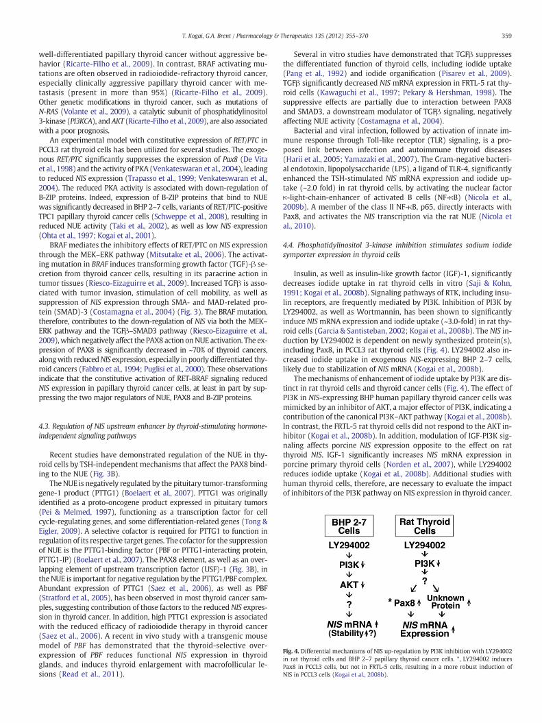
Fig. 4. Differential mechanisms of NIS up-regulation by PI3K inhibition with LY294002in rat thyroid cells and BHP 2–7 papillary thyroid cancer cells. *, LY294002 inducesPax8 in PCCL3 cells, but not in FRTL-5 cells, resulting in a more robust induction ofNIS in PCCL3 cells (Kogai et al., 2008b).
4.3. Regulation of NIS upstream enhancer by thyroid‐stimulating hormone-independent signaling pathways
Recent studies have demonstrated regulation of the NUE in thy-roid cells by TSH-independent mechanisms that affect the PAX8 bind-ing to the NUE (Fig. 3B).
TheNUE is negatively regulated by the pituitary tumor-transforminggene-1 product (PTTG1) (Boelaert et al., 2007). PTTG1 was originallyidentified as a proto-oncogene product expressed in pituitary tumors(Pei & Melmed, 1997), functioning as a transcription factor for cellcycle-regulating genes, and some differentiation-related genes (Tong &Eigler, 2009). A selective cofactor is required for PTTG1 to function inregulation of its respective target genes. The cofactor for the suppressionof NUE is the PTTG1-binding factor (PBF or PTTG1-interacting protein,PTTG1-IP) (Boelaert et al., 2007). The PAX8 element, as well as an over-lapping element of upstream transcription factor (USF)-1 (Fig. 3B), intheNUE is important for negative regulation by the PTTG1/PBF complex.Abundant expression of PTTG1 (Saez et al., 2006), as well as PBF(Stratford et al., 2005), has been observed in most thyroid cancer sam-ples, suggesting contribution of those factors to the reduced NIS expres-sion in thyroid cancer. In addition, high PTTG1 expression is associatedwith the reduced efficacy of radioiodide therapy in thyroid cancer(Saez et al., 2006). A recent in vivo study with a transgenic mousemodel of PBF has demonstrated that the thyroid-selective over-expression of PBF reduces functional NIS expression in thyroidglands, and induces thyroid enlargement with macrofollicular le-sions (Read et al., 2011).
Several in vitro studies have demonstrated that TGFβ suppressesthe differentiated function of thyroid cells, including iodide uptake(Pang et al., 1992) and iodide organification (Pisarev et al., 2009).TGFβ significantly decreased NIS mRNA expression in FRTL-5 rat thy-roid cells (Kawaguchi et al., 1997; Pekary & Hershman, 1998). Thesuppressive effects are partially due to interaction between PAX8and SMAD3, a downstream modulator of TGFβ signaling, negativelyaffecting NUE activity (Costamagna et al., 2004).
Bacterial and viral infection, followed by activation of innate im-mune response through Toll-like receptor (TLR) signaling, is a pro-posed link between infection and autoimmune thyroid diseases(Harii et al., 2005; Yamazaki et al., 2007). The Gram-negative bacteri-al endotoxin, lipopolysaccharide (LPS), a ligand of TLR-4, significantlyenhanced the TSH-stimulated NIS mRNA expression and iodide up-take (~2.0 fold) in rat thyroid cells, by activating the nuclear factorκ-light-chain-enhancer of activated B cells (NF-κB) (Nicola et al.,2009b). A member of the class II NF-κB, p65, directly interacts withPax8, and activates the NIS transcription via the rat NUE (Nicola etal., 2010).
4.4. Phosphatidylinositol 3‐kinase inhibition stimulates sodium iodidesymporter expression in thyroid cells
Insulin, as well as insulin-like growth factor (IGF)-1, significantlydecreases iodide uptake in rat thyroid cells in vitro (Saji & Kohn,1991; Kogai et al., 2008b). Signaling pathways of RTK, including insu-lin receptors, are frequently mediated by PI3K. Inhibition of PI3K byLY294002, as well as Wortmannin, has been shown to significantlyinduce NISmRNA expression and iodide uptake (~3.0-fold) in rat thy-roid cells (Garcia & Santisteban, 2002; Kogai et al., 2008b). The NIS in-duction by LY294002 is dependent on newly synthesized protein(s),including Pax8, in PCCL3 rat thyroid cells (Fig. 4). LY294002 also in-creased iodide uptake in exogenous NIS-expressing BHP 2–7 cells,likely due to stabilization of NIS mRNA (Kogai et al., 2008b).
The mechanisms of enhancement of iodide uptake by PI3K are dis-tinct in rat thyroid cells and thyroid cancer cells (Fig. 4). The effect ofPI3K in NIS-expressing BHP human papillary thyroid cancer cells wasmimicked by an inhibitor of AKT, a major effector of PI3K, indicating acontribution of the canonical PI3K–AKT pathway (Kogai et al., 2008b).In contrast, the FRTL-5 rat thyroid cells did not respond to the AKT in-hibitor (Kogai et al., 2008b). In addition, modulation of IGF-PI3K sig-naling affects porcine NIS expression opposite to the effect on ratthyroid NIS. IGF-1 significantly increases NIS mRNA expression inporcine primary thyroid cells (Norden et al., 2007), while LY294002reduces iodide uptake (Kogai et al., 2008b). Additional studies withhuman thyroid cells, therefore, are necessary to evaluate the impactof inhibitors of the PI3K pathway on NIS expression in thyroid cancer.

360 T. Kogai, G.A. Brent / Pharmacology & Therapeutics 135 (2012) 355–370
Most RTK inhibitors, clinically used for treatment of non-thyroidcancers, induce hypothyroidism in 20–50% of patients, by severalmechanisms. These mechanisms include attenuating thyroid bloodflow and increased metabolism of thyroid hormone by type 3deiodinase (Hamnvik et al., 2011). A multi-targeted RTK inhibitor,sunitinib, transiently induces hypothyroidism, in part due to reducediodide uptake in the thyroid (Mannavola et al., 2007). The effect,however, does not likely require the suppression of NIS expression,but other mechanisms, such as impairment of iodide organification(Salem et al., 2008). In contrast, an in vitro study with BHP 2–7papillary thyroid cancer cells demonstrated stimulatory effects ofsunitinib on NIS mRNA expression in the presence of an adenylylcyclase activator, forskolin (Fenton et al., 2010). Since sunitinibdown-regulates the PI3K–AKT pathway (Keefe et al., 2010), it likelymimics the effects of PI3K inhibition on NIS expression, at least par-tially through PAX8 induction (Fenton et al., 2010).
4.5. Effects of histone deacetylase inhibitors and combination treatmentswith signal transduction inhibitors in less-differentiated thyroid cancer cells
Epigenetic modifications of chromatin, including histone de-acetylation and hypermethylation, are associated with poorly differ-entiated cancer cells. Histone deacetylase (HDAC) inhibitors inducedifferentiation and expression of thyroid-selective genes in poorlydifferentiated thyroid cancer cells (Kitazono et al., 2001; Furuya etal., 2004). A member of the bicyclic peptide class of HDAC inhibitor,FR901228 (or depsipeptide), significantly induces NIS mRNA expres-sion and iodide uptake in thyroid cancer cell lines, including BHP18-21v papillary thyroid cancer cells (Furuya et al., 2004), FTC-133 fol-licular thyroid cancer cells, and SW-1736 undifferentiated thyroid can-cer cells (Kitazono et al., 2001). Depsipeptide significantly inducedexpression of Tg and TPO, resulting in recovery of iodide organificationin BHP 18-21v cells (Furuya et al., 2004), favorable for increasingradioiodide retention.
Most papillary thyroid cancer expresses RET/PTC or BRAF mutants,which activate theMAPKpathway ofMEK–ERK. The PI3K–AKT signalingalso plays a role in tumor progression in thyroid cancer (Shinohara et al.,2007). Modulation of these signaling pathways may reconstitute thethyroid-specific functions in poorly differentiated thyroid cancer cells.Recently, combination treatments with anHDAC inhibitor, aMEK inhib-itor, and/or an AKT inhibitor have been tested in several thyroid cancercell lines with successful recovery of NIS expression (Hou et al., 2010),although the combination required for NIS induction varied among thecell lines (Table 1). An HDAC inhibitor, SAHA, was required forsignificant NIS induction in the tested cell lines, including K1 papillarycancer cells, FTC-133 follicular cancer cells, and OCUT1 and C643undifferentiated cancer cell lines. Since TSHR was also induced bySAHA, treatment with TSH further enhanced the SAHA-induced NIS ex-pression in some cell lines. The addition of a MEK inhibitor RDEA119and/or an AKT inhibitor perifosine, variably affected the SAHA-inducedNIS expression among the tested cell lines. The triple combination ofSAHA, RDEA119, and perifosine significantly (4 to 8-fold) induced theiodide uptake in K1 cells, as well as two undifferentiated cancer celllines, C643 and KAT18.
These findings have raised the possibility of radioiodide therapyin some aggressive radioiodide-refractory thyroid cancer aftertreatment with an HDAC inhibitor. The inhibition of MAPK path-way and/or AKT possibly enhances the effects of HDAC inhibitoron the NIS expression. K1 cells, harboring the BRAF mutation butnot RET/PTC (Schweppe et al., 2008), responded well to the MEKinhibitor RDEA119 to enhance the SAHA-induced NIS expression(Hou et al., 2010). In contrast, another MEK inhibitor, PD98059, signif-icantly decreased the iodide uptake in the exogenous RET/PTC-expressingPCCL3 cells (Vadysirisack et al., 2007). The difference of genetic back-ground may confer the differential responses to signal transductioninhibitors.
5. Potential application of radioiodide therapy to non-thyroidalcancers
5.1. Sodium iodide symporter gene therapy for non-thyroidal cancers
Due to the success of radioiodide therapy for thyroid cancer, theNIS gene has been introduced into other cancers to achieve sufficient131I accumulation for tumor shrinkage. Early studies of the antitumoreffects of 131I after NIS gene therapy, however, did not show a consis-tent response of increased uptake (reviewed in Riesco-Eizaguirre &Santisteban, 2006). This may have been the result of insufficient de-livery or expression of NIS. Recent improvements in NIS delivery sys-tems, as well as the addition of radiation-sensitizing agents andoncolytic treatments, have resulted in greater tumor shrinkage andsignificant tumor growth inhibition in models of many types of can-cer (summarized in Tables 2 and 3).
Previous studies of NIS gene therapy, without adjuvant therapies,have shown that the antitumor efficacy of 131I is dependent on themagnitude of NIS gene expression. To achieve complete tumordestruction, more than 20% of the injected radioiodide dose, pergram of tumor (%ID/g), needs to accumulate in a tumor. A successfulprostate cancer xenograft model has been described that accumulates25 to 30%ID/g in the tumors (Spitzweg et al., 2000). For comparison,poorly differentiated thyroid cancer xenografts accumulated only4.9–9.3%ID/g and were not effectively treated with radioiodine(Shimura et al., 1997). A NIS gene delivered with an adenovirus vectorand a tissue specific gene promoter, the prostate-specific antigengene (PSA) promoter, conferred efficient functional NIS expressionin prostate cancer xenografts (Spitzweg et al., 2001; Dwyer et al.,2005), and phase 2 trials are currently being conducted in prostatecancer patients. Other tumor specific promoters, such as the human tel-omerase reverse transcriptase (hTERT) promoter, the carcinoembryonicantigen (CEA) promoter, and the alpha-fetoprotein (AFP) promoter,have also directed robust and selective NIS expression in the tumor,leading to remarkable inhibition of tumor growth by 131I (Table 2).
To achieve synergistic or additive cytotoxic effects, combined treat-ments with NIS gene therapy and a tumor targeting strategy, such as uti-lization of an oncolytic vector (Goel et al., 2007; Hakkarainen et al., 2009;Peerlinck et al., 2009; Li et al., 2010), vaccination against a tumor-specificantigen (Jeon et al., 2011), or inhibition of intracellular glucose metabo-lism by knockdown of hexokinase II (Kim et al., 2011), have been studied,resulting in significant growth inhibition or eradication of tumor (Tables 2and 3). Inhibition of DNA repair by theDNA-dependent protein kinase in-hibitor (DNA-PKi) enhanced the antitumor effects by combination treat-ment with 131I and external beam radiotherapy in colorectal cancercells, as well as head and neck cancer cells (Hingorani et al., 2010b).
Conventionally, NIS gene therapy has been performed with virusvectors. Results of initial clinical studies of gene therapy for X-linked se-vere combined immunodeficiency (X-SCID), however, have indicated ahigh incidence of leukemia due to unexpected integration of viral DNAto the host genomes, raising safety concerns about the gene deliveryby virus vectors. The majority of recent experimental NIS genetherapies have been performed with replication-defective adeno-viruses (Table 3) preventing unfavorable genomic integration.Some oncolytic viruses used for NIS gene therapy (Dingli et al.,2004; Goel et al., 2007; Carlson et al., 2009; Hakkarainen et al.,2009) are negative-sense single-stranded RNA viruses, not gener-ally integrated into the host genomes. A plasmid vector conjugat-ed with polyplex targeted to EGF receptor (Klutz et al., 2009) isalso a promising strategy for safe and highly selective delivery ofNIS into the targeted tumor.
5.2. Induction of endogenous sodium iodide symporter in breast cancer
The majority (70–80%) of breast cancers express NIS (Tazebay et al.,2000; Wapnir et al., 2003), while only 20–30% take-up radioiodide, due
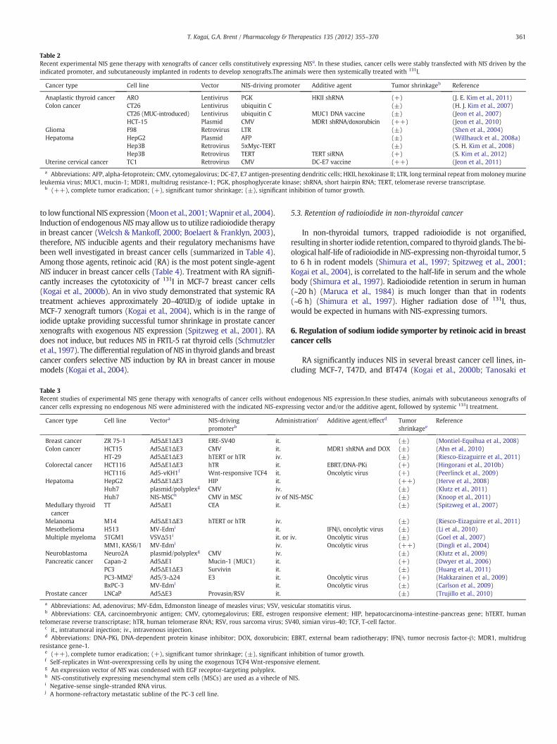
Table 2Recent experimental NIS gene therapy with xenografts of cancer cells constitutively expressing NISa. In these studies, cancer cells were stably transfected with NIS driven by theindicated promoter, and subcutaneously implanted in rodents to develop xenografts.The animals were then systemically treated with 131I.
Cancer type Cell line Vector NIS-driving promoter Additive agent Tumor shrinkageb Reference
Anaplastic thyroid cancer ARO Lentivirus PGK HKII shRNA (+) (J. E. Kim et al., 2011)Colon cancer CT26 Lentivirus ubiquitin C (±) (H. J. Kim et al., 2007)
CT26 (MUC-introduced) Lentivirus ubiquitin C MUC1 DNA vaccine (±) (Jeon et al., 2007)HCT-15 Plasmid CMV MDR1 shRNA/doxorubicin (++) (Jeon et al., 2010)
Glioma F98 Retrovirus LTR (±) (Shen et al., 2004)Hepatoma HepG2 Plasmid AFP (±) (Willhauck et al., 2008a)
Hep3B Retrovirus 5xMyc-TERT (±) (S. H. Kim et al., 2008)Hep3B Retrovirus TERT TERT siRNA (+) (S. Kim et al., 2012)
Uterine cervical cancer TC1 Retrovirus CMV DC-E7 vaccine (++) (Jeon et al., 2011)
a Abbreviations: AFP, alpha-fetoprotein; CMV, cytomegalovirus; DC-E7, E7 antigen-presenting dendritic cells; HKII, hexokinase II; LTR, long terminal repeat frommoloneymurineleukemia virus; MUC1, mucin-1; MDR1, multidrug resistance-1; PGK, phosphoglycerate kinase; shRNA, short hairpin RNA; TERT, telomerase reverse transcriptase.
b (++), complete tumor eradication; (+), significant tumor shrinkage; (±), significant inhibition of tumor growth.
361T. Kogai, G.A. Brent / Pharmacology & Therapeutics 135 (2012) 355–370
to low functionalNIS expression (Moon et al., 2001;Wapnir et al., 2004).Induction of endogenousNISmay allow us to utilize radioiodide therapyin breast cancer (Welcsh & Mankoff, 2000; Boelaert & Franklyn, 2003),therefore, NIS inducible agents and their regulatory mechanisms havebeen well investigated in breast cancer cells (summarized in Table 4).Among those agents, retinoic acid (RA) is the most potent single-agentNIS inducer in breast cancer cells (Table 4). Treatment with RA signifi-cantly increases the cytotoxicity of 131I in MCF-7 breast cancer cells(Kogai et al., 2000b). An in vivo study demonstrated that systemic RAtreatment achieves approximately 20–40%ID/g of iodide uptake inMCF-7 xenograft tumors (Kogai et al., 2004), which is in the range ofiodide uptake providing successful tumor shrinkage in prostate cancerxenografts with exogenous NIS expression (Spitzweg et al., 2001). RAdoes not induce, but reduces NIS in FRTL-5 rat thyroid cells (Schmutzleret al., 1997). The differential regulation ofNIS in thyroid glands and breastcancer confers selective NIS induction by RA in breast cancer in mousemodels (Kogai et al., 2004).
Table 3Recent studies of experimental NIS gene therapy with xenografts of cancer cells without ecancer cells expressing no endogenous NIS were administered with the indicated NIS-expre
Cancer type Cell line Vectora NIS-drivingpromoterb
Admi
Breast cancer ZR 75-1 Ad5ΔE1ΔE3 ERE-SV40 it.Colon cancer HCT15 Ad5ΔE1ΔE3 CMV it.
HT-29 Ad5ΔE1ΔE3 hTERT or hTR iv.Colorectal cancer HCT116 Ad5ΔE1ΔE3 hTR it.
HCT116 Ad5-vKH1f Wnt-responsive TCF4 it.Hepatoma HepG2 Ad5ΔE1ΔE3 HIP it.
Huh7 plasmid/polyplexg CMV iv.Huh7 NIS-MSCh CMV in MSC iv of
Medullary thyroidcancer
TT Ad5ΔE1 CEA it.
Melanoma M14 Ad5ΔE1ΔE3 hTERT or hTR iv.Mesothelioma H513 MV-Edmi it.Multiple myeloma 5TGM1 VSVΔ51i it. or
MM1, KAS6/1 MV-Edmi iv.Neuroblastoma Neuro2A plasmid/polyplexg CMV iv.Pancreatic cancer Capan-2 Ad5ΔE1 Mucin-1 (MUC1) it.
PC3 Ad5ΔE1ΔE3 Survivin it.PC3-MM2j Ad5/3-Δ24 E3 it.BxPC-3 MV-Edmi it.
Prostate cancer LNCaP Ad5ΔE3 Provasin/RSV it.
a Abbreviations: Ad, adenovirus; MV-Edm, Edmonston lineage of measles virus; VSV, vesb Abbreviations: CEA, carcinoembryonic antigen; CMV, cytomegalovirus; ERE, estrogen
telomerase reverse transcriptase; hTR, human telomerase RNA; RSV, rous sarcoma virus; SVc it., intratumoral injection; iv., intravenous injection.d Abbreviations: DNA-PKi, DNA-dependent protein kinase inhibitor; DOX, doxorubicin;
resistance gene-1.e (++), complete tumor eradication; (+), significant tumor shrinkage; (±), significantf Self-replicates in Wnt-overexpressing cells by using the exogenous TCF4 Wnt-responsivg An expression vector of NIS was condensed with EGF receptor-targeting polyplex.h NIS-constitutively expressing mesenchymal stem cells (MSCs) are used as a vihecle ofi Negative-sense single-stranded RNA virus.j A hormone-refractory metastatic subline of the PC-3 cell line.
5.3. Retention of radioiodide in non-thyroidal cancer
In non-thyroidal tumors, trapped radioiodide is not organified,resulting in shorter iodide retention, compared to thyroid glands. Thebi-ological half-life of radioiodide inNIS-expressing non-thyroidal tumor, 5to 6 h in rodent models (Shimura et al., 1997; Spitzweg et al., 2001;Kogai et al., 2004), is correlated to the half-life in serum and the wholebody (Shimura et al., 1997). Radioiodide retention in serum in human(~20 h) (Maruca et al., 1984) is much longer than that in rodents(~6 h) (Shimura et al., 1997). Higher radiation dose of 131I, thus,would be expected in humans with NIS-expressing tumors.
6. Regulation of sodium iodide symporter by retinoic acid in breastcancer cells
RA significantly induces NIS in several breast cancer cell lines, in-cluding MCF-7, T47D, and BT474 (Kogai et al., 2000b; Tanosaki et
ndogenous NIS expression.In these studies, animals with subcutaneous xenografts ofssing vector and/or the additive agent, followed by systemic 131I treatment.
nistrationc Additive agent/effectd Tumorshrinkagee
Reference
(±) (Montiel-Equihua et al., 2008)MDR1 shRNA and DOX (±) (Ahn et al., 2010)
(±) (Riesco-Eizaguirre et al., 2011)EBRT/DNA-PKi (+) (Hingorani et al., 2010b)Oncolytic virus (+) (Peerlinck et al., 2009)
(++) (Herve et al., 2008)(±) (Klutz et al., 2011)
NIS-MSC (±) (Knoop et al., 2011)(±) (Spitzweg et al., 2007)
(±) (Riesco-Eizaguirre et al., 2011)IFNβ, oncolytic virus (±) (Li et al., 2010)
iv. Oncolytic virus (±) (Goel et al., 2007)Oncolytic virus (++) (Dingli et al., 2004)
(±) (Klutz et al., 2009)(+) (Dwyer et al., 2006)(±) (Huang et al., 2011)
Oncolytic virus (+) (Hakkarainen et al., 2009)Oncolytic virus (±) (Carlson et al., 2009)
(±) (Trujillo et al., 2010)
icular stomatitis virus.responsive element; HIP, hepatocarcinoma-intestine-pancreas gene; hTERT, human40, simian virus-40; TCF, T-cell factor.
EBRT, external beam radiotherapy; IFNβ, tumor necrosis factor-β; MDR1, multidrug
inhibition of tumor growth.e element.
NIS.
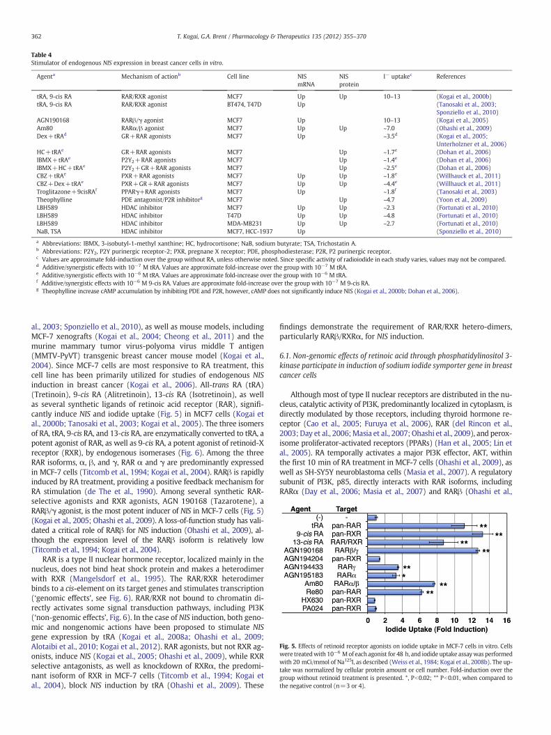
Table 4Stimulator of endogenous NIS expression in breast cancer cells in vitro.
Agenta Mechanism of actionb Cell line NISmRNA
NISprotein
I− uptakec References
tRA, 9-cis RA RAR/RXR agonist MCF7 Up Up 10–13 (Kogai et al., 2000b)tRA, 9-cis RA RAR/RXR agonist BT474, T47D Up (Tanosaki et al., 2003;
Sponziello et al., 2010)AGN190168 RARβ/γ agonist MCF7 Up 10–13 (Kogai et al., 2005)Am80 RARα/β agonist MCF7 Up Up ~7.0 (Ohashi et al., 2009)Dex+tRAd GR+RAR agonists MCF7 Up ~3.5d (Kogai et al., 2005;
Unterholzner et al., 2006)HC+tRAe GR+RAR agonists MCF7 Up ~1.7e (Dohan et al., 2006)IBMX+tRAe P2Y2+RAR agonists MCF7 Up ~1.4e (Dohan et al., 2006)IBMX+HC+tRAe P2Y2+GR+RAR agonists MCF7 Up ~2.5e (Dohan et al., 2006)CBZ+tRAe PXR+RAR agonists MCF7 Up Up ~1.8e (Willhauck et al., 2011)CBZ+Dex+tRAe PXR+GR+RAR agonists MCF7 Up Up ~4.4e (Willhauck et al., 2011)Troglitazone+9cisRAf PPARγ+RAR agonists MCF7 Up ~1.8f (Tanosaki et al., 2003)Theophylline PDE antagonist/P2R inhibitorg MCF7 Up ~4.7 (Yoon et al., 2009)LBH589 HDAC inhibitor MCF7 Up Up ~2.3 (Fortunati et al., 2010)LBH589 HDAC inhibitor T47D Up Up ~4.8 (Fortunati et al., 2010)LBH589 HDAC inhibitor MDA-MB231 Up Up ~2.7 (Fortunati et al., 2010)NaB, TSA HDAC inhibitor MCF7, HCC-1937 Up (Sponziello et al., 2010)
a Abbreviations: IBMX, 3-isobutyl-1-methyl xanthine; HC, hydrocortisone; NaB, sodium butyrate; TSA, Trichostatin A.b Abbreviations: P2Y2, P2Y purinergic receptor-2; PXR, pregnane X receptor; PDE, phosphodiesterase; P2R, P2 purinergic receptor.c Values are approximate fold-induction over the group without RA, unless otherwise noted. Since specific activity of radioiodide in each study varies, values may not be compared.d Additive/synergistic effects with 10−7 M tRA. Values are approximate fold-increase over the group with 10−7 M tRA.e Additive/synergistic effects with 10−6 M tRA. Values are approximate fold-increase over the group with 10−6 M tRA.f Additive/synergistic effects with 10−6 M 9-cis RA. Values are approximate fold-increase over the group with 10−7 M 9-cis RA.g Theophylline increase cAMP accumulation by inhibiting PDE and P2R, however, cAMP does not significantly induce NIS (Kogai et al., 2000b; Dohan et al., 2006).
Fig. 5. Effects of retinoid receptor agonists on iodide uptake in MCF-7 cells in vitro. Cellswere treatedwith 10−6 Mof each agonist for 48 h, and iodide uptake assaywas performedwith 20 mCi/mmol of Na125I, as described (Weiss et al., 1984; Kogai et al., 2008b). The up-take was normalized by cellular protein amount or cell number. Fold-induction over thegroup without retinoid treatment is presented. *, Pb0.02; ** Pb0.01, when compared tothe negative control (n=3 or 4).
362 T. Kogai, G.A. Brent / Pharmacology & Therapeutics 135 (2012) 355–370
al., 2003; Sponziello et al., 2010), as well as mouse models, includingMCF-7 xenografts (Kogai et al., 2004; Cheong et al., 2011) and themurine mammary tumor virus-polyoma virus middle T antigen(MMTV-PyVT) transgenic breast cancer mouse model (Kogai et al.,2004). Since MCF-7 cells are most responsive to RA treatment, thiscell line has been primarily utilized for studies of endogenous NISinduction in breast cancer (Kogai et al., 2006). All-trans RA (tRA)(Tretinoin), 9-cis RA (Alitretinoin), 13-cis RA (Isotretinoin), as wellas several synthetic ligands of retinoic acid receptor (RAR), signifi-cantly induce NIS and iodide uptake (Fig. 5) in MCF7 cells (Kogai etal., 2000b; Tanosaki et al., 2003; Kogai et al., 2005). The three isomersof RA, tRA, 9-cis RA, and 13-cis RA, are enzymatically converted to tRA, apotent agonist of RAR, as well as 9-cis RA, a potent agonist of retinoid-Xreceptor (RXR), by endogenous isomerases (Fig. 6). Among the threeRAR isoforms, α, β, and γ, RAR α and γ are predominantly expressedin MCF-7 cells (Titcomb et al., 1994; Kogai et al., 2004). RARβ is rapidlyinduced by RA treatment, providing a positive feedback mechanism forRA stimulation (de The et al., 1990). Among several synthetic RAR-selective agonists and RXR agonists, AGN 190168 (Tazarotene), aRARβ/γ agonist, is the most potent inducer of NIS in MCF-7 cells (Fig. 5)(Kogai et al., 2005; Ohashi et al., 2009). A loss-of-function study has vali-dated a critical role of RARβ for NIS induction (Ohashi et al., 2009), al-though the expression level of the RARβ isoform is relatively low(Titcomb et al., 1994; Kogai et al., 2004).
RAR is a type II nuclear hormone receptor, localized mainly in thenucleus, does not bind heat shock protein and makes a heterodimerwith RXR (Mangelsdorf et al., 1995). The RAR/RXR heterodimerbinds to a cis-element on its target genes and stimulates transcription(‘genomic effects’, see Fig. 6). RAR/RXR not bound to chromatin di-rectly activates some signal transduction pathways, including PI3K(‘non-genomic effects’, Fig. 6). In the case of NIS induction, both geno-mic and nongenomic actions have been proposed to stimulate NISgene expression by tRA (Kogai et al., 2008a; Ohashi et al., 2009;Alotaibi et al., 2010; Kogai et al., 2012). RAR agonists, but not RXR ag-onists, induce NIS (Kogai et al., 2005; Ohashi et al., 2009), while RXRselective antagonists, as well as knockdown of RXRα, the predomi-nant isoform of RXR in MCF-7 cells (Titcomb et al., 1994; Kogai etal., 2004), block NIS induction by tRA (Ohashi et al., 2009). These
findings demonstrate the requirement of RAR/RXR hetero-dimers,particularly RARβ/RXRα, for NIS induction.
6.1. Non-genomic effects of retinoic acid through phosphatidylinositol 3‐kinase participate in induction of sodium iodide symporter gene in breastcancer cells
Although most of type II nuclear receptors are distributed in the nu-cleus, catalytic activity of PI3K, predominantly localized in cytoplasm, isdirectly modulated by those receptors, including thyroid hormone re-ceptor (Cao et al., 2005; Furuya et al., 2006), RAR (del Rincon et al.,2003;Day et al., 2006;Masia et al., 2007; Ohashi et al., 2009), and perox-isome proliferator-activated receptors (PPARs) (Han et al., 2005; Lin etal., 2005). RA temporally activates a major PI3K effector, AKT, withinthe first 10 min of RA treatment in MCF-7 cells (Ohashi et al., 2009), aswell as SH-SY5Y neuroblastoma cells (Masia et al., 2007). A regulatorysubunit of PI3K, p85, directly interacts with RAR isoforms, includingRARα (Day et al., 2006; Masia et al., 2007) and RARβ (Ohashi et al.,
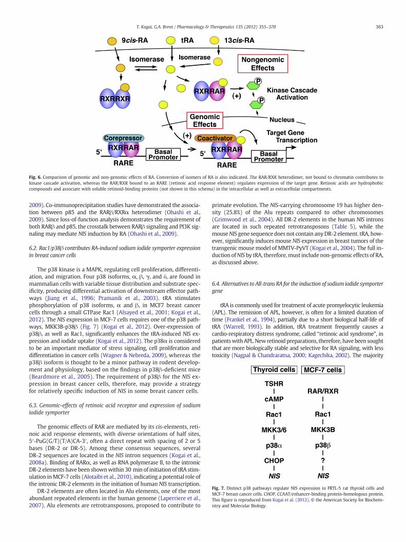
Fig. 6. Comparison of genomic and non-genomic effects of RA. Conversion of isomers of RA is also indicated. The RAR/RXR heterodimer, not bound to chromatin contributes tokinase cascade activation, whereas the RAR/RXR bound to an RARE (retinoic acid response element) regulates expression of the target gene. Retinoic acids are hydrophobiccompounds and associate with soluble retinoid-binding proteins (not shown in this schema) in the intracellular as well as extracellular compartments.
Fig. 7. Distinct p38 pathways regulate NIS expression in FRTL-5 rat thyroid cells andMCF-7 breast cancer cells. CHOP, CCAAT/enhancer-binding protein-homologous protein.This figure is reproduced from Kogai et al. (2012). © the American Society for Biochem-istry and Molecular Biology.
363T. Kogai, G.A. Brent / Pharmacology & Therapeutics 135 (2012) 355–370
2009). Co-immunoprecipitation studies have demonstrated the associa-tion between p85 and the RARβ/RXRα heterodimer (Ohashi et al.,2009). Since loss-of-function analysis demonstrates the requirement ofboth RARβ and p85, the crosstalk between RARβ signaling and PI3K sig-naling may mediate NIS induction by RA (Ohashi et al., 2009).
6.2. Rac1/p38β contributes RA-induced sodium iodide symporter expressionin breast cancer cells
The p38 kinase is a MAPK, regulating cell proliferation, differenti-ation, and migration. Four p38 isoforms, α, β, γ, and δ, are found inmammalian cells with variable tissue distribution and substrate spec-ificity, producing differential activation of downstream effector path-ways (Jiang et al., 1996; Pramanik et al., 2003). tRA stimulatesphosphorylation of p38 isoforms, α and β, in MCF7 breast cancercells through a small GTPase Rac1 (Alsayed et al., 2001; Kogai et al.,2012). The NIS expression in MCF-7 cells requires one of the p38 path-ways, MKK3B-p38β (Fig. 7) (Kogai et al., 2012). Over-expression ofp38β, as well as Rac1, significantly enhances the tRA-induced NIS ex-pression and iodide uptake (Kogai et al., 2012). The p38α is consideredto be an important mediator of stress signaling, cell proliferation anddifferentiation in cancer cells (Wagner & Nebreda, 2009), whereas thep38β isoform is thought to be a minor pathway in rodent develop-ment and physiology, based on the findings in p38β-deficient mice(Beardmore et al., 2005). The requirement of p38β for the NIS ex-pression in breast cancer cells, therefore, may provide a strategyfor relatively specific induction of NIS in some breast cancer cells.
6.3. Genomic-effects of retinoic acid receptor and expression of sodiumiodide symporter
The genomic effects of RAR are mediated by its cis-elements, reti-noic acid response elements, with diverse orientations of half sites,5′-PuG(G/T)(T/A)CA-3′, often a direct repeat with spacing of 2 or 5bases (DR-2 or DR-5). Among these consensus sequences, severalDR-2 sequences are located in the NIS intron sequences (Kogai et al.,2008a). Binding of RARα, as well as RNA polymerase II, to the intronicDR-2 elements have been shownwithin 30 min of initiation of tRA stim-ulation inMCF-7 cells (Alotaibi et al., 2010), indicating a potential role ofthe intronic DR-2 elements in the initiation of human NIS transcription.
DR-2 elements are often located in Alu elements, one of the mostabundant repeated elements in the human genome (Laperriere et al.,2007). Alu elements are retrotransposons, proposed to contribute to
primate evolution. The NIS-carrying chromosome 19 has higher den-sity (25.8%) of the Alu repeats compared to other chromosomes(Grimwood et al., 2004). All DR-2 elements in the human NIS intronsare located in such repeated retrotransposons (Table 5), while themouseNIS gene sequence does not contain anyDR-2 element. tRA, how-ever, significantly induces mouse NIS expression in breast tumors of thetransgenic mousemodel of MMTV-PyVT (Kogai et al., 2004). The full in-duction ofNIS by tRA, therefore,must includenon-genomic effects of RA,as discussed above.
6.4. Alternatives to All‐trans RA for the induction of sodium iodide symportergene
tRA is commonly used for treatment of acute promyelocytic leukemia(APL). The remission of APL, however, is often for a limited duration oftime (Frankel et al., 1994), partially due to a short biological half-life oftRA (Warrell, 1993). In addition, tRA treatment frequently causes acardio-respiratory distress syndrome, called “retinoic acid syndrome”, inpatientswithAPL.Newretinoidpreparations, therefore, havebeen soughtthat are more biologically stable and selective for RA signaling, with lesstoxicity (Nagpal & Chandraratna, 2000; Kagechika, 2002). The majority
364 T. Kogai, G.A. Brent / Pharmacology & Therapeutics 135 (2012) 355–370
of synthetic retinoids, however, have only been used for topical skintreatment.
The 50% effective concentration (EC50) of tRA in vitro for NIS induc-tion is ~10−7 M (Kogai et al., 2005), consistent with that for transcrip-tional regulation of other tRA-regulated genes (Idres et al., 2002). Thesystemic dose of tRA required for maximum NIS induction in rodentmodels, however, is likely higher than would be tolerated for routinetreatment in humans (Kogai et al., 2004). In addition, several in vivostudies have demonstrated variable effects of systemic tRA treatmenton radioiodide uptake (~1.2 to ~15-fold) in MCF-7 xenografts (Kogai etal., 2004; Willhauck et al., 2008b; Cheong et al., 2011), possibly due toclonal variation of MCF-7 cells (Seibert et al., 1983; Kogai et al., 2004;Lacroix & Leclercq, 2004). To search for more efficient agents, severalother retinoids have been tested for the ability to induce NIS in MCF-7cells in vitro (Fig. 5) (Kogai et al., 2005; Ohashi et al., 2009).
Among the retinoids that markedly induce NIS, 13-cis RA is the onlyretinoid, other than tRA, commonly used for systemic administration.13-cis RA iswidely used for treatment of cystic acne, aswell as neuroblas-toma and other cancers. Endogenous isomerases, such as glutathioneS-transferases (Chen & Juchau, 1998), convert 13-cis RA to tRA in targetcells so 13-cis RA works as a prodrug of tRA (Fig. 6) with less side effectsand a longer biological half-life. The efficacy of enzymatic conversion isdistinct in each tissue, and isomerase activity is relatively low in tumor tis-sues. After systemic administration of 13-cis RA inMCF-7 xenograft mice,the fraction converted to tRA in the xenograft tumors (~20%) is signifi-cantly smaller than that in liver (~68%) (Conley et al., 1999). The magni-tude of NIS mRNA induction by 13-cis RA is actually lower (~65%) thanthat by tRA in MCF-7 cells in vitro (Kogai et al., 2005).
An in vitro study has demonstrated that AGN190168 (Tazarotene)is the most effective synthetic retinoid for the NIS induction in MCF-7cells (Fig. 5) (Kogai et al., 2005). AGN190168 is clinically used foracne and psoriasis, but limited to topical application, due to a veryshort half-life (b1 h) of its active metabolite AGN 190299 in theserum (Chien et al., 1992; Hsyu et al., 1994). Selective RARβ agonistswith longer half-lives would establish more effective and less toxictreatment for the NIS induction.
6.5. Enhancement of All‐trans RA-induced sodium iodide symporterexpression by other nuclear receptor ligands
A nuclear receptor dimmer recognizes a specific response element,typically containing two common half-sites, 5′-AGGTCA-3′, or itsvariants. The selectivity of cis-element to each receptor dimmer isdependent on spacing and orientation of the two half-sites. The consen-sus half-site, therefore, is occasionally shared by different receptors.Co-activators and co-repressors are also often shared among variousnuclear receptors. In addition, RXR is shared among type II nuclear
Table 5Putative retinoic acid response elements in human NIS intron sequences andretrotransposon.
Position Sequence RARE Stranda Repeatedelementb
1st intron AGGTCAggAGTTCA DR-2c (+) AluSx1AGGTCAggAGTTCA DR-2c (+) AluSg
5th intron TCACCTgAGGTCAcAGTTCA DR-1, ER-8d (−) AluSx17th intron AGGTCAatGGGCAA DR-2 (+) MIRc8th intron AGGTCAggAGTTCA DR-2c (−) AluSx12th intron AGGTCAggAGTTCA DR-2c (+) AluSg
AGGTCAggAGTTCA DR-2c (−) AluSxAGGTCAggAGTTCA DR-2c (−) AluSx
13th intron AGGTCAggAGTTCA DR-2c (+) AluSx114th intron GGTTCAatTGTTCA DR-2 (−) AluSx2
a (+), top strand; (−), bottom strand.b According to RepeatMasker (http://www.repeatmasker.org).c The sequence is equivalent.d ER-8, everted repeat with 8-base pair separation.
receptors to form hetero-dimers. These mechanisms result in crosstalkamong nuclear receptor signaling pathways (Yen, 2001). To potentiallyenhance the effects of RAR agonists on NIS expression, a number of nu-clear receptor ligands have been tested in breast cancer cells (Table 4).
Agonists of glucocorticoid receptor (GR), such as dexamethasone(Dex) and hydrocortisone, synergistically increase expression of genesinduced by tRA (Medh & Schmidt, 1997; Tsai et al., 2000), includingNIS (Kogai et al., 2005; Dohan et al., 2006; Unterholzner et al., 2006).Dex significantly increases iodide uptake (>3-fold with 10−7 M tRA)in MCF-7 cells both in the presence and absence of tRA (Kogai et al.,2005). Significant reduction of the EC50 of tRA (from ~10−7 M to6.8×10−9 M) for iodide uptake by Dex (10−7 M), shown in an in vitrostudy (Kogai et al., 2005), provides an approach to decrease the in vivodose of tRA for NIS induction.
The combination of Dex and AGN190168 is effective for NIS induc-tion in MCF-7 breast cancer cells. Sustained treatment with tRA is asso-ciated with significant attenuation of iodide uptake after the peak ofinduction at 48 h (Kogai et al., 2005). The addition of Dex toAGN190168 prolonged the peak period of iodide-uptake for up to4 days, while the addition of Dex to tRA did not extend the peak period(Kogai et al., 2005). It is likely, therefore, that the combination ofAGN190168 and Dex could confer an increased cumulative radiationdose of 131I, although in vivo use of AGN190168 is not feasible becauseof its rapid metabolism (Chien et al., 1992; Hsyu et al., 1994).
Addition of carbamazepine (CBZ), an agonist of pregnane X recep-tor (PXR), has been described to significantly (~1.8-fold) enhancetRA-induced iodide uptake both in the presence and absence of Dex(Willhauck et al., 2011), although a relatively high concentration(100 μM) of CBZ is required for the maximum stimulatory effect.The addition of Dex, as well as CBZ, significantly enhances the cyto-toxic effects of 131I induced by tRA in MCF-7 cells (Kogai et al.,2005; Willhauck et al., 2008b, 2011).
Troglitazone, a PPARγ agonist, has also been reported to significantly(~1.8-fold) enhance 9-cis RA-induced NIS expression in MCF-7 cells(Tanosaki et al., 2003). Other PPARγ agonists, pioglitazone androsiglitazone, however, did not significantly enhance iodide uptake orNIS mRNA expression in MCF-7 cells (Tanosaki et al., 2003; Kogai etal., 2005). The enhancement of NIS expression by troglitazone is likelydue to PPARγ-independent off target effects, as is the case in its effectson cell growth and apoptosis (Wei et al., 2009).
A GR agonist is the most effective enhancer of tRA-induced NIS ex-pression in MCF-7 cells. The combination of AGN190168 and Dex(Kogai et al., 2005), as well as the triple combination of tRA, Dexand CBZ (Willhauck et al., 2011), are the most effective for iodide up-take in vitro. An in vivo study with MCF-7 xenografts has demonstrat-ed significant enhancement of tRA-stimulated tumor radioiodideuptake by systemic Dex treatment (Willhauck et al., 2008b). Themagnitude of induction, however, is modest (~3.5-fold), achievingiodide accumulation with only 25% or less activity of the radioiodiderequired for tumor shrinkage (Willhauck et al., 2008b). In contrast,another in vivo study with only tRA demonstrated robust inductionof iodide uptake (up to 15-fold) (Kogai et al., 2004). The discrepancycould be due to differential responses of MCF7 cells to systemic tRAtreatment in NIS mRNA induction, almost no significant induction(Willhauck et al., 2008b) vs. ~40-fold induction (Kogai et al., 2004).The difference in findings may be due to the heterogeneity of MCF-7cells (Lacroix & Leclercq, 2004).
6.6. Effects of retinoic acid and/or dexamethasone on the sodium iodidesymporter expression in normal breast cells
To establish a potential therapeutic and diagnostic application ofNIS induction by RA in breast cancer, the effects of RA on NIS expres-sion in normal breast tissues are important. In vitro studies withhuman normal breast-derived cells have demonstrated no significanteffects of tRA on the NIS mRNA and iodide uptake in MCF12A cells

Table 6Differential regulation of NIS expression in MCF7 cells and FRTL5 cells.
Agent Mechanismof actiona
MCF7cells
FRTL5cells
References
FSK AC stimulator Down Up (Kogai et al., 2000b;Dohan et al., 2006)
tRA RAR agonist Up Down (Schmutzler et al., 1997;Kogai et al., 2000b)
Insulin IR agonist Up Down (Saji & Kohn, 1991;Arturi et al., 2005;Kogai et al., 2008b)
Dex GR agonist Up Down (Saji & Kohn, 1990;Kogai et al., 2005)
Estradiol ER agonist Down Down (Furlanetto et al., 1999;Kogai et al., 2005)
LY294002 PI3K inhibitor Down Up (Kogai et al., 2008a,b)SB203580 p38α/β inhibitor Down
(p38β)Down(p38α)
(Pomerance et al., 2000;Kogai et al., 2012)
a Abbreviations: AC, adenylyl cyclase; IR, insulin receptor; ER, estrogen receptor.
365T. Kogai, G.A. Brent / Pharmacology & Therapeutics 135 (2012) 355–370
(Kogai et al., 2000b), as well as in HB-2 cells (Willhauck et al., 2008b).Although systemic tRA treatment does not promote significant iodideuptake in breast tissues in severe combined immunodeficient (SCID)/beige mice (Kogai et al., 2004), another study with sensitive imaginghas demonstrated radioiodide uptake in normal mammary glands in~75% of tRA/Dex-treated CD1 mice (Willhauck et al., 2008b). The ef-fects of tRA/Dex combination treatment on normal breast tissueswill need to be investigated in other animal models, as well ashuman primary cell models.
7. Differential regulation of sodium iodide symporter expressionin thyroid cells and breast cancer cells
Since iodide accumulation is critical for thyroid hormone synthesis,NIS expression is persistentlymaintained in thyroid glands by TSH stim-ulation. In contrast, NIS expression in breast tissue is not dependent onTSH and is transient, just during lactation due to stimulation by oxyto-cin, prolactin, and estradiol (Dohan et al., 2003). RA significantly in-duces NIS in some breast cancer cells (Kogai et al., 2006) and thyroidfollicular cancer cells (Schmutzler & Kohrle, 2000), but not other normaltissues, including thyroid (Schmutzler et al., 1997; Kogai et al., 2004).Such differential regulation of NIS (summarized in Table 6) allows forselective induction or reduction of endogenous NIS expression in targettissue(s). An example is the thyroid-specific regulation of NIS by theTSHR signaling pathway brings about the selective induction of NIS inthyroid cancer required for 131I therapy.
In the 131I therapy proposed for non-thyroidal cancer treatment, 131Iaccumulation by thyroid glands should be minimized to avoid thyroiddamage and to maximize 131I content available to target the tumor. Ele-vated serum thyroid hormones suppress secretion of TSH from the pitu-itary gland, followed by suppression of thyroidNIS expression. Thyroxinedoes not significantly affect the iodide uptake in tRA-stimulated MCF-7breast cancer xenografts (Kogai et al., 2004), orNIS-introduced xenografttumors (Shimura et al., 1997; Boland et al., 2000). An inhibitor of iodideorganification, methimazole (MMI), reduces retention of 131I in thyroidglands. A pilot clinical trial (Wapnir et al., 2004) demonstrated that thecombination treatment with triiodothyronine and MMI markedly re-duced the estimated radiation dose of 131I in thyroid glands after inges-tion of 100 mCi from ~270 Gy to a cumulative dose of ~3 Gy in thyroid(Wapnir et al., 2004).
The RA signaling for NIS induction in breast cancer cells is mediatedby the PI3K and p38β MAPK pathways (Ohashi et al., 2009; Kogai etal., 2012). The PI3K pathway inhibits expression of NIS in thyroid cancer,opposite to the effect in breast cancer (Kogai et al., 2008a,b), as shown inTable 6. Thyroid cells, aswell as breast cancer cells, require the Rac1-p38MAPK pathway for the full induction of NIS (Pomerance et al., 2000;Kogai et al., 2012). Distinct isoforms of p38 and MKK, as well as down-stream effectors, however, mediate the signaling toward NIS expressionin those cell types (Fig. 7) (Kogai et al., 2012). These differential regula-tory mechanisms could allow stimulation of a selective NIS-inducingpathway in target tumors expectedly with less side effects.
8. Posttranslational regulation of sodium iodide symporter
NIS functions as a transporter, only when it is properly distrib-uted to the cell surface membrane. Posttranslational regulatorymechanisms, especially translocation of NIS, have been proposedas an important factor determining the functionality of NIS, andof interest as a target to augment iodide uptake in NIS-expressingcancer cells.
8.1. Regulation of sodium iodide symporter translocation in thyroid cellsby thyroid‐stimulating hormone
When FRTL-5 rat thyroid cells were stimulated by TSH, iodide up-take, as well as NIS protein production, was significantly induced in
24 h (Kogai et al., 1997). NIS protein induction reached ~80% of themaximum at 36 h, while iodide uptake at 36 h was still 30–40% ofthe maximum reached in 72 h (Kogai et al., 1997). The time lag be-tween iodide uptake and NIS protein induction has suggested theposttranslational regulation of NIS by TSH (Kogai et al., 1997). In thepresence of TSH, NIS in FRTL-5 cells is mainly distributed to the cellsurface membrane, while when TSH is removed NIS is mainlylocalized in the intracellular compartments (Riedel et al., 2001). Athyroid-specific NIS translocation mechanism, therefore, has beenproposed, which is responsive to TSH stimulation (Kogai et al.,1997; Riedel et al., 2001). In Graves' disease thyroid tissues, NIS ispredominantly expressed on the basolateral membrane (Dohan etal., 2001), likely due to the activation of TSHR signaling by circulatingstimulating antibody associated with Graves' disease.
8.2. Impairment of sodium iodide symporter translocation in cancer cells
NIS mRNA expression is decreased in some differentiated thyroidcancer tissues, likely due to failure of transcriptional regulation ofNIS (Kogai et al., 2001; Taki et al., 2002; Puppin et al., 2004). Severalstudies, however, have reported abundant expression of NIS in differ-entiated thyroid cancer (Saito et al., 1998; Dohan et al., 2001; Wapniret al., 2003), demonstrating abundant NIS expression in the cyto-plasm, but little on the cell surface membrane. Similar observationshave been described in breast tissues. Lactating breast alveolar cellsexpress intense membrane NIS (Cho et al., 2000; Tazebay et al.,2000), while the majority of breast cancer NIS is localized in the cyto-plasm (Wapnir et al., 2003; Kogai et al., 2004). The failure of NIStranslocation to the cell surface membrane, therefore, has been pro-posed to contribute to reduced radioiodide accumulation in thosecancers.
8.3. PTTG1‐binding factor as a regulator of sodium iodide symportertranslocation
PBF, one of the NUE regulators (Fig. 3B), also has been character-ized as a protein interacting and co-localizing with NIS protein inthe cytoplasm (Smith et al., 2009). Exogenous PBF in Cos-7 cells ispredominantly expressed in CD63-positive late endosome withNIS, co-localizing with NIS in clathrin-coated vesicles (Smith etal., 2009). In fact, NIS has a dileucine motif, which is able to directlyinteract with the clathrin-coated machinery (Bonifacino & Traub,2003), at the intracellular C-terminal portion (Dohan et al., 2003).In NIS-introduced Cos-7 cells, exogenous PBF expression significantly

366 T. Kogai, G.A. Brent / Pharmacology & Therapeutics 135 (2012) 355–370
reduced iodide uptake and cell surface NIS expression (Smith et al.,2009). Expression of PBF is significantly increased in thyroid cancer,compared to normal thyroid (Stratford et al., 2005). Most breast cancersexpress abundant PBF, while expression of PBF in normal breast tissuesis only modest (Watkins et al., 2010). The abundant expression of PBF,therefore, is likely associated with the reduced cell surface NIS expres-sion in those cancers.
8.4. Signal transduction pathways and translocation of sodium iodidesymporter
Cultures of the rat thyroid cell lines, FRTL-5 and PCCL3, requireboth TSH and insulin to maintain cell differentiation and proliferation.The stimulatory effects of PI3K inhibitor LY294002 on iodide uptake inFRTL-5 rat thyroid cells are, at least partially, due to the up-regulationof NIS mRNA expression (Kogai et al., 2008b). Removal of insulin fromculture media completely abolished the augmentation of NIS mRNAand protein expression by LY294002. Meanwhile, iodide uptake was in-creased by LY294002 evenwithout insulin (Kogai et al., 2008b). The dis-crepancy between the effects on NIS protein expression and iodideuptake indicates some posttranslational mechanism(s), including NIStranslocation to the cell surface membrane, in the regulation by PI3K in-hibition. Our preliminary study has indicated stimulation of NIS translo-cation by a PI3K–AKT–mTOR signaling inhibitor, PP242, in BHP 2–7thyroid cancer cells (unpublished observation).
Regulation of NIS translocation by PI3K has also been reported inbreast cancer cells. A constitutively active mutant of PI3K, p110αCAAX,suppressed the expression of cell surface NIS, as well as iodide uptake,in MCF7 cells (Knostman et al., 2007). The over-expression of PI3K in-creased expression of unglycosylated forms of NIS (~50 kDa) in theNIS-induced MCF-7 cells (Knostman et al., 2007). Consistently, PI3K in-hibition abolished the expression of unglycosylated NIS in FRTL-5 ratthyroid cells (Kogai et al., 2008b). Mutation of NIS at the glycosylationsites reduced the iodide uptake up to ~50%, likely due to reducedNIS ex-pression on the cell surfacemembrane (Levy et al., 1998). PI3Kmay reg-ulate the NIS translocation by modulating the glycosylation status ofNIS.
The NIS translocation to the cell surface membrane is enhanced byEGF (epidermal growth factor) receptor stimulation in NIS-introducednon-thyroidal cancer cells (Jung et al., 2008). Treatment with epidermalgrowth factor increased iodide uptake in NIS-transfected T47D humanbreast cancer cells, as well as PC12 rat pheochromocytoma cells. Thiseffectwas abolished by PD98059 (Jung et al., 2008), aMEK-1 inhibitor, in-dicating a role of theMEK–ERK signaling cascade in the NIS translocation.
9. Conclusion
Over 60 years of experience validates significant efficacy of 131Itherapy in most differentiated thyroid cancer. TSH stimulation inthyroid cancer maximizes the effect of 131I, likely by enhancingNIS gene expression and facilitating the translocation of NIS to thecell surface membrane. Despite these actions, more than 90 mCi of131I, however, is still typically required to achieve the sufficient ef-fective radiation dose in the target tumors. High doses of radio-iodine are associated with adverse effects, including dysfunctionof salivary and lacrimal glands, and a small increased risk of second-ary cancers and leukemia (Alexander et al., 1998). Recent progressin the study of NIS regulation has brought about possibilities ofnew therapeutic approaches, which may decrease the ingesteddose in 131I therapy, and expand application of 131I therapy tosome radioiodide-refractory thyroid cancers.
PI3K inhibitors induceNIS in rat thyroid cells aswell as RET/PTC-pos-itive papillary thyroid cancer cells. To enhance the NIS expression inwell-differentiated thyroid cancer, modulation of PI3K-AKT pathway isa promising strategy (Kogai et al., 2008b; de Souza et al., 2010), especial-ly in cancer that retains TSH-responsiveness. HDAC inhibitors restore
NIS expression in poorly differentiated thyroid cancer cells (Kitazonoet al., 2001; Furuya et al., 2004). Since over-activation of theMAPK path-way and the PI3K–AKT pathway is critical for development and progres-sion of aggressive thyroid cancers (Fagin, 2004; Shinohara et al., 2007),inhibition of these pathways may also induce re-differentiation and re-store NIS expression. A very recent clinical pilot study has actuallydemonstrated increased radioiodide uptake with a MEK inhibitorSelumetinib in 11 of 17 cases with metastatic thyroid cancer (Ho et al.,2011). The triple combination treatment with inhibitors of HDAC,MAPK, and AKT is a new approach to restore NIS expression andradioiodide accumulation in the poorly differentiated thyroid cancer(Hou et al., 2010). Previous observations, however, have shown variableeffects in different cell lines (Hou et al., 2010), possibly due to differentgenetic backgrounds and culture conditions. Elucidation of detailedmechanisms of NIS induction, including isoform specificity of targetedkinases, as well as gene expression profiles in those cells, will be re-quired to establish the efficient NIS induction in various types of thyroidcancer.
A number of animal studies of exogenous NIS introduction intonon-thyroidal cancer have demonstrated efficient tumor shrinkagewith 131I (Hingorani et al., 2010a). Breast cancer expresses endoge-nous NIS, which can be markedly induced by RA in some experimen-tal models (Table 4). To achieve sufficient radioiodide uptake fortumor shrinkage, however, a high dose of RA is required that wouldnot likely be tolerated in human (Kogai et al., 2004). Addition ofsome other nuclear hormone receptor ligands, such as Dex and CBZ,significantly enhances the tRA-induced NIS expression (Kogai et al.,2005; Unterholzner et al., 2006; Willhauck et al., 2011), and prolongsNIS induction by some RAR isoform-specific agonists (Kogai et al.,2005). The magnitude of NIS induction, however, has varied amongexperimental systems, especially in animal studies (Kogai et al.,2004; Willhauck et al., 2008b; Cheong et al., 2011). Genetic and epi-genetic differences could influence the responsiveness to RA.
Recent studies elucidating RA signaling toNIS transcription have dem-onstrated significant roles for what were considered minor signal trans-duction mediators, such as RARβ, and p38β, in the NIS induction by RA(Ohashi et al., 2009; Kogai et al., 2012). Comprehensive studies of signaltransduction, such as phospho-proteomics, may provide more detailedinformation of NIS-inducing pathways. Targeted modulation of such sig-naling pathways to NIS transcription would provide more selective, andhence more efficient and less toxic, treatment for the induction of iodideuptake in some breast cancer cells.
Stimulation of NIS translocation to the cell surface membrane is anovel approach to achieve higher iodide uptake in NIS-expressingcells. Targeting NIS-interacting protein(s), such as PBF (Smith etal., 2009), in the intracellular compartment would stimulate cellsurface NIS expression. Signal transduction inhibitors, includingPI3K/AKT inhibitors and MEK/ERK inhibitors, also have the potentialto enhance the functional NIS expression in some thyroid cancer, aswell as non-thyroid cancer cells (Jung et al., 2008; Kogai et al.,2008b).
Functional NIS expression can be augmented by up-regulation ofboth the transcriptional and post-translational pathways. Someisoform-specific signal transduction pathways play critical roles inthe tissue-specific NIS regulation. Dissection of such signaling path-ways should lead to methods to further enhance the functional NISexpression in thyroid and breast cancer, expanding the applicationof radioiodide therapy to radioiodide-refractory thyroid cancer andNIS-expressing breast cancer.
Acknowledgment
Wewould like to thank Drs. Jerome Hershman, Masahiro Sugawara,and Yan-Yun Liu for helpful discussions. This study was supported byNIH RO1 CA089364 and VA merit review funds.
367T. Kogai, G.A. Brent / Pharmacology & Therapeutics 135 (2012) 355–370
References
Ahn, S. J., Jeon, Y. H., Lee, Y. J., Lee, Y. L., Lee, S. W., Ahn, B. C., et al. (2010). Enhancedanti-tumor effects of combined MDR1 RNA interference and human sodium/iodidesymporter (NIS) radioiodine gene therapy using an adenoviral system in a coloncancer model. Cancer Gene Ther 17, 492–500.
Alexander, C., Bader, J. B., Schaefer, A., Finke, C., & Kirsch, C. M. (1998). Intermediateand long-term side effects of high-dose radioiodine therapy for thyroid carcinoma.J Nucl Med 39, 1551–1554.
Alotaibi, H., Yaman, E., Salvatore, D., Di Dato, V., Telkoparan, P., Di Lauro, R., et al.(2010). Intronic elements in the Na+/I− symporter gene (NIS) interact withretinoic acid receptors and mediate initiation of transcription. Nucleic Acids Res38, 3172–3185.
Alsayed, Y., Uddin, S., Mahmud, N., Lekmine, F., Kalvakolanu, D. V., Minucci, S., et al.(2001). Activation of Rac1 and the p38 mitogen-activated protein kinase pathwayin response to all-trans-retinoic acid. J Biol Chem 276, 4012–4019.
Altorjay, A., Dohan, O., Szilagyi, A., Paroder, M., Wapnir, I. L., & Carrasco, N. (2007). Ex-pression of the Na+/I− symporter (NIS) is markedly decreased or absent in gastriccancer and intestinal metaplastic mucosa of Barrett esophagus. BMC Cancer 7, 5.
Arturi, F., Ferretti, E., Presta, I., Mattei, T., Scipioni, A., Scarpelli, D., et al. (2005). Regu-lation of iodide uptake and sodium/iodide symporter expression in the MCF-7human breast cancer cell line. J Clin Endocrinol Metab 90, 2321–2326.
Beardmore, V. A., Hinton, H. J., Eftychi, C., Apostolaki, M., Armaka, M., Darragh, J., et al.(2005). Generation and characterization of p38beta (MAPK11) gene-targetedmice. Mol Cell Biol 25, 10454–10464.
Boelaert, K., & Franklyn, J. A. (2003). Sodium iodide symporter: a novel strategy to tar-get breast, prostate, and other cancers? Lancet 361, 796–797.
Boelaert, K., Smith, V. E., Stratford, A. L., Kogai, T., Tannahill, L. A., Watkinson, J. C., et al.(2007). PTTG and PBF repress the human sodium iodide symporter. Oncogene 26,4344–4356.
Boland, A., Ricard, M., Opolon, P., Bidart, J. M., Yeh, P., Filetti, S., et al. (2000).Adenovirus-mediated transfer of the thyroid sodium/iodide symporter gene intotumors for a targeted radiotherapy. Cancer Res 60, 3484–3492.
Bonifacino, J. S., & Traub, L. M. (2003). Signals for sorting of transmembrane proteins toendosomes and lysosomes. Annu Rev Biochem 72, 395–447.
Bosch, E. H., van Doorne, H., & de Vries, S. (2000). The lactoperoxidase system: the in-fluence of iodide and the chemical and antimicrobial stability over the period ofabout 18 months. J Appl Microbiol 89, 215–224.
Bricker, N. S., & Hlad, C. J., Jr. (1955). Observations on the mechanism of the renal clear-ance of I131. J Clin Invest 34, 1057–1072.
Brown-Grant, K. (1961). Extrathyroidal iodide concentrating mechanisms. Physiol Rev41, 189–213.
Burgi, H., Schaffner, T. H., & Seiler, J. P. (2001). The toxicology of iodate: a review of theliterature. Thyroid 11, 449–456.
Cao, X., Kambe, F., Moeller, L. C., Refetoff, S., & Seo, H. (2005). Thyroid hormone inducesrapid activation of Akt/protein kinase B-mammalian target of rapamycin-p70S6Kcascade through phosphatidylinositol 3-kinase in human fibroblasts. Mol Endocrinol19, 102–112.
Carlson, S. K., Classic, K. L., Hadac, E. M., Dingli, D., Bender, C. E., Kemp, B. J., et al. (2009).Quantitative molecular imaging of viral therapy for pancreatic cancer using anengineered measles virus expressing the sodium-iodide symporter reporter gene.AJR Am J Roentgenol 192, 279–287.
Castro, M. R., Bergert, E. R., Goellner, J. R., Hay, I. D., & Morris, J. C. (2001). Immunohis-tochemical analysis of sodium iodide symporter expression in metastatic differen-tiated thyroid cancer: correlation with radioiodine uptake. J Clin Endocrinol Metab86, 5627–5632.
Cavalieri, R. R. (1997). Iodine metabolism and thyroid physiology: current concepts.Thyroid 7, 177–181.
Chen, H., & Juchau, M. R. (1998). Recombinant human glutathione S-transferasescatalyse enzymic isomerization of 13-cis-retinoic acid to all-trans-retinoic acid invitro. Biochem J 336(Pt 1), 223–226.
Cheong, S. J., Jang, D., Jeong, H. J., Lim, S. T., Sohn, M. H., Katzenellenbogen, J. A., et al.(2011). Reduction of stimulated sodium iodide symporter expression by estrogenreceptor ligands in breast cancer cells. Nucl Med Biol 38, 287–294.
Chien, D. S., Sandri, R. B., & Tang-Liu, D. S. (1992). Systemic pharmacokinetics ofacitretin, etretinate, isotretinoin, and acetylenic retinoids in guinea pigs andobese rats. Drug Metab Dispos 20, 211–217.
Cho, J. Y., Leveille, R., Kao, R., Rousset, B., Parlow, A. F., Burak, W. E., Jr., et al. (2000).Hormonal regulation of radioiodide uptake activity and Na+/I− symporterexpression in mammary glands. J Clin Endocrinol Metab 85, 2936–2943.
Chun, J. T., Di Dato, V., D'Andrea, B., Zannini, M., & Di Lauro, R. (2004). The CRE-likeelement inside the 5′-upstream region of the rat sodium/iodide symporter gene in-teracts with diverse classes of b-Zip molecules that regulate transcriptional activi-ties through strong synergy with Pax-8. Mol Endocrinol 18, 2817–2829.
Conley, B. A., Ramsland, T. S., Sentz, D. L., Wu, S., Rosen, D. M., Wollman, M., et al.(1999). Antitumor activity, distribution, and metabolism of 13-cis-retinoic acidas a single agent or in combination with tamoxifen in established human MCF-7xenografts in mice. Cancer Chemother Pharmacol 43, 183–197.
Costamagna, E., Garcia, B., & Santisteban, P. (2004). The functional interaction betweenthe paired domain transcription factor Pax8 and Smad3 is involved in transforminggrowth factor-beta repression of the sodium/iodide symporter gene. J Biol Chem279, 3439–3446.
Dai, G., Levy, O., & Carrasco, N. (1996). Cloning and characterization of the thyroidiodide transporter. Nature 379, 458–460.
Day, R. M., Lee, Y. H., Park, A. M., & Suzuki, Y. J. (2006). Retinoic acid inhibits airwaysmooth muscle cell migration. Am J Respir Cell Mol Biol 34, 695–703.
de Carvalho, F. D., & Quick, M. (2011). Surprising substrate versatility in SLC5A6:Na+‐coupled I− transport by the human Na+/multivitamin transporter(hSMVT). J Biol Chem 286, 131–137.
de Souza, E. C., Padron, A. S., Braga, W. M., de Andrade, B. M., Vaisman, M., Nasciutti, L.E., et al. (2010). MTOR downregulates iodide uptake in thyrocytes. J Endocrinol 206,113–120.
de The, H., Vivanco-Ruiz, M. M., Tiollais, P., Stunnenberg, H., & Dejean, A. (1990). Iden-tification of a retinoic acid responsive element in the retinoic acid receptor betagene. Nature 343, 177–180.
De Vita, G., Zannini, M., Cirafici, A. M., Melillo, R. M., Di Lauro, R., Fusco, A., et al. (1998).Expression of the RET/PTC1 oncogene impairs the activity of TTF-1 and Pax-8thyroid transcription factors. Cell Growth Differ 9, 97–103.
del Rincon, S. V., Rousseau, C., Samanta, R., & Miller, W. H., Jr. (2003). Retinoicacid-induced growth arrest of MCF-7 cells involves the selective regulation of theIRS-1/PI 3-kinase/AKT pathway. Oncogene 22, 3353–3360.
Dingli, D., Peng, K.W., Harvey,M. E., Greipp, P. R., O'Connor,M. K., Cattaneo, R., et al. (2004).Image-guided radiovirotherapy for multiple myeloma using a recombinant measlesvirus expressing the thyroidal sodium iodide symporter. Blood 103, 1641–1646.
Dohan, O., Baloch, Z., Banrevi, Z., Livolsi, V., & Carrasco, N. (2001). Rapid communication:predominant intracellular overexpression of the Na(+)/I(−) symporter (NIS) in alarge sampling of thyroid cancer cases. J Clin Endocrinol Metab 86, 2697–2700.
Dohan, O., De la Vieja, A., Paroder, V., Riedel, C., Artani, M., Reed, M., et al. (2003). Thesodium/iodide symporter (NIS): characterization, regulation, and medical signifi-cance. Endocr Rev 24, 48–77.
Dohan, O., De la Vieja, A., & Carrasco, N. (2006). Hydrocortisone and purinergic signal-ing stimulate sodium/iodide symporter (NIS)-mediated iodide transport in breastcancer cells. Mol Endocrinol 20, 1121–1137.
Durante, C., Haddy, N., Baudin, E., Leboulleux, S., Hartl, D., Travagli, J. P., et al. (2006).Long-term outcome of 444 patientswith distantmetastases frompapillary and follicularthyroid carcinoma: benefits and limits of radioiodine therapy. J Clin Endocrinol Metab 91,2892–2899.
Dwyer, R. M., Schatz, S. M., Bergert, E. R., Myers, R. M., Harvey, M. E., Classic, K. L., et al.(2005). A preclinical large animal model of adenovirus-mediated expression of thesodium-iodide symporter for radioiodide imaging and therapy of locally recurrentprostate cancer. Mol Ther 12, 835–841.
Dwyer, R. M., Bergert, E. R., O'Connor, M. K., Gendler, S. J., & Morris, J. C. (2006).Adenovirus-mediated and targeted expression of the sodium-iodide symporterpermits in vivo radioiodide imaging and therapy of pancreatic tumors. Hum GeneTher 17, 661–668.
Fabbro, D., Di Loreto, C., Beltrami, C. A., Belfiore, A., Di Lauro, R., & Damante, G. (1994).Expression of thyroid-specific transcription factors TTF-1 and PAX-8 in human thy-roid neoplasms. Cancer Res 54, 4744–4749.
Fagin, J. A. (2004). Challenging dogma in thyroid cancer molecular genetics—role ofRET/PTC and BRAF in tumor initiation. J Clin Endocrinol Metab 89, 4264–4266.
Fenton, M. S., Marion, K. M., & Hershman, J. M. (2008). Identification of cyclic adenosine3′,5′-monophosphate response element modulator as an activator of the humansodium/iodide symporter upstream enhancer. Endocrinology 149, 2592–2606.
Fenton, M. S., Marion, K. M., Salem, A. K., Hogen, R., Naeim, F., & Hershman, J. M. (2010).Sunitinib inhibits MEK/ERK and SAPK/JNK pathways and increases sodium/iodidesymporter expression in papillary thyroid cancer. Thyroid 20, 965–974.
Field, J. B., Larsen, P. R., Yamashita, K., Mashiter, K., & Dekker, A. (1973). Demonstrationof iodide transport defect but normal iodide organification in nonfunctioningnodules of human thyroid glands. J Clin Invest 52, 2404–2410.
Fortunati, N., Catalano, M. G., Marano, F., Mugoni, V., Pugliese, M., Bosco, O., et al.(2010). The pan-DAC inhibitor LBH589 is a multi-functional agent in breast cancercells: cytotoxic drug and inducer of sodium-iodide symporter (NIS). Breast CancerRes Treat 124, 667–675.
Fozzatti, L., Velez, M. L., Lucero, A. M., Nicola, J. P., Mascanfroni, I. D., Maccio, D. R., et al.(2007). Endogenous thyrocyte-produced nitric oxide inhibits iodide uptake andthyroid-specific gene expression in FRTL-5 thyroid cells. J Endocrinol 192, 627–637.
Frankel, S. R., Eardley, A., Heller, G., Berman, E., Miller, W. H., Jr., Dmitrovsky, E., et al.(1994). All-trans retinoic acid for acute promyelocytic leukemia. Results of theNew York Study. Ann Intern Med 120, 278–286.
Frohlich, E., Brossart, P., & Wahl, R. (2009). Induction of iodide uptake in transformedthyrocytes: a compound screening in cell lines. Eur J Nucl Med Mol Imaging 36,780–790.
Furlanetto, T.W., Nguyen, L. Q., & Jameson, J. L. (1999). Estradiol increases proliferation anddown-regulates the sodium/iodide symporter gene in FRTL-5 cells. Endocrinology 140,5705–5711.
Furuya, F., Shimura, H., Suzuki, H., Taki, K., Ohta, K., Haraguchi, K., et al. (2004). Histonedeacetylase inhibitors restore radioiodide uptake and retention in poorly differen-tiated and anaplastic thyroid cancer cells by expression of the sodium/iodidesymporter thyroperoxidase and thyroglobulin. Endocrinology 145, 2865–2875.
Furuya, F., Hanover, J. A., & Cheng, S. Y. (2006). Activation of phosphatidylinositol3-kinase signaling by a mutant thyroid hormone beta receptor. Proc Natl Acad SciU S A 103, 1780–1785.
Garcia, B., & Santisteban, P. (2002). PI3K is involved in the IGF-I inhibition of TSH-inducedsodium/iodide symporter gene expression.Mol Endocrinol 16, 342–352.
Gerard, A. C., Daumerie, C., Mestdagh, C., Gohy, S., De Burbure, C., Costagliola, S., et al.(2003). Correlation between the loss of thyroglobulin iodination and the expres-sion of thyroid-specific proteins involved in iodine metabolism in thyroid carcino-mas. J Clin Endocrinol Metab 88, 4977–4983.
Goel, A., Carlson, S. K., Classic, K. L., Greiner, S., Naik, S., Power, A. T., et al. (2007). Radioiodideimaging and radiovirotherapy of multiple myeloma using VSV(Delta51)-NIS, an attenu-ated vesicular stomatitis virus encoding the sodium iodide symporter gene. Blood 110,2342–2350.
368 T. Kogai, G.A. Brent / Pharmacology & Therapeutics 135 (2012) 355–370
Grimwood, J., Gordon, L. A., Olsen, A., Terry, A., Schmutz, J., Lamerdin, J., et al. (2004).The DNA sequence and biology of human chromosome 19. Nature 428, 529–535.
Hakkarainen, T., Rajecki, M., Sarparanta, M., Tenhunen, M., Airaksinen, A. J., Desmond,R. A., et al. (2009). Targeted radiotherapy for prostate cancer with an oncolyticadenovirus coding for human sodium iodide symporter. Clin Cancer Res 15, 5396–5403.
Hamnvik, O. P., Larsen, P. R., & Marqusee, E. (2011). Thyroid dysfunction from antineo-plastic agents. J Natl Cancer Inst 103, 1572–1587.
Han, S., Ritzenthaler, J. D., Wingerd, B., & Roman, J. (2005). Activation of peroxisomeproliferator-activated receptor beta/delta (PPARbeta/delta) increases the expressionof prostaglandin E2 receptor subtype EP4. The roles of phosphatidylinositol 3-kinaseand CCAAT/enhancer-binding protein beta. J Biol Chem 280, 33240–33249.
Harii, N., Endo, T., Ohmori, M., & Onaya, T. (1999). Extracellular adenosine increasesNa+/I− symporter gene expression in rat thyroid FRTL-5 cells. Mol Cell Endocrinol157, 31–39.
Harii, N., Lewis, C. J., Vasko, V., McCall, K., Benavides-Peralta, U., Sun, X., et al. (2005).Thyrocytes express a functional toll-like receptor 3: overexpression can be inducedby viral infection and reversed by phenylmethimazole and is associated withHashimoto's autoimmune thyroiditis. Mol Endocrinol 19, 1231–1250.
Haugen, B. R., Pacini, F., Reiners, C., Schlumberger, M., Ladenson, P. W., Sherman, S. I.,et al. (1999). A comparison of recombinant human thyrotropin and thyroid hor-mone withdrawal for the detection of thyroid remnant or cancer. J Clin EndocrinolMetab 84, 3877–3885.
Herve, J., Cunha, A. S., Liu, B., Valogne, Y., Longuet, M., Boisgard, R., et al. (2008). Internalradiotherapy of liver cancer with rat hepatocarcinoma–intestine–pancreas gene asa liver tumor-specific promoter. Hum Gene Ther 19, 915–926.
Hingorani, M., Spitzweg, C., Vassaux, G., Newbold, K., Melcher, A., Pandha, H., et al.(2010). The biology of the sodium iodide symporter and its potential for targetedgene delivery. Curr Cancer Drug Targets 10, 242–267.
Hingorani, M., White, C. L., Zaidi, S., Pandha, H. S., Melcher, A. A., Bhide, S. A., et al.(2010). Therapeutic effect of sodium iodide symporter gene therapy combinedwith external beam radiotherapy and targeted drugs that inhibit DNA repair. MolTher 18, 1599–1605.
Ho, A. L., Grewal, R., Leboeuf, R., Sherman, E., Fury, M., Pfister, D., et al. (2011).Reacquisition of RAI uptake in RAI-refractory metastatic thyroid cancers by pre-treatment with the selective MEK inhibitor selumetinib. Thyroid 21(S1), A-111.
Hou, P., Bojdani, E., & Xing, M. (2010). Induction of thyroid gene expression and radio-iodine uptake in thyroid cancer cells by targeting major signaling pathways. J ClinEndocrinol Metab 95, 820–828.
Hsyu, P. H., Bowen, B., & Tang-Liu, D. (1994). Pharmacokinetics of a novel retinoid AGN190168 and its metabolite AGN 190299 after intravenous administration of AGN190168 to rats. Biopharm Drug Dispos 15, 347–357.
Huang, R., Zhao, Z., Ma, X., Li, S., Gong, R., & Kuang, A. (2011). Targeting of tumor radio-iodine therapy by expression of the sodium iodide symporter under control of thesurvivin promoter. Cancer Gene Ther 18, 144–152.
Idres, N., Marill, J., Flexor, M. A., & Chabot, G. G. (2002). Activation of retinoic acidreceptor-dependent transcription by all-trans-retinoic acid metabolites and iso-mers. J Biol Chem 277, 31491–31498.
Jeon, Y. H., Choi, Y., Kim, H. J., Kim, C. W., Jeong, J. M., Lee, D. S., et al. (2007). Humansodium iodide symporter gene adjunctive radiotherapy to enhance the preventiveeffect of hMUC1 DNA vaccine. Int J Cancer 121, 1593–1599.
Jeon, Y. H., Ahn, S. J., Lee, Y. J., Lee, Y. L., Lee, S. W., Park, S. Y., et al. (2010). Humansodium iodide symporter added to multidrug resistance 1 small hairpin RNA in asingle gene construct enhances the therapeutic effects of radioiodine in a nudemouse model of multidrug resistant colon cancer. Cancer Biother Radiopharm 25,671–679.
Jeon, Y. H., Lee, H.W., Lee, Y. L., Kim, J. E., Hwang, M. H., Jeong, S. Y., et al. (2011). CombinedE7-dendritic cell-based immunotherapy and human sodium/iodide symporter radio-iodine gene therapy with monitoring of antitumor effects by bioluminescent imagingin a mouse model of uterine cervical cancer. Cancer Biother Radiopharm 26, 671–679.
Jiang, Y., Chen, C., Li, Z., Guo, W., Gegner, J. A., Lin, S., et al. (1996). Characterization ofthe structure and function of a new mitogen-activated protein kinase (p38beta).J Biol Chem 271, 17920–17926.
Josefsson, M., Grunditz, T., Ohlsson, T., & Ekblad, E. (2002). Sodium/iodide-symporter:distribution in different mammals and role in entero-thyroid circulation of iodide.Acta Physiol Scand 175, 129–137.
Josefsson, M., Evilevitch, L., Westrom, B., Grunditz, T., & Ekblad, E. (2006). Sodium-iodidesymporter mediates iodide secretion in rat gastric mucosa in vitro. Exp BiolMed (Maywood) 231, 277–281.
Jung, K. H., Paik, J. Y., Ko, B. H., & Lee, K. H. (2008). Mitogen-activated protein kinase sig-naling enhances sodium iodide symporter function and efficacy of radioiodidetherapy in nonthyroidal cancer cells. J Nucl Med 49, 1966–1972.
Kagechika, H. (2002). Novel synthetic retinoids and separation of the pleiotropicretinoidal activities. Curr Med Chem 9, 591–608.
Kambe, F., Nomura, Y., Okamoto, T., & Seo, H. (1996). Redox regulation of thyroid-transcription factors, Pax-8 and TTF-1, is involved in their increased DNA-bindingactivities by thyrotropin in rat thyroid FRTL-5 cells. Mol Endocrinol 10, 801–812.
Kawaguchi, A., Ikeda, M., Endo, T., Kogai, T., Miyazaki, A., & Onaya, T. (1997). Trans-forming growth factor-beta1 suppresses thyrotropin-induced Na+/I− symportermessenger RNA and protein levels in FRTL-5 rat thyroid cells. Thyroid 7,789–794.
Keefe, S. M., Cohen, M. A., & Brose, M. S. (2010). Targeting vascular endothelial growthfactor receptor in thyroid cancer: the intracellular and extracellular implications.Clin Cancer Res 16, 778–783.
Kim,H. J., Jeon, Y.H., Kang, J. H., Lee, Y. J., Kim, K. I., Chung,H. K., et al. (2007). In vivo long-termimaging and radioiodine therapy by sodium-iodide symporter gene expression using alentiviral system containing ubiquitin C promoter. Cancer Biol Ther 6, 1130–1135.
Kim, J. E., Ahn, B. C., Hwang, M. H., Jeon, Y. H., Jeong, S. Y., Lee, S. W., et al. (2011). Com-bined RNA interference of hexokinase II and (131)I-sodium iodide symporter genetherapy for anaplastic thyroid carcinoma. J Nucl Med 52, 1756–1763.
Kim, S. H., Chung, H. K., Kang, J. H., Kim, K. I., Jeon, Y. H., Jin, Y. N., et al. (2008).Tumor-targeted radionuclide imaging and therapy based on human sodium iodidesymporter gene driven by a modified telomerase reverse transcriptase promoter.Hum Gene Ther 19, 951–957.
Kim, S., Youn, H., Song, M. G., Kang, J. H., Chung, H. K., Lee, D. S., et al. (2012). Comple-mentary treatment of siTERT for improving the antitumor effect of TERT-specificI-131 therapy. Cancer Gene Ther 19, 263–270.
Kitazono, M., Robey, R., Zhan, Z., Sarlis, N. J., Skarulis, M. C., Aikou, T., et al. (2001). Lowconcentrations of the histone deacetylase inhibitor, depsipeptide (FR901228), in-crease expression of the Na(+)/I(−) symporter and iodine accumulation in poorlydifferentiated thyroid carcinoma cells. J Clin Endocrinol Metab 86, 3430–3435.
Klutz, K., Russ, V., Willhauck, M. J., Wunderlich, N., Zach, C., Gildehaus, F. J., et al. (2009).Targeted radioiodine therapy of neuroblastoma tumors following systemic non-viral delivery of the sodium iodide symporter gene. Clin Cancer Res 15, 6079–6086.
Klutz, K., Willhauck, M. J., Dohmen, C., Wunderlich, N., Knoop, K., Zach, C., et al. (2011).Image-guided tumor-selective radioiodine therapy of liver cancer after systemic non-viral delivery of the sodium iodide symporter gene. Hum Gene Ther 22, 1563–1574.
Knoop, K., Kolokythas, M., Klutz, K., Willhauck, M. J., Wunderlich, N., Draganovici, D.,et al. (2011). Image-guided, tumor stroma-targeted 131I therapy of hepatocellularcancer after systemic mesenchymal stem cell-mediated NIS gene delivery.Mol Ther19, 1704–1713.
Knostman, K. A., McCubrey, J. A., Morrison, C. D., Zhang, Z., Capen, C. C., & Jhiang, S. M.(2007). PI3K activation is associated with intracellular sodium/iodide symporterprotein expression in breast cancer. BMC Cancer 7, 137.
Kogai, T., Endo, T., Saito, T., Miyazaki, A., Kawaguchi, A., & Onaya, T. (1997). Regulationby thyroid-stimulating hormone of sodium/iodide symporter gene expression andprotein levels in FRTL-5 cells. Endocrinology 138, 2227–2232.
Kogai, T., Curcio, F., Hyman, S., Cornford, E. M., Brent, G. A., & Hershman, J. M. (2000).Induction of follicle formation in long-term cultured normal human thyroid cellstreated with thyrotropin stimulates iodide uptake but not sodium/iodidesymporter messenger RNA and protein expression. J Endocrinol 167, 125–135.
Kogai, T., Schultz, J. J., Johnson, L. S., Huang, M., & Brent, G. A. (2000). Retinoic acidinduces sodium/iodide symporter gene expression and radioiodide uptake in theMCF-7 breast cancer cell line. Proc Natl Acad Sci U S A 97, 8519–8524.
Kogai, T., Hershman, J.M., Motomura, K., Endo, T., Onaya, T., & Brent, G. A. (2001). Differen-tial regulation of the human sodium/iodide symporter gene promoter in papillary thy-roid carcinoma cell lines and normal thyroid cells. Endocrinology 142, 3369–3379.
Kogai, T., Kanamoto, Y., Che, L. H., Taki, K., Moatamed, F., Schultz, J. J., et al. (2004).Systemic retinoic acid treatment induces sodium/iodide symporter expressionand radioiodide uptake in mouse breast cancer models. Cancer Res 64, 415–422.
Kogai, T., Kanamoto, Y., Li, A. I., Che, L. H., Ohashi, E., Taki, K., et al. (2005). Differentialregulation of sodium/iodide symporter gene expression by nuclear receptor li-gands in MCF-7 breast cancer cells. Endocrinology 146, 3059–3069.
Kogai, T., Taki, K., & Brent, G. A. (2006). Enhancement of sodium/iodide symporterexpression in thyroid and breast cancer. Endocr Relat Cancer 13, 797–826.
Kogai, T., Ohashi, E., Jacobs, M. S., Sajid-Crockett, S., Fisher, M. L., Kanamoto, Y., et al.(2008). Retinoic acid stimulation of the sodium/iodide symporter in MCF-7 breastcancer cells is mediated by the insulin growth factor-I/phosphatidylinositol3-kinase and p38 mitogen-activated protein kinase signaling pathways. J ClinEndocrinol Metab 93, 1884–1892.
Kogai, T., Sajid-Crockett, S., Newmarch, L. S., Liu, Y. Y., & Brent, G. A. (2008).Phosphoinositide-3-kinase inhibition induces sodium/iodide symporter expressionin rat thyroid cells and human papillary thyroid cancer cells. J Endocrinol 199, 243–252.
Kogai, T., Liu, Y. Y., Mody, K., Shamsian, D. V., & Brent, G. A. (2012). Regulation of sodiumiodide symporter gene expression by the Rac1/p38betaMAP kinase signaling pathwayin MCF-7 breast cancer cells. J Biol Chem 287, 3292–3300.
Lacroix, M., & Leclercq, G. (2004). Relevance of breast cancer cell lines as models forbreast tumours: an update. Breast Cancer Res Treat 83, 249–289.
Ladenson, P. W., Braverman, L. E., Mazzaferri, E. L., Brucker-Davis, F., Cooper, D. S.,Garber, J. R., et al. (1997). Comparison of administration of recombinant humanthyrotropin with withdrawal of thyroid hormone for radioactive iodine scanningin patients with thyroid carcinoma. N Engl J Med 337, 888–896.
Laperriere, D., Wang, T. T., White, J. H., & Mader, S. (2007). Widespread Alu repeat-driven expansion of consensus DR2 retinoic acid response elements during pri-mate evolution. BMC Genomics 8, 23.
Levy, O., De la Vieja, A., Ginter, C. S., Riedel, C., Dai, G., & Carrasco, N. (1998). N-linkedglycosylation of the thyroid Na+/I− symporter (NIS). Implications for its second-ary structure model. J Biol Chem 273, 22657–22663.
Li, H., Peng, K. W., Dingli, D., Kratzke, R. A., & Russell, S. J. (2010). Oncolytic measlesviruses encoding interferon beta and the thyroidal sodium iodide symporter gene formesothelioma virotherapy. Cancer Gene Ther 17, 550–558.
Lin, C. Y., Gurlo, T., Haataja, L., Hsueh, W. A., & Butler, P. C. (2005). Activation ofperoxisome proliferator-activated receptor-gamma by rosiglitazone protects humanislet cells against human islet amyloid polypeptide toxicity by a phosphatidylinositol3′-kinase-dependent pathway. J Clin Endocrinol Metab 90, 6678–6686.
Majerus, P. M., & Courtois, P. A. (1992). Susceptibility of Candida albicans toperoxidase-catalyzed oxidation products of thiocyanate, iodide and bromide.J Biol Buccale 20, 241–245.
Mangelsdorf, D. J., Thummel, C., Beato, M., Herrlich, P., Schutz, G., Umesono, K., et al.(1995). The nuclear receptor superfamily: the second decade. Cell 83, 835–839.
Mannavola, D., Coco, P., Vannucchi, G., Bertuelli, R., Carletto, M., Casali, P. G., et al.(2007). A novel tyrosine-kinase selective inhibitor, sunitinib, induces transient hy-pothyroidism by blocking iodine uptake. J Clin Endocrinol Metab 92, 3531–3534.
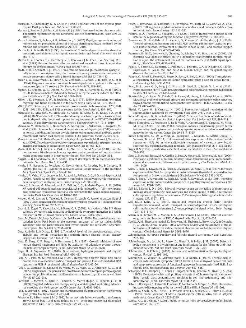
369T. Kogai, G.A. Brent / Pharmacology & Therapeutics 135 (2012) 355–370
Mansouri, A., Chowdhury, K., & Gruss, P. (1998). Follicular cells of the thyroid glandrequire Pax8 gene function. Nat Genet 19, 87–90.
Maruca, J., Santner, S., Miller, K., & Santen, R. J. (1984). Prolonged iodine clearance witha depletion regimen for thyroid carcinoma: concise communication. J Nucl Med 25,1089–1093.
Masia, S., Alvarez, S., de Lera, A. R., & Barettino, D. (2007). Rapid, nongenomic actions ofretinoic acid on phosphatidylinositol-3-kinase signaling pathway mediated by theretinoic acid receptor. Mol Endocrinol 21, 2391–2402.
Maxon, H. R., & Smith, H. S. (1990). Radioiodine-131 in the diagnosis and treatment ofmetastatic well differentiated thyroid cancer. Endocrinol Metab Clin North Am 19,685–718.
Maxon, H. R., Thomas, S. R., Hertzberg, V. S., Kereiakes, J. G., Chen, I. W., Sperling, M. I.,et al. (1983). Relation between effective radiation dose and outcome of radioiodinetherapy for thyroid cancer. N Engl J Med 309, 937–941.
Medh, R. D., & Schmidt, T. J. (1997). Trans-retinoic acid and glucocorticoids synergisti-cally induce transcription from the mouse mammary tumor virus promoter inhuman embryonic kidney cells. J Steroid Biochem Mol Biol 62, 129–142.
Meier, C. A., Braverman, L. E., Ebner, S. A., Veronikis, I., Daniels, G. H., Ross, D. S., et al.(1994). Diagnostic use of recombinant human thyrotropin in patients with thyroidcarcinoma (phase I/II study). J Clin Endocrinol Metab 78, 188–196.
Menzel, C., Kranert, W. T., Dobert, N., Diehl, M., Fietz, T., Hamscho, N., et al. (2003).rhTSH stimulation before radioiodine therapy in thyroid cancer reduces the effec-tive half-life of (131)I. J Nucl Med 44, 1065–1068.
Miller, J. K., Swanson, E. W., & Spalding, G. E. (1975). Iodine absorption, excretion,recycling, and tissue distribution in the dairy cow. J Dairy Sci 58, 1578–1593.
MIRD (1975). Summary of current radiation dose estimates to humans from 123I, 124I,125I, 126I, 130I, 131I, and 132I as sodium iodide. J Nucl Med 16, 857–860.
Mitsutake, N., Miyagishi, M., Mitsutake, S., Akeno, N., Mesa, C., Jr., Knauf, J. A., et al.(2006). BRAF mediates RET/PTC-induced mitogen-activated protein kinase activa-tion in thyroid cells: functional support for requirement of the RET/PTC-RAS-BRAFpathway in papillary thyroid carcinogenesis. Endocrinology 147, 1014–1019.
Mizukami, Y., Hashimoto, T., Nonomura, A., Michigishi, T., Nakamura, S., Noguchi, M.,et al. (1994). Immunohistochemical demonstration of thyrotropin (TSH)-receptorin normal and diseased human thyroid tissues using monoclonal antibody againstrecombinant human TSH-receptor protein. J Clin Endocrinol Metab 79, 616–619.
Montiel-Equihua, C. A., Martin-Duque, P., de la Vieja, A., Quintanilla,M., Burnet, J., Vassaux, G.,et al. (2008). Targeting sodium/iodide symporter gene expression for estrogen-regulatedimaging and therapy in breast cancer. Cancer Gene Ther 15, 465–473.
Moon, D. H., Lee, S. J., Park, K. Y., Park, K. K., Ahn, S. H., Pai, M. S., et al. (2001). Correla-tion between 99mTc-pertechnetate uptakes and expressions of human sodiumiodide symporter gene in breast tumor tissues. Nucl Med Biol 28, 829–834.
Nagpal, S., & Chandraratna, R. A. (2000). Recent developments in receptor-selectiveretinoids. Curr Pharm Des 6, 919–931.
Nicola, J. P., Basquin, C., Portulano, C., Reyna-Neyra, A., Paroder, M., & Carrasco, N.(2009). The Na+/I− symporter mediates active iodide uptake in the intestine.Am J Physiol Cell Physiol 296, C654–C662.
Nicola, J. P., Velez, M. L., Lucero, A. M., Fozzatti, L., Pellizas, C. G., & Masini-Repiso, A. M.(2009). Functional toll-like receptor 4 conferring lipopolysaccharide responsive-ness is expressed in thyroid cells. Endocrinology 150, 500–508.
Nicola, J. P., Nazar, M., Mascanfroni, I. D., Pellizas, C. G., & Masini-Repiso, A. M. (2010).NF-kappaB p65 subunit mediates lipopolysaccharide-induced Na(+)/I(−) symportergene expression by involving functional interaction with the paired domain transcrip-tion factor Pax8.Mol Endocrinol 24, 1846–1862.
Norden, M. M., Larsson, F., Tedelind, S., Carlsson, T., Lundh, C., Forssell-Aronsson, E., et al.(2007). Down-regulation of the sodium/iodide symporter explains 131I-induced thyroidstunning. Cancer Res 67, 7512–7517.
Ohashi, E., Kogai, T., Kagechika, H., & Brent, G. A. (2009). Activation of the PI3 kinasepathway by retinoic acid mediates sodium/iodide symporter induction and iodidetransport in MCF-7 breast cancer cells. Cancer Res 69, 3443–3450.
Ohno, M., Zannini, M., Levy, O., Carrasco, N., & di Lauro, R. (1999). The paired-domain tran-scription factor Pax8 binds to the upstream enhancer of the rat sodium/iodidesymporter gene and participates in both thyroid-specific and cyclic-AMP-dependenttranscription.Mol Cell Biol 19, 2051–2060.
Ohta, K., Endo, T., & Onaya, T. (1991). The mRNA levels of thyrotropin receptor, thyro-globulin and thyroid peroxidase in neoplastic human thyroid tissues. BiochemBiophys Res Commun 174, 1148–1153.
Ohta, K., Pang, X. P., Berg, L., & Hershman, J. M. (1997). Growth inhibition of newhuman thyroid carcinoma cell lines by activation of adenylate cyclase throughthe beta-adrenergic receptor. J Clin Endocrinol Metab 82, 2633–2638.
Ohye, H., & Sugawara, M. (2010). Dual oxidase, hydrogen peroxide and thyroiddiseases. Exp Biol Med (Maywood) 235, 424–433.
Pang, X. P., Park, M., & Hershman, J. M. (1992). Transforming growth factor-beta blocksprotein kinase-A-mediated iodide transport and protein kinase-C-mediated DNAsynthesis in FRTL-5 rat thyroid cells. Endocrinology 131, 45–50.
Park, J. W., Zarnegar, R., Kanauchi, H., Wong, M. G., Hyun, W. C., Ginzinger, D. G., et al.(2005). Troglitazone, the peroxisome proliferator-activated receptor-gamma agonist,induces antiproliferation and redifferentiation in human thyroid cancer cell lines.Thyroid 15, 222–231.
Peerlinck, I., Merron, A., Baril, P., Conchon, S., Martin-Duque, P., Hindorf, C., et al.(2009). Targeted radionuclide therapy using a Wnt-targeted replicating adenovi-rus encoding the Na/I symporter. Clin Cancer Res 15, 6595–6601.
Pei, L., & Melmed, S. (1997). Isolation and characterization of a pituitary tumor-transforminggene (PTTG).Mol Endocrinol 11, 433–441.
Pekary, A. E., & Hershman, J. M. (1998). Tumor necrosis factor, ceramide, transforminggrowth factor-beta1, and aging reduce Na+/I− symporter messenger ribonucleicacid levels in FRTL-5 cells. Endocrinology 139, 703–712.
Pesce, L., Bizhanova, A., Caraballo, J. C., Westphal, W., Butti, M. L., Comellas, A., et al.(2012). TSH regulates pendrin membrane abundance and enhances iodide effluxin thyroid cells. Endocrinology 153, 512–521.
Pisarev, M. A., Thomasz, L., & Juvenal, G. J. (2009). Role of transforming growth factorbeta in the regulation of thyroid function and growth. Thyroid 19, 881–892.
Pomerance, M., Abdullah, H. B., Kamerji, S., Correze, C., & Blondeau, J. P. (2000).Thyroid-stimulating hormone and cyclic AMP activate p38 mitogen-activated pro-tein kinase cascade. Involvement of protein kinase A, rac1, and reactive oxygenspecies. J Biol Chem 275, 40539–40546.
Pramanik, R., Qi, X., Borowicz, S., Choubey, D., Schultz, R. M., Han, J., et al. (2003). p38isoforms have opposite effects on AP-1-dependent transcription through regula-tion of c-Jun. The determinant roles of the isoforms in the p38 MAPK signal spec-ificity. J Biol Chem 278, 4831–4839.
Puglisi, F., Cesselli, D., Damante, G., Pellizzari, L., Beltrami, C. A., & Di Loreto, C. (2000).Expression of Pax-8, p53 and bcl-2 in human benign and malignant thyroiddiseases. Anticancer Res 20, 311–316.
Puppin, C., Arturi, F., Ferretti, E., Russo, D., Sacco, R., Tell, G., et al. (2004). Transcription-al regulation of human sodium/iodide symporter gene: a role for redox factor-1.Endocrinology 145, 1290–1293.
Read, M. L., Lewy, G. D., Fong, J. C., Sharma, N., Seed, R. I., Smith, V. E., et al. (2011).Proto-oncogene PBF/PTTG1IP regulates thyroid cell growth and represses radioiodidetreatment. Cancer Res 71, 6153–6164.
Ricarte-Filho, J. C., Ryder, M., Chitale, D. A., Rivera, M., Heguy, A., Ladanyi, M., et al. (2009).Mutational profile of advanced primary and metastatic radioactive iodine-refractorythyroid cancers reveals distinct pathogenetic roles for BRAF, PIK3CA, and AKT1. CancerRes 69, 4885–4893.
Riedel, C., Levy, O., & Carrasco, N. (2001). Post-transcriptional regulation of thesodium/iodide symporter by thyrotropin. J Biol Chem 276, 21458–21463.
Riesco-Eizaguirre, G., & Santisteban, P. (2006). A perspective view of sodium iodidesymporter research and its clinical implications. Eur J Endocrinol 155, 495–512.
Riesco-Eizaguirre, G., Rodriguez, I., De la Vieja, A., Costamagna, E., Carrasco, N., Nistal,M., et al. (2009). The BRAFV600E oncogene induces transforming growth factorbeta secretion leading to sodium iodide symporter repression and increased malig-nancy in thyroid cancer. Cancer Res 69, 8317–8325.
Riesco-Eizaguirre, G., De la Vieja, A., Rodriguez, I., Miranda, S., Martin-Duque, P.,Vassaux, G., et al. (2011). Telomerase-driven expression of the sodium iodidesymporter (NIS) for in vivo radioiodide treatment of cancer: a new broad-spectrumNIS-mediated antitumor approach. J Clin Endocrinol Metab 96, E1435–E1443.
Riggs, D. S. (1952). Quantitative aspects of iodine metabolism in man. Pharmacol Rev 4,284–370.
Saez, C., Martinez-Brocca, M. A., Castilla, C., Soto, A., Navarro, E., Tortolero, M., et al. (2006).Prognostic significance of human pituitary tumor-transforming gene immunohisto-chemical expression in differentiated thyroid cancer. J Clin Endocrinol Metab 91,1404–1409.
Saito, T., Endo, T., Kawaguchi, A., Ikeda, M., Nakazato, M., Kogai, T., et al. (1997). Increasedexpression of the Na+/I− symporter in cultured human thyroid cells exposed to thy-rotropin and in Graves' thyroid tissue. J Clin Endocrinol Metab 82, 3331–3336.
Saito, T., Endo, T., Kawaguchi, A., Ikeda, M., Katoh, R., Kawaoi, A., et al. (1998). Increasedexpression of the sodium/iodide symporter in papillary thyroid carcinomas. J ClinInvest 101, 1296–1300.
Saji, M., & Kohn, L. D. (1990). Effect of hydrocortisone on the ability of thyrotropin toincrease deoxyribonucleic acid synthesis and iodide uptake in FRTL-5 rat thyroidcells: opposite regulation of adenosine 3′,5′-monophosphate signal action. Endo-crinology 127, 1867–1876.
Saji, M., & Kohn, L. D. (1991). Insulin and insulin-like growth factor-I inhibitthyrotropin-increased iodide transport in serum-depleted FRTL-5 rat thyroidcells: modulation of adenosine 3′,5′-monophosphate signal action. Endocrinology128, 1136–1143.
Salem, A. K., Fenton, M. S., Marion, K. M., & Hershman, J. M. (2008). Effect of sunitinibon growth and function of FRTL-5 thyroid cells. Thyroid 18, 631–635.
Sawka, A. M., Thephamongkhol, K., Brouwers, M., Thabane, L., Browman, G., & Gerstein,H. C. (2004). Clinical review 170: a systematic review and metaanalysis of the ef-fectiveness of radioactive iodine remnant ablation for well-differentiated thyroidcancer. J Clin Endocrinol Metab 89, 3668–3676.
Schlumberger, M. (1998). Papillary and follicular thyroid carcinoma. N Engl J Med 338,297–306.
Schlumberger, M., Lacroix, L., Russo, D., Filetti, S., & Bidart, J. M. (2007). Defects iniodide metabolism in thyroid cancer and implications for the follow-up and treat-ment of patients. Nat Clin Pract Endocrinol Metab 3, 260–269.
Schmutzler, C., & Kohrle, J. (2000). Retinoic acid redifferentiation therapy for thyroidcancer. Thyroid 10, 393–406.
Schmutzler, C., Winzer, R., Meissner-Weigl, J., & Kohrle, J. (1997). Retinoic acid in-creases sodium/iodide symporter mRNA levels in human thyroid cancer cell linesand suppresses expression of functional symporter in nontransformed FRTL-5 ratthyroid cells. Biochem Biophys Res Commun 240, 832–838.
Schweppe, R. E., Klopper, J. P., Korch, C., Pugazhenthi, U., Benezra, M., Knauf, J. A., et al.(2008). Deoxyribonucleic acid profiling analysis of 40 human thyroid cancer celllines reveals cross-contamination resulting in cell line redundancy and mis-identification. J Clin Endocrinol Metab 93, 4331–4341.
Sebai, H., Hovsepian, S., Ristorcelli, E., Aouani, E., Lombardo, D., & Fayet, G. (2010). Resveratrolincreases iodide trapping in the rat thyroid cell line FRTL-5. Thyroid 20, 195–203.
Seibert, K., Shafie, S. M., Triche, T. J., Whang-Peng, J. J., O'Brien, S. J., Toney, J. H., et al.(1983). Clonal variation of MCF-7 breast cancer cells in vitro and in athymicnude mice. Cancer Res 43, 2223–2239.
Semba, R. D., & Delange, F. (2001). Iodine in human milk: perspectives for infant health.Nutr Rev 59, 269–278.
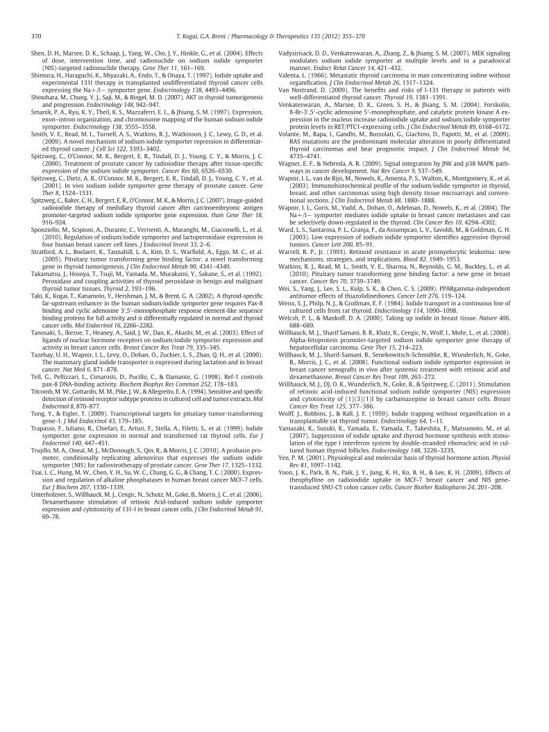
370 T. Kogai, G.A. Brent / Pharmacology & Therapeutics 135 (2012) 355–370
Shen, D. H., Marsee, D. K., Schaap, J., Yang, W., Cho, J. Y., Hinkle, G., et al. (2004). Effectsof dose, intervention time, and radionuclide on sodium iodide symporter(NIS)-targeted radionuclide therapy. Gene Ther 11, 161–169.
Shimura, H., Haraguchi, K., Miyazaki, A., Endo, T., & Onaya, T. (1997). Iodide uptake andexperimental 131I therapy in transplanted undifferentiated thyroid cancer cellsexpressing the Na+/I− symporter gene. Endocrinology 138, 4493–4496.
Shinohara, M., Chung, Y. J., Saji, M., & Ringel, M. D. (2007). AKT in thyroid tumorigenesisand progression. Endocrinology 148, 942–947.
Smanik, P. A., Ryu, K. Y., Theil, K. S., Mazzaferri, E. L., & Jhiang, S. M. (1997). Expression,exon–intron organization, and chromosome mapping of the human sodium iodidesymporter. Endocrinology 138, 3555–3558.
Smith, V. E., Read, M. L., Turnell, A. S., Watkins, R. J., Watkinson, J. C., Lewy, G. D., et al.(2009). A novel mechanism of sodium iodide symporter repression in differentiat-ed thyroid cancer. J Cell Sci 122, 3393–3402.
Spitzweg, C., O'Connor, M. K., Bergert, E. R., Tindall, D. J., Young, C. Y., & Morris, J. C.(2000). Treatment of prostate cancer by radioiodine therapy after tissue-specificexpression of the sodium iodide symporter. Cancer Res 60, 6526–6530.
Spitzweg, C., Dietz, A. B., O'Connor, M. K., Bergert, E. R., Tindall, D. J., Young, C. Y., et al.(2001). In vivo sodium iodide symporter gene therapy of prostate cancer. GeneTher 8, 1524–1531.
Spitzweg, C., Baker, C. H., Bergert, E. R., O'Connor,M. K., &Morris, J. C. (2007). Image-guidedradioiodide therapy of medullary thyroid cancer after carcinoembryonic antigenpromoter-targeted sodium iodide symporter gene expression. Hum Gene Ther 18,916–924.
Sponziello, M., Scipioni, A., Durante, C., Verrienti, A., Maranghi, M., Giacomelli, L., et al.(2010). Regulation of sodium/iodide symporter and lactoperoxidase expression infour human breast cancer cell lines. J Endocrinol Invest 33, 2–6.
Stratford, A. L., Boelaert, K., Tannahill, L. A., Kim, D. S., Warfield, A., Eggo, M. C., et al.(2005). Pituitary tumor transforming gene binding factor: a novel transforminggene in thyroid tumorigenesis. J Clin Endocrinol Metab 90, 4341–4349.
Takamatsu, J., Hosoya, T., Tsuji, M., Yamada, M., Murakami, Y., Sakane, S., et al. (1992).Peroxidase and coupling activities of thyroid peroxidase in benign and malignantthyroid tumor tissues. Thyroid 2, 193–196.
Taki, K., Kogai, T., Kanamoto, Y., Hershman, J. M., & Brent, G. A. (2002). A thyroid-specificfar-upstream enhancer in the human sodium/iodide symporter gene requires Pax-8binding and cyclic adenosine 3′,5′-monophosphate response element-like sequencebinding proteins for full activity and is differentially regulated in normal and thyroidcancer cells.Mol Endocrinol 16, 2266–2282.
Tanosaki, S., Ikezoe, T., Heaney, A., Said, J. W., Dan, K., Akashi, M., et al. (2003). Effect ofligands of nuclear hormone receptors on sodium/iodide symporter expression andactivity in breast cancer cells. Breast Cancer Res Treat 79, 335–345.
Tazebay, U. H., Wapnir, I. L., Levy, O., Dohan, O., Zuckier, L. S., Zhao, Q. H., et al. (2000).The mammary gland iodide transporter is expressed during lactation and in breastcancer. Nat Med 6, 871–878.
Tell, G., Pellizzari, L., Cimarosti, D., Pucillo, C., & Damante, G. (1998). Ref-1 controlspax-8 DNA-binding activity. Biochem Biophys Res Commun 252, 178–183.
Titcomb,M.W., Gottardis,M.M., Pike, J.W., & Allegretto, E. A. (1994). Sensitive and specificdetection of retinoid receptor subtype proteins in cultured cell and tumor extracts.MolEndocrinol 8, 870–877.
Tong, Y., & Eigler, T. (2009). Transcriptional targets for pituitary tumor-transforminggene-1. J Mol Endocrinol 43, 179–185.
Trapasso, F., Iuliano, R., Chiefari, E., Arturi, F., Stella, A., Filetti, S., et al. (1999). Iodidesymporter gene expression in normal and transformed rat thyroid cells. Eur JEndocrinol 140, 447–451.
Trujillo, M. A., Oneal, M. J., McDonough, S., Qin, R., & Morris, J. C. (2010). A probasin pro-moter, conditionally replicating adenovirus that expresses the sodium iodidesymporter (NIS) for radiovirotherapy of prostate cancer. Gene Ther 17, 1325–1332.
Tsai, L. C., Hung, M.W., Chen, Y. H., Su, W. C., Chang, G. G., & Chang, T. C. (2000). Expres-sion and regulation of alkaline phosphatases in human breast cancer MCF-7 cells.Eur J Biochem 267, 1330–1339.
Unterholzner, S., Willhauck, M. J., Cengic, N., Schutz, M., Goke, B., Morris, J. C., et al. (2006).Dexamethasone stimulation of retinoic Acid-induced sodium iodide symporterexpression and cytotoxicity of 131-I in breast cancer cells. J Clin Endocrinol Metab 91,69–78.
Vadysirisack, D. D., Venkateswaran, A., Zhang, Z., & Jhiang, S. M. (2007). MEK signalingmodulates sodium iodide symporter at multiple levels and in a paradoxicalmanner. Endocr Relat Cancer 14, 421–432.
Valenta, L. (1966). Metastatic thyroid carcinoma in man concentrating iodine withoutorganification. J Clin Endocrinol Metab 26, 1317–1324.
Van Nostrand, D. (2009). The benefits and risks of I-131 therapy in patients withwell-differentiated thyroid cancer. Thyroid 19, 1381–1391.
Venkateswaran, A., Marsee, D. K., Green, S. H., & Jhiang, S. M. (2004). Forskolin,8-Br-3′,5′-cyclic adenosine 5′-monophosphate, and catalytic protein kinase A ex-pression in the nucleus increase radioiodide uptake and sodium/iodide symporterprotein levels in RET/PTC1-expressing cells. J Clin Endocrinol Metab 89, 6168–6172.
Volante, M., Rapa, I., Gandhi, M., Bussolati, G., Giachino, D., Papotti, M., et al. (2009).RAS mutations are the predominant molecular alteration in poorly differentiatedthyroid carcinomas and bear prognostic impact. J Clin Endocrinol Metab 94,4735–4741.
Wagner, E. F., & Nebreda, A. R. (2009). Signal integration by JNK and p38 MAPK path-ways in cancer development. Nat Rev Cancer 9, 537–549.
Wapnir, I. L., van de Rijn, M., Nowels, K., Amenta, P. S., Walton, K., Montgomery, K., et al.(2003). Immunohistochemical profile of the sodium/iodide symporter in thyroid,breast, and other carcinomas using high density tissue microarrays and conven-tional sections. J Clin Endocrinol Metab 88, 1880–1888.
Wapnir, I. L., Goris, M., Yudd, A., Dohan, O., Adelman, D., Nowels, K., et al. (2004). TheNa+/I− symporter mediates iodide uptake in breast cancer metastases and canbe selectively down-regulated in the thyroid. Clin Cancer Res 10, 4294–4302.
Ward, L. S., Santarosa, P. L., Granja, F., da Assumpcao, L. V., Savoldi, M., & Goldman, G. H.(2003). Low expression of sodium iodide symporter identifies aggressive thyroidtumors. Cancer Lett 200, 85–91.
Warrell, R. P., Jr. (1993). Retinoid resistance in acute promyelocytic leukemia: newmechanisms, strategies, and implications. Blood 82, 1949–1953.
Watkins, R. J., Read, M. L., Smith, V. E., Sharma, N., Reynolds, G. M., Buckley, L., et al.(2010). Pituitary tumor transforming gene binding factor: a new gene in breastcancer. Cancer Res 70, 3739–3749.
Wei, S., Yang, J., Lee, S. L., Kulp, S. K., & Chen, C. S. (2009). PPARgamma-independentantitumor effects of thiazolidinediones. Cancer Lett 276, 119–124.
Weiss, S. J., Philp, N. J., & Grollman, E. F. (1984). Iodide transport in a continuous line ofcultured cells from rat thyroid. Endocrinology 114, 1090–1098.
Welcsh, P. L., & Mankoff, D. A. (2000). Taking up iodide in breast tissue. Nature 406,688–689.
Willhauck, M. J., Sharif Samani, B. R., Klutz, K., Cengic, N., Wolf, I., Mohr, L., et al. (2008).Alpha-fetoprotein promoter-targeted sodium iodide symporter gene therapy ofhepatocellular carcinoma. Gene Ther 15, 214–223.
Willhauck, M. J., Sharif-Samani, B., Senekowitsch-Schmidtke, R., Wunderlich, N., Goke,B., Morris, J. C., et al. (2008). Functional sodium iodide symporter expression inbreast cancer xenografts in vivo after systemic treatment with retinoic acid anddexamethasone. Breast Cancer Res Treat 109, 263–272.
Willhauck, M. J., DJ, O. K., Wunderlich, N., Goke, B., & Spitzweg, C. (2011). Stimulationof retinoic acid-induced functional sodium iodide symporter (NIS) expressionand cytotoxicity of (1)(3)(1)I by carbamazepine in breast cancer cells. BreastCancer Res Treat 125, 377–386.
Wolff, J., Robbins, J., & Rall, J. E. (1959). Iodide trapping without organification in atransplantable rat thyroid tumor. Endocrinology 64, 1–11.
Yamazaki, K., Suzuki, K., Yamada, E., Yamada, T., Takeshita, F., Matsumoto, M., et al.(2007). Suppression of iodide uptake and thyroid hormone synthesis with stimu-lation of the type I interferon system by double-stranded ribonucleic acid in cul-tured human thyroid follicles. Endocrinology 148, 3226–3235.
Yen, P. M. (2001). Physiological and molecular basis of thyroid hormone action. PhysiolRev 81, 1097–1142.
Yoon, J. K., Park, B. N., Paik, J. Y., Jung, K. H., Ko, B. H., & Lee, K. H. (2009). Effects oftheophylline on radioiodide uptake in MCF-7 breast cancer and NIS gene-transduced SNU-C5 colon cancer cells. Cancer Biother Radiopharm 24, 201–208.