the soluble form of a disintegrin and metalloprotease 33 promotes angiogenesis: implications for...
TRANSCRIPT
The soluble form of a disintegrin and metalloprotease 33promotes angiogenesis: Implications for airway remodelingin asthma
Ilaria Puxeddu, MD, PhD,a* Yun Yun Pang, PhD,a* Anna Harvey, BSc,a Hans Michael Haitchi, MD,a Ben Nicholas, PhD,a
Hajime Yoshisue, PhD,a Domenico Ribatti, MD,c Geraldine Clough, PhD,a Rob M. Powell, PhD,a Gillian Murphy, PhD,d
Neil A. Hanley, MD,b David I. Wilson, MD,b Peter H. Howarth, MD,a Stephen T. Holgate, DSc,a and Donna E. Davies, PhDa
Southampton and Cambridge, United Kingdom, and Bari, Italy
Background: A disintegrin and metalloprotease (ADAM)–33is a susceptibility gene for asthma and chronic obstructivepulmonary disease whose function remains unknown.Objective: Because asthmatic bronchoalveolar lavage fluidcontains high levels of soluble ADAM33 (sADAM33), whichincludes the catalytic domain, we postulated that its releasefrom cell membranes might play functional roles in airwayremodeling by promoting angiogenesis.Methods: The proangiogenic activity of the highly purifiedcatalytic domain of ADAM33 or a catalytically inactive mutantwas studied in vitro (Matrigel assay), ex vivo (human embryonic/fetal lung explants) and in vivo (chorioallantoic membraneassay). The regulation of sADAM33 release from cellsoverexpressing full-length ADAM33 and its biological activitywere characterized.Results: We show that the purified catalytic domain ofADAM33, but not its inactive mutant, causes rapid induction ofendothelial cell differentiation in vitro, and neovascularizationex vivo and in vivo. We also show that TGF-b2 enhancessADAM33 release from cells overexpressing full-lengthADAM33 and that this truncated form is biologically active.Conclusion: The discovery that sADAM33 promotesangiogenesis defines it as a tissue remodeling gene with potentialto affect airflow obstruction and lung function independently ofinflammation. As TGF-b2 enhances sADAM33 release,environmental factors that cause epithelial damage maysynergize with ADAM33 in asthma pathogenesis, resulting in a
From the Divisions of aInfection, Inflammation, and Repair and bHuman Genetics,
School of Medicine, Southampton General Hospital, University of Southampton;cthe Department of Anatomy, Faculty of Medicine, University of Bari; and dthe De-
partment of Oncology, University of Cambridge.
*These authors contributed equally to this article.
Supported by the Medical Research Council UK; the Rayne Foundation; the British Lung
Foundation; the Asthma, Allergy, and Inflammation Research Charity; the Roger
Brooke Charitable Trust; and the Wellcome Trust. S.T.H. is supported by a Medical
Research Council Clinical Professorship. I.P. was supported by a European Respira-
tory Society Short Term Traveling Fellowship and a European Academy of Allergy
and Clinical Immunology Fellowship.
Disclosure of potential conflict of interest: G. Clough has received research support from
the British Heart Foundation, Unilever, and Probe Scientific UK. N. Hanley has
received research support from Novo Nordisk. D. I. Wilson has received research
support from the British Heart Foundation and the Department of Health UK. The rest
of the authors have declared that they have no conflict of interest.
Received for publication November 2, 2007; revised February 12, 2008; accepted for
publication March 5, 2008.
Available online April 14, 2008.
Reprint requests: Donna E. Davies, PhD, Mailpoint 810, Southampton General Hospital,
Southampton, SO16 6YD, United Kingdom. E-mail: [email protected].
0091-6749/$34.00
� 2008 American Academy of Allergy, Asthma & Immunology
doi:10.1016/j.jaci.2008.03.003
1400
disease-related gain of function. This highlights the potential forinterplay between genetic and environmental factors in thiscomplex disease. (J Allergy Clin Immunol 2008;121:1400-6.)
Key words: ADAM33, asthma, angiogenesis, airway remodeling
A disintegrin and metalloprotease (ADAM)–33 is an asthmasusceptibility gene whose polymorphic variation has been linkedto asthma and bronchial hyperresponsiveness, but not to indices ofallergic inflammation.1 These findings have been replicated inseveral other studies, including a meta-analysis2 that includedall literature-reported studies, including those that have shownweak or no association. ADAM33 polymorphism also predicts im-paired lung function in young children,3 suggesting that ADAM33may contribute to the early-life origins of asthma. A similar asso-ciation of ADAM33 with impaired lung function has also beenreported in asthma,4 the general population,5 and chronic obstruc-tive pulmonary disease (COPD).6
ADAM33 belongs to the a disintegrin and metalloproteasefamily of membrane-anchored glycoproteins that play diverseroles in cell surface remodeling, ectodomain shedding of growthfactors and receptors, and mediating cell-cell and cell-matrixinteractions.7 It is made up of a full-length molecule of 813 aminoacids including 7 domains: pro-, metalloprotease, disintegrin,cysteine-rich, epidermal growth factor, transmembrane, and cyto-plasmic, serving the functions of activation, proteolysis, adhesion,fusion, and signaling.1 Since its discovery in 2002, ADAM33 hasbeen extensively characterized at molecular and structurallevels,8-12 but its biological role and its contribution to asthmapathogenesis have remained elusive.
Expression of ADAM33 mRNA and protein is restricted tomesenchymal cells, which include airway fibroblasts, myofibro-blasts, and smooth muscle.1,13,14 In bronchial biopsies from nor-mal and asthmatic airways, ADAM33 colocalizes to smoothmuscle and fibroblastlike cells in the submucosa, supporting itslink with bronchial hyperresponsiveness and airway remodeling,but no disease-related differences in total levels of expressionwere found.13 In human embryonic lung, ADAM33 is expressedin mesenchymal progenitor cells,13 suggesting a potential role inlung development. A number of alternatively spliced mRNA tran-scripts of ADAM33 have been detected in bronchial biopsies13
and mesenchymal cells,14 but again, no disease-related differencesin splicing have been detected. These disease-derived findingscontrast with the suggestion that the disease associated intronicsingle nucleotide polymorphism (SNP) between exons B and C(BC+1), which has a marked repressive effect on ADAM33 ex-pression in an in vitro reporter gene system, might affect pre-mRNA alternative splicing in asthma.15 Although high levelsof ADAM33 and aberrant expression of ADAM33 in airway

J ALLERGY CLIN IMMUNOL
VOLUME 121, NUMBER 6
PUXEDDU ET AL 1401
Abbreviations used
ADAM: A disintegrin and metalloprotease
BALF: Bronchoalveolar lavage fluid
CAM: Chick embryo chorioallantoic membrane
CHO: Chinese hamster ovary
COPD: Chronic obstructive pulmonary disease
FGF: Fibroblast growth factor
HEK: Human embryonic kidney
HUVEC: Human umbilical vein endothelial cell
SNP: Single nucleotide polymorphism
sADAM33: Soluble a disintegrin and metalloprotease 33
UK: United Kingdom
VEGF: Vascular endothelial growth factor
epithelium of patients with severe asthma have been suggested asa possible pathogenetic mechanism,16 we have not been able toreproduce these findings.17 ADAM33 knockout mice have alsofailed to yield any functional clues, showing normal growth anddevelopment and no abnormalities in a model of allergic airwaysensitization.18 In contrast with these negative findings, the occur-rence of high levels of a 55-kDa soluble form of ADAM33 (sA-DAM33) in bronchoalveolar lavage fluid (BALF) of subjectswith asthma, detected using an antibody to the ADAM33 metal-loprotease domain, and its correlation with disease severity andreduced lung function19 are one of the few pieces of nongeneticevidence showing a disease-related effect.
In children and adults, angiogenesis is believed to be animportant aspect of asthma pathogenesis. Increased expressionof angiogenic mediators and their receptors has been correlatedwith disease severity and accelerated lung function decline.20-22
In the current study, we hypothesized that loss of the membraneanchor and regulatory cytoplasmic domain of ADAM33 resultsin a disease-related gain of function whereby the aberrant locali-zation of the ADAM33 metalloprotease results in inappropriatecleavage of substrates to cause angiogenesis. In vivo, endothelialcells are supported by a vascular basement membrane thatoverlies layers of mesenchymal cells such as pericytes, fibro-blasts, and smooth muscle cells. Because endothelial cells donot express ADAM33,1 we postulated that their proximity to themesenchymal cells that do express ADAM331,13,14 would makethem a target for sADAM33. Here, we describe the produc-tion and regulation of sADAM33 and its ability to promoteangiogenesis.
METHODS
Recombinant cells expressing ADAM33 and
purification of ADAM33 proteinsRecombinant human embryonic kidney (HEK)–293 and Chinese hamster
ovary (CHO) cells were generated by transfection with either full-length
ADAM33 cDNA or ADAM33 signal, Pro-, and metalloprotease domains
fused to a C-terminal Fc tag; empty pcDNA3 vector or Signal pIg plus (R&D
Systems, Abingdon, United Kingdom [UK]) was used as control. Drosophila
S2 cells were cotransfected by using the pCoHygro vector and pMT/Bip/V5-
His vector (Invitrogen, Carlsbad, Calif) containing the ADAM33 Pro- and
metalloprotease domains (residues 31-409) fused to a V5 and 6xHis tag.
The E346A mutant was prepared by site-directed mutagenesis. ADAM33 ex-
pression was confirmed by SDS-PAGE and Western blotting using antibodies
to the Pro-, metalloprotease, and cytoplasmic domains of ADAM33 (Abcam
plc, Cambridge, UK). Conditioned medium from S2 cells was harvested
and the ADAM33 Pro-metalloprotease purified through a series of
chromatographic steps involving lectin affinity chromatography, immobilized
metal ion affinity chromatography, cation exchange, and gel filtration.
Concanavalin A-Sepharose 4B beads (Sigma, Poole, UK) were used to pull
down sADAM33 from the supernatants of recombinant HEK293 cells and
BALF obtained by fiberoptic bronchoscopy, as described in this article’s
Online Repository at www.jacionline.org. The bound proteins were either lysed
into 1x SDS sample buffer for SDS-PAGE and Western blotting or eluted with
0.5 mol/L methyl a-D-mannopyranoside for analysis of protease activity.
ADAM33 biochemical activityA fluorescence resonance energy transfer peptide cleavage assay based on
that described by Zou et al9 was used to assess the enzyme activity of
ADAM33. Synthetic inhibitors of matrix metalloproteases were tested in
this assay for their potency against the catalytic activity of ADAM33.
In vitro and in vivo angiogenesis assaysHuman umbilical vein endothelial cells (HUVECs) were isolated from
umbilical cords collected after ethical approval and informed consent.
Endothelial cells were plated into wells precoated with growth factor–reduced
Matrigel (BD Biosciences, Oxford, UK) and incubated with recombinant cell
culture supernatants (minus G418) or ADAM33 proteins for 18 hours. The
3-dimensional organization of the endothelial cells was recorded and topolog-
ical parameters measured by computer-aided image analysis using QWin
image analysis software (Leica Microsystems Imaging Solutions Ltd,
Cambridge, UK).
The chick embryo chorioallantoic membrane (CAM) assay was performed
as previously described.23 CAMs were treated after 8 days of culture with
ADAM33 Pro-metalloprotease, ADAM Pro-metalloprotease E346A, or re-
combinant CHO cells contained in 1-mm3 sterilized gelatin sponges (Gel-
foam; Upjohn Co, Kalamazoo, Mich). CAMs were examined daily until day
12 and photographed in ovo. Blood vessels entering the sponge within the fo-
cal plane of the CAM were counted by 2 observers in a double-blind fashion at
350 magnification.
Human embryonic/fetal lung explant culturesHuman embryonic lungs were harvested according to the Polkinghorne
Committee guidelines and after ethical approval and informed consent from
the donor. They were dissected into pieces 1 to 2 mm and cultured in transwells
in Matrigel in the presence of 60 ng/well ADAM33 Pro-metalloprotease or
ADAM33 Pro-metalloprotease E346A for 12 days, replenishing media and
ADAM33 protein every second day. The tissue was harvested and processed
for immunohistochemistry with the antibody EN-4 recognizing the vessel
marker platelet endothelial cell adhesion molecule 1 (PECAM-1). All sections
were examined by 3 blind investigators and expression-quantified by com-
puter-assisted image analysis.
Statistical analysisParametric ANOVA, followed by the Tukey-Kramer multiple comparisons
test, was used to compare groups in the in vitro and in vivo experiments. Non-
parametric ANOVA followed by Kruskal-Wallis test was used to compare
groups in the ex vivo experiments.
Online supplemental materialFurther details of each protocol can be found in this article’s Online
Repository.
RESULTS
The ADAM33 metalloprotease domain
is proangiogenic in vitroAngiogenesis is a process that critically involves proteolysis.24
Because sADAM33 is made up of several functional domains,we tested whether the metalloprotease domain specifically was
J ALLERGY CLIN IMMUNOL
JUNE 2008
1402 PUXEDDU ET AL
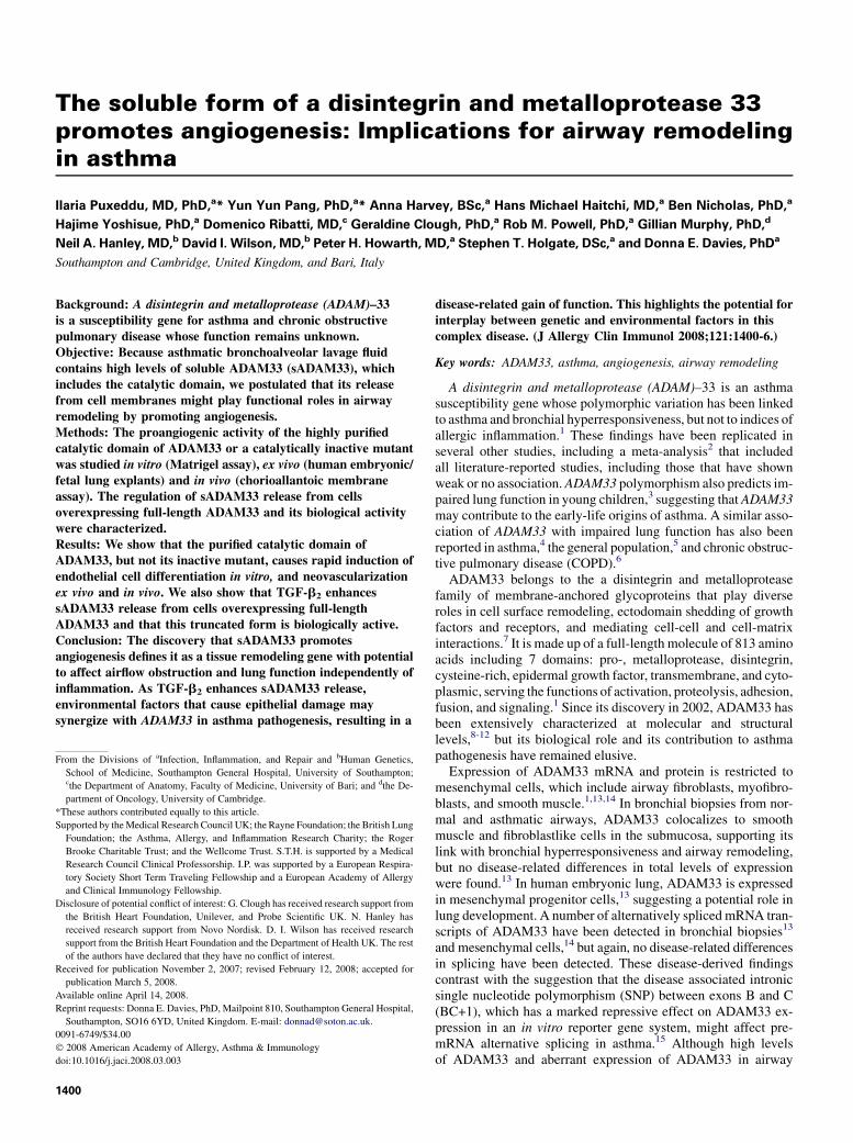
able to promote angiogenesis. When supernatants derived fromCHO cells transfected with cDNA encoding the ADAM33 Pro-and metalloprotease domains with an Fc tag were tested in theMatrigel assay, they promoted endothelial cell differentiation, asassessed by an increase in both the number of branching points(597 6 40 vs 336 6 30; P < .05; n 5 3) and meshes (20 6 3 vs13 6 0.5; P < .05; n 5 3) per field compared with control superna-tants from pcDNA3.1 mock-transfected cells. Supernatant fromSignal pIg plus mock-transfected cells expressing an Ig Fc frag-ment gave comparable results to the supernatant from pcDNA3.1mock-transfected cells. These findings suggested that theADAM33 metalloprotease domain possessed angiogenic potential.
FIG 1. ADAM33 promotes angiogenesis in vitro. HUVEC differentiation
was tested with buffer (A, i, B, C, white bars), ADAM33 Pro-metalloprotease
(MP) (A, ii, B, C, black bars), ADAM33 Pro-MP E346A (A, iii, B, C, light gray
bars), or FGF-2 (A, iv, C, dark gray bars). In C, the effect of (3R)-(1)-[2-(4-me-
thoxybenzenesulfonyl)-1,2,3,4-tetrahydroisoquinoline-3-hydroxamate (5 mmol/
L, hatched bars) was tested. Topological parameters of capillary structure
(branch points and meshes) were quantified by computer-aided image analysis
(B, C). Data are means 6 SDs; n 5 3; *P < .05; **P < .01.
Because the CHO ADAM33 Pro-metalloprotease cells did notproduce enough recombinant protein to allow their purificationfor further functional testing, we used an insect cell expressionsystem to generate large quantities of enzymatically activeADAM33 Pro-metalloprotease and an inactive mutant E346Aprotein (ADAM33 Pro-metalloprotease E346A) targeting thecatalytic residue in the active site. Isolation of the recombinantproteins was achieved by multistep chromatography yieldinghighly purified preparations with the anticipated enzymaticactivities (see this article’s Fig E1 in the Online Repository atwww.jacionline.org). As was found with the crude supernatantsfrom ADAM33-expressing cells, purified ADAM33 Pro-metallo-protease protein stimulated endothelial cell differentiation andformation of tubelike structures on Matrigel (Fig 1, A, ii vs i).This effect was similar to the effect of fibroblast growth factor(FGF)–2 (Fig 1, A, iv) but was not reproduced by ADAM33Pro-metalloprotease E346A (Fig 1, A, iii). The effect ofADAM33 Pro-metalloprotease was dose-dependent, as deter-mined by assessment of branch points and meshes usingcomputer-aided image analysis (Fig 1, B). As ADAM33 Pro-met-alloprotease E346Awas inactive, it not only controlled for any ef-fects of the V5 and 6xHis tags that had been engineered into theprotein to aid purification but also demonstrated that the angio-genic activity of ADAM33 Pro-metalloprotease was unlikely tobe a result of a copurified contaminant. Because these data sug-gested that the enzymatic activity of ADAM33 was required forthe angiogenic effect, 4 synthetic carboxylate and hydroxamatecompounds that are known inhibitors of metalloproteases25 weretested for their ability to inhibit ADAM33 Pro-metalloprotease.These were found to exhibit different potencies againstADAM33-mediated peptide cleavage activity, with (3R)-(1)-[2-(4-methoxybenzenesulfonyl)-1,2,3,4-tetrahydroisoquinoline-3-hydroxamate being the most potent (inhibitory concentration 50%[IC50] of 330 nmol/L; Fig E1, D). When tested in the Matrigel as-say, this inhibitor was also able to abrogate the proangiogenic effectof ADAM33 Pro-metalloprotease (Fig 1, C). Although this inhibi-tor is also known to inhibit matrix metalloprotease 8,25 it did notaffect the biological potency of FGF-2. This suggested that the in-hibitor was selective for ADAM33 in our assays, although we can-not completely exclude the possibility that it affected anotherprotease involved in the response to ADAM33.
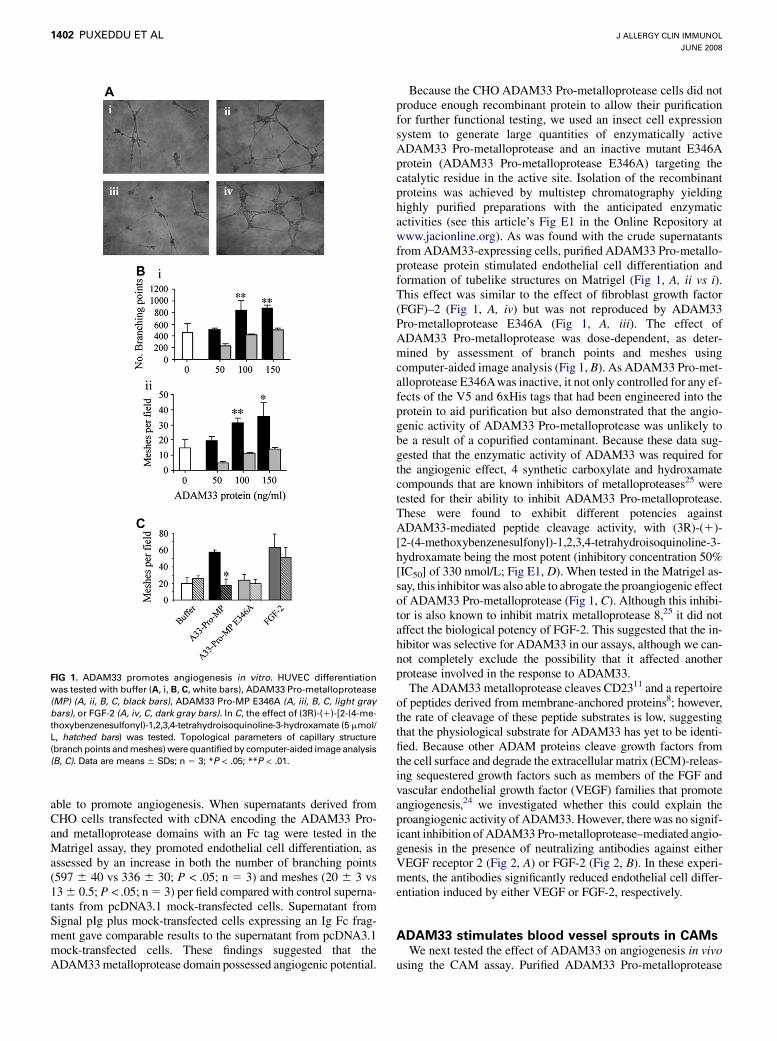
The ADAM33 metalloprotease cleaves CD2311 and a repertoireof peptides derived from membrane-anchored proteins8; however,the rate of cleavage of these peptide substrates is low, suggestingthat the physiological substrate for ADAM33 has yet to be identi-fied. Because other ADAM proteins cleave growth factors fromthe cell surface and degrade the extracellular matrix (ECM)-releas-ing sequestered growth factors such as members of the FGF andvascular endothelial growth factor (VEGF) families that promoteangiogenesis,24 we investigated whether this could explain theproangiogenic activity of ADAM33. However, there was no signif-icant inhibition of ADAM33 Pro-metalloprotease–mediated angio-genesis in the presence of neutralizing antibodies against eitherVEGF receptor 2 (Fig 2, A) or FGF-2 (Fig 2, B). In these experi-ments, the antibodies significantly reduced endothelial cell differ-entiation induced by either VEGF or FGF-2, respectively.
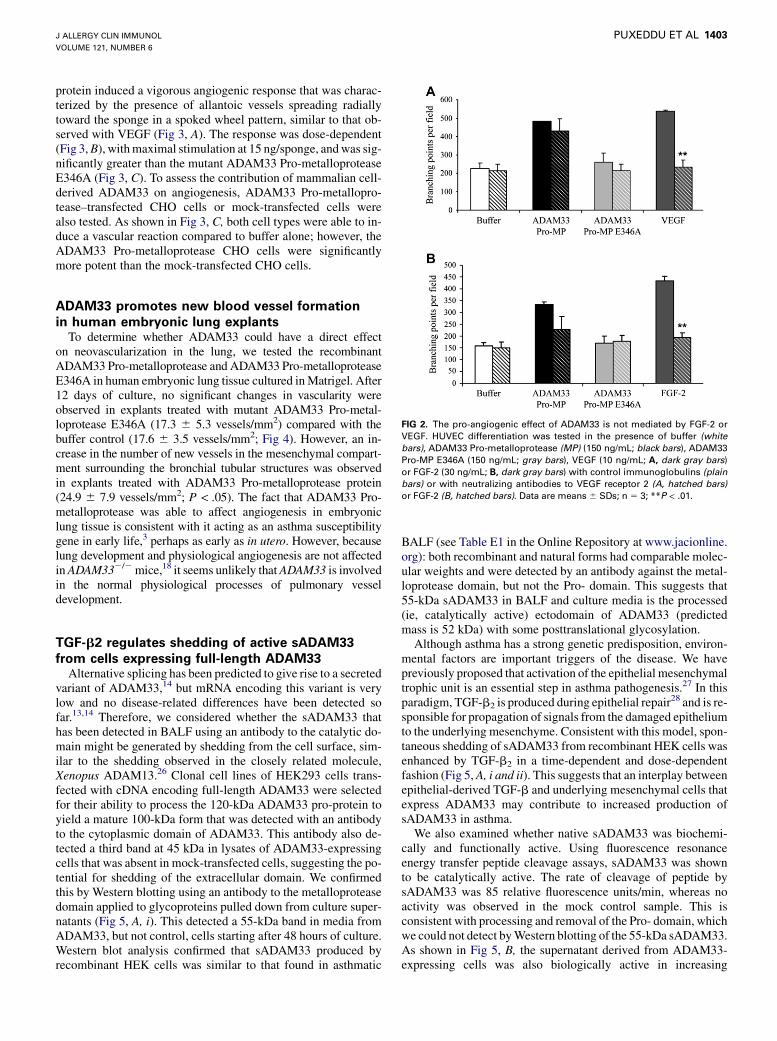
ADAM33 stimulates blood vessel sprouts in CAMsWe next tested the effect of ADAM33 on angiogenesis in vivo
using the CAM assay. Purified ADAM33 Pro-metalloprotease
J ALLERGY CLIN IMMUNOL
VOLUME 121, NUMBER 6
PUXEDDU ET AL 1403
protein induced a vigorous angiogenic response that was charac-terized by the presence of allantoic vessels spreading radiallytoward the sponge in a spoked wheel pattern, similar to that ob-served with VEGF (Fig 3, A). The response was dose-dependent(Fig 3, B), with maximal stimulation at 15 ng/sponge, and was sig-nificantly greater than the mutant ADAM33 Pro-metalloproteaseE346A (Fig 3, C). To assess the contribution of mammalian cell-derived ADAM33 on angiogenesis, ADAM33 Pro-metallopro-tease–transfected CHO cells or mock-transfected cells werealso tested. As shown in Fig 3, C, both cell types were able to in-duce a vascular reaction compared to buffer alone; however, theADAM33 Pro-metalloprotease CHO cells were significantlymore potent than the mock-transfected CHO cells.
ADAM33 promotes new blood vessel formation
in human embryonic lung explantsTo determine whether ADAM33 could have a direct effect
on neovascularization in the lung, we tested the recombinantADAM33 Pro-metalloprotease and ADAM33 Pro-metalloproteaseE346A in human embryonic lung tissue cultured in Matrigel. After12 days of culture, no significant changes in vascularity wereobserved in explants treated with mutant ADAM33 Pro-metal-loprotease E346A (17.3 6 5.3 vessels/mm2) compared with thebuffer control (17.6 6 3.5 vessels/mm2; Fig 4). However, an in-crease in the number of new vessels in the mesenchymal compart-ment surrounding the bronchial tubular structures was observedin explants treated with ADAM33 Pro-metalloprotease protein(24.9 6 7.9 vessels/mm2; P < .05). The fact that ADAM33 Pro-metalloprotease was able to affect angiogenesis in embryoniclung tissue is consistent with it acting as an asthma susceptibilitygene in early life,3 perhaps as early as in utero. However, becauselung development and physiological angiogenesis are not affectedin ADAM332/2 mice,18 it seems unlikely that ADAM33 is involvedin the normal physiological processes of pulmonary vesseldevelopment.
TGF-b2 regulates shedding of active sADAM33
from cells expressing full-length ADAM33Alternative splicing has been predicted to give rise to a secreted
variant of ADAM33,14 but mRNA encoding this variant is verylow and no disease-related differences have been detected sofar.13,14 Therefore, we considered whether the sADAM33 thathas been detected in BALF using an antibody to the catalytic do-main might be generated by shedding from the cell surface, sim-ilar to the shedding observed in the closely related molecule,Xenopus ADAM13.26 Clonal cell lines of HEK293 cells trans-fected with cDNA encoding full-length ADAM33 were selectedfor their ability to process the 120-kDa ADAM33 pro-protein toyield a mature 100-kDa form that was detected with an antibodyto the cytoplasmic domain of ADAM33. This antibody also de-tected a third band at 45 kDa in lysates of ADAM33-expressingcells that was absent in mock-transfected cells, suggesting the po-tential for shedding of the extracellular domain. We confirmedthis by Western blotting using an antibody to the metalloproteasedomain applied to glycoproteins pulled down from culture super-natants (Fig 5, A, i). This detected a 55-kDa band in media fromADAM33, but not control, cells starting after 48 hours of culture.Western blot analysis confirmed that sADAM33 produced byrecombinant HEK cells was similar to that found in asthmatic
BALF (see Table E1 in the Online Repository at www.jacionline.org): both recombinant and natural forms had comparable molec-ular weights and were detected by an antibody against the metal-loprotease domain, but not the Pro- domain. This suggests that55-kDa sADAM33 in BALF and culture media is the processed(ie, catalytically active) ectodomain of ADAM33 (predictedmass is 52 kDa) with some posttranslational glycosylation.
Although asthma has a strong genetic predisposition, environ-mental factors are important triggers of the disease. We havepreviously proposed that activation of the epithelial mesenchymaltrophic unit is an essential step in asthma pathogenesis.27 In thisparadigm, TGF-b2 is produced during epithelial repair28 and is re-sponsible for propagation of signals from the damaged epitheliumto the underlying mesenchyme. Consistent with this model, spon-taneous shedding of sADAM33 from recombinant HEK cells wasenhanced by TGF-b2 in a time-dependent and dose-dependentfashion (Fig 5, A, i and ii). This suggests that an interplay betweenepithelial-derived TGF-b and underlying mesenchymal cells thatexpress ADAM33 may contribute to increased production ofsADAM33 in asthma.
We also examined whether native sADAM33 was biochemi-cally and functionally active. Using fluorescence resonanceenergy transfer peptide cleavage assays, sADAM33 was shownto be catalytically active. The rate of cleavage of peptide bysADAM33 was 85 relative fluorescence units/min, whereas noactivity was observed in the mock control sample. This isconsistent with processing and removal of the Pro- domain, whichwe could not detect by Western blotting of the 55-kDa sADAM33.As shown in Fig 5, B, the supernatant derived from ADAM33-expressing cells was also biologically active in increasing
FIG 2. The pro-angiogenic effect of ADAM33 is not mediated by FGF-2 or
VEGF. HUVEC differentiation was tested in the presence of buffer (white
bars), ADAM33 Pro-metalloprotease (MP) (150 ng/mL; black bars), ADAM33
Pro-MP E346A (150 ng/mL; gray bars), VEGF (10 ng/mL; A, dark gray bars)
or FGF-2 (30 ng/mL; B, dark gray bars) with control immunoglobulins (plain
bars) or with neutralizing antibodies to VEGF receptor 2 (A, hatched bars)
or FGF-2 (B, hatched bars). Data are means 6 SDs; n 5 3; **P < .01.
J ALLERGY CLIN IMMUNOL
JUNE 2008
1404 PUXEDDU ET AL
FIG 3. ADAM33 promotes angiogenesis in vivo. A, Photomicrographs of CAMs, treated with buffer,
ADAM33 Pro-metalloprotease (MP) (45 ng/sponge); ADAM Pro-MP E346A (45 ng/sponge); or VEGF (500
ng/sponge). B, Dose-response for the angiogenic effect of ADAM33 Pro-MP, and C, comparison of the
effects of ADAM33 Pro-MP (15 ng/sponge) with ADAM33 Pro-MP E346A (15 ng/sponge), ADAM33
Pro-MP–expressing CHO cells, mock-transfected cells (each with 40,000 cells/sponge), or VEGF (500 ng/
sponge). Data are means 6 SDs from 8 CAMs per condition, from 2 independent experiments.
FIG 4. ADAM33 promotes angiogenesis in lung tissue. Human embryonic lung pieces (8 wk postconcep-
tion) were cultured with buffer alone, 60 ng/well ADAM33 Pro-metalloprotease (MP) E346A or ADAM33
Pro-MP for 12 days. The lungs were fixed and stained for the endothelium marker, PECAM-1, using immu-
nohistochemistry. The arrows indicate the presence of vessels showing both positive brown immunostain-
ing and an identifiable lumen. Scale bar 5 100 mm.
endothelial cell differentiation into capillary-like structures inMatrigel compared with the supernatants from the mock-trans-fected cells.
DISCUSSIONAlthough several factors are involved in airway vascular
remodeling,29 the ability of sADAM33 to promote angiogenesisis consistent with the increased vascularity observed in asth-matic airways and its correlation with disease severity.21 Al-though not as well studied, there is evidence that the vasculararea of the airway is also significantly increased in COPD andcorrelates with the degree of airflow obstruction.22 Angiogenesis
is fundamental for supporting many aspects of tissue inflamma-tion and remodelling.30 During angiogenesis, blood vesselsenlarge and increase in number, enhancing the production ofinflammatory mediators, augmenting and sustaining cellular in-filtration, and contributing to edema through microvascular leak-age. The new vessels can also supply nutrients and oxygen to theinflamed tissue, facilitating proliferation of structural cells suchas smooth muscle cells, fibroblasts, and myofibroblasts. Thus,increased angiogenesis has the potential to contribute to abnor-mal airway function including fixed airflow obstruction andconsequent accelerated lung function decline through directand indirect mechanisms.

J ALLERGY CLIN IMMUNOL
VOLUME 121, NUMBER 6
PUXEDDU ET AL 1405
FIG 5. Characterization of sADAM33. A, Culture supernatants from control or TGF-b2–treated HEK293-
expressing full-length recombinant ADAM33 (A33) or empty vector (Mock) were analyzed for sADAM33
by Western blotting (left panel) and the 55-kDa sADAM33 band quantified by densitometry (right panel);
data are representative of 3 individual experiments. B, Culture supernatants from recombinant ADAM33
or mock-transfected cells were tested for their ability to promote HUVEC differentiation in vitro (left panel)
and branching points quantified (right panel). Data are means 6 SDs (n 5 3 experiments).
Proteolysis has been recognized as one of most important activitiesduring angiogenesis. Members of the matrix metalloprotease,ADAM and ADAMTS (a disintegrin and metalloproteasedomain with thrombospondin motifs) families, as well as cys-teine and serine proteases, have been implicated in this regu-lation.24 These enzymes mediate their effect by degradation ofthe endothelial basement membrane and extracellular matrixproteins; release of angiogenic factors; processing of cyto-kines, growth factors, and growth factor receptors; and theproduction of endogenous inhibitors. Although it is likelythat the substrate for ADAM33 falls into one of these cate-gories, we have shown that its effect is not mediated by re-lease of VEGF or FGF-2. Even though both of these factorsare strong angiogenic mediators, it is noteworthy that the po-tency of sADAM33 in the CAM assay exceeded that ofVEGF, whereas it was quantitatively less potent than eitherFGF-2 or VEGF in the endothelial tube formation assay. Al-though many factors can influence the outcomes of the indi-vidual assays (eg, human vs chicken receptors), it is possiblethat the more complex CAM assay contained better substratesfor sADAM33. It is also important to note that most of ourstudies were performed with the purified metalloprotease do-main without the more proximal disintegrin and cysteine-richdomains. Because these domains are involved in substrate pre-sentation for other ADAMs,31,32 the activity of the purifiedcatalytic domain may be an underestimate of the true activityof shed ectodomain of ADAM33 present in BALF.
Our results showing TGF-b–mediated ectodomain shedding ofADAM33 led us to propose that loss of its membrane anchorallows ADAM33 to gain the capacity to contribute to patholog-ical neovascularization. Because the proangiogenic effect of
sADAM33 is dependent on its catalytic activity, our data furthersuggest that therapeutic interventions that are able to targetspecifically the ADAM33 metalloprotease may be effectivedisease modifiers that can affect the natural history of the disease.ADAM15, a close relative of ADAM33, has also been shown topromote angiogenesis.24 However, unlike ADAM15, ADAM33 isnot expressed by endothelial cells,1 even though it can act on themto promote angiogenesis. In this respect, the gain of functionachieved by sADAM33 may enable it to mimic the proangiogenicproperties of endothelium-derived ADAM15. This gain of func-tion may explain why there was no obvious angiogenic phenotypein ADAM332/2 mice (ie, loss of function), even after antigensensitization and airway challenge.18
Identification of a function for ADAM33 and the involvementof TGF-b in ectodomain shedding of ADAM33 should pave theway for identifying how polymorphic variation in the ADAM33gene is linked to asthma and COPD pathogenesis. Although thereare a large number of SNPs in ADAM33, 4 SNPs (S1, S2, T1, andT2) encoding amino acid substitutions in the transmembrane andcytoplasmic tail of ADAM33 have been associated with asthma.1
It is conceivable that these SNPs may potentiate trafficking ofADAM33 to the cell surface and/or modulate its ability to beshed from the cell surface in a TGF-b–dependent manner. Alter-natively, the ADAM33 locus that is associated with reduced lungfunction in young children lies at the 59 end of the gene within theregions encoding the Pro- and metalloprotease domains,3 whichmay directly affect catalytic activity.
In conclusion, we have shown that a truncated, soluble form ofADAM33 containing the catalytic domain causes rapid inductionof endothelial cell differentiation in vitro, and angiogenesis bothex vivo and in vivo. By demonstrating a role of ADAM33 in

J ALLERGY CLIN IMMUNOL
JUNE 2008
1406 PUXEDDU ET AL
angiogenesis, our study is the first to demonstrate that ADAM33acts as a remodeling gene that can act independently of airway in-flammation. The ability of TGF-b2 to augment sADAM33 releasefurther suggests that environmental factors that cause epithelialdamage may synergize with ADAM33 in the pathogenesis ofasthma and COPD, highlighting the potential for interplay be-tween genetic and environmental factors in this complex disease.The importance of angiogenesis in many physiological and path-ological processes broadens the range of diseases in whichADAM33 could be implicated, the most obvious being cancerand atherosclerosis.
Clinical implications: The proangiogenic potential of the solu-ble form of ADAM33 suggests that therapeutic interventionsthat are able to target specifically the ADAM33 metalloproteasemay be effective disease modifiers.
REFERENCES
1. Van Eerdewegh P, Little RD, Dupuis J, Del Mastro RG, Falls K, Simon J, et al.
Association of the ADAM-33 gene with asthma and bronchial hyper-responsive-
ness. Nature 2002;418:426-30.
2. Blakey J, Halapi E, Bjornsdottir US, Wheatley A, Kristinsson S, Upmanyu R, et al.
The contribution of ADAM 33 polymorphisms to the population risk of asthma.
Thorax 2005;60:274-6.
3. Simpson A, Maniatis N, Jury F, Cakebread JA, Lowe LA, Holgate ST, et al. Pol-
ymorphisms in a disintegrin and metalloprotease 33 (ADAM33) predict impaired
early-life lung function. Am J Respir Crit Care Med 2005;172:55-60.
4. Jongepier H, Boezen HM, Dijkstra A, Howard TD, Vonk JM, Koppelman GH,
et al. Polymorphisms of the ADAM33 gene are associated with accelerated lung
function decline in asthma. Clin Exp Allergy 2004;34:757-60.
5. van Diemen CC, Postma DS, Vonk JM, Bruinenberg M, Schouten JP, Boezen HM.
A disintegrin a metalloprotease 33 polymorphisms and lung function decline in the
general population. Am J Respir Crit Care Med 2005;172:329-33.
6. Gosman MM, Boezen HM, van Diemen CC, Snoeck-Stroband JB, Lapperre TS,
Hiemstra PS, et al. A disintegrin and metalloproteinase 33 and chronic obstructive
pulmonary disease pathophysiology. Thorax 2007;62:242-7.
7. Blobel CP. Remarkable roles of proteolysis on and beyond the cell surface. Curr
Opin Cell Biol 2000;12:606-12.
8. Zou J, Zhu F, Liu J, Wang W, Zhang R, Garlisi CG, et al. Catalytic activity of
human ADAM33. J Biol Chem 2004;279:9818-30.
9. Zou J, Zhang R, Zhu F, Liu J, Madison V, Umland SP. ADAM33 enzyme proper-
ties and substrate specificity. Biochemistry 2005;44:4247-56.
10. Bridges LC, Sheppard D, Bowditch RD. ADAM disintegrin-like domain recognition by
the lymphocyte integrins alpha4beta1 and alpha4beta7. Biochem J 2005;387:101-8.
11. Meng JF, McFall C, Rosenwasser LJ. Polymorphism R62W results in resistance of
CD23 to enzymatic cleavage in cultured cells. Genes Immun 2007;8:215-23.
12. Orth P, Reichert P, Wang W, Prosise WW, Yarosh-Tomaine T, Hammond G, et al.
Crystal structure of the catalytic domain of human ADAM33. J Mol Biol 2004;335:
129-37.
13. Haitchi HM, Powell RM, Shaw TJ, Howarth PH, Wilson SJ, Wilson DI, et al.
ADAM33 expression in asthmatic airways and human embryonic lungs. Am J
Respir Crit Care Med 2005;171:958-65.
14. Powell RM, Wicks J, Holloway JW, Holgate ST, Davies DE. The splicing and fate
of ADAM33 transcripts in primary human airways fibroblasts. Am J Respir Cell
Mol Biol 2004;31:13-21.
15. Del Mastro RG, Turenne L, Giese H, Kieth TP, Van EP, May KJ, et al. Mechanistic
role of a disease-associated genetic variant within the ADAM33 asthma suscepti-
bility gene. BMC Med Genet 2007;8:46.
16. Foley SC, Mogas AK, Olivenstein R, Fiset PO, Chakir J, Bourbeau J, et al. In-
creased expression of ADAM33 and ADAM8 with disease progression in asthma.
J Allergy Clin Immunol 2007;119:863-71.
17. Yang Y, Haitchi H-M, Cakebread J, Sammut D, Harvey A, Powell RM, et al. Ep-
igenetic mechanisms silence a disintegrin and metalloprotease 33 expression in
bronchial epithelial cells. J Allergy Clin Immunol 2008;121:1393-9.
18. Chen C, Huang X, Sheppard D. ADAM33 is not essential for growth and develop-
ment and does not modulate allergic asthma in mice. Mol Cell Biol 2006;26:
6950-6.
19. Lee JY, Park SW, Chang HK, Kim HY, Rhim T, Lee JH, et al. A disintegrin and
metalloproteinase 33 protein in patients with asthma: relevance to airflow limita-
tion. Am J Respir Crit Care Med 2006;173:729-35.
20. Li X, Wilson JW. Increased vascularity of the bronchial mucosa in mild asthma.
Am J Respir Crit Care Med 1997;156:229-33.
21. Orsida BE, Li X, Hickey B, Thien F, Wilson JW, Walters EH. Vascularity in asth-
matic airways: relation to inhaled steroid dose. Thorax 1999;54:289-95.
22. Hashimoto M, Tanaka H, Abe S. Quantitative analysis of bronchial wall vascularity
in the medium and small airways of patients with asthma and COPD. Chest 2005;
127:965-72.
23. Ribatti D, Nico B, Vacca A, Presta M. The gelatin sponge-chorioallantoic mem-
brane assay. Nat Protoc 2006;1:85-91.
24. Roy R, Zhang B, Moses MA. Making the cut: protease-mediated regulation of
angiogenesis. Exp Cell Res 2006;312:608-22.
25. Matter H, Schwab W. Affinity and selectivity of matrix metalloproteinase inhibi-
tors: a chemometrical study from the perspective of ligands and proteins. J Med
Chem 1999;42:4506-23.
26. Gaultier A, Cousin H, Darribere T, Alfandari D. ADAM13 disintegrin and cyste-
ine-rich domains bind to the second heparin-binding domain of fibronectin.
J Biol Chem 2002;277:23336-44.
27. Davies DE, Wicks J, Powell RM, Puddicombe SM, Holgate ST. Airway remodel-
ling in asthma: new insights. J Allergy Clin Immunol 2003;111:215-25.
28. Puddicombe SM, Polosa R, Richter A, Krishna MT, Howarth PH, Holgate ST, et al.
Involvement of the epidermal growth factor receptor in epithelial repair in asthma.
FASEB J 2000;14:1362-74.
29. Simcock DE, Kanabar V, Clarke GW, O’Connor BJ, Lee TH, Hirst SJ. Proangio-
genic activity in bronchoalveolar lavage fluid from patients with asthma. Am J
Respir Crit Care Med 2007;176:146-53.
30. Jackson JR, Seed MP, Kircher CH, Willoughby DA, Winkler JD. The code-
pendence of angiogenesis and chronic inflammation. FASEB J 1997;11:
457-65.
31. Primakoff P, Myles DG. The ADAM gene family: surface proteins with adhesion
and protease activity. Trends Genet 2000;16:83-7.
32. Janes PW, Saha N, Barton WA, Kolev MV, Wimmer-Kleikamp SH, Nievergall E,
et al. Adam meets Eph: an ADAM substrate recognition module acts as a molec-
ular switch for ephrin cleavage in trans. Cell 2005;123:291-304.

J ALLERGY CLIN IMMUNOL
VOLUME 121, NUMBER 6
PUXEDDU ET AL 1406.e1
METHODS
Recombinant cells expressing ADAM33Recombinant HEK293 cells were generated by lipid (Effectene; Qiagen,
Crawley, UK) transfection with the pcDNA3 vector containing full-length
ADAM33 cDNA or empty vector as control. Recombinant CHO cells were
also generated using the pcDNA3.1 vector that contained the ADAM33 signal,
Pro- and metalloprotease domains fused to a C-terminal Fc tag. Stable
transfectants were selected by using G418 and cell lines (HEK293 cells)
generated by 2 rounds of cloning by limiting dilution. HEK293 cells were
routinely cultured in Dulbecco’s modified Eagle medium supplemented with
10% vol/vol heat-inactivated FBS and antibiotics, whereas CHO cells were in
Ultraculture (Lonza Biologics plc, Slough, UK). Conditioned media prepared
for use in angiogenesis assays were generated without the addition of G418.
ADAM33 expression was confirmed by SDS-PAGE and Western blotting
using antibodies to the Pro-, metalloprotease, and cytoplasmic domains of
ADAM33 (Abcam plc, Cambridge, UK).
Drosophila S2 cells were maintained in Schneider’s S2 medium supple-
mented with 10% FBS and 10 U/mL penicillin and 10 mg/mL streptomycin,
or Drosophila Serum Free medium (all from Invitrogen, Paisley, UK) supple-
mented with 10 U/mL penicillin and 10 mg/mL streptomycin. Cells were cul-
tured at 278C and were cotransfected by using the pCoHygro vector and pMT/
Bip/V5-His vector containing the ADAM33 Pro- and metalloprotease domains
(residues 31-409) fused to a V5 and 6xHis tag. Transfected S2 cells were seeded
at 4 3 106 cells/mL in Drosophila Serum Free medium supplemented with
500 mmol/L copper sulfate, which induced expression of ADAM33 Pro-metal-
loprotease-His or ADAM33 Pro-metalloprotease E346A-His proteins. Condi-
tioned medium was collected by centrifugation after 5 days.
Expression and purification of the recombinant
human ADAM33 Pro-metalloproteaseConditioned medium from S2 cells expressing recombinant ADAM33
Pro- and metalloprotease domains (Pro-metalloprotease) fused to a C
terminal 6xHis tag was harvested and the ADAM33 purified through a
series of 5 chromatographic steps. Steps i to iv involved loading the sample
onto a column, washing away the unbound proteins using the binding buffer,
and elution of the unbound proteins. The ADAM33 containing fractions
after each step were identified by SDS-PAGE and dialyzed into the binding
buffer of the relevant column of the proceeding step before continuing. The
column type, binding buffer, and elution details of each step are as follows.
Step i, 20 mL Concanavalin A-Sepharose 4B column (Sigma, Poole, UK),
binding buffer: 20 mmol/L TrisHCl pH7.4, 0.5 mol/L NaCl. Proteins were
eluted using a 1-step elution with 0.5 mol/L methyl a-D mannopyranoside
(Sigma). Step ii, His Trap column (Amersham Biosciences, Chalfont St
Giles, UK), binding buffer 25 mmol/L HEPES pH 7.9, 0.5 mol/L NaCl, 5
mmol/L imidazole, 5 mmol/L CaCl2, 10% glycerol. Proteins were eluted
by using a gradient of imidazole (5-250 mmol/L over a period of 50 min-
utes). Step iii, SP Sepharose Fast Flow column (Amersham Biosciences),
binding buffer 25 mmol/L HEPES pH 7.0, 50 mmol/L NaCl, 20 mmol/L im-
idazole, 5 mmol/L CaCl2, 10% glycerol. Proteins were eluted using a gradi-
ent of NaCl (50-500 mmol/L over a period of 20 minutes). Step iv, Same as
step ii with the exception of the elution step. Elution was carried as a 1-step
elution, which was performed to concentrate the sample. Step v, Superose 12
column (Amersham Biosciences), binding/elution buffer 25 mmol/L HEPES
pH 7.5, 150 mmol/L NaCl, 50 mmol/L imidazole, 5 mmol/L CaCl2, 10%
glycerol.
sADAM33 pull down with concanavalin A beadsConcanavalin A-Sepharose 4B beads (Sigma) were used to pull down sA-
DAM33 from the supernatants of recombinant HEK293 clones or BALF. The
supernatants (2 mL) or BALF samples (1 mL) were adjusted to contain 20
mmol/L TrisHCl pH 7.4, 0.5mol/L NaCl, and protease inhibitors (Roche Ap-
plied Science, Burgess Hill, UK) before addition of activated concanavalin A
beads (20 mL beads per mL sample). After binding overnight at 48C, the beads
were washed twice in equilibration buffer (20 mmol/L TrisHCl pH 7.4, 0.5
mol/L NaCl) and then twice in 20 mmol/L TrisHCl pH 7.4 to remove excess
salt. The beads were then lysed into 1x SDS sample buffer for SDS-PAGE
and Western blotting with antibodies against the ADAM33 Pro- or metallopro-
tease domains (Abcam plc), or the protein was eluted from the beads by using
0.5 mol/L methyl a-D-mannopyranoside in 20 mmol/L Tris HCl, 0.5 mol/L
NaCl, pH 7.4 for analysis of protease activity using the fluorescence resonance
energy transfer (FRET) peptide cleavage assay.
FRET peptide cleavage assayA FRET peptide cleavage assay based on that described by Zou et alE1
was used to assess the enzyme activity of ADAM33. All assay measurements
were made in real time by using the Bio-Rad iCycler (Bio-Rad Laboratories,
Herts, UK) with a 490-nm filter, allowing determination of initial rates. The
ADAM33 substrate peptide was based on a modification of amyloid precursor
protein known to be cleaved by ADAM33.E1 Typically 20-mL reactions con-
taining 4.4 mmol/L FRET peptide (DABCYL-YRVAFQKLAE[FAM]K-NH2)
(kindly synthesized by Dr JJ Dıaz-Mochon, University of Edinburgh) were
incubated with sADAM33 (pulled down from HEK cell medium by using
ConA beads), purified ADAM33 Pro-metalloprotease-His, or ADAM33
Pro-metalloprotease(E346A)-His, at 378C in assay buffer containing 20
mmol/L HEPES pH 7.0, 0.5 mol/L NaCl, 10 mmol/L CaCl2, 10 mmol/L
ZnCl2, and 0.2 mg/mL BSA. Synthetic inhibitors of matrix metalloproteases
(Merck Chemicals Ltd, Nottingham, UK), were tested for their potency
against the catalytic activity of ADAM33. The inhibitors were serially di-
luted to cover a concentration range from 1.6 nmol/L to 5 mmol/L.
In vitro angiogenesis assayHuman umbilical cords were collected after ethical approval from South-
ampton and South West Hampshire Research Ethics Committee and informed
consent from the donor. HUVECs were isolated from human umbilical cord
vein. The cells were grown in gelatin-coated flasks and maintained in M199
media supplemented with 10% FBS, 10% newborn calf serum, 100 U/mL
penicillin, 0.1 mg/mL streptomycin, 30 mg/mL endothelial cell growth
supplement, and 0.1 mg/mL heparin (both from Sigma, Poole, UK). All the
experiments were conducted with HUVECs from passage 2 to 7.
Ninety-six–well plates were precoated with 50 mL growth factor reduced
Matrigel solution (BD Biosciences, Oxford, UK), and the gel was allowed to
solidify at 378C for 1 hour. Endothelial cells were then plated at a density of 8
3 103 cells/well and further incubated for 18 hours with supernatants (minus
G418) obtained from either full-length ADAM33-transfected HEK293 cells or
ADAM33 Pro-metalloprotease–transfected CHO cells. Supernatants from
mock-transfected HEK293 cells or CHO cells were used as negative controls.
In another set of experiments, endothelial cells were stimulated with the re-
combinant active ADAM33 Pro-metalloprotease domain or its inactive mutant
at different concentrations. Buffer alone and recombinant human FGF-2
(30 ng/mL) or VEGF (10 ng/mL; R&D Systems, Abingdon, UK) were used
as negative and positive controls, respectively. Where required, antibodies
against VEGF receptor 2 or FGF-2 (R&D Systems) were used according to
the manufacturer’s recommendations and were compared with responses in
the presence of isotype control immunoglobulins. The 3-dimensional organi-
zation was examined under an inverted, phase contrast microscope, and the to-
pological parameters of the capillary mesh (mesh areas, vessel length, and
mesh branching points) were measured by computer aided image analysisE2
using QWin image analysis software (Leica Microsystems Imaging Solutions
Ltd, Cambridge, UK). Inhibitors were tested alone or together with either
ADAM33 Pro-metalloprotease or ADAM33 Pro-metalloprotease E346A or
FGF-2.
In vivo angiogenesis assaysThe contribution of ADAM33 to angiogenesis in vivo was investigated us-
ing the CAM assay.E3 Briefly, fertilized White Leghorn chicken eggs (30/
group) were incubated under constant humidity at 378C. On the third day of
incubation, a square window was opened in the shell after removal of 2 to 3
mL albumen to detach the developing CAM from the shell. The window
was sealed with a glass of the same dimension, and eggs were returned to the
incubator. CAMs were treated after 8 days of culture with either different con-
centrations of ADAM33 Pro-metalloprotease or ADAM Pro-metalloprotease
E346A (4.5-45 ng/sponge) dissolved in 3 mL serum-free medium and adsorbed

J ALLERGY CLIN IMMUNOL
JUNE 2008
1406.e2 PUXEDDU ET AL
on 1-mm3 sterilized gelatin sponges (Gelfoam; Upjohn Co, Kalamazoo, Mich).
CHO cells expressing ADAM33 Pro-metalloprotease and mock-transfected
cells were applied to sponges, using 4 3 104 cells per sponge. Sponges con-
taining buffer alone and VEGF (500 ng/embryo) were used as negative and
positive controls, respectively. CAMs were examined daily until day 12 and
photographed in ovo with a stereomicroscope SR equipped with a Zeiss Cam-
era System MC63 (Carl Zeiss S.p.A., Lombardia, Italy). Two observers in a
double-blind fashion at 350 magnification counted blood vessels entering
the sponge within the focal plane of the CAM.
Human embryonic/fetal lung explant cultures
and immunohistochemical analysisHuman embryonic lungs were harvested according to the Polkinghorne
Committee guidelines and after ethical approval from the Southampton and
South West Hampshire Joint local research ethics committee and informed
consent from the donor. Human embryonic lungs, development age 7 to 11
weeks, were dissected into pieces of tissue of 1 to 2 mm and cultured in
transwells in Matrigel in the presence of 60 ng/well ADAM33 Pro-metallopro-
tease or ADAM33 Pro-metalloprotease E346A for 12 days, replenishing
the media and ADAM33 protein every second day. The tissue was then
harvested and processed into glycol methacrylate resin for immunohisto-
chemistry, as previously described.E4 Semithin 2-mm sections were cut
and immunostained using a streptavidin-biotin-peroxidase detection system
with aminoethylcarbazole as a chromogen. To investigate the expression
of blood vessels, serial sections were stained by using the mAb EN-4
(dilution 1/200; Bradshaw Biologicals, Loughborough, UK) that detects
the vessel marker PECAM-1. All sections were examined simultaneously
by 3 blind investigators and expression-quantified by computer-assisted
image analysis.
Fiberoptic bronchoscopyAfter ethical approval by the Southampton and South West Hampshire
Local Research Ethics Committees and written informed consent, subjects
underwent fiberoptic bronchoscopy under local anesthesia as previously
described.E1 BALF was obtained by wedging the bronchoscope into a segmen-
tal bronchus and introducing six 20-mL aliquots of 0.9% saline, prewarmed to
378C. Gentle suction was used to collect the fluid into a 100-mL plastic trap.
The lavage fluid was centrifuged at 400g for 10 minutes at 48C and the super-
natant aliquoted and stored at 2808C until assayed for sADAM33. BALF was
obtained from 2 normal subjects and 2 subjects with severe asthma, whose
details are provided in Table E1.
Statistical analysisData are expressed as means 6 SDs. Parametric ANOVA, followed by the
Tukey-Kramer multiple comparisons test, was used to compare groups in the
in vitro and in vivo experiments. Nonparametric ANOVA followed by the
Kruskal-Wallis test was used to compare groups in the ex vivo experiments.
A probability of <.05 was considered statistically significant.
RESULTS
Purification and catalytic activity of the
recombinant ADAM33 Pro-metalloproteaseBecause the CHO ADAM33 Pro-metalloprotease cells did not
produce enough recombinant protein to allow its purification forfurther functional testing, we used an insect cell expressionsystem to generate large quantities of the ADAM33 Pro-metallo-protease and a mutant E346A protein (ADAM33 Pro-metallopro-tease E346A) targeting the catalytic residue in the active site.Isolation of the recombinant proteins was achieved by multistepchromatography yielding highly purified preparations composedof the pro- and catalytic domains, which copurified under nativeconditions. However, SDS gel electrophoresis and mass spectrom-etry revealed that this association was noncovalent, because 2individual proteins with the expected masses of the Pro- and metal-loprotease domains were detected (Fig E1, A and B). This ability ofthe Pro- and metalloprotease domains to remain associated aftercleavage has also been reported for ADAM12.E5 A third, minorband was found to be an alternative glycoform of ADAM33 Pro-metalloprotease, as determined by enzymatic deglycosylation(data not shown). As shown in Fig E1, C, ADAM33 Pro-metallopro-teasewas catalytically active, even though the Pro-domainremainednoncovalently associated; in contrast, the E346A mutant was com-pletely inactive. Synthetic carboxylate and hydroxamate com-pounds, which are known inhibitors of metalloproteases,E6
exhibited different potencies against ADAM33-mediated peptidecleavage activity, the most potent being (3R)-(1)-[2-(4-methoxy-benzenesulfonyl)-1,2,3,4-tetrahydroisoquinoline-3-hydroxamatewith an IC50 of 330 nmol/L (Fig E1, D).
REFERENCES
E1. Zou J, Zhang R, Zhu F, Liu J, Madison V, Umland SP. ADAM33 enzyme proper-
ties and substrate specificity. Biochemistry 2005;44:4247-56.
E2. Guidolin D, Vacca A, Nussdorfer GG, Ribatti D. A new image analysis method
based on topological and fractal parameters to evaluate the angiostatic activity
of docetaxel by using the Matrigel assay in vitro. Microvasc Res 2004;67:117-24.
E3. Ribatti D, Nico B, Vacca A, Presta M. The gelatin sponge-chorioallantoic mem-
brane assay. Nat Protoc 2006;1:85-91.
E4. Britten KM, Howarth PH, Roche WR. Immunohistochemistry on resin sections: a
comparison of resin embedding techniques for small mucosal biopsies. Biotech
Histochem 1993;68:271-80.
E5. Wewer UM, Morgelin M, Holck P, Jacobsen J, Lydolph MC, Johnsen AH, et al.
ADAM12 is a four-leafed clover: the excised prodomain remains bound to the
mature enzyme. J Biol Chem 2006;281:9418-22.
E6. Matter H, Schwab W. Affinity and selectivity of matrix metalloproteinase inhibi-
tors: a chemometrical study from the perspective of ligands and proteins. J Med
Chem 1999;42:4506-23.

J ALLERGY CLIN IMMUNOL
VOLUME 121, NUMBER 6
PUXEDDU ET AL 1406.e3
FIG E1. Purification and catalytic activity of the recombinant ADAM33 Pro-metalloprotease (MP). A and B,
Purified ADAM33 was analyzed by SDS-PAGE and staining with EZBlue (Sigma, Poole, UK) or by mass spec-
trographic analysis. The bands representing the Pro- and metalloprotease domains are indicated. C, The
proteolytic activity of ADAM33 Pro-MP and ADAM33 Pro-MP E346A was determined in FRET peptide cleav-
age assays. Values are expressed as relative fluorescence units (RFUs). D, The potencies of synthetic MP
inhibitors against ADAM33 activity in FRET peptide cleavage assays. Data are means 6 SDs (n 5 3).

J ALLERGY CLIN IMMUNOL
JUNE 2008
1406.e4 PUXEDDU ET AL
TABLE E1. Subject characteristics for BALF study
Control Asthma
No. of subjects 2 2
Sex (female/male) 2/0 1/1
Age (y), mean (range) 39 (19-59) 30 (17-43)
FEV1, % predicted (range) 106 (79-134) 60 (46-71)
Atopy (yes/no) 1/1 1/1
Inhaled corticosteroids
(beclomethasone dipropionate
equivalent), mg/d (range)
0 3400 (2800,4000)
Long-acting b-agonists (yes/no) 0 2/0