the sound science of cartilage regeneration - smith & … advanced bioscaffold technology for...
TRANSCRIPT
The sound science of cartilage regeneration
The advanced bioscaffold technology for enhancing cartilage regeneration
A minimally invasive one-step cartilage regeneration system
Suited for most cartilage lesion cases
Greater quantity and better quality of tissue compared to Microfracture alone
The highest standard in cartilage regeneration randomized clinical trials
BST-CarGel®, naturally enhancing the process of cartilage regeneration
BST-CarGel® Description BST-CarGel® is an easy to use, off-the-shelf product
applied during a one-step cartilage repair procedure
BST-CarGel® is easily prepared by combining two components — a chitosan solution and a buffer
BST-CarGel® is mixed with fresh autologous whole blood just before its application to a lesion surgically-prepared by Bone Marrow Stimulation
4
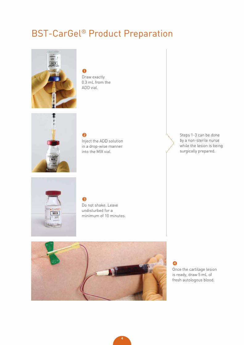
BST-CarGel® Product Preparation
Draw exactly 0.3 mL from the ADD vial.
Inject the ADD solution in a drop-wise manner into the MIX vial.
Do not shake. Leave undisturbed for a minimum of 10 minutes.
Steps 1-3 can be done by a non-sterile nurse while the lesion is being surgically prepared.
Once the cartilage lesion is ready, draw 5 mL of fresh autologous blood.
5
Physiotherapy Program Summary
standard knee immobilizer for the first 24 hours, and thereafter for 14 days at night and during all movement
non-weight-bearing on the treated knee for 6-8 weeks
frequent physiotherapy for 12 weeks, using typical modalities for joint health
no high impact activities requiring pivoting or shifting for 12 months
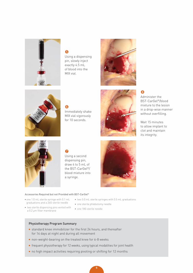
Using a dispensing pin, slowly inject exactly 4.5 mL of blood into the MIX vial.
Immediately shake MIX vial vigorously for 10 seconds.
Using a second dispensing pin, draw 4 to 5 mL of the BST-CarGel®/blood mixture into a syringe.
Administer the BST-CarGel®/blood mixture to the lesion in a drop-wise manner without overfilling.
Wait 15 minutes to allow implant to clot and maintain its integrity.
Accessories Required but not Provided with BST-CarGel®
one 1.0 mL sterile syringe with 0.1 mL graduations and a 26G sterile needle
two sterile dispensing pins vented with a 0.2 μm filter membrane
two 5.0 mL sterile syringes with 0.5 mL graduations
one sterile phlebotomy needle
one 18G sterile needle
6
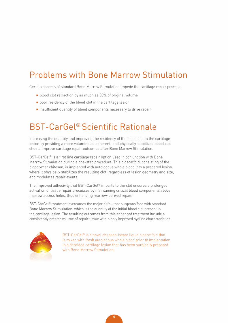
BST-CarGel® is a novel chitosan-based liquid bioscaffold that is mixed with fresh autologous whole blood prior to implantation in a debrided cartilage lesion that has been surgically prepared with Bone Marrow Stimulation.
Problems with Bone Marrow StimulationCertain aspects of standard Bone Marrow Stimulation impede the cartilage repair process:
blood clot retraction by as much as 50% of original volume
poor residency of the blood clot in the cartilage lesion
insufficient quantity of blood components necessary to drive repair
BST-CarGel® Scientifi c RationaleIncreasing the quantity and improving the residency of the blood clot in the cartilage lesion by providing a more voluminous, adherent, and physically-stabilized blood clot should improve cartilage repair outcomes after Bone Marrow Stimulation.
BST-CarGel® is a first line cartilage repair option used in conjunction with Bone Marrow Stimulation during a one-step procedure. This bioscaffold, consisting of the biopolymer chitosan, is implanted with autologous whole blood into a prepared lesion where it physically stabilizes the resulting clot, regardless of lesion geometry and size, and modulates repair events.
The improved adhesivity that BST-CarGel® imparts to the clot ensures a prolonged activation of tissue repair processes by maintaining critical blood components above marrow access holes, thus enhancing marrow-derived repair.
BST-CarGel® treatment overcomes the major pitfall that surgeons face with standard Bone Marrow Stimulation, which is the quantity of the initial blood clot present in the cartilage lesion. The resulting outcomes from this enhanced treatment include a consistently greater volume of repair tissue with highly improved hyaline characteristics.
7
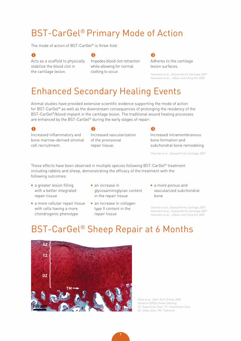
Enhanced Secondary Healing EventsAnimal studies have provided extensive scientific evidence supporting the mode of action for BST-CarGel® as well as the downstream consequences of prolonging the residency of the BST-CarGel®/blood implant in the cartilage lesion. The traditional wound healing processes are enhanced by the BST-CarGel® during the early stages of repair:
Increased inflammatory and bone marrow-derived stromal cell recruitment.
Increased vascularization of the provisional repair tissue.
Increased intramembranous bone formation and subchondral bone remodeling.
Chevrier et al., Osteoarthritis Cartilage 2007
BST-CarGel® Primary Mode of ActionThe mode of action of BST-CarGel® is three-fold:
Acts as a scaffold to physically stabilize the blood clot in the cartilage lesion.
Impedes blood clot retraction while allowing for normal clotting to occur.
Adheres to the cartilage lesion surfaces.
Hoemann et al., Osteoarthritis Cartilage 2007Hoemann et al., J Bone Joint Surg Am 2005
BST-CarGel® Sheep Repair at 6 Months
Shive et al., Oper Tech Orthop 2006Safranin-O/Fast Green StainingSZ: Superficial Zone, TZ: Transitional Zone DZ: Deep Zone, TM: Tidemark
a greater lesion filling with a better integrated repair tissue
a more cellular repair tissue with cells having a more chondrogenic phenotype
an increase in glycosaminoglycan content in the repair tissue
an increase in collagen type II content in the repair tissue
a more porous and vascularized subchondral bone
These effects have been observed in multiple species following BST-CarGel® treatment including rabbits and sheep, demonstrating the efficacy of the treatment with the following outcomes:
Chevrier et al., Osteoarthritis Cartilage 2007 Hoemann et al., Osteoarthritis Cartilage 2007Hoemann et al., J Bone Joint Surg Am 2005
8
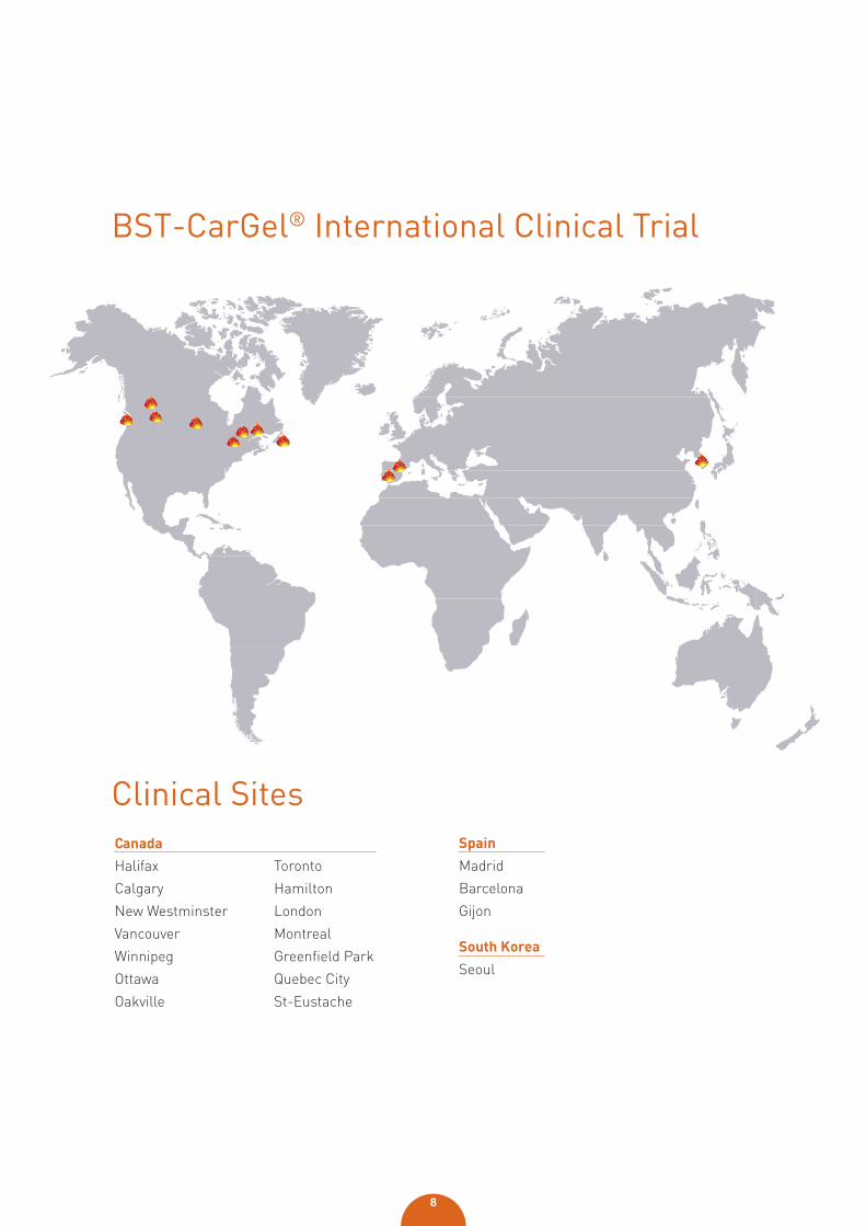
BST-CarGel® International Clinical Trial
Halifax
Calgary
New Westminster
Vancouver
Winnipeg
Ottawa
Oakville
Toronto
Hamilton
London
Montreal
Greenfield Park
Quebec City
St-Eustache
Canada Spain
Madrid
Barcelona
Gijon
South Korea
Seoul
Clinical Sites
9
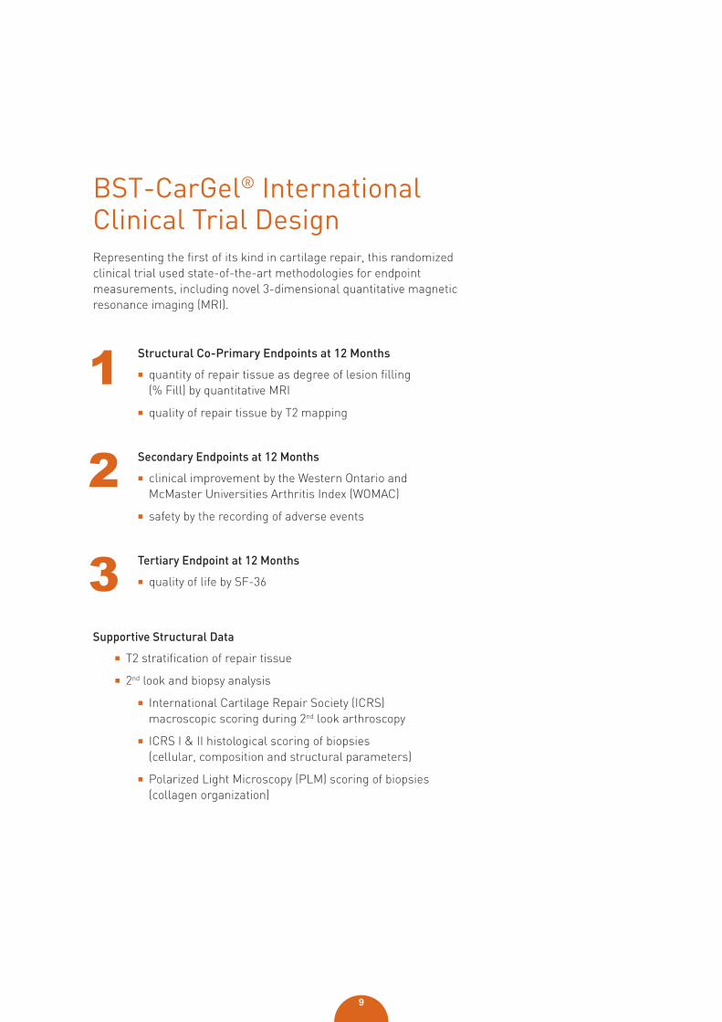
BST-CarGel® International Clinical Trial DesignRepresenting the first of its kind in cartilage repair, this randomized clinical trial used state-of-the-art methodologies for endpoint measurements, including novel 3-dimensional quantitative magnetic resonance imaging (MRI).
Structural Co-Primary Endpoints at 12 Months
quantity of repair tissue as degree of lesion filling (% Fill) by quantitative MRI
quality of repair tissue by T2 mapping
Secondary Endpoints at 12 Months
clinical improvement by the Western Ontario and McMaster Universities Arthritis Index (WOMAC)
safety by the recording of adverse events
Tertiary Endpoint at 12 Months
quality of life by SF-36
Supportive Structural Data
T2 stratification of repair tissue
2nd look and biopsy analysis
International Cartilage Repair Society (ICRS) macroscopic scoring during 2nd look arthroscopy
ICRS I & II histological scoring of biopsies (cellular, composition and structural parameters)
Polarized Light Microscopy (PLM) scoring of biopsies(collagen organization)
10
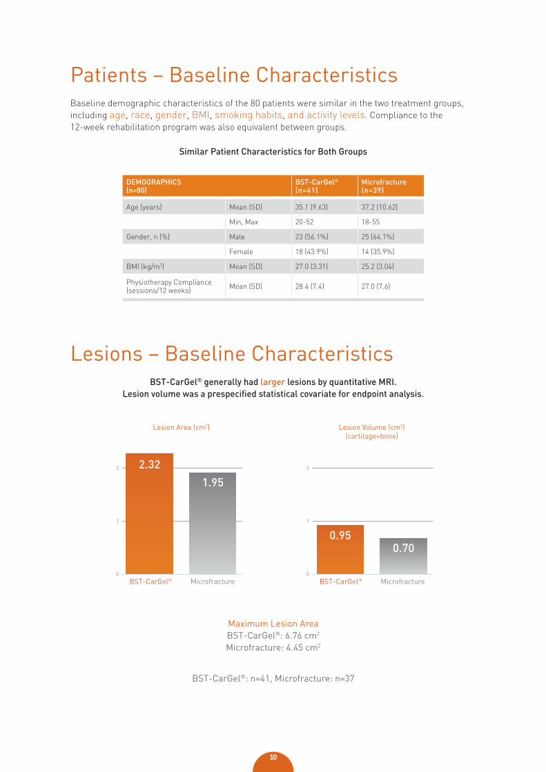
DEMOGRAPHICS(n=80)
BST-CarGel®
(n=41)Microfracture(n=39)
Age (years) Mean (SD) 35.1 (9.63) 37.2 (10.62)
Min, Max 20-52 18-55
Gender, n (%) Male 23 (56.1%) 25 (64.1%)
Female 18 (43.9%) 14 (35.9%)
BMI (kg/m2) Mean (SD) 27.0 (3.31) 25.2 (3.04)
Physiotherapy Compliance (sessions/12 weeks) Mean (SD) 28.4 (7.4) 27.0 (7.6)
Patients – Baseline CharacteristicsBaseline demographic characteristics of the 80 patients were similar in the two treatment groups, including age, race, gender, BMI, smoking habits, and activity levels. Compliance to the 12-week rehabilitation program was also equivalent between groups.
Lesions − Baseline CharacteristicsBST-CarGel® generally had larger lesions by quantitative MRI.
Lesion volume was a prespecifi ed statistical covariate for endpoint analysis.
Maximum Lesion AreaBST-CarGel®: 6.76 cm2
Microfracture: 4.45 cm2
BST-CarGel®: n=41, Microfracture: n=37
0
1
2
Lesion Area (cm2)
1.95
2.32
BST-CarGel® Microfracture
1
2
Lesion Volume (cm3)(cartilage+bone)
0.700.95
0BST-CarGel® Microfracture
Similar Patient Characteristics for Both Groups
11
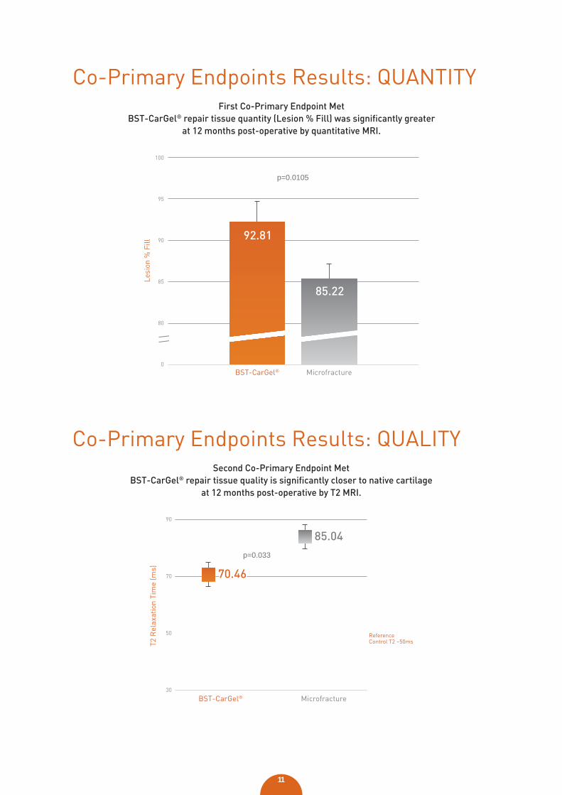
Co-Primary Endpoints Results: QUANTITYFirst Co-Primary Endpoint Met
BST-CarGel® repair tissue quantity (Lesion % Fill) was signifi cantly greater at 12 months post-operative by quantitative MRI.
0
80
85
90
95
100
Lesi
on %
Fill
92.81
85.22
BST-CarGel® Microfracture
p=0.0105
T2 R
elax
atio
n Ti
me
(ms)
BST-CarGel®
ReferenceControl T2 ~50ms
Microfracture30
50
70
90
85.04
70.46
Co-Primary Endpoints Results: QUALITYSecond Co-Primary Endpoint Met
BST-CarGel® repair tissue quality is signifi cantly closer to native cartilage at 12 months post-operative by T2 MRI.
p=0.033
12
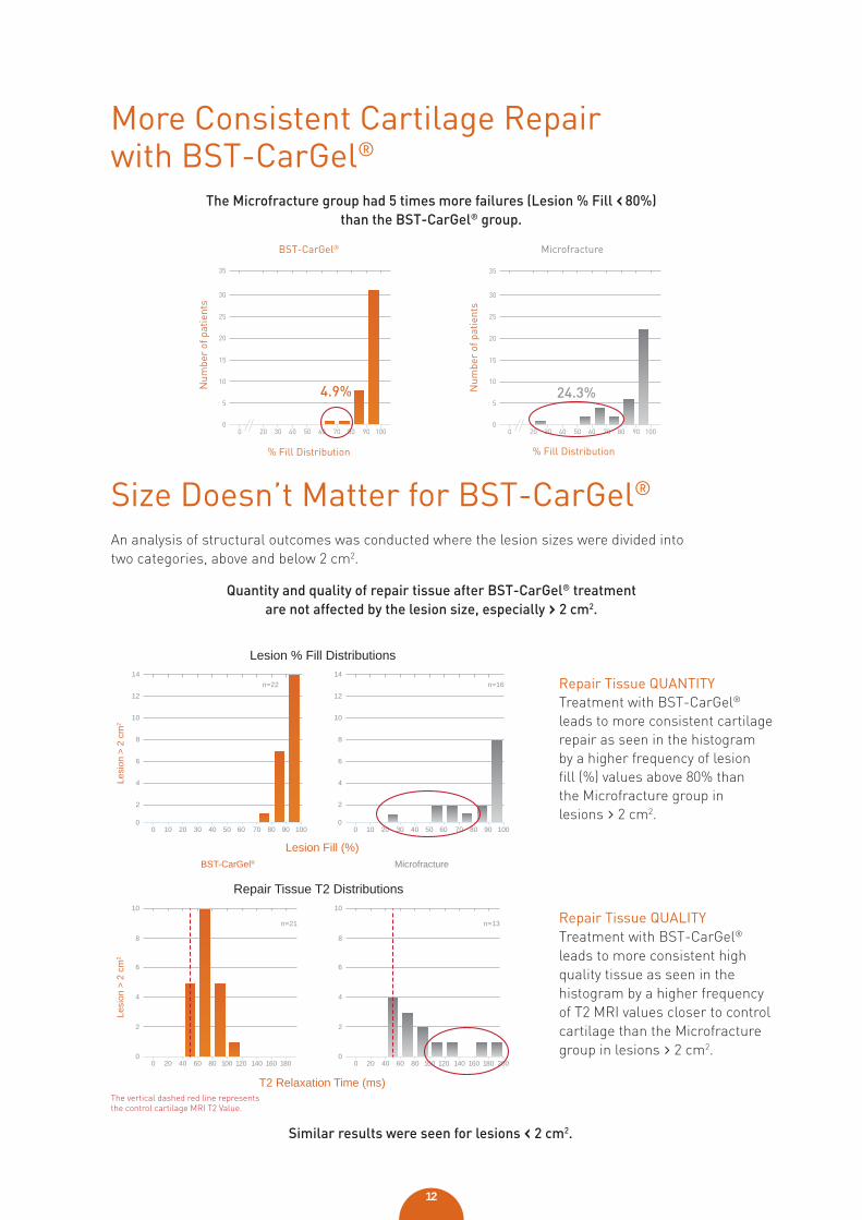
Size Doesn’t Matter for BST-CarGel®
An analysis of structural outcomes was conducted where the lesion sizes were divided into two categories, above and below 2 cm2.
Quantity and quality of repair tissue after BST-CarGel® treatment are not affected by the lesion size, especially › 2 cm2.
T2 Relaxation Time (ms)
Lesion Fill (%)
Lesion % Fill Distributions
Repair Tissue T2 Distributions
Lesi
on >
2 c
m2
2
4
0
6
10
8
0 20 40 60 80 100 120 140 160 180
n=21
2
4
0
6
10
8
0 20 40 60 80 100 120 140 160 180 200
n=13
Lesi
on >
2 c
m2
12
2
4
8
10
6
0
14
0 10 20 30 40 50 60 70 80 90 100
n=2212
2
4
8
10
6
0
14
0 10 20 30 40 50 60 70 80 90 100
n=16
BST-CarGel® Microfracture
Repair Tissue QUALITYTreatment with BST-CarGel® leads to more consistent high quality tissue as seen in the histogram by a higher frequency of T2 MRI values closer to control cartilage than the Microfracture group in lesions › 2 cm2.
Repair Tissue QUANTITYTreatment with BST-CarGel® leads to more consistent cartilage repair as seen in the histogram by a higher frequency of lesion fill (%) values above 80% than the Microfracture group in lesions › 2 cm2.
The vertical dashed red line represents the control cartilage MRI T2 Value.
More Consistent Cartilage Repair with BST-CarGel®
The Microfracture group had 5 times more failures (Lesion % Fill ‹ 80%) than the BST-CarGel® group.
4.9%
BST-CarGel®
% Fill Distribution
0
5
10
15
20
30
25
35
0 20 30 40 50 60 70 80 90 100
Num
ber
of p
atie
nts
0
5
10
15
20
30
25
35
0 20 30 40 50 60 70 80 90 100
% Fill Distribution
24.3%
Microfracture
Num
ber
of p
atie
nts
Similar results were seen for lesions ‹ 2 cm2.
13
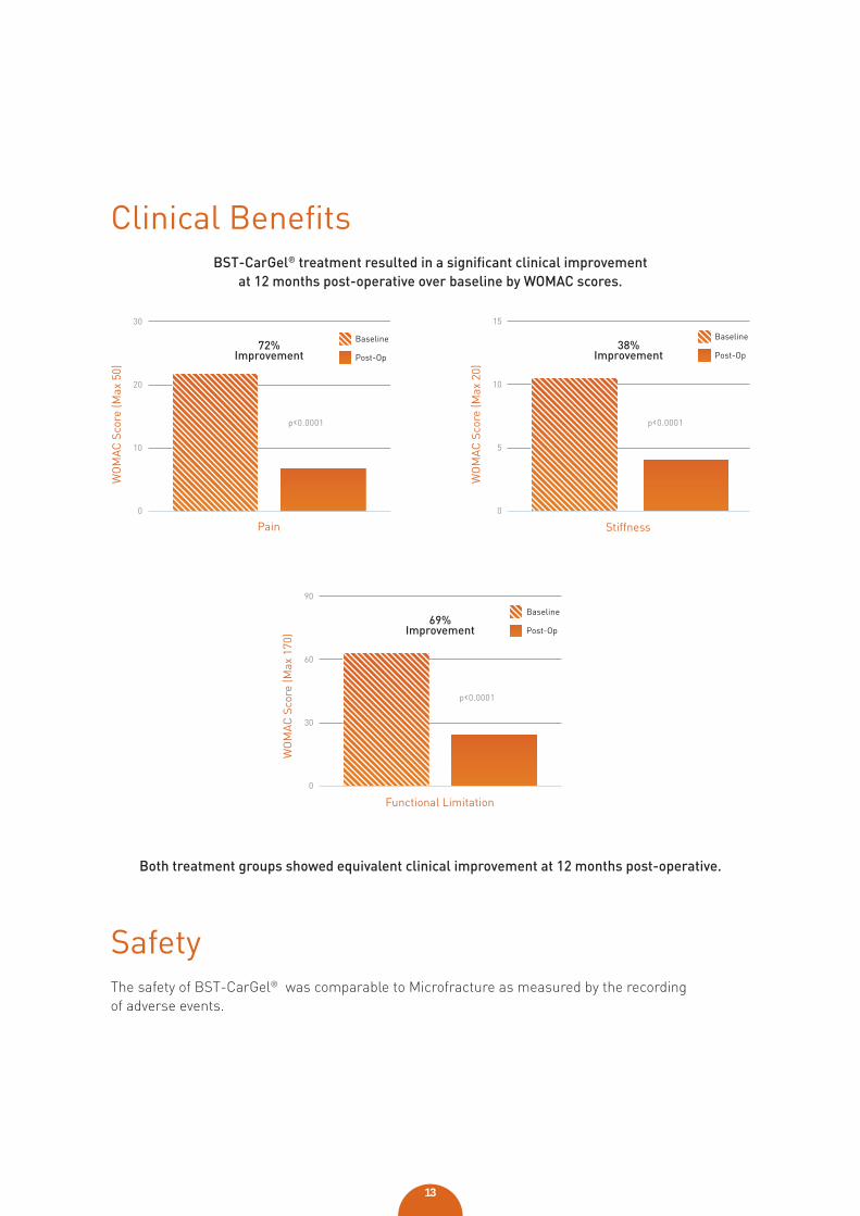
Clinical Benefi tsBST-CarGel® treatment resulted in a signifi cant clinical improvement
at 12 months post-operative over baseline by WOMAC scores.
Both treatment groups showed equivalent clinical improvement at 12 months post-operative.
0
10
20
30
WO
MAC
Sco
re (M
ax 5
0)
p‹0.0001
72%Improvement
Pain
Baseline
Post-Op
0
5
10
15
WO
MAC
Sco
re (M
ax 2
0)
p‹0.0001
38%Improvement
Stiffness
Baseline
Post-Op
0
30
60
90
WO
MAC
Sco
re (M
ax 1
70)
p‹0.0001
69%Improvement
Functional Limitation
Baseline
Post-Op
SafetyThe safety of BST-CarGel® was comparable to Microfracture as measured by the recording of adverse events.
14
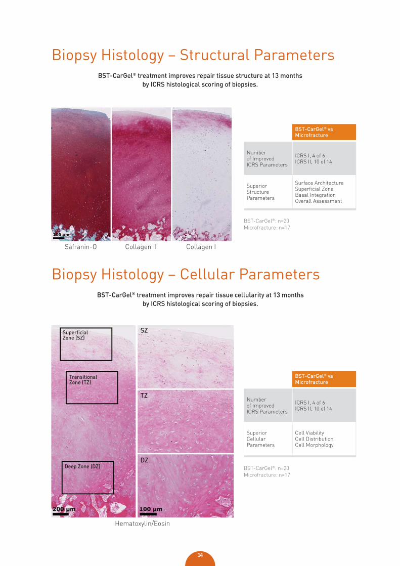
Biopsy Histology – Structural ParametersBST-CarGel® treatment improves repair tissue structure at 13 months
by ICRS histological scoring of biopsies.
Biopsy Histology – Cellular ParametersBST-CarGel® treatment improves repair tissue cellularity at 13 months
by ICRS histological scoring of biopsies.
Safranin-O Collagen II Collagen I
Hematoxylin/Eosin
BST-CarGel® vs Microfracture
Number of Improved ICRS Parameters
ICRS I, 4 of 6ICRS II, 10 of 14
Superior Structure Parameters
Surface ArchitectureSuperficial ZoneBasal IntegrationOverall Assessment
BST-CarGel® vs Microfracture
Number of Improved ICRS Parameters
ICRS I, 4 of 6ICRS II, 10 of 14
Superior Cellular Parameters
Cell ViabilityCell DistributionCell Morphology
Superfi cial Zone (SZ)
SZ
TZ
DZ
Transitional Zone (TZ)
Deep Zone (DZ)
BST-CarGel®: n=20Microfracture: n=17
BST-CarGel®: n=20Microfracture: n=17
15
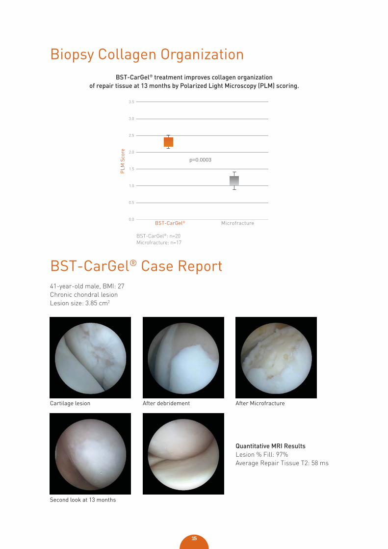
PLM
Sco
re
BST-CarGel® Microfracture0.0
0.5
1.0
1.5
2.0
3.0
2.5
3.5
Biopsy Collagen OrganizationBST-CarGel® treatment improves collagen organization
of repair tissue at 13 months by Polarized Light Microscopy (PLM) scoring.
BST-CarGel®: n=20Microfracture: n=17
p=0.0003
Second look at 13 months
Cartilage lesion After debridement After Microfracture
BST-CarGel® Case Report41-year-old male, BMI: 27Chronic chondral lesionLesion size: 3.85 cm2
Quantitative MRI ResultsLesion % Fill: 97%Average Repair Tissue T2: 58 ms
Intended UseBST-CarGel® is a medical device intended to promote hyaline cartilage regeneration when used in conjunction with the bone marrow stimulation technique for the repair of focal articular cartilage lesions.
Treatment with BST-CarGel® should be a one-time application administered through a mini-arthrotomy or by arthroscopic delivery performed by an orthopaedic surgeon properly trained in the technique.
BST-CarGel® is a fi rst line therapy for most cartilage lesion sizes
Novel scaffold-based BST-CarGel® treatment results in superior cartilage repair
is a fi rslagfi a s a fi
lagis a fi riss
lssis a fi r


0086