the statement on death and organ donation
TRANSCRIPT

The Statement on Death and Organ DonationEDITION 4.1 | 2021

2
Published by the Australian and New Zealand Intensive Care Society Suite 1.01, Level 1, 277 Camberwell Road, Camberwell VIC 3124 Phone: +61 3 9340 3400 Email: [email protected] Website: anzics.com.au
© Australian and New Zealand Intensive Care Society 2021
This work is copyright. It may be reproduced in whole or in part for study or training purposes, subject to the inclusion of an acknowledgment of the source. Requests and enquiries concerning reproduction and rights for purposes other than those indicated above require the written permission of the Australian and New Zealand Intensive Care Society (ANZICS) - email: [email protected]
ANZICS requests that you attribute this publication (and any material sourced from it) using the following citation: The Australian and New Zealand Intensive Care Society Statement on Death and Organ Donation. Melbourne. Edition 4.1 2021. ISBN 4978-1-876980-39-9.
Disclaimer: The statement on death and organ donation (Edition 4.1) has been authored by the ANZICS Death and Organ Donation Committee using all care and appropriate diligence according to the information available at the time of preparation of this statement. The practitioner should, therefore, have regard to any information, research or material which may have been published or become available subsequently. The Committee has endeavored to ensure that the document is as current as possible at the time of its preparation, and it takes no responsibility for matters arising from changed circumstances, information or material which may have become available subsequently. This statement has been prepared for information purposes with regard to general circumstances only. Ultimately, it is the responsibility of the practitioner to have regard to the particular circumstances of each case. ANZICS does not warrant the information contained in this statement.
ANZICS | The Statement on Death and Organ Donation
3
ContentsMembership of the ANZICS Death and Organ Donation Committee 4Foreword 5ANZICS recommendations 6Introduction 9
1 Determination of death 101.1 Death 101.2 Neurological determination of death 131.3 Circulatory determination of death 23
2 Organ and tissue donation after death 252.1 Organ and tissue donation in perspective 252.2 When donation should be considered 292.3 Donation after neurological determination of death (also called brain death) 312.4 Donation after circulatory determination of death (circulatory death) 342.5 Paediatric donation 382.6 Tissue-only donation 39
3 Patient and family-centred care 403.1 Family meetings in the context of end-of-life care 403.2 Discussing organ and tissue donation 423.3 Ongoing support of the family 443.4 Confidentiality 453.5 Cultural humility in discussing death and organ donation 45
4 Best practice in organ donation 494.1 Educational requirements 49 4.2 Care of the dying patient 504.3 Supporting organ donation 504.4 Support of hospital staff 53
Appendices 54A Glossary of terms 54B Acronyms and abbreviations 55C Systolic and mean blood pressure in children (95th percentile for age) 56D Sample documentation for neurological determination of death 57E Sample documentation for circulatory determination of death (in the context of organ donation) 59F Clinical and physiological support of a patient after neurological determination of death 60
References 61
List of tablesTable 1.1: Medical practitioners permitted to determine death by neurological criteria, by jurisdiction 13Table 1.2: Process for neurological determination of death 16Table 2.1: Legislative basis for the removal of tissues (including organs) after death 27Table 2.2: Maastricht classification 30Table 2.3: Hormonal treatment 33Table 2.4: Principles for donation after circulatory determination of death150,151 34Table 3.1: Elements that can be covered in the family donation conversation 43Table 3.2: Essential aspects to discuss with the family regarding donation after neurological
determination of death 44Table 3.3: Essential aspects to discuss with the family regarding donation after circulatory
determination of death 44Table 3.4: Selected considerations in caring for Aboriginal and Torres Strait Islander families 46Table F1: Checklist for clinical support of a patient with permanent loss of brain function 60

4
Membership of the ANZICS Death and Organ Donation Committee
A/Prof William Silvester Chair
Dr Rob Bevan College of Intensive Care Medicine
Dr Jorge Brieva New South Wales
A/Prof David Cook Queensland
Dr Rohit D’Costa Victoria
Prof Geoffrey Dobb Western Australia
Dr Ben Gelbart Paediatric
Dr Sarah Jones Northern Territory
Dr James Judson New Zealand (Auckland)
Dr Lucy Modra Trainee representative
Dr Stewart Moodie South Australia
Dr Helen Opdam Australian Organ and Tissue Authority
Dr Chris Poynter New Zealand (Wellington)
Dr Stephen Streat Organ Donation New Zealand
ANZICS | The Statement on Death and Organ Donation 5
Foreword
This is the fourth edition of the Statement on death and organ donation. The Statement was last substantially revised in 2013. Changes in both intensive care practice and in donation since 2013 have led us to a more fundamental revision of the Statement including both structure and content. The number of sections has been reduced from six to four, now entitled 1) Death, 2) Organ and tissue donation after death, 3) Patient and family-centred care and 4) Best practice in organ donation. We have also aimed to ensure that the document is culturally sensitive with specific relevance to First Nations peoples of Australia and New Zealand. We believe that the fourth edition is simpler and more logically coherent. Despite containing considerable new material the size of the Statement has not increased.
Significantly, the Statement is now preceded by a table of 29 ‘ANZICS Recommendations’ which presents the key elements of the process, legal and ethical issues, best practice and care of the patient and family. Acting as an executive summary, this quick reference guide also directs the reader to the relevant section for greater detail, explanation or evidence. The Committee wishes to draw attention to changes in the sections on neurological and circulatory determination of death.
The revision process took place by email, teleconference and face-to-face meetings over two years, and included a critical review of the previous edition by the ANZICS Death and Organ Donation Committee, a review of relevant literature, complete rewriting of the document in the restructured format, consultation with ANZICS members, medical and nursing colleges, Australian and New Zealand organ donation agencies, other societies and associations, assessment and incorporation of comments received and, finally, a further review and final revision by the Committee. The process involved extensive work by all Committee members and many others who contributed, and these contributions are very gratefully acknowledged. We have been greatly assisted with the management, formatting, style and language of the document by our professional writer, Jenny Ramson. A five-year horizon for the next revision seems reasonable.
Donation is increasing in both Australia and New Zealand and the Committee acknowledges the professionalism and commitment of the intensive care, organ donation and transplantation communities in this. The Committee believes that there is potential for donation to increase further while ensuring that donation always accords with good clinical practice, ethical standards and the law.
There are ethical and clinical challenges in donation as both medical practice and societal expectations continue to change. ICU mortality is falling, despite increasing patient case-complexity and at the same time the recipient outcomes of organ transplantation continue to improve. There is an increase in the number of potential recipients who could benefit from transplantation, particularly kidney transplantation, a trend that is likely to continue. Donation now often occurs in circumstances which are more complex than previously (e.g. donation after circulatory determination of death, donor co-morbidity and older age, donors with acute but reversible organ dysfunction, or when admission to intensive care has been solely for possible future donation) and the ANZICS Statement supports clinicians who must necessarily address these issues in their donation practice.
ANZICS is proud to have always taken a leading position of responsibility for providing guidance and support to the membership and in maintaining the trust of the profession and society at large in the processes of deceased donation. ANZICS re-iterates its strong commitment to these values and goals in this edition of the Statement. It will be made available to all Australian and New Zealand hospitals, intensive care units and emergency departments. The most up-to-date version will be the version available on the ANZICS website, with significant changes appropriately highlighted.
Finally, the Committee pays tribute to Dr James Judson and Professor Geoffrey Dobb who have been involved in this work ever since the first “ANZICS Guidelines” in 1993 and contributed to this fourth Edition 25 years later. Their wisdom and institutional knowledge have been most valued.
A/Prof William (Bill) Silvester Chair, ANZICS Death and Organ Donation Committee

6
ANZICS Recommendations
The table below lists the recommendations developed by the ANZICS Death and Organ Donation Committee based on review of the law, medical literature and Committee consensus. The table also notes the section where the evidence or context for each recommendation is discussed.
ANZICS recommends that: Section
Neurological determination of death
1 Neurological determination of death is carried out by two doctors, one of whom should be a specialist, who must each independently determine death according to this Statement, and meet the requirements of jurisdictional legislation.
1.1.5
2 For neurological determination of death to be conducted, there must be definite clinical or neuroimaging evidence of acute brain pathology consistent with deterioration to permanent loss of all neurological function. In cases of hypoxic-ischaemic encephalopathy, clinical history alone may provide sufficient explanation of the acute brain pathology and not require neuroimaging prior to neurological determination of death by clinical examination.
1.2
3 There is a minimum 4-hour observation period prior to neurological determination of death using clinical examination alone. Throughout this observation period, all preconditions are met, the patient has a Glasgow Coma Scale of 3, with pupils non-reactive to light, absent cough/tracheal reflex and apparent apnoea on a ventilator. Following an acute hypoxic-ischaemic encephalopathy or hypothermia (<35°C) of duration greater than 6 hours, there should be a waiting period of 24 hours before determination of death using clinical examination alone.
1.2.2
4 The opportunity to observe a clinical examination of brain function should be offered to family members. Appropriate explanation and support for the family should be provided.
1.2.2
5 When imaging to demonstrate absence of brain perfusion is required, it must be preceded by performance of those parts of the clinical examination that are possible. Responsiveness, all testable brainstem reflexes and breathing effort must be absent.
1.2.5
6 If assessment of brain perfusion is required, three- or four-vessel angiography or radionuclide imaging are preferred. Computed tomography angiography is acceptable if recommended radiological guidelines are followed. Magnetic resonance imaging or angiography and transcranial Doppler should not be used.
1.2.5
7 Neurological determination of death cannot be conducted with certainty by clinical examination in preterm neonates (<37 weeks’ gestation) or in term neonates in the first 24 hours after birth.
1.2.6
8 Neurological determination of death by clinical examination can be made in term neonates (≥37 weeks’ gestation). In term neonates between 24 hours and 30 days old, an observation period of 24 hours should occur before the first clinical examination, followed by a 24-hour interval before the second clinical examination.
1.2.6
9 The criteria for neurological determination of death in children above the age of 30 days are the same as those in adults.
1.2.6
10 Brain perfusion studies can be used to assist in the neurological determination of death in children of all ages including term and preterm neonates, when preconditions for neurological testing cannot be met.
1.2.6
11 Documentation of neurological determination of death should be made using a specific form (see Appendix D) to demonstrate explicitly that all criteria set out in this Statement are met, whether or not organ or tissue donation occurs. The same criteria should be listed in local hospital forms.
1.2.7

ANZICS | The Statement on Death and Organ Donation 7
ANZICS recommends that: Section
Circulatory determination of death in the context of organ donation
12 Circulatory determination of death in the context of organ donation requires the absence of spontaneous movement, breathing and circulation. Absence of circulation is evidenced by absent arterial pulsatility for 5 minutes, using intra-arterial pressure monitoring and confirmed by clinical examination (absent heart sounds and/or absent central pulse). In cases without an arterial line, electrical asystole should be observed for 5 minutes on the electrocardiogram and confirmed by clinical examination.
1.3.1
13 For the purposes of organ donation, circulatory determination of death should be documented using a specific form (see Appendix E) to demonstrate explicitly that all criteria set out in this Statement are met. The same criteria should be listed in local hospital forms.
1.3.2
Organ donation — Legal requirements
14 Processes should be in place so that relevant consents and authorisations for organ and tissue donation, including those provided verbally, are adequately documented.
2.1.3
15 Irrespective of the patient’s known wishes about donation, all possible attempts should be made to contact the family to discuss donation and to ascertain their agreement or otherwise. Donation should not proceed if the family disagrees.
2.1.3
16 Donation may proceed in situations where the family either does not exist or cannot be contacted after reasonable attempts, in those Australian jurisdictions where legislation permits. If donation is authorised when the family exists but cannot be contacted, continued attempts to locate the family should occur after donation. In New Zealand, organ donation is not legal without the informed consent and absence of objection by the family.
2.1.3
17 The specific physiological changes associated with brain death should be treated early following the general principles of critical care management, with expert advice sought from a medical donation specialist through the donor coordinator if instability persists despite treatment or if specific treatments are requested by the retrieval team.
2.3.1
Tissue donation
18 Tissue donation should be considered and, if donation is locally feasible, offered to the family for all patients dying in hospital.
2.6
Patient and family centred care
19 Organ donation should not be raised prior to family understanding of the patient’s death or likely impending death. Introducing the topic of organ donation should not be rushed and it is often best raised as a separate discussion.
3.2.2
20 Donation should be discussed with all families where donation is a possibility. Discussions about organ and tissue donation should be respectful of the patient and sensitive to the family’s emotional, psychological, spiritual, cultural and religious needs.
3.2.3
21 Support and the opportunity to provide feedback should be offered to the family of every patient who dies in the intensive care unit.
3.3
22 Intensivists and all other hospital, donation and transplantation staff should safeguard the anonymity of the donor, the donor’s family and the recipient during and after the donation process.
3.4

8
ANZICS recommends that: Section
Organ donation — Best practice
23 All intensivists and intensive care trainees who discuss donation with families should have completed, as a minimum, the Australian Organ and Tissue Authority Core Family Donation Conversation workshop, or the equivalent in New Zealand, and maintain skills in this area. Intensive care trainees, with support and supervision by intensivists and other involved staff, should be involved when the opportunity to consider organ donation arises.
4.1
24 Organ and tissue donation should be considered in all patients once there is medical consensus that the patient is near the end of life and ongoing treatment is not in his or her best interests. The possibility of donation can then be assessed in consultation with donation staff, ideally before donation is discussed with the family.
4.3.1
25 In Australia, the Australian Organ Donor Register should be accessed once there is medical consensus that the patient is near the end of life and prior to raising donation. Any information recorded in the Register should be communicated to the family.
4.3.2
26 The potential for organ donation should be supported until organ donation has been formally discussed with the family. This includes physiological support, simple tests and collection (but not processing) of blood samples.
4.3.5
27 Complex, invasive and resource-intensive donor investigations should only be performed with family agreement and if the transplant teams require the information to determine suitability of specific organs for transplantation.
4.3.5
28 Intensivists, in collaboration with donation staff, should develop local pathways so that patients with potential for organ donation who are near the end of life in other hospital departments or remote centres are referred to an intensive care unit for exploration of the possibility of organ donation.
4.3.6
29 Intensive care units should develop systems so that all staff involved with organ and tissue donation have access to support.
4.3.7
ANZICS | The Statement on Death and Organ Donation 9
Introduction
This is the fourth edition of the Australian and New Zealand Intensive Care Society (ANZICS) Statement on death and organ donation. The Statement is intended to provide a relevant and accessible resource for intensive care specialists (intensivists) and other health professionals involved in the determination of death and in the care of potential organ and tissue donors and their families. It encourages consistency of approach in addressing clinical issues, caring for families, and engaging with other expert opinion in Australia and New Zealand.
The Statement has been developed by the ANZICS Committee on Death and Organ Donation, which comprises intensivists with expertise in end-of-life care, determination of death and organ donation. It draws on the best available scientific evidence, the extensive experience of the Committee, and consultation with other organisations concerned with organ and tissue donation in Australia and New Zealand.
ANZICS endorses the Statement and the high standard of medical practice it documents.
The full Statement will be reviewed in 2022. The electronic version of the Statement, available on the ANZICS website, will be updated to reflect significant changes in evidence and practice as they occur.
Purpose and scopeThe main purposes of the Statement are:
• to provide a standard for intensivists and other health professionals in relation to the determination of death and the conduct of organ and tissue donation, including donation after circulatory determination of death; and
• to provide assurance to the Australian and New Zealand communities that determination of death and the conduct of organ and tissue donation are undertaken with diligence, integrity, respect and compassion, and in accordance with available medical evidence and societal expectations.
The Statement includes some discussion of the ethical issues surrounding death and organ and tissue donation. It does not address end-of-life care or the ethics of withdrawal of treatment, which are discussed separately in the ANZICS Statement on care and decision-making at the end of life for the critically ill.1 The National Health and Medical Research Council (NHMRC) 2016 publication Ethical Guidelines for organ transplantation from deceased donors2 provides further discussion of the ethics of organ and tissue donation.
TerminologyThe language used during discussion of death and organ and tissue donation is important and needs to be precise. Recommended language is explained in Sections 3.1.1 (see page 41) and 3.2.1 (see page 42) and terms used in the Statement are defined in the Glossary (page 54).
StructureThe Statement includes discussion of:
• neurological and circulatory determination of death (Chapter 1);
• the legal framework for organ and tissue donation and the processes involved in organ donation after neurological or circulatory determination of death in adults and children (Chapter 2);
• the provision of patient and family-centred care in the context of organ donation (Chapter 3); and
• responsibilities of intensive care staff in supporting best practice in organ and tissue donation (Chapter 4).

10
1Determination of Death
This chapter provides information on:
• death from a cultural and religious perspective, evolution of biological concepts of death, and the legal definitions of death (see Section 1.1);
• the neurological determination of death (in adults and children), including preconditions, process of clinical examination, and studies recommended to demonstrate absence of brain perfusion when clinical examination cannot be used alone to determine death (see Section 1.2; page 13); and
• the criteria for circulatory determination of death (see Section 1.3; page 23).
1.1 Death1.1.1 Death in perspectiveDeath is a concept and a reality that humans have sought to understand over time from a number of different perspectives, initially cultural, spiritual and religious and, in recent centuries, from a biological viewpoint.
It is essential to have a clear distinction between life and death in any society, to determine when loss of personhood and individual rights occurs, wills may be executed, efforts to preserve life can cease, organs can be donated, burial or cremation can be undertaken, and religious or social ceremonies that mark the end of a life can be held.
Dying is foremost a biological process and the determination of death is an event in that process. Dying is the process during which cellular activities and organ functions progressively cease. The determination and certification of death indicate that an irrevocable point in the dying process has been reached. The precise time of death is somewhat arbitrary and represents a societal consensus that is informed by biological understanding.
Technological advances in the fields of resuscitation and intensive care challenge historical concepts of death by interrupting the normal interlinkages between brain, respiratory and circulatory function. These advances, including practices seeking to optimise the opportunity for donation for transplantation after death, have led to the requirement for death to be diagnosed in a precise and timely manner.
The determination of death is, in most countries, the legal responsibility of a medical practitioner.
1.1.2 Death from a cultural and religious perspectiveDeath has always had immense cultural, religious and mystical significance to humans. Cultural ideas and beliefs about death are many and varied and include the ideas that a soul has departed from the body for a spirit world, purgatory, hell, heaven or paradise; has been reincarnated into another living being; is in an indeterminate state awaiting bodily resurrection; or that in the absence of life there is merely oblivion.
Consideration of cultural and religious perspectives in discussing death is of great importance and is discussed in Section 3.5; page 45.

ANZICS | The Statement on Death and Organ Donation 11
1.1.3 Evolution of the biological understanding of deathHistorically, recognition of death relied upon observations of absence of signs of life including immobility, absence of breathing, and subsequent cooling of the body, rigor mortis, putrefaction and decay.
A biological understanding of death was prompted after William Harvey in the 17th century described the circulation of blood and the function of the heart as a pump, stating that ‘…the heart is the principle of life…from which heat and life are dispersed to all parts…’.3 Under this concept, death occurred when the heart and circulation stopped.
By the end of the 19th century it was known that, during an increase in intracranial pressure, respirations suddenly stopped whereas the heart continued to beat for some time. It was also recognised that the heart could continue to beat if artificial respiration was performed.4-6
During the 1952 Copenhagen poliomyelitis epidemic, it was found that, with positive-pressure ventilatory support, inadequate breathing did not automatically lead to coma and cardiac arrest.7,8 The success of mechanical ventilation and the subsequent development of intensive care units (ICUs) rapidly led to positive-pressure ventilation being used in other conditions, including respiratory arrest associated with devastating brain injury.
In the 1950s, medical teams reported absence of blood flow in the cerebral arteries9,10 and isoelectric electro-encephalogram11 in patients with respiratory arrest from neurological catastrophes. A Lyon University doctoral thesis mentioned several patients with fixed dilated pupils and apnoea treated with positive pressure ventilation in a neurosurgical unit.12 Wertheimer and his colleagues described the syndrome of coma, areflexia, apnoea and called it death of the central nervous system.13 They described signs that indicated the definitive absence of all central nervous system activity in order to define criteria for abandoning ongoing intensive therapy. Mollaret and Goulon described 23 patients with deep coma with no spontaneous respiration, areflexia, polyuria and hypotension (if noradrenaline was not given continuously), in the absence of all electroencephalographic activity and called it le coma depassé (beyond coma).14
In 1968, an ad hoc committee of Harvard Medical School15 agreed that mechanical ventilatory support could be withdrawn from patients diagnosed with ‘irreversible coma’ or ‘brain death’ (terms were used interchangeably) and that, with consent, organs could be removed from such patients for transplantation. Their primary concern was to provide an acceptable mechanism to permit withdrawal of mechanical ventilatory support from such patients. The sanction this gave to removal of organs for transplantation was a secondary consideration.
In the same year, the 22nd World Medical Assembly (WMA) announced the Declaration of Sydney on Human Death,5,16 which stated that ‘the point of death of the different cells and organs is not so important as the certainty that the process has become irreversible by whatever techniques of resuscitation that may be employed’ and that ‘clinical interest lies not in the state of preservation of isolated cells but in the fate of a person’. In 1983, the 35th WMA in Venice amended the 1968 declaration, adding that, for diagnosis of brain death, ‘it is essential to determine the irreversible cessation of all functions of the entire brain, including the brainstem’.17
There is no world-wide consensus on the definition or criteria for determining death. In 2012, an invitational forum in Montreal proposed an international consensus definition of death which emphasised the cessation of neurological or circulatory function, and focussed on the centrality of brain function for the determination of death.18 A single operational definition of human death was proposed ‘The permanent loss of capacity for consciousness and all brainstem functions, as a consequence of permanent cessation of circulation or catastrophic brain injury’. This definition has not been universally adopted and efforts continue towards greater international consistency and consensus on death determination.
1.1.4 Death from a legal perspectiveSome countries have enacted laws or codes of medical practice and, while they are broadly similar, they differ in detail. Relevant to this Statement are those of Australia and New Zealand; discussion of those in the United States and the United Kingdom is also included.
Australia and New Zealand
In 1972, a New Zealand High Court judge submitted that doctors should endeavour to agree among themselves on the criteria by which death can be determined and should ensure that their criteria accord with the concept that the ordinary man has of death.19 In New Zealand, death and organ donation are covered by the Human Tissue Act 2008, which does not define death (except in the case of a foetus) and uses the words ‘satisfied… that the individual concerned is dead’. New Zealand legislation has no definition of death.
12
In 1977, the Australian Law Reform Commission addressed the absence of definition of death in Australian law, recommending that a statutory definition of death should be introduced. They recommended that death be defined as:
• irreversible* cessation of all function of the brain of the person; or• irreversible cessation of circulation of blood in the body of the person.
They did not provide detailed criteria on the grounds that ‘the creation and prescription of techniques of diagnosis should be the responsibility of the medical profession’. They specified that, although it appeared in the context of transplantation, the definition should have general application. The Commission stated ‘...the practice of transplantation does not govern or regulate the definition of death. Despite this, it is true that the practice of transplantation forces the close attention of the community to the subject of death and in many cases requires greater accuracy and care to be brought to bear in determining that death has occurred’.
Current Australian state and territory laws vary but all are closely based on the recommendations of the Australian Law Reform Commission and include a legal definition of death.20 The definition of death is included in the Acts governing tissue† donation in all jurisdictions apart from South Australia (SA) and Western Australia (WA), where the definition of death is contained in separate legislation: in the SA Death (Definition) Act 1983 and the WA Interpretation Act 1984 respectively. The Queensland (QLD) Transplantation and Anatomy Act 1979 states that its definition is only for the purposes of the Act.
United States
Lack of consistent criteria for defining death led to the appointment of a President’s Commission to recommend a uniform definition of death. It declared in 1981 that ‘An individual who has sustained either irreversible cessation of circulatory and respiratory functions, or irreversible cessation of all functions of the entire brain, including the brainstem is dead and that a determination of death must be made in accordance with accepted medical standards’.21,22 Subsequently, this declaration has been enacted into law in most states.23
United Kingdom
The United Kingdom does not have a statutory definition of death.
In 1976, a Conference of the Royal Colleges and Faculties of the United Kingdom published a statement called Diagnosis of brain death, which set out preconditions and diagnostic criteria for establishing when death had occurred in patients whose vital functions were being maintained mechanically.24 In 1979, this statement was supplemented with a memorandum, called Diagnosis of death,25 which emphasised that death of the brain is the final common pathway to death either resulting, in the majority of cases, from failure of vital organ functions and systems or, in the minority of cases, from the direct result of severe damage to the brain itself. It concluded that the identification of brain death means that the patient is dead, whether or not the function of some organs, such as a heartbeat, is still maintained by artificial means.
In 1995, the United Kingdom Medical Royal Colleges defined death to be the ‘irreversible loss of the capacity for consciousness, combined with irreversible loss of the capacity to breathe’.26 (They also recommended use of the term ‘brainstem death’ rather than ‘brain death’.) This definition is used in some Commonwealth countries but not in Australia or New Zealand. The 2008 Code of Practice for the Diagnosis and Confirmation of Death refers to death following irreversible cessation of brainstem function and death following cessation of cardiorespiratory function.27
1.1.5 The legal processes for determination of death in Australia and New Zealand
Neurological determination of death
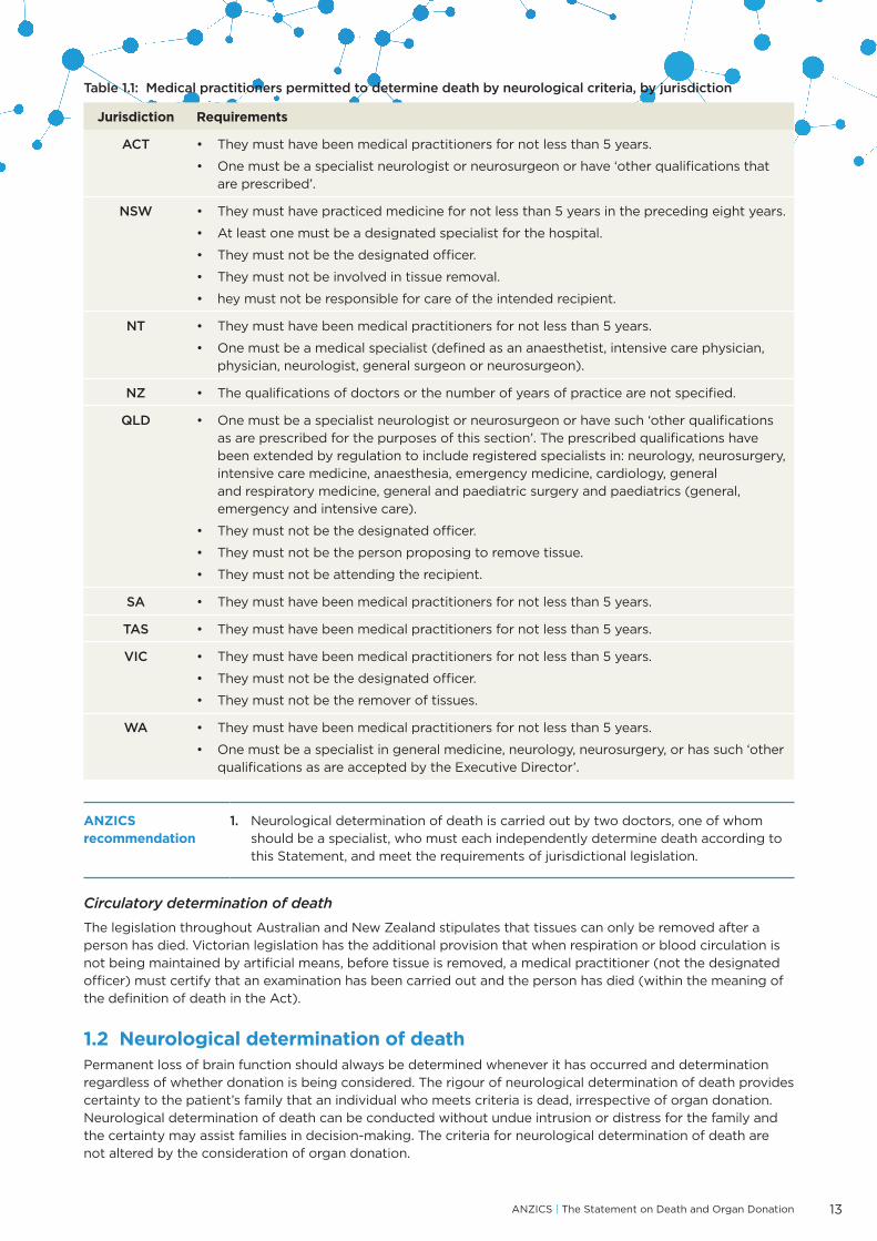
Although the Australian and New Zealand jurisdictional laws do not stipulate how death is to be determined, legislation in all jurisdictions except New Zealand requires that, for the purposes of tissue donation from patients whose death is defined by ‘irreversible cessation of all function of the brain’, the determination of death be carried out by two medical practitioners who have sufficient qualifications and experience and who have made the determination based on clinical examination. The ‘sufficient qualifications and experience’ stipulated for each jurisdiction are outlined in Table 1.1.
Although it is not a legal requirement that two medical practitioners determine death when tissue (including organ) donation is not being considered, ANZICS recommends that the same standard for neurological determination of death is followed, whether donation is being considered or not. When death by neurological criteria has been (apparently) determined by one doctor only, it does not meet the ANZICS standard.
* The wording used in legislation is not ideal, particularly the use of the word ‘irreversible’, which may be taken to mean either that the relevant vital function is not able to be reversed or that no attempt will be made to reverse it. This is particularly relevant to circulatory determination of death because it is sometimes possible, if attempts are made, to restart a heart and circulation that have ceased. Arguably, the word ‘permanent’ would have been better than ‘irreversible’.
† Note that, in the context of the legislation, ‘tissue’ refers to both organs and tissues.
ANZICS | The Statement on Death and Organ Donation 13
Table 1.1: Medical practitioners permitted to determine death by neurological criteria, by jurisdiction
Jurisdiction Requirements
ACT • They must have been medical practitioners for not less than 5 years.
• One must be a specialist neurologist or neurosurgeon or have ‘other qualifications thatare prescribed’.
NSW • They must have practiced medicine for not less than 5 years in the preceding eight years.
• At least one must be a designated specialist for the hospital.
• They must not be the designated officer.
• They must not be involved in tissue removal.
• hey must not be responsible for care of the intended recipient.
NT • They must have been medical practitioners for not less than 5 years.
• One must be a medical specialist (defined as an anaesthetist, intensive care physician,physician, neurologist, general surgeon or neurosurgeon).
NZ • The qualifications of doctors or the number of years of practice are not specified.
QLD • One must be a specialist neurologist or neurosurgeon or have such ‘other qualificationsas are prescribed for the purposes of this section’. The prescribed qualifications havebeen extended by regulation to include registered specialists in: neurology, neurosurgery,intensive care medicine, anaesthesia, emergency medicine, cardiology, generaland respiratory medicine, general and paediatric surgery and paediatrics (general,emergency and intensive care).
• They must not be the designated officer.
• They must not be the person proposing to remove tissue.
• They must not be attending the recipient.
SA • They must have been medical practitioners for not less than 5 years.
TAS • They must have been medical practitioners for not less than 5 years.
VIC • They must have been medical practitioners for not less than 5 years.
• They must not be the designated officer.
• They must not be the remover of tissues.
WA • They must have been medical practitioners for not less than 5 years.
• One must be a specialist in general medicine, neurology, neurosurgery, or has such ‘otherqualifications as are accepted by the Executive Director’.
ANZICS recommendation
1. Neurological determination of death is carried out by two doctors, one of whomshould be a specialist, who must each independently determine death according tothis Statement, and meet the requirements of jurisdictional legislation.
Circulatory determination of death
The legislation throughout Australian and New Zealand stipulates that tissues can only be removed after a person has died. Victorian legislation has the additional provision that when respiration or blood circulation is not being maintained by artificial means, before tissue is removed, a medical practitioner (not the designated officer) must certify that an examination has been carried out and the person has died (within the meaning of the definition of death in the Act).
1.2 Neurological determination of deathPermanent loss of brain function should always be determined whenever it has occurred and determination regardless of whether donation is being considered. The rigour of neurological determination of death provides certainty to the patient’s family that an individual who meets criteria is dead, irrespective of organ donation. Neurological determination of death can be conducted without undue intrusion or distress for the family and the certainty may assist families in decision-making. The criteria for neurological determination of death are not altered by the consideration of organ donation.
14
Neurological determination of death cannot be conducted without clinical or neuroimaging evidence of sufficient intracranial pathology. Cases have been reported in which the brainstem has been the primary site of injury and death of the brainstem has occurred without death of the cerebral hemispheres.28 Thus death cannot be determined through clinical examination alone when the condition causing coma and loss of all brainstem function has affected only the brainstem and there might still be cerebral blood flow to, and function of, the supratentorial part of the brain.
In cases of hypoxic-ischaemic encephalopathy the cause of the precipitating event is often obvious from the clinical history (e.g. cardiorespiratory arrest from asthma, drowning, hanging or other form of asphyxia; drug overdose, or a cardiac event). In these situations, neuroimaging is not required. In circumstances where the underlying cause is not evident (e.g. unexplained cardiac arrest or collapse) neuroimaging should be performed in case this is an otherwise clinically silent neurologic event (e.g. intracranial haemorrhage, acute hydrocephalus). If the neuroimaging shows an alternative intracranial pathology sufficient to cause brain death, then the cause is no longer hypoxic-ischaemic encephalopathy and a 24-hour waiting period will not be required prior to determination of death (see Section 1.2.2).
ANZICS recommendation
2. For neurological determination of death to be conducted, there must be definite clinical or neuroimaging evidence of acute brain pathology consistent with deterioration to permanent loss of all neurological function. In cases of hypoxic-ischaemic encephalopathy, clinical history alone may provide sufficient explanation of the acute brain pathology and not require neuroimaging prior to neurological determination of death by clinical examination.
1.2.1 PreconditionsTo determine death by clinical examination there must be evidence of sufficient intracranial pathology to deteriorate to loss of all brain function and all of the following preconditions must be met prior to and throughout clinical examination.
• Normothermia — temperature ≥35°C.
• Normotension — as a guide, systolic blood pressure ≥90 mmHg, mean arterial pressure (MAP) ≥60 mmHg in an adult and age appropriate systolic blood pressure and MAP in children (see Appendix C).
• Exclusion of effects of sedative medications (self-administered or otherwise) — the time taken for plasma concentrations of sedative medications to fall below levels with clinically significant effects depends on the dose and pharmacokinetics of medications used and on hepatic and renal function.
– Particular care should be taken to ensure the absence of continued sedative medication effect in patients who have been hypothermic (e.g. post cardiac arrest).
– In the case of barbiturates, which take many days to metabolise, including thiopentone in high dose, blood levels should be shown to be below that of clinically significant effects (<10 mg/L for thiopentone29). If there is any doubt about the persisting effects of opioids or benzodiazepines, an appropriate medication antagonist should be administered at the time of examination.
– Clinical judgement is advised if the patient has kidney or liver failure or if a longer-acting agent has been used.
• Absence of severe electrolyte, metabolic or endocrine disturbances — these include marked derangements in plasma concentrations of glucose (<3 mmol/L or >25 mmol/L), sodium (<125 mmol/L or >160 mmol/L), phosphate (<0.5 mmol/L) or magnesium (<0.5 mmol/L), urea >40 mmol/L; and severe endocrine dysfunction (untreated severe hypothyroidism or severe hypoadrenalism). Marked derangements (as defined) should be corrected before clinical examination.
• Absence of acute liver failure or decompensated chronic liver disease.
• Absence of neuromuscular-blocking drugs — unless it is known for certain that neuromuscular-blocking medications have not been administered, a peripheral nerve stimulator or other recognised method (e.g. electromyography) should always be used to confirm that neuromuscular conduction is normal.
• Ability to adequately examine the brainstem reflexes — it must be possible to examine all brainstem reflexes, with at least one ear and one eye examination.
• Cervical level spinal cord injury may also preclude reliable sensory and motor assessment — as a minimum it must be possible to assess the motor response in the facial nerve (VII) to painful stimulus in the upper limbs and to assess the motor response in the upper limbs to painful stimulus in the trigeminal (V) sensory region.
• Ability to perform apnoea testing — this may be precluded by severe hypoxic respiratory failure or a high cervical spinal cord injury.

ANZICS | The Statement on Death and Organ Donation 15
If any of these preconditions cannot be met, brain perfusion studies should be used to inform neurological determination of death (see Section 1.2.5; page 20).
1.2.2 Clinical examinationClinical examination is a requirement for neurological determination of death.
Observation period
There must be a minimum of 4 hours observation and mechanical ventilation throughout which the patient has been unresponsive to stimuli (Glasgow Coma Score [GCS] of 3), with pupils non-reactive to light, an absent cough/tracheal reflex and no spontaneous breathing effort prior to undertaking the first set of tests. In patients who are unresponsive with non-reactive pupils but appear to be spontaneously breathing, care must be taken that the ventilator is not auto-cycling/auto-triggering on a spontaneous mode.30 Temporary disconnection from the ventilator will identify any true spontaneous breaths. All preconditions must be fulfilled throughout the 4-hour observation period, before clinical examination can begin. These observations are recorded by attending nursing or medical staff.
Waiting period
Return of brain function may be delayed after resuscitation from cardiorespiratory arrest. It is therefore recommended that, in cases of acute hypoxic-ischaemic encephalopathy or post cardiac arrest, clinical examination be delayed for at least 24 hours subsequent to the restoration of spontaneous circulation.
Prolonged hypothermia (<35°C) (whether induced or accidental) may modify outcome prediction after cardiac arrest31,32 and there are published case reports suggesting that determination of death might be confounded either by hypothermia itself or by impaired clearance of associated medications.33-35 If the patient has been hypothermic for more than 6 hours, it is recommended that, clinical examination be delayed for at least 24 hours after rewarming. If the temperature has not been below 35°C, or has fallen below 35°C for less than 6 hours, the 24-hour waiting period is not required.
The 4-hour observation period and the 24-hour waiting period can finish at the same time.
Death may be determined before 24 hours if brain perfusion is absent (see Section 1.2.5; page 20).
ANZICS recommendation
3. There is a minimum 4-hour observation period prior to neurological determination of death using clinical examination alone. Throughout this observation period, all preconditions are met, the patient has a Glasgow Coma Scale of 3, with pupils non-reactive to light, absent cough/tracheal reflex and apparent apnoea on a ventilator. Following an acute hypoxic-ischaemic encephalopathy or hypothermia (<35°C) of duration greater than 6 hours, there should be a waiting period of 24 hours before determination of death using clinical examination alone.
Process of clinical examination
Clinical examination is carried out by two medical practitioners with specific experience and qualifications (see Table 1.1; page 13). The two clinical examinations are performed separately, so that the doctors and the tests are truly independent (and seen to be so). One medical practitioner performs a complete set of tests, including an apnoea test and a blood gas, and reinstitutes mechanical ventilation. Then, the other medical practitioner performs a complete set of tests, including an apnoea test and a blood gas, and reinstitutes mechanical ventilation. No fixed interval between the two clinical examinations is required, except where age-related criteria apply (see Section 1.2.6; page 22). The examinations may be done consecutively but not simultaneously. It is acceptable, but not required, for one of the doctors to be present during the examination by the other doctor, but each doctor must be responsible for performing a complete clinical examination.
The following three criteria need to be established for the neurological determination of death by clinical examination:
• absence of responsiveness; and
• absence of brainstem reflexes; and
• absence of breathing (note that apparent spontaneous breathing may result from ventilator auto-triggering/auto-cycling‡ and this should be excluded).
‡ Auto-triggering and auto-cycling occur when the ventilator, while connected to the patient, misinterprets small changes in airway pressure, caused by cardiac contraction or by pressure oscillations in the ventilator tubing respectively, thereby causing the ventilator to record a ‘patient breath’ and to provide gas flow.

16
Table 1.2 sets out the process for testing, with response and cautionary remarks for each test.
Table 1.2: Process for neurological determination of death
Responsiveness
Test Apply noxious stimuli in the cranial nerve distribution and all four limbs and trunk, observing for motor responses (e.g. pressure over the supra-orbital nerve, sternal rub, and deep nail bed pressure).
Response There should be no responsiveness. This equates to a GCS of 3.
Any motor response within the cranial nerve distribution, or any response in the limbs in response to cranial nerve stimulation: stop clinical testing as this precludes neurological determination of death.
Cautionary remarks
Spinal reflexes may be present in patients with permanent loss of brain function (see Section 1.2.3; page 19). Spinal reflexes are not to be confused with a pathological flexion or extension response.
Throughout this Statement, due to the context of intubation, GCS 3 = GCS 2T
Brainstem Reflexes
General remarks
Testing of the brainstem reflexes comprises examination of the cranial nerves: pupils, eye movements, facial sensation and movement, pharyngeal and tracheal response. These are tested sequentially and bilaterally when possible. Not all cranial nerves have a testable reflex associated with them in the context of severe brain injury.
All testable brainstem reflexes must be absent for neurological determination of death.
Pupillary light reflex — cranial nerves II & III
Test Shine a bright light into the eye and look for a pupillary constrictor response.
Response No pupillary constriction response: proceed with testing other brainstem reflexes.
Pupillary light reflex is observed: stop clinical testing, as this precludes neurological determination of death.
Cautionary remarks
The pupils must be at least midsize in diameter.36,37
Anti-cholinergic medications such as atropine can cause pupillary dilatation.
Cataract or iris surgery does not preclude the test.
Corneal reflex — cranial nerves V & VII
Test Touch the corneas with sterile soft cotton wool or gauze and examine the eyes for blinking or other response.
Response No blinking or other response: proceed with testing other brainstem reflexes
Blink reflex is observed: stop clinical testing, as this precludes neurological determination of death.
Cautionary remarks
Touching the sclera is not sufficient. Examine the cornea gently, by touching rather than scraping, as it is easily damaged.
Reflex response to pain in the trigeminal distribution — cranial nerves V & VII
Test Apply pain over the trigeminal distribution, e.g. pressure over the supra-orbital nerve.
Response No facial or limb movement: proceed with testing other brainstem reflexes.
Facial or limb movement is observed: stop clinical testing, as this precludes neurological determination of death.
Vestibulo-ocular reflex — cranial nerves III, IV, VI & VIII
Test Inspect the external auditory canal with an otoscope to confirm that the eardrum is visible. If the eardrum is not visible, the canal must be cleared before testing can occur. Elevate the head to 30° to place the horizontal semicircular canal in a horizontal position. Instil 50 mL of ice-cold water (less for a child) into the ear canal using a syringe. Hold eyelids open and observe for eye movement for a minimum of 60 seconds.

ANZICS | The Statement on Death and Organ Donation 17
Response No eye movement in response to the cold water; the eyes remain in the midline within the socket: proceed with testing other brainstem reflexes.
Presence of any movement, including tonic deviation or nystagmus: stop clinical testing, as this precludes neurological determination of death.
Cautionary remarks
A ruptured eardrum does not preclude the test. Fractures to base of skull or petrous temporal bone may obliterate the response on the side of the fracture.
Testing for the oculocephalic reflex (head turning) examines the same reflex pathways but is a sub-maximal stimulus and is not recommended. It may also aggravate a pre-existing cervical spinal injury.
Gag reflex — cranial nerves IX & X
Test Touch the posterior pharyngeal wall, on both sides, with a tongue depressor or cotton swab. A laryngoscope or video laryngoscope may assist in obtaining a good view of the pharynx for stimulation.
Response No gag response: proceed with testing other brainstem reflexes.
Gag response: do not proceed with clinical testing, as this precludes neurological determination of death.
Cautionary remarks
If the patient is orally intubated, the gag reflex may be difficult to discern. It is important to view the posterior pharyngeal wall and/or uvula.
Cough/ tracheal reflex — cranial nerve X
Test Stimulate the tracheobronchial wall with a soft suction catheter.
Response No cough response is seen: proceed with testing other brainstem reflexes.
Cough response is observed: do not proceed with clinical testing, as this precludes neurological determination of death.
Cautionary remarks
The efferent limbs for this reflex are the phrenic nerve and the nerves of the thoracic and abdominal muscles. Therefore, it cannot be assessed in patients with high cervical cord injury.
Breathing
ONLY if all the above reflexes are absent, proceed with testing for apnoea.
The apnoea test should be conducted last so that a high pressure of carbon dioxide (PaCO₂) could not be potentially confounding if brain perfusion was present.
General remarks
Apnoeic oxygenation is used to demonstrate lack of ventilatory drive. This involves the supply of 100 per cent oxygen to the trachea, without providing ventilatory assistance. Through gas mass-movement, oxygen reaches the alveoli, allowing for transfer to the blood. In normal circumstances, in the absence of ventilation, PaCO₂ rises and pHa falls, and associated changes in PCO₂ and pH stimulate the brainstem respiratory centres via peripheral chemoreceptors, or by direct effects on the respiratory centres. Usually PaCO₂ rises by ~3 mmHg (0.4 kPa) for every minute of apnoea.38,39 As the PaCO₂ rises, the ventilatory centre is maximally stimulated by a PaCO₂ of ~60 mmHg (a pH <7.30).
Attempt at breathing is defined as any respiratory muscle activity that results in abdominal or chest excursions or activity of accessory respiratory muscles.

18
Breathing (continued)
Test Throughout the procedure, monitor the patient’s percentage blood oxygenation saturation (SpO₂). Pre-oxygenate the patient with 100% oxygen for at least 5 minutes40 to eliminate nitrogen in the respiratory tract and prevent hypoxaemia during the test. An option to minimise the time required for the PaCO₂ to rise to the desired level is to mechanically ventilate to mild hypercarbia (PaCO₂ ~45 mmHg [6 kPa]) before disconnecting the patient from the ventilator. Disconnect the patient from the mechanical ventilator. While mechanical ventilation is temporarily stopped, supply oxygen via a self-inflating bag with a positive end-expiratory pressure (PEEP) valve to prevent atelectasis.
It is recommended not to deliver oxygen via a catheter inserted through the endotracheal tube and placed above the carina. A continuous positive air pressure (CPAP) circuit may also be used to deliver apnoeic oxygenation. The use of a T-piece is not recommended.
In patients receiving ECMO, pre-oxygenate the patient with 100% oxygen for at least 5 minutes via both the ventilator and the ECMO circuit and ensure that the PaCO₂ is ~45 mm Hg [6 KPa] before connecting the patient to a CPAP circuit on 100% oxygen (CPAP level of 8-10 cm H₂O or the same as the previous level of PEEP on ventilator if this was higher). Reduce the sweep gas flow on the ECMO circuit to 0.5–1 L/min (for paediatric patients reduce sweep gas flow rate to 10-25% of the blood flow rate) and be prepared to increase this somewhat should desaturation occur before hypercarbia. PaCO₂ can be expected to rise 3-5 mm Hg per minute.
Expose the chest and abdomen and observe continuously for any spontaneous breathing.
Request an arterial blood gas be taken to document the rise in PaCO₂. If the patient is stable, await the (rapid) return of the arterial blood gas result before reconnecting to the ventilator in case the required change in PaCO₂ or pH has not been achieved.
At end of testing, return the patient to mechanical ventilation or restore ECMO settings.
Response No breathing effort is seen with testing: this concludes the clinical testing of brain function.
Spontaneous breathing is observed during the test: stop testing, as this precludes neurological determination of death.
Cautionary remarks
At the end of the period without mechanical ventilation, apnoea must persist in the presence of an adequate stimulus to spontaneous ventilation, i.e. an arterial PaCO₂ >60 mmHg (8 kPa) and an arterial pH <7.30. In patients with pre-existing hypercapnia, it is recommended to wait for a PaCO₂ rise of >20 mmHg (2.7 Kpa) above the chronic level, with a pH <7.30.
If starting from normocapnoea, the PaCO₂ is likely to be >60 mmHg (8 kPa) after 10 minutes. If this is not the case, wait a further 5 minutes and repeat the arterial blood gas.
The period of observation to achieve an adequate threshold of stimulus of the respiratory centre is variable. Failure of the PaCO₂ to rise is most likely due to an inappropriately high oxygen flow rate via a tracheal catheter.
Patients may become hypoxic or develop haemodynamic instability during this process. Adequate pre-oxygenation usually avoids this problem. If hypoxia (<SaO₂ 88) does occur, give 1–2 mandatory breaths and continue apnoea testing. Although some degree of hypoxia may be well-tolerated, if the patient develops malignant dysrhythmia, then testing may need to be abandoned.
The CPAP circuit on the ventilator should not be used due to the known risk of auto-triggering or auto-cycling, which is then misinterpreted by the clinician as spontaneous breathing. Delivery of oxygen via a catheter in the endotracheal tube is not recommended because:
• the flow of oxygen will reduce the required rise in the PaCO₂• wedging of the catheter may raise intrapulmonary pressure and cause barotrauma41• CPAP cannot be maintained• it is difficult to give the patient 1-2 mandatory breaths if the patient desaturates significantly.

ANZICS | The Statement on Death and Organ Donation 19
Family presence during clinical examination
The opportunity to observe the clinical examination of brain function should be offered to family members.42 If the family are to be present, the intensivist should explain the tests and responses, particularly forewarning them of the possibility of spinal reflexes. There must be someone available (e.g. a nurse) to support the family. It may also be helpful if there is a designated person able to explain the process as it is carried out.
Diagrams or imaging demonstrating absent brain perfusion (see Section 1.2.5) may help the family understand permanent loss of brain function.
ANZICS recommendation
4. The opportunity to observe a clinical examination of brain function should beoffered to family members. Appropriate explanation and support for the familyshould be provided.
Repeat testing
If clinical examination or imaging (see Section 1.2.5) demonstrate that permanent loss of brain function has not yet occurred, consideration should be given to repeating these tests after a suitable interval.
1.2.3 Observations that are compatible with permanent loss of brain functionSpinal reflexes can be either spontaneous or elicited by stimulation outside the cranial nerve distribution. They occur in up to 50% of patients with permanent loss of brain function and are the result of a functioning spinal arc with loss of higher centre inhibitory control. Reflex movements generally occur within the first 24 hours of death being determined but can occur hours or days after a period of flaccid paralysis.43
Spinal reflexes can be quite complex in form and can be confronting when observed by family members and staff. It is essential that these movements are acknowledged and their origin explained. It is recommended that a cerebral blood flow study be performed if there is any doubt about whether movements are spinal in origin.
Examples of spinal reflexes include:44-47
• extension-pronation movements of the upper limbs or non-specific flexion of the lower limbs;
• undulating toe reflex (plantar flexion of great toe, followed by brief plantar flexion sequentially of second tofifth toes);
• Lazarus sign (bilateral arm flexion, shoulder adduction, hand raising to above the chest, and may includeflexion of trunk, hips and knees);48,49
• deep tendon reflexes;
• plantar responses, either flexor or extensor; and
• head turning.50
Other physiological signs that do not preclude permanent loss of brain function include:
• sweating, blushing, tachycardia;
• normal blood pressure without the need for pharmacological support;
• absence of diabetes insipidus; and
• having intracranial pressure less than mean arterial pressure.
1.2.4 Observations that are incompatible with permanent loss of brain functionThe following are incompatible with permanent loss of brain function:
• decerebrate or decorticate posturing;
• true extensor or flexor motor responses to painful stimuli;
• seizures; and
• limb movement elicited by stimulation of the cranial sensory nerves or facial movement elicited bystimulation of torso/limbs.
20
1.2.5 Demonstrating the absence of brain perfusionIn situations where the clinical examination cannot be solely relied upon for neurological determination of death, it is essential to undertake imaging to demonstrate the absence of brain perfusion. When imaging is required, it must be preceded by undertaking those parts of the clinical examination that are possible. Testing for brain perfusion should be deferred until responsiveness, examinable brainstem reflexes and breathing effort are all absent.
Imaging should only be performed if the systemic blood pressure is adequate (as a guide, systolic blood pressure >90 mmHg, MAP >60 mmHg in an adult) and should be performed by a specialist in radiology or nuclearmedicine. It is recommended (but not mandatory) that there is at least 4 hours observation of unresponsiveness,absence of brainstem reflexes and no spontaneous breathing effort before imaging is performed. This willincrease the likelihood that the test will indicate absent brain perfusion.
In the pathogenesis of permanent loss of brain function from any cause, injury to brain tissue and subsequent oedema cause the intracranial pressure to rise to equal or exceed the systemic arterial pressure, thus occluding intracranial blood flow. This occlusion is hydrodynamic, occurs where the cerebral arteries become intracranial and is not due to arterial thrombosis.9,10,51 When intracranial pressure is not consistently greater than blood pressure, or subsequently falls below it, delayed filling of major vessels may be seen on arteriography/imaging. This may occur in infants, in patients with massive skull fractures, or if craniotomy with extensive bone removal has occurred.52 It may then take some time before brain blood flow stops completely and permanently.
Although the absence of brain perfusion is determined by a radiologist or nuclear physician, it is the responsibility of two medical practitioners who have clinically examined the patient to determine that the patient has died.
If an imaging study shows that perfusion is still present, the findings should be discussed with the family, along with the various options that exist and their implications. These include continuing ventilatory support and testing neurological function at a later stage, either by a second study showing absent brain perfusion or by clinical examination if the clinical examination is no longer confounded. These options preserve the possibility of organ donation after neurological determination of death. The other option is that treatment might be withdrawn prior to cessation of brain function, and this might include the possibility of organ donation after circulatory determination of death.
Imaging techniques for assessing brain perfusion
Imaging tests must have a high sensitivity and, most importantly, a specificity of 100% to avoid the false conclusion that brain perfusion is absent in a person who does not meet neurological criteria for death.
The three acceptable imaging techniques for demonstrating absent brain perfusion are intra-arterial catheter angiography, radionuclide imaging and computed tomography angiography (CTA).
Intra-arterial catheter angiography, with digital subtraction
• Intra-arterial catheter angiography is regarded as the gold standard test for absence of perfusion.
• Intra-arterial contrast must be absent above the level of the carotid siphon in the anterior circulation andabove the foramen magnum in the posterior circulation.53
• Four-vessel angiography is direct injection of contrast medium into both carotid arteries and both vertebralarteries.54 Three-vessel angiography refers to the injection of contrast medium into both carotid arteries andthe basilar artery.
Radionuclide imaging
• Tc-99m HMPAO (technetium 99m radiolabelled hexamethyl propylene amine oxime) is a radionuclide thatdemonstrates perfusion and crosses the blood–brain barrier to then be retained by brain parenchyma(by conversion from a lipophilic to a hydrophilic form).55,56 The absence of radionuclide intracranially iscompared to the presence of radionuclide extra cranially.
• Blood pool or blood-flow agents, such as Tc-99m pertechnetate, Tc-99 DTPA (diethylene-triamine-penta-acetate) or Tc-99m glucoheptonate, are not acceptable radionuclides to demonstrate absence of perfusion,because they do not cross the blood–brain barrier and do not remain within the intracranial cavity longenough for static gamma camera imaging, leading to false positive and negative results.
• Although two-planar imaging is still used, single photon emission computerised tomography (SPECT)provides superior imaging in adults57-59 and in children.60
ANZICS | The Statement on Death and Organ Donation 21
Computed tomography angiography
• Computed tomography angiography (CTA) to demonstrate absent brain perfusion is acceptable (subject tothe radiologic guidelines set out below) where intra-arterial catheter angiography or radionuclide imagingis not readily available, because more proximal intracranial arteries (Circle of Willis, A1, M1, P1) may showcontrast enhancement when there is permanent loss of brain function. This enhancement is a reflection ofthe high sensitivity of CTA to small amounts of contrast that may admix within the Circle of Willis in theabsence of brain perfusion.
• If CTA is performed, ANZICS recommends the use of a four-point scale61,62 to assess absent enhancementof peripheral intracranial arteries at 20 seconds and central veins at 60 seconds after an intravenous bolusinjection of 120 mL of >300 mg/mL non-ionic contrast injected at 3 mL/sec via a power injector through atleast an 18 g intravenous catheter.
• Before assessing brain perfusion with the four-point scale, the presence of contrast enhancement ofexternal carotid artery branches is required to confirm that the study is technically adequate.
• Criteria for absent brain perfusion under the four-point scale are:
– absent enhancement of both middle cerebral artery (MCA) cortical branches (i.e. beyond the Sylvianbranches); and
– absent enhancement of both internal cerebral veins.
• The four-point scale has demonstrated 100% specificity and 85.7% sensitivity.61 A subsequent meta-analysisof CTA in 322 patients with permanent loss of brain function using the four-point scale showed a sensitivityof 87%.62 Specificity was not assessed in the meta-analysis. A seven-point scale (based on absence ofopacification in the cortical segments of bilateral MCAs, bilateral internal cerebral veins, bilateral pericallosalarteries and the great cerebral vein) used in France since 1998 has a demonstrated specificity of 100% but asensitivity of only 68.2%.63
• Cautionary notes
– Solely relying on absent enhancement of the bilateral internal cerebral veins, while sensitive, is ofquestionable specificity given the possibility of venous sinus thrombosis in a patient without permanentloss of brain function.
– Assessment of the specificity of CTA has been limited by the small number of normal controls in thepublished literature.
Magnetic resonance imaging
• Magnetic resonance imaging (MRI),64 flow sensitive gradient-echo MRI,65 post-gadolinium MRI66 andmagnetic resonance angiography67 (MRA) have all been used to demonstrate absence of brain perfusion.
• Reduced sensitivity of some MRI techniques to slow flow may mimic occlusion and lead to a false positiveneurological determination of death. Furthermore, flow detection with MRI is too dependent on a largenumber of variables including field strength, sequence type, sequence parameters such as slice thicknessand echo time, and physiological variables such as direction of flow relative to image slice and flowvelocity and pulsatility.
• Assessment of brain perfusion by MRI is not recommended due to the potential for false positives, variablesin flow detection, the lack of large studies with matched controls and the logistical difficulties in performingMRI on monitored intensive care patients.
Transcranial Doppler
Transcranial Doppler (TCD) is not an acceptable technique because of operator dependence, difficulty with assessing the posterior intracranial circulation and diagnostic inaccuracy.68,69 It may be used as a screening test to optimise the timing of the imaging (i.e. to avoid performing imaging at a time when TCD indicates persisting cerebral artery flow and reduce the need to repeat the study).
ANZICS recommendation
5. When imaging to demonstrate absence of brain perfusion is required, it must bepreceded by performance of those parts of the clinical examination that are possible.Responsiveness, all testable brainstem reflexes and breathing effort must be absent.
6. If assessment of brain perfusion is required, three- or four-vessel angiography orradionuclide imaging are preferred. Computed tomography angiography is acceptableif recommended radiological diagnostic guidelines are followed. Magnetic resonanceimaging or angiography and transcranial Doppler should not be used.
22
1.2.6 Neurological determination of death in childrenThe requirements for neurological determination of death, including preconditions, in neonates, infants and children are similar to those in adults. This is supported by current guidelines from Canada in 2006,70 the United States in 201171 and the United Kingdom in 2015.72 It is approached with caution in neonates and infants due to its relative infrequency and immaturity of brainstem reflexes.
While previous guidelines in the United Kingdom suggested that neurological determination of death, by clinical means, could not be made in infants less than 2 months old, the 2015 revised guidelines of the Royal College of Paediatrics and Child Health accept neurological determination of death in neonates.72 The current guidelines from Canada and the United States also support this position. All recommend an increasingly cautious approach with decreasing age.
Neurological determination of death by clinical examination in preterm neonates is unreliable due to the relative immaturity of brainstem reflexes
Thus, the ANZICS recommendations below are similar to those in Canada, the United States and the United Kingdom.
Additional considerations in term neonates
For term (≥37 weeks’ gestation) neonates, a prolonged period of observation before clinical examination is warranted in light of the frequent inability to define the timing, severity and duration of the initial insult leading to coma.73 International guidelines range in their recommended minimum period of observation prior to neurological determination of death of between 24 and 48 hours.70-72 Limited data from neonates referred for organ donation following neurological determination of death, support this position.74 Concerns about the initial insult, along with the particular difficulties of performing clinical examination in the neonate,73-75 give rise to the recommendation that two clinical examinations be conducted. The recommended minimum separations have been either 24 and 48 hours.70-72,75-77
Preterm neonates
Below 37 weeks of gestation, caution is needed as many of the brainstem reflexes to be tested are developing or have only recently developed.78 For example, the pupillary response to light appears at 30 weeks but is only consistently present at 32–35 weeks of gestation,79 and the central respiratory response to carbon dioxide (CO₂) is relatively poorly developed below 33 weeks of gestation.80,81 Furthermore, clinical examination is technically more difficult in these very small infants75 and there is an isolated report of good neurological recovery after apparent fulfilment of neurological criteria for death.82
As a result of this uncertainty, it is recommended that neurological determination of death in preterm neonates utilise brain perfusion imaging rather than rely on clinical examination alone. In preterm neonates who have some preservation of brain perfusion in the setting of absent brainstem reflexes, a repeat study will usually show loss of brain perfusion within 48 hours.75
ANZICS recommendation
7. Neurological determination of death cannot be conducted with certainty by clinicalexamination in preterm neonates (<37 weeks’ gestation) or in term neonates in thefirst 24 hours after birth.
8. Neurological determination of death by clinical examination can be made in termneonates (≥37 weeks’ gestation). In term neonates between 24 hours and 30days old, an observation period of 24 hours should occur before the first clinicalexamination, followed by a 24-hour interval before the second clinical examination.
9. The criteria for neurological determination of death in children above the age of 30days are the same as those in adults.
10. Brain perfusion studies can be used to assist in the neurological determination of deathin children of all ages including term and preterm neonates, when preconditions forneurological testing cannot be met.

ANZICS | The Statement on Death and Organ Donation 23
1.2.7 Documentation of neurological determination of deathThe process of clinical examination alone or with ancillary brain perfusion imaging which leads to neurological determination of death must be documented in the medical record. Death is certified when the two medical practitioners defined by local legislation have both completed the process required for neurological determination of death. This requirement was a recommendation of the Australian Law Reform Commission, which intended the neurological determination of death to have general application, irrespective of whether or not organ or tissue donation is to follow.20
The time of death should be recorded as the time the second medical practitioner determines that death has occurred whether this is by clinical examination alone or with the assistance of imaging. The rationale for this recommendation is that the process of determining death is only complete at this time.
An example of a form recommended for the documentation of neurological determination of death is included as Appendix D, page 57.
ANZICS recommendation
11. Documentation of neurological determination of death should be made using aspecific form (see Appendix D) to demonstrate explicitly that all criteria set out inthis Statement are met, whether or not organ or tissue donation occurs. The samecriteria should be listed in local hospital forms.
1.3 Circulatory determination of death1.3.1 Criteria for circulatory determination of deathAs discussed in Section 1.1.4, the legal definition of death in all Australian jurisdictions is either ‘irreversible cessation of all function of the brain of the person’ or the ‘irreversible cessation of circulation of blood in the body of the person’. Almost all determinations of death, whether in hospitals or in the community, are by confirmation that the person is unresponsive, not breathing, is not moving and has no pulse. This is circulatory determination of death.
Duration of circulatory arrest to satisfy determination of death
Death cannot be determined until there is certainty regarding the permanence of the circulatory arrest. Permanence means that circulation ‘will not resume spontaneously and will not be restored through intervention’. Certainty is assured when the duration of cessation of circulation has extended beyond the possibility of spontaneous resumption of cardiac contraction causing antegrade circulation, known as autoresuscitation.
Previous research had concluded that autoresuscitation after withdrawal of cardiorespiratory support did not occur after 2 minutes of cessation of circulation.18 In 2018, an updated review concluded that autoresuscitation between 2 and 5 minutes was rare.83 An Australian audit conducted in Queensland of the last 10 years of donation after circulatory death reported 2 cases out of 176 (1%) in which autoresuscitation occurred between 2 and 3 minutes.84
A prospective observational study in 20 ICUs in 3 countries of 631 adults who died following withdrawal of cardiorespiratory support revealed a spontaneous resumption of cardiac activity after a period of pulselessness in 14% of patients, with the longest duration before resumption of 4 minutes 20 seconds.85 Although all patients in the study died, 49% of the resumptions were for only one QRS cycle and the median duration of resumption was 3.9 seconds, nevertheless the resumptions occurred up to 4 minutes 20 seconds. ANZICS, therefore, has concluded that the circulatory determination of death requires absence of circulation for 5 minutes. It is not necessary, or desirable, to extend the period of observation beyond 5 minutes of circulatory arrest.
In the context of donation after circulatory determination of death, it is important to determine death as soon as possible to minimise warm ischaemia of the organs to be retrieved. For this reason, no more than 5 minutes of circulatory arrest is recommended.
This timeframe for circulatory determination of death only applies in the context of withdrawal of cardiorespiratory support, where there is a subsequent monitored circulatory arrest or where there is no intention to restore or re-establish cardiorespiratory function. It does not apply following cardiopulmonary resuscitative attempts or where it is reasonable to provide such interventions. After failed resuscitation, there are reports of spontaneous resumption of the circulation up to 10 minutes post cessation of cardiopulmonary resuscitation, which is thought to result from cardiopulmonary interactions such as dynamic hyperinflation from mechanical ventilation.83
In the context of donation after circulatory death, once circulatory arrest has occurred, interventions that may restore circulation must not be undertaken. This includes cardiac compressions and ventilation. After determination of death, however, reintubation without ventilation to prevent aspiration into lungs intended for donation, is necessary and permissible.
24
Method of determination
The usual manner of determining death is to confirm that the patient is unresponsive, is not breathing, has no pulse or heart sounds and, for some doctors, that the patient’s pupils are fixed and dilated. In the context of donation after circulatory death, due to the requirement to detect the exact onset of cessation of circulation and due to the presence of family during this period of observation, the monitoring of the patient is different.
Although there are some international variations in the circulatory determination of death,18 there is general consensus that coma and sustained absence of palpable pulse, heart sounds, breath sounds and chest wall movement are necessary criteria. Electrical asystole is not required for this determination as electrical activity may continue for minutes after cessation of circulation.
Arterial line monitoring is the recommended method to determine the loss of pulsatile arterial blood pressure in the context of donation. Absent pulse on the arterial line for 5 minutes is observed and then confirmed by clinical examination (absent heart sounds and/or absent central pulse).18,86-89 Reliance on an isoelectric ECG may unnecessarily prolong the observation period so the ECG should not be monitored if an arterial line is present.
In the 2021 observational study 7% of the patients had ECG activity for greater than 30 minutes post cessation of a pulsatile arterial blood pressure.
When the use of an arterial line is unacceptable to the patient or family or is not technically possible, electrical asystole must be observed for 5 minutes on the ECG. Absent circulation is then confirmed by clinical examination (absent heart sounds and/or absent central pulse).
ANZICS recommendation
12. Circulatory determination of death in the context of organ donation requires theabsence of spontaneous movement, breathing and circulation. Absence of circulationis evidenced by absent arterial pulsatility for 5 minutes using intra-arterialpressure monitoring and confirmed by clinical examination (absent heart soundsand/or absent central pulse). In cases without an arterial line, electrical asystoleshould be observed for 5 minutes on the electrocardiogram and confirmed byclinical examination.
1.3.2 Documentation of circulatory determination of death in the context of organ donationThe doctor responsible for determining death should document the time and date of determination of death, along with his or her name and signature. A proforma (see Appendix E; page 59) will facilitate this. If organ donation is to take place, it is important that only one clock be used to time events, to ensure consistency in time keeping throughout the process. This will allow accurate and consistent recording of time on all documentation (including time of death, warm ischaemic time) and ensure that operating theatre documentation does not inadvertently record organ removal as having begun before the time of determination of death.90
ANZICS recommendation
13. For the purposes of organ donation, circulatory determination of death shouldbe documented using a specific form (see Appendix E) to demonstrate explicitlythat all criteria set out in this Statement are met. The same criteria should belisted in local hospital forms.

ANZICS | The Statement on Death and Organ Donation 25
2Organ and Tissue Donation After Death
Chapter 1 outlined the legal framework and processes for neurological and circulatory determination of death, using terminology that clearly reflects that death does not differ in type, rather in the way in which it is determined. In this chapter, these terms are used interchangeably with ‘brain death’ and ‘circulatory death’ for ease and familiarity. The chapter discusses:
• organ and tissue donation from an ethical and cultural perspective and the legalframework for donation in Australia and New Zealand (see Section 2.1; page 25);
• donation potential in general and after brain death or circulatory death (seeSection 2.2; page 29);
• medical management of patients with permanent loss of brain function beforeand during organ retrieval (see Section 2.3; page 31);
• the principles and evidence supporting donation after circulatory determinationof death and the process of proceeding to donation (see Section 2.4; page 34);
• paediatric donation (see Section 2.5; page 38); and
• tissue-only donation (see Section 2.6; page 39).
2.1 Organ and tissue donation in perspectiveThis Statement provides a standard for donation of organs and tissues after death based on good medical practice (see Sections 2.3; page 31 and 2.4; page 34), ethics and the law.
2.1.1 Organ and tissue donation from an ethical perspectiveCurrent Australian and New Zealand practice is based on donation of organs and tissues being an unconditional altruistic, non-commercial act. Donated organs are allocated to the most suitable recipients on the transplant waiting list.
This Statement supports organ donation in accordance with the following.
• The World Health Organization guiding principles on human cell, tissue and organ transplantation,90which states ‘The allocation of organs, cells and tissues processes should be guided by clinical criteria andethical norms, not financial or other considerations. Allocation rules, defined by appropriately constitutedcommittees, should be equitable, externally justified, and transparent.’
• The Declaration of Istanbul,91 which emphasises that organ trafficking and transplant tourism should beprohibited because they violate the principles of equity, justice and respect for human dignity.
• The Australian and New Zealand Human Tissue Acts governing organ and tissue donation (see Section 2.1.3;page 27), which prohibit trading in human organs or tissue.

26
• The Australian NHMRC in its 2016 Ethical Guidelines for organ transplantation from deceased donors,2which state as a general principle that: ‘Donation of organs is an act of altruism, solidarity and communityreciprocity that provides significant benefits to those in medical need’.
Altruism
ANZICS believes that:
• no person, organisation or company should profit financially from organ or tissue donation; and
• neither the estate of an organ or tissue donor nor his or her family should incur any cost from the processesthat occur primarily to facilitate organ and tissue donation.
When the family wishes to place conditions on donation (conditional donation)
Sometimes the family expresses interest or a wish to place conditions on the donation of organs or tissues by their family member. The condition may be expressed negatively (e.g. not to persons of a specific race or life-style) or positively (e.g. only to somebody of a specific religion). Some families may refuse to donate at all if their condition is not accepted. Subject to the following discussion of directed donation, ANZICS opposes donation with conditions even if it means that no organs are donated. In such situations, it is recommended that the situation be discussed with donation staff as soon as possible.
When the family wishes to direct donation to a specific recipient (directed donation)
Directed donation is only a consideration when no conditions are imposed, there is consent from the family and donation is in accordance with the following guidelines.
In Australia, the NHMRC ethical guidelines2 consider that it may be ethical for deceased directed donation to occur when:
• “there is evidence that the person was prepared to be an organ donor after death;
• there is evidence (e.g. through a living will, advanced care directive or prior planning with a transplantteam) that the person expressed a preference for certain organs to be donated to a close relative in needof a transplant;
• the potential recipient is considered eligible for transplantation and consents to receiving organs from thatdonor.”
In New Zealand, Organ Donation New Zealand (ODNZ) released a Standard Operating Procedure for Directed Donation from Deceased Donors in 2018,93 which outlines the following.
• ODNZ will not support donation when donors or their families request that organs be offered only tospecific groups of persons (e.g. members of a specific ethnicity or religion) or request the exclusion ofmembers of such groups as recipients.
• ODNZ will support donation to a specific named individual subject to the following conditions.
– The named individual has had a personal, long-standing relationship with the donor or the donor family.
– The named individual is currently listed for transplantation within New Zealand or Australia.
– The proposed donation would not conflict with the existing Australian and New Zealand organ allocationagreements for patients listed for liver or heart transplantation at ‘Urgent’ status.94
– The relevant senior transplant clinician agrees that the proposal is medically appropriate, feasible andwill be supported by the transplant service despite not conforming to the normal process for allocationof that organ.
– In recognition of their conflict of interest, the proposed recipient is not physically present when theformal decision about donation of the proposed organ is made.
– The proposed recipient is free to accept or decline the organ in discussion with the relevant seniortransplant clinician.
– The outcome of the family decision and/or the proposed recipient’s decision will be relayed to the familyby the intensivist (or by the ODNZ co-ordinator is he/she is involved in the consent process).
If the proposed donation does not proceed, either because the family decide against it in the absenceof the proposed recipient, or the proposed recipient declines the organ in the absence of the rest of thefamily, there is risk of disruption within the donor family.
In this situation there needs to be careful discussion between the ODNZ medical specialist and intensivistabout how to inform the family that the proposed donation will not go ahead. The family and the proposedrecipient both have the right to keep their reasons confidential. It should be explained, without goinginto specific details, that either the donor or the recipient is unsuitable.

ANZICS | The Statement on Death and Organ Donation 27
• If ODNZ does not support directed donation under either of the points above:
– ODNZ will ensure that ICU staff give the family the opportunity to donate without conditions
– the decision not to support directed donation will apply even if the outcome is that the family do notagree to any donation at all.
Consent models
Australia and New Zealand have an ‘opt in’ model of organ donation. In contrast, an ‘opt out’, ‘presumed consent’ or ‘deemed consent’ model is commonly proposed as a legislative change to increase organ donation consent rates. In an opt out model, it is assumed that each member of the community would be prepared to donate unless they register a decision to the contrary. Usually this model allows for families to decline donation. Many countries, including most recently Wales,95,96 have not reported an increase in actual donors with a change to a presumed consent model. Some comparative studies between countries have reported an association between consent model and actual donors and suggest it is causal, while others have not found an association at all.97,98
The evidence about whether consent models by themselves influence the number of actual donors is confounded by association with other factors in various countries and, in those countries which have changed to ‘presumed consent’ and similar models, there has not been consistent evidence that the number of actual donors has increased. Risks of legislative government intervention in such an emotionally complex issue and a reduced focus on excellent family care and communication are realistic concerns. There is no evidence that changing to presumed consent internationally or in Australia or New Zealand would result in an increase in donation.99,100
ANZICS does not support a presumed consent system for organ donation. It believes the focus should remain on providing compassionate communication, adequate information about donation and the highest quality of care so that every family can make an informed and enduring decision that is right for them and their family member.
2.1.2 Organ and tissue donation from a cultural perspectiveAustralia and New Zealand both have multicultural populations. They have distinct indigenous populations and a large proportion of the population of both countries was born overseas.101,102 Intensivists therefore need to be aware that an individual or family’s cultural or religious affiliation can affect health-care decisions. This is vitally important in considering their views on organ donation and how these affect their decisions. Rather than assume knowledge, it is best to adopt an approach of cultural humility.103 The ANZICS Statement on care and decision-making at the end of life for the critically ill1 contains practical information on the use of cultural leaders who may be helpful when exploring end-of-life issues including organ and tissue donation. The Australian Organ and Tissue Authority (AOTA)104 and ODNZ have resources that may also be helpful. A more detailed consideration of deceased donation in indigenous cultures in both Australia and New Zealand is included in Section 3.5. There are no cultures in which organ donation is known to be strictly prohibited and all patients and their families have the right to be offered the opportunity to donate.
2.1.3 Organ and tissue donation: the legal frameworkIn all Australian jurisdictions and in New Zealand, there is a legislative basis for the removal of tissues (including organs) after death, for the purpose of transplantation. Legislation in the various jurisdictions uses the word ‘tissue’ in a legal sense (see Glossary, page 54), which includes what health care professionals customarily refer to as organs. Accordingly, in reading this section, wherever the word ‘tissue’ appears, it should be read as ‘tissues, including organs’.
The relevant Acts are as follows (most of the Acts have been amended over the years and several jurisdictions also have statutory regulations under the relevant Act).
Table 2.1: Legislative basis for the removal of tissues (including organs) after death
Jurisdiction Legislation
ACT Transplantation and Anatomy Act 1978 and Transplantation and Anatomy Regulation 2001
NSW Human Tissue Act 1983
NZ Human Tissue Act 2008
NT Transplantation and Anatomy Act 2014
QLD Transplantation and Anatomy Act 1979 and Transplantation and Anatomy Regulation 2017
SA Transplant and Anatomy Act 1983 and Death (Definition) Act 1983
TAS Human Tissue Act 1985
VIC Human Tissue Act 1982 and Human Tissue Regulations 2006
WA Human Tissue and Transplant Act 1982 and Human Tissue and Transplant Regulations 2006

28
The New Zealand legislation is quite detailed in its consent provisions. It defines informed consent, informed objection and over-riding objection and states that they must take into account the cultural and spiritual needs, values, and beliefs of the immediate family of the individual. It further states that informed consent may be given by the individual before death; or after death by the individual’s nominee, a member of the individual’s immediate family, or a close available relative. It allows for over-riding objection by a close available relative. It states that a person who proposes to collect or use tissue from a dead human body must:
• ascertain what informed consent is required, whether it has been given and whether it has been over-ridden by objection; and
• consult with the person lawfully in possession of the body (specified as the person in charge of the hospital).
Legislation in the various jurisdictions specifies who may authorise removal of tissue and when it may be authorised. In different ways, all legislation provides a sound legal basis for the removal of organs and tissues after death. However, there is considerable variation in how far legislation goes in defining death and specifying criteria and requirements for the determination of death. As a consequence, there is scope for variation in interpretation and clinical practice across the different jurisdictions, and between individuals within the same jurisdiction, which is one of the reasons that ANZICS has developed this Statement.
In the legislation of the various jurisdictions, the following matters relating to the removal of tissue after death in hospital are particularly relevant to this ANZICS Statement.
The responsibility for authorising removal of tissue (including organs)
The legislation in all jurisdictions recognises a specific role within the hospital of an officer responsible for authorising the removal of tissue for the purpose of transplantation, and other therapeutic, medical or scientific purposes.
• In all Australian jurisdictions, the role is referred to as ‘the designated officer’. ACT, SA, VIC, TAS, and WA legislation specifies that the designated officer must be a medical practitioner. In TAS legislation it is stipulated that the designated officer may not act in that role if, before the patient’s death, he or she was involved in the clinical care of the patient.
• New Zealand legislation refers to the role as ‘the person lawfully in possession of the body’, who is, in effect, the intensive care doctor in charge. In NZ, it is ODNZ that ensures that documentation is correct and that relevant consents have been obtained; the intensive care doctor in charge authorises removal of tissue.
Most (but not all) legislation states that authorisation for removal of tissue must be in writing.
ANZICS recommendation
14. Processes should be in place so that relevant consents and authorisations for organ and tissue donation, including those provided verbally, are adequately documented.
The wishes of the deceased patient
In general, Australian legislation states that the designated officer may authorise removal of tissue if the deceased patient had in advance expressed a wish or given consent to donation of tissue, which had not been revoked, or at least had not expressed an objection to donation.
Some variations to this exist:
• QLD legislation refers to the wish or consent as being in writing and signed;
• NT legislation refers to the wish or consent as being by signed in writing, and not withdrawn or revoked, and authorised; and
• VIC legislation refers to the wish or consent as being in writing at any time, or orally in the presence of two witnesses during the last illness.
New Zealand legislation states that prior informed consent must be given either in writing (with or without witnesses) or orally in the presence of two or more witnesses present at the same time.
Notwithstanding these legislative provisions, the practice in Australia and New Zealand is always to seek family agreement.
ANZICS recommendation
15. Irrespective of the patient’s known wishes about donation, all possible attempts should be made to contact the family to discuss donation and ascertain their agreement or otherwise. Donation should not proceed if the family disagrees.

ANZICS | The Statement on Death and Organ Donation 29
The wishes of the family
In Australia, legislation states that the designated officer may authorise removal of tissue if, after reasonable enquiries, the deceased patient did not object and next-of-kin do not object. Australian jurisdictional legislation refers to the ‘senior available next-of-kin’ and most state that, where there are two or more senior next-of-kin, the objection of any one of them has effect. New Zealand law refers to ‘immediate family’ and ‘close available relative’ without using the term ‘next-of-kin’.
QLD legislation states that next-of-kin consent must be in writing or, if given orally, reasonable attempts made to confirm in writing. NSW and TAS legislation state that it should be in writing. VIC legislation states that, where two equally senior next-of-kin are available, the consent of one of them has effect.
When family cannot be located and the wishes of the deceased patient are unknown
ACT, SA and VIC legislation state that the designated officer may authorise removal of tissue if the next-of-kin cannot be located and the wishes of the deceased patient are unknown and there is no reason to believe that he or she would object. The legislation of other jurisdictions does not contain this provision.
In New Zealand, organ donation is not legal without the informed consent and absence of objection by the family.
ANZICS recommendation
16. Donation may proceed in situations where the family either does not exist or cannot be contacted after reasonable attempts, in those Australian jurisdictions where legislation permits. If donation is authorised when the family exists but cannot be contacted, continued attempts to locate the family should occur after donation. In New Zealand, organ donation is not legal without the informed consent and absence of objection by the family.
2.2 When donation should be considered2.2.1 Patient characteristics and donation potentialThe patient must be medically suitable for organ or tissue donation to proceed but suitability is far from an absolute concept and donor suitability is a dynamic area. As with any therapy, the decision to proceed with organ and tissue transplantation needs to take into consideration risks and potential benefits for individual recipients. Criteria for donor suitability are expanding over time as evidenced by trends revealing a greater proportion of donors with medical comorbidities than in previous decades.105 For this reason, the possibility of organ and tissue donation should routinely be considered in all patients as part of the care provided near the end of life. Chapter 4 discusses identification and referral processes in more detail. These are important as they ensure that the intensive care team works in consultation with donation staff (who may further liaise with transplant teams) to collectively determine whether donation is a possibility.
There are very few absolute contraindications to donation proceeding. While metastatic and haematological malignancies generally pose an unacceptably high risk of disease transmission to most potential recipients, donors with a history of cancer should not be automatically ruled out as potential donors. Many suitable donors have localised cancers, or have had a long disease-free interval since treatment (including childhood leukaemia and lymphoma).104 Those with treated bacterial infection (including meningococcal infection), infection with hepatitis B or C virus, or risk factors for human immunodeficiency virus (HIV) and viral hepatitis may also be suitable organ donors. For example, patients with evidence of past or treated hepatitis C infection are increasingly considered for donation to non-infected recipients106 and the recent availability of effective direct-acting antiviral therapies may further broaden medical acceptability to those donors with active viraemia.107 There is some early experience internationally with HIV positive to positive liver108 and kidney109 donation.
Acute organ dysfunction, in particular acute kidney injury in a patient with previously normal kidney function, is not a contraindication to donation. Hypertension and diabetes mellitus do not necessarily preclude organ donation, nor does older age (persons in their late seventies or early eighties may be suitable kidney and/or liver donors). Criteria for tissue-only donation differ from those for organ donation (see Section 2.6; page 39).
In all cases, it is important that the potential donor is referred to donation staff for assessment of donation potential which usually involves detailed history, review of operative findings and histology (if relevant), expert opinion and consultation with transplantation clinicians.

30
2.2.2 When is donation after neurological determination of death a consideration?Donation after neurological determination of death (DNDD)§ can only occur in a patient who has been mechanically ventilated and physiologically supported in the context of severe brain injury. Donation occurs after a period of ongoing intensive care. During this period, continued attention to physiological goals using the usual spectrum of intensive care treatments and monitoring is required to optimise organ function for potential recipients (see Section 3.2.1; page 42). For patients in whom end-of-life care is being planned, the possibility that they may deteriorate to brain death and the potential for DNDD should always be considered.
2.2.3 When is donation after circulatory determination of death a consideration?Donation after circulatory determination of death (DCDD)** should be considered for a ventilated patient from whom treatment is to be withdrawn (e.g. with severe irreversible brain injury, severe cardiac or respiratory failure, or ventilator-dependent quadriplegia) when:
• death is likely to occur within a time frame following treatment withdrawal that permits organ retrieval fortransplantation;
• the patient is potentially medically suitable to donate one or more organs.
The Maastricht classifications (see Table 2.2) were defined in 1995 to categorise potential DCDD donors and a new Category 5 was included in 2000.110,111
In Australia and New Zealand, only Maastricht category 3 and 4 patients are currently regarded as suitable for DCDD. Categories 1 and 2 are not currently undertaken in Australia and New Zealand due to the challenges inobtaining appropriate consents, the logistic difficulties and the prolonged or unknown warm ischaemic time.
Category 3 is the most likely setting for DCDD. Persons in this category include patients from whom active treatment is being withdrawn as it is no longer in their best interests. Conditions that lead to death in these patients include severe brain injuries (traumatic, cerebrovascular or hypoxic), but also non-neurological causes, such as respiratory failure, liver failure or high cervical cord lesion, where there is little or no possibility of deterioration to brain death. Category 4 includes persons with unexpected or expected cardiac arrest following neurological determination of death. An emergency Category 4 retrieval is realistic in cases where the family has already consented to donation and logistic factors are favourable.
Table 2.2: Maastricht classification
Category 1 Dead on arrival at hospital — unknown warm ischaemic time
Uncontrolled
Category 2 Failed resuscitation (in emergency department or ICU) — known warm ischaemic time
Uncontrolled
Category 3 Withdrawal of treatment in ICU — known and limited warm ischaemic time
Controlled
Category 4 Cardiac arrest following neurological determination of death but before planned organ retrieval — known and potentially limited warm ischaemic time
Uncontrolled (controlled in circumstances where a person is determined to be brain dead and the family wish to be present at the time of withdrawal of cardiorespiratory support).
Category 5 Unexpected cardiac arrest in intensive care Uncontrolled
§ The term neurological determination of death is now used in preference to the term brain death because it avoids the implication that thereare two different types of death, and is consistent with the more contemporary idea that there is only one “death” but two different ways of determining it. The use of the term brain death is likely to persist (including in this document) because it is short and convenient.
** As above, the term circulatory determination of death is now used in preference to the terms circulatory death or cardiac death as it avoids the implication that there are two different types of death.

ANZICS | The Statement on Death and Organ Donation 31
2.3 Donation after neurological determination of death (also called brain death)DNDD generally allows for more organs to be donated for transplantation than DCDD, and without the additional warm organ ischaemia of DCDD.112,113
2.3.1 Physiological support in brain deathThe transition from critical illness to brain death is due to the refractory intracranial pressure being greater than arterial perfusion pressure, occurring with mass lesions, cerebral oedema or vascular insufficiency. While there is clinical variability, a crescendo sympathetic autonomic response (‘autonomic storm’) often occurs at this time. Transient hypertension with vasoconstriction, tachyarrhythmia and less often, tachypnoea, seizures, sweating and pupillary dilation, is followed by collapse of autonomic and reflex brainstem activity including respiratory drive. Apnoea, loss of regulation of cardiac rate and vascular tone with hypotension, loss of temperature regulation, and diabetes insipidus follow. Not all patients exhibit these classic features. On rare occasions, brain death can occur without any instability and is only recognised when the clinical examination is performed.
Early treatment of the physical changes associated with brain death and the use of coordinated care guidelines increase the likelihood of medical suitability of donors and maximises successful organ transplantation.114 Patients should be provided with the same expert treatment as is provided to all patients in intensive care following the general principles of critical care management.
The pathophysiology of brain death and the patient’s underlying condition determine the treatments that provide the best ongoing care to optimise the potential for organ donation. The challenges can be anticipated, and the physiological goals are similar to those in any other critically ill patient. The physiology in these patients is optimally managed with central venous and arterial access. Appendix F (see page 60) provides a checklist and physiological goals for the clinical management of patients with permanent loss of brain function in the context of donation.
Where physiological instability persists despite treatment, expert advice should be sought from a medical donation specialist via the donor coordinator. Routine contact between the intensive care team and the retrieval team should be through the donor coordinator and involve the medical donation specialist as required.
ANZICS recommendation
17. The physiological changes associated with brain death should be treated early following the general principles of critical care management, with expert advice sought from a medical donation specialist through the donor coordinator if instability persists despite treatment or if specific treatments are requested by the retrieval team.
Cardiovascular effects and support
The ‘autonomic storm’ is usually of short duration but can cause myocardial ischaemia, ECG changes, cardiac dysfunction and myocyte necrosis.115,116 If antihypertensive agents are used, they should be short-acting (e.g. esmolol, nitroglycerin, sodium nitroprusside). Longer-acting agents exacerbate subsequent hypotension. It is unknown whether reducing hypertension and tachycardia protects the heart or other organs from catecholamine-mediated injury. Vasodilation and hypotension with loss of sympathetic outflow is exacerbated by pre-existing hypovolaemia, cardiac dysfunction and polyuria from diabetes insipidus. Arrhythmias can be minimised by normal serum electrolyte concentrations, body temperature, blood pressure and fluid management. Standard treatment (e.g. amiodarone, cardioversion) is used for atrial and ventricular arrhythmias. In the event of cardiac arrest, cardiopulmonary resuscitation may result in recovery of cardiac function and successful transplantation. Bradycardia in a patient without brain function is resistant to vagolytic effects of atropine, so adrenaline, isoprenaline, salbutamol or pacing are recommended.117
Arterial pressure goals should maintain organ perfusion (e.g. MAP >70 mmHg). This is achieved by volume expansion and vasopressor agents. There is little evidence to guide the use of vasopressor agents. In Australia and New Zealand, noradrenaline is used in 85 per cent of donors118 and vasopressin is also used. A single prospective multi-centre study demonstrated improved kidney graft function following transplantation when the donor received a dopamine infusion before donation,119 but this finding has not been repeated.
Echocardiography or invasive cardiac function monitoring may be necessary in unstable patients or those for whom cardiac function assessment and optimisation is a high priority.

32
Respiratory effects and support
Patients who have lost brain function have central apnoea. Using a mandatory mode of ventilation, a tidal volume of 6-8 mL/kg of predicted body weight, plateau pressure less than 30 cmH₂O and PEEP 5-10 cmH₂0 with the fraction of inspired oxygen (FiO₂) at the lowest level to keep an oxygen saturation (SpO₂) of 92-95% should be maintained.120-122
Routine respiratory care should continue: suctioning and physiotherapy, positioning side to side and 30° head up positioning and turning, ventilator techniques that reduce atelectasis (e.g. PEEP, recruitment manoeuvres) and avoidance of interstitial fluid overload should continue. Fibre-optic bronchoscopy can be considered for suctioning and clearing bronchial obstructions and may be requested by retrieval teams. Secretions should undergo microscopy and culture and antibiotics be started if there is suspicion of a pulmonary infection, but there is no role for routine antibiotic prophylaxis. The tracheal cuff should remain inflated to avoid aspiration.
Diabetes insipidus and fluid therapy
The goals of treatment of diabetes insipidus are to control free water loss and prevent hypovolaemia and hyperosmolality. Diabetes insipidus can be expected in up to 80% of patients around the time of loss of brain-stem function and before organ retrieval. It is easily recognisable and should be treated promptly on clinical grounds to prevent hyperosmolality and hypovolaemia and the adverse consequences that might otherwise develop. Donor hypernatraemia has been associated with poorer outcomes for liver transplant recipients,123,124 particularly if left uncorrected, and has been reported to adversely affect kidney allograft function.125 Hypovolaemia increases vasoconstrictor requirements and compromises organ perfusion so it must be avoided.
Desmopressin (1-desamino-8-D-arginine vasopressin) should be given early if diabetes insipidus is suspected. After brain death, polyuria (≥3mL/kg/hr) and/or rising serum sodium are sufficient triggers to begin treatment because hypernatraemia can develop very quickly. Desmopressin is given as an IV bolus 4–8 μg (paediatric dose: 0.25–2 μg)126,127 and repeated if polyuria recurs. In patients with hypotension,vasopressin (arginine vasopressin) can be given as an IV infusion at a dose of 0.5-2.0 U/h (paediatric dose: 0.002-0.04 U/kg/h),128-130 titrated to urine output goals of 0.5–3 mL/kg/h for adults and children. Vasopressin at doses that raise blood pressure does not always control diabetes insipidus. Some protocols for diabetes insipidus treatment in children add vasopressin to IV low-sodium fluid, again titrated to urine output.126,129 The aims of fluid management should be prevention or treatment of diabetes insipidus, aiming for a urine output <3/mL/kg/hr and correction of fluid deficit due to diabetes insipidus. Maintenance fluid requirements should be provided as enteral feeding and/or salt-free IV fluid.
The assessment of adequacy of volume status is similar to that in patients with competent haemodynamic control but more care is required as the central reflexes and neurohumoral homeostasis of volume and electrolytes may be lacking. Higher rates of lung donation are associated with a minimal donor positive fluid balance.131 Euvolaemia is compatible with donation of all organs including lung and kidneys.
Transfusion of blood and blood products is occasionally necessary. Anaemia is commonly due to inter-current bleeding, exacerbated by coagulopathy, dilution from fluid administration and repeated blood sampling. Coagulopathy should be actively treated. It may occur secondary to tissue damage in trauma, particularly neurotrauma, inducing fibrinolysis and thrombocytopenia, or can be due to dilution and is worsened by hypothermia.
Hormonal treatment
The use of insulin to control glucose levels is part of routine treatment and desmopressin or vasopressin to treat diabetes insipidus is not controversial. There is no randomised controlled trial (RCT) evidence to support the use of thyroid hormones or steroids in the donor awaiting retrieval surgery. Nevertheless, hormonal replacement protocols using thyroid hormone and steroids continue to debated by some. Research does not justify routine use of hormone replacement or supra-physiological treatments. Evidence on the occurrence and clinical significance of hypothalamic-pituitary-adrenal/thyroid dysfunction after brain death is conflicting. It is suggested that thyroid hormone deficiency may decrease mitochondrial function and impair cardiac function, and that this, along with cortisol deficiency, contributes to haemodynamic instability.132 However, most human studies show that anterior pituitary function is partially preserved, with normal levels of cortisol and thyroid hormone, or low thyroid hormone with normal or increased levels of thyroid stimulating hormone (TSH) consistent with the sick euthyroid syndrome.133
While routine use of hormone treatment is not justified by evidence,134 it may be an option for potential donors with haemodynamic instability in conjunction with invasive monitoring or echocardiography. As there is an association between steroid use, donor lung oxygenation, lung recovery and transplantation,135 some lung retrieval teams will request methylprednisolone 15 mg/kg,135 but this lacks support from RCTs. Hormone supplement regimens that have been used include those listed in Table 2.3.114,118,127,132,136,137

ANZICS | The Statement on Death and Organ Donation 33
Table 2.3: Hormonal treatment
Adult Paediatric
Vasopressin 0.5–4.0 U/h* 0.02–0.04 U/kg/h
Tri-iodothyronine (T3) 4 mcg IV bolus, then 3 μg/h 0.05–0.2 μg/kg/h
Methylprednisolone 15 mg/kg IV single bolus 15 mg/kg IV single bolus
* Most protocols recommend a dose of 0.5–4 U/h,114,118,132 although it has been suggested that vasopressin doses greater than 2.4 U/h may cause regional ischaemia.137
Hypothermia
Hypothermia is a risk after loss of brain function because hypothalamic thermoregulation is absent and body temperature will therefore tend to approach that of the environment. The tendency to hypothermia is accentuated by reduction of body heat production (from loss of brain metabolism, resting muscle tone and shivering) and increase in heat loss (from vasodilatation, exposure, high urine output and by administration of cold fluids). Adverse effects to the potential donor include cardiac dysfunction, arrhythmias, coagulopathy and a leftward shift of the oxyhaemoglobin dissociation curve, with reduced oxygen delivery to tissues. Temperatures <35°C preclude or delay the determination of death using clinical examination alone. Hypothermia is better prevented than treated after it has occurred by limiting exposure, using surface warming, warm intravenous fluids and warm humidified gases.
A recent multi-centre RCT in donors after neurological determination of death showed a reduction in the incidence of delayed kidney graft function in recipients (requirement for dialysis in the first week after transplantation) when mild hypothermia (34-35°C) was induced and maintained until retrieval surgery.139 However, effects on other organ functions were not reported and may be clinically unfavourable140 and the context and donor populations differ sufficiently for it not to be routinely recommended. Outside of possible use in kidney-only donors, hypothermia should only be used in further RCTs looking at the effect of hypothermia on non-renal organs.
General care and nutrition
The high level of care provided to the critically ill patient should be continued after determination of death has occurred and in the period leading up to organ retrieval surgery. Continuing enteral feeding in the patient up until the time of organ retrieval surgery may have beneficial effects on organ function in transplant recipients by restoring energy reserves, reducing cytokine generation, and protecting against ischaemia and reperfusion injury,141,142 but the risk of pulmonary aspiration in the setting of gastroparesis and feed intolerance is increased. Thromboprophylaxis, infection control measures (catheters, devices and respiratory care) and mouth hygiene are continued. Anticonvulsant, anti-emetic, analgesic and anxiolytic medications are not required. Occasionally, after brain death, neuromuscular blockade may be necessary to suppress exaggerated spinal reflexes. Serum electrolytes (sodium and potassium) should be monitored 6 hourly or as necessary to guide fluid replacement and electrolyte supplementation. Insulin may be given by infusion to maintain blood glucose within the normal range.
Extracorporeal membrane oxygenation
There is little evidence on the use of extracorporeal membrane oxygenation (ECMO) in ICUs in the context of organ donation.143-145 For patients already established on ECMO, with the exception of the apnoea test, there are no specific considerations for the neurological determination of death, donor care and organ support.146
2.3.2 Physiological support during organ retrieval surgeryDuring the organ retrieval surgery, the donor’s physiology is managed by an anaesthetist. The anaesthetist should ensure that there is adequate monitoring and intravenous access. Blood products should be available, if required, and normal ventilatory and circulatory parameters maintained. It is usual to administer a neuromuscular blocking medication and volatile anaesthetic agent.147 These agents serve to prevent spinal reflex movements and ablate sympathetic responses such as tachycardia and hypertension that may occur during organ removal surgery. Opioid agents are also sometimes used for this purpose although they may not be useful in suppressing catecholamine-mediated sympathetic activity.148 This is not anaesthesia, as anaesthesia is not required in individuals who are dead. These agents are used to prevent reflex movements and sympathetic responses to noxious stimuli that can be disconcerting for operating theatre staff. In addition, excessive sympathetic responses could result in myocardial injury and exacerbate bleeding, with subsequent haemodynamic instability and detrimental effects on graft function. Whether agents that more directly block the effects of sympathetic activity (e.g. beta-blockers and vasodilators) have advantages over a combination of volatile agent and high-dose opioid remains untested.
34
2.4 Donation after circulatory determination of death (circulatory death)DCDD has increased in the last decade in both Australia and New Zealand to comprise 25% and 10% of deceased organ donation, respectively.149 This has been partly led by families of patients who wished to donate but for whom donation following brain death was not possible. Community acceptance of DCDD and excellent transplantation outcomes have made DCDD one of the main advancements to address the ongoing shortage of organs for transplantation. This has been supported by national and local initiatives in the intensive care and donation sectors, development of protocols, advances in transplantation medicine and adoption of evolving technologies. This is an international trend, with similar growth in the United Kingdom, the Netherlands, Belgium, Canada and the United States.150
DNDD generally allows for a greater number of organs suitable for transplantation and, in some circumstances, somewhat better recipient outcomes. If end-of-life decision-making is occurring and loss of brain function might occur in the near future, the option to continue to wait (e.g. for 12–24 hours before another review) rather than withdraw treatment should be discussed with the patient’s family. This discussion should include an explanation of the differences in recipient outcomes that might occur under DNDD and DCDD conditions. If a decision is made to wait, there should be a further family discussion after the agreed waiting period, at which the clinical circumstances should be reviewed.
2.4.1 Principles underpinning donation after circulatory determination of deathIn Australia and New Zealand DCDD occurs predominantly in the context of a critical illness from which the patient is unable to recover and where there has been agreement between the family and treating teams to withdraw cardiorespiratory support because ongoing treatment is not in the patient’s best interests. The ANZICS Statement on care and decision-making at the end of life for the critically ill (2014),1 describes the principles to be followed in withdrawing treatment. Table 2.4 describes principles to be followed when DCDD is a consideration.
Table 2.4: Principles for donation after circulatory determination of death151,152
To prevent any actual or apparent conflict of interest, it is important that consideration of organ donation only occurs after medical consensus has been reached that active treatment is no longer in the patient’s best interests.
For similar reasons, it is inappropriate for members of the donation, retrieval or transplantation teams to be involved in decision-making concerning withdrawal of active treatment or in any aspect of treatment withdrawal.
It is the responsibility of the treating team, and no-one else, to ensure that sedatives and opioids are administered in the same way (“no more and no less”) that they would be used for a patient in a similar end-of-life situation who was not donating organs.
2.4.2 Graft and recipient survival
Kidney
Long-term transplant outcomes from standard criteria kidneys donated following circulatory and brain death are equivalent. There is an overall survival of 86% at 5 years, with a higher incidence of delayed graft function in kidneys donated after circulatory death.87,153-156
Lung
Early and midterm DCDD lung recipient survival rates are comparable to those of recipients of organs donated after brain death,157,158 with survival rates of 97% and 90% at 1 and 5 years, respectively.159,160 New preservation techniques and ex-vivo lung perfusion are expanding the pool of donors and improving outcomes.161,162
Liver
The number of DCDD liver transplants is increasing and there is a 5-year recipient survival rate of 70–75% with careful donor selection.154,163 Compared to livers donated after brain death, liver graft and patient survival are lower, at 1 year and 3 years, among recipients of DCDD livers due to the increased incidence of primary non-function, and ischaemic type biliary strictures.164,165 Ischaemic cholangiopathy after transplant of DCDD livers occurs in 15–37% of recipients.166
ANZICS | The Statement on Death and Organ Donation 35
Heart
Vulnerability of the heart to warm ischaemia after circulatory cessation has led to DNDD being the predominant source of donor hearts. More recently, development and utilisation of ex-vivo perfusion systems for preservation, resuscitation and transportation of grafts has led to interest in donation of hearts after circulatory determination of death. Considering the relatively recent initiation of DCDD heart programs, the current outcomes seem very promising.167-170
Although the practice has generated debate,171-174 heart donation in the context of circulatory determination of death is ethical and consistent with the law and this Statement.
Pancreas
Pancreas graft function remains in 83% and 72% of recipients of DCDD organs at 1 and 5 years respectively and graft function and patient survival are comparable to recipients of pancreatic tissues donated after brain death. Important complications include ischaemia–reperfusion injury with graft pancreatitis and a higher incidence of venous thrombosis.175,176
2.4.3 Warm ischaemic injuryWarm ischaemic injury in DCDD results from inadequate organ perfusion during the dying process leading to delayed graft function, primary non-function and organ specific ischaemic injury such as biliary strictures in liver grafts.177 The term ‘warm ischaemic time’ is not well defined and is used generally to refer to the duration of inadequate organ perfusion, as well as more specifically to the time between circulatory arrest and organ perfusion with cold preservation fluid at retrieval surgery.178 Two frequently measured time intervals that are considered important for warm ischaemic injury are the agonal period and functional warm ischaemic time. The agonal period is the time from withdrawal of cardiorespiratory support until circulatory arrest.177 It is limited to approximately 90 minutes in Australia and New Zealand so that if death has not occurred within this time frame the donation process is usually aborted, although other countries accept longer agonal periods (2 hours in Spain,177 Canada179 and the Netherlands and 3 hours in the UK). The functional warm ischaemic time is the time from when the systolic blood pressure falls below 50 mmHg after withdrawal of cardiorespiratory support to cold perfusion and may be a better measure of ischaemic injury.177
Organs have different susceptibility to warm ischaemia with the liver and heart being particularly vulnerable. A guide to current acceptable ischaemic times in Australia and New Zealand is as follows, although individual donor and recipient factors may influence this on a case-by-case basis.
Australia:155
• liver, pancreas: <30 minutes from withdrawal of cardiorespiratory support to cold perfusion
• heart: <30 minutes from when systolic blood pressure falls below 90 mmHg following withdrawal of cardiorespiratory support to cold perfusion
• kidneys: <60 minutes from when systolic blood pressure falls below 50 mmHg post withdrawal of cardiorespiratory support to cold perfusion
• lungs: <90 minutes from when systolic blood pressure falls below 50 mmHg post withdrawal of cardiorespiratory support to cold perfusion
New Zealand:
• liver: ≤30 minutes from withdrawal of cardiorespiratory support to cold perfusion
• kidneys, Lungs: ≤90 minutes from withdrawal of cardiorespiratory support to death
New developments in the area of machine organ perfusion, both normothermic and hypothermic, could have the potential to improve graft selection and survival by mitigating warm ischaemic injury, and enabling organ conditioning and functional assessment prior to transplantation. This is standard in DCDD heart preservation169,170 and is evolving for lung, liver and kidney transplantation.165,180,181
2.4.4 Prediction of circulatory arrestIt can be difficult to predict whether circulatory arrest after treatment withdrawal will occur within the timeframes required for organ donation and subsequent transplantation. Several predictive tools have been described, though none are highly reliable.182-184 Predicted likelihood of death occurring soon after treatment withdrawal is one of several factors considered when the donation staff decide whether to proceed with donation workup. Intensivists should seek advice from donation staff in all situations where treatment is being withdrawn.
36
Currently in Australia if death has not occurred within approximately 90 minutes of withdrawal of treatment, the donation process is stood down. If this time is reached and death is imminent, the time may be extended if the patient is still within the warm ischaemic time for kidneys and/or lungs. Guidance from attending donation staff and liaison between retrieval staff in the operating theatre and those attending the patient is important at this time. Currently in Australia 30% (and in New Zealand 15%) of patients who have treatment withdrawn in the context of possible DCDD, do not die within a timeframe permitting organ donation.149
Consistent predictors of time to death were identified in a recent systematic review. The variables associated with rapid circulatory arrest were controlled ventilation, poor oxygenation, vasopressor use, low GCS and number of brainstem reflexes absent.185 Additional variables (such as metabolic derangements, age, abnormal motor responses, medical background) have been described over the last few years.186-189 When medications are used to manage pain and anxiety and to promote comfort, there is no indication that time to death is shortened. Following withdrawal of ventilatory support, rapid deterioration in poor oxygenation is associated with early progression to death.190 Therefore, absent or inadequate ventilation or oxygenation pre-extubation are reliable predictors. It is a consistent finding that the predictions of intensive care specialists are as reliable as those from statistically derived models.182,191
2.4.5 Agreement to proceed to organ donationThere are some aspects of ‘agreement to proceed’ that are specific to DCDD.
• For coroners’ cases, consent for retrieval of organs for transplantation is required from the coroner. In some jurisdictions, the coroner may wish to withhold consent until circulatory arrest, although conditional approval will have been given before withdrawal of treatment.
• The designated officer (in Australia) needs to be fully aware of all relevant details before treatment is withdrawn, in order to authorise removal of tissue (including organs) immediately after death has been determined.
2.4.6 Proceeding to organ donationDCDD is an infrequent event in most hospitals and not all hospital staff will be familiar with the process. However, donation agencies in Australia and New Zealand are involved with all cases and have considerable experience. The local treating teams should work closely with donation staff to facilitate the process. Some of this is ideally done as an integrated local hospital plan with development of protocols and processes involving the ICU, operating theatre and donation staff, before the event takes place.
The order of proceedings should be as follows.
• The intensivist caring for the patient ensures that the family (or, rarely, the patient if competent) understands the poor prognosis and agrees to withdrawal of treatment. This should occur prior to donation being discussed.
• The intensivist, in collaboration with donation staff, determines whether the patient is a realistic potential DCDD donor.
• Donation is raised with the family according to the principles outlined in this statement, ensuring involvement of staff who have undertaken communication skills training specific to donation. This may be a collaborative approach involving both treating clinical staff and donation staff. The outcome of the discussion is documented in the patient medical record.
• If the patient or the family have consented to organ donation in anticipation of circulatory determination of death, donation staff undertake the workup and coordinate the logistics of the process. This includes obtaining formal consents. When coronial consent is required, in some jurisdictions donation staff approach the coroner directly and in others this is a role of the intensivist (see Section 4.3.7). It is recommended that donation agencies provide or hospitals create a specific ‘checklist’ for staff to follow. The intensivist remains responsible for clinical care of the patient throughout the process.
• The donor assessment process must occur before withdrawal of treatment. This includes taking blood samples for serology and tissue typing. Other blood tests and imaging may be required to assess donor and organ suitability. Liaison with transplant units to determine organs to be retrieved, obtaining of formal consents, arrival of retrieval surgeons and preparation of the operating theatre must all occur before withdrawal of treatment. Occasionally the retrieval process can be expedited if the donor is physiologically unstable.
• It is recommended that a brief meeting of donation staff and relevant personnel from the ICU, retrieval team and operating theatre be held in a suitable private location, before cardiorespiratory support is withdrawn. This meeting should cover the specifics of the situation, so that all personnel are aware of the plan and their individual responsibilities and are prepared for them.

ANZICS | The Statement on Death and Organ Donation 37
• The time and place of withdrawal of cardiorespiratory support is negotiated by the intensivist and donation staff with the donor family, the retrieval surgeons, the operating theatre and ICU staff. Withdrawal of cardiorespiratory support may occur in three different locations.
– In the ICU, which provides the maximum opportunity for family members to be with the patient, including at the time of death. The patient is not moved to the operating theatre until immediately after death. If death does not occur within the predetermined time frame in which organ donation is feasible, unnecessary patient movement is avoided. However, if death does occur within such a time frame, rapid transfer of the deceased patient to the operating theatre is required. In some instances, the additional warm ischaemic time may make some organs (e.g. the liver) unsuitable for transplantation.
– In an appropriate room near the operating theatre, which involves moving the living patient but enables similar family access to the patient at the time of withdrawal of cardiorespiratory support as would occur in the ICU, while reducing the time between death and organ removal. However, this approach still requires rapid transfer of the deceased patient to the operating theatre if death occurs within the predetermined time frame, or transfer back to the ICU if this does not occur.
– In the operating theatre, which requires moving the living patient and may limit family access at the time of death as well as requiring the return of the patient to the ICU should death not occur in the predetermined time frame.
• Any evidence of patient distress is treated with such analgesia and sedation as would be given in any other circumstances of end-of-life care and should be directed by the treating intensivist and not donation or transplantation staff. Medication must not be administered with the intention of hastening death nor should it be withheld when deemed necessary.
• The surgical retrieval team should not be in the proximity of the patient for the treatment withdrawal process or until the departure of the ICU staff and family from the patient’s bedside following death. ICU staff should not witness the post-mortem process of organ removal.
• Following determination of death and consent from the designated officer (in Australia) or other appropriate person (in New Zealand), organ removal surgery can proceed. Manoeuvres that may inadvertently restore circulation in the body of the donor, such as cardiac compressions or repeated lung insufflation, should be avoided. It is permissible for the trachea to be re-intubated to protect the airway when lung donation is occurring. This is the responsibility of the anaesthetist who is a member of the lung retrieval team.
• If circulatory arrest does not occur within the required time frame, organ donation will not proceed. The intensivist and/or the donation specialist will then inform the family and care will be continued in an appropriate location. Tissue donation can still occur following death.
Retrieval-related antemortem interventions in Australia
In Australia, the AOTA national protocol152 and the NHMRC ethical guidelines on organ and tissue donation191 support antemortem interventions to maintain organ viability under the following circumstances, providing there is no legal impediment:
• there is evidence that the individual wanted to be an organ donor;
• the individual or their family has been provided with sufficient information and time to make an informed decision;
• consent for the specific intervention has been obtained from the individual or their family;
• such interventions do not contribute to the individual’s death or compromise their care; and
• appropriate measures are taken to prevent any pain or discomfort.
Interventions considered in the documents include:
• administering heparin (e.g. 25,000 units [or 300 u/kg]) to prevent small-vessel thrombosis193-195 — if there is any concern than heparin may foreshorten the patient’s life, the heparin can be given when the patient is apnoeic;164
• moving a patient to the operating theatre before withdrawal of treatments; and
• bronchoscopy, which is also commonly performed.
Antemortem interventions are not currently permissible in NSW because the necessary conditions for consent by donors are not present, and the laws relating to substitute consent do not have the scope to enable non-therapeutic procedures in incompetent patients.196 However, a process of review is underway. The wording of relevant acts elsewhere in Australia considers the best interests of the individual and published ethical and legal opinion contends that antemortem procedures are supported by such consideration.197
38
Retrieval-related antemortem interventions in New Zealand
In New Zealand, the ODNZ national protocol requires the informed consent of the family for antemortem interventions of no benefit to the patient, including the administration of heparin 300 u/kg at the time of withdrawal of treatment.
2.5 Paediatric donationThe principles for paediatric donation are essentially the same as those for adults, however some differences exist. Experience in Australia and New Zealand, and internationally is smaller than for adults primarily due to the low paediatric mortality rate. In Australia and New Zealand since 2000, the rate of donation in children has been 5.9% of intensive care deaths compared to 1.2% for infants younger than 2 years old.198 The paediatric donation rate, however, is similar to the adult rate as a percentage of ICU deaths. Neonatal and infant donation is more challenging and less common. Approximately 40% of paediatric intensive care deaths occur in children under 1 year old.199 In this population, particularly those in the first 6 months of life, feasibility of donation can be limited by infrequency of determination of brain death and medical suitability, but primarily due to technical aspects of organ retrieval and transplantation.
As in adults, DCDD is becoming an increasingly frequent mode of donation in children. The potential for DCDD has been reported as between 9 and 20% of paediatric ICU deaths with wide regional variation.200 Internationally, practice guidelines exist201 and international collaborations to describe jurisdictional patterns of DCDD are being developed. In Australia and New Zealand, DCDD in children increased from 0.7% to 17% of all donations between 2000 and 2017.198
Currently, in Australia and New Zealand, the lower age limit criteria for transplantation of some organs such as the kidneys, pancreas and lungs, may preclude the consideration of infant and neonatal donation.94 However, there are no lower donor age criteria for the heart or liver.
Specific ethical considerations in paediatric organ donation primarily relate to the ability of children to make decisions regarding their wishes and best interests and the role of parents as surrogate decision makers. In the UK and US, position statements support a family-centred approach whereby parents are appropriately placed to make decisions on behalf of their child for procedures relating to donation.202,203 In Australia, adolescents aged 16 and 17 may register intent to donate on the Australian Organ Donor Register (AODR).
2.5.1 Neonatal donationNeonatal donation is rare in Australia and New Zealand, however its feasibility is increasingly being considered. The rare occurrence of brain death in neonates and the technical limitations and complications of transplantation are the main barriers. Nevertheless, neonatal donation does occur204,205 and broader experience exists in the US compared to Europe and Canada. Other considerations for neonatal and infant donation at the lower extremes of age include logistic and resource allocation. Neonates who die in perinatal centres where paediatric surgical services may not exist, will require mobilisation of specialist anaesthetic, surgical and theatre staff. The necessary blood tests required prior to donation also present challenges.
Over the past decade, a number of retrospective studies have described some potential for neonatal donation. These indicate that, in tertiary level neonatal ICUs, brain death is rare and that donation may be possible in anywhere between 8 and 36% of all neonatal deaths if potential for DCDD is included.206-208 Neonatal death following treatment withdrawal occurred within a timeframe permissible for donation.209 Recently, cases in the UK of ‘en bloc’ kidney donation from a neonate and the donation of kidneys from a 7-week-old infant have been reported.210,211 Outcomes of kidney transplantation from infant donors suggest equivalent graft and overall survival to older donors despite a lower utilisation rate.212 However, the international literature regarding outcomes are based on small series as well as donor populations beyond neonatal age and size and thus interpretation warrants caution.212-216 For neonatal organ donation to eventuate in Australia and New Zealand, collaboration between paediatric intensive care physicians, neonatologists, donation staff and TSANZ is desirable.
Children born with anencephaly are unlikely to satisfy the preconditions for determining brain death at birth due to intact brainstem function and death determined by circulatory criteria is unpredictable. For these reasons, organ donation cannot be considered in this group in Australia and New Zealand at present

ANZICS | The Statement on Death and Organ Donation 39
2.6 Tissue-only donationThere are more opportunities for tissue donation than for organs217 because tissues can be retrieved up to 24 hours after cessation of circulation.218 Many families appreciate the opportunity to donate their family member’s tissues, including when organ donation is not possible. For many families donation of any type is of value, whether it be tissues or organs. It is therefore important that, when a patient dies in the hospital (whether in the ICU, emergency department or hospital ward), the possibility of tissue-only donation is considered and offered to families.
Tissues that can be donated include:
• cornea and sclera (usually the whole eye is removed)
• skin (split thickness from back, anterior and posterior thigh and posterior calf)
• heart valves (aortic and pulmonary valves)
• other cardiovascular tissues including pericardium, ascending or descending aorta, saphenous veins
• bone: may include hip (iliac crest), long leg bones (femur, tibia), patella and humerus
• other musculoskeletal tissue including tendon (may include achilles, patella, anterior and posterior tibialis, semitendinosus, quadriceps, peroneus longus and extensor hallicus longus; note that these are often retrieved with subjacent bone to aid the subsequent attachment surgery) and cartilage (knee meniscus).
In Australia, the safety and quality of tissue donation is regulated by the Therapeutic Goods Administration (TGA). In New Zealand, similar regulation is under development; in the meantime, the various tissue banks follow their own policies and procedures based on the Human Tissue Act 2008. Information on the general and tissue-specific contraindications to tissue donation is available from donation staff or directly from the tissue banks to which donated tissues will be referred. Coronial referral does not exclude tissue donation. In WA, a full post-mortem examination is required for musculoskeletal tissue donation.
Intensive care staff should contact and work with donation staff to minimise the number of separate contacts made with the families of recently deceased patients. In some jurisdictions, automatic notification of death occurs electronically to either the donation staff or the tissue banks. It is, therefore, imperative that any discussion with the family regarding tissue donation is documented in the medical record.
Early notification of a possible tissue donation to donation staff or to the tissue bank (according to local practice), enables earlier suitability assessment, provision of informed consent and the collection of blood specimens required for the exclusion of transmissible infections. Ante-mortem blood samples are preferred and, if available, should be provided to the donation staff or tissue bank.
The retrieval of any donated tissue is performed with care and respect. Utmost consideration is given to the donor’s appearance following donation; for example, a prosthesis is used to replace a removed eye or long bone, to restore normal bodily appearance.
ANZICS recommendation
18. Tissue donation should be considered and, if donation is locally feasible, offered to the family for all patients dying in hospital.
40
3Patient and family-centred care
Sensitive communication is important in intensive care practice. The perceptions, thoughts and feelings of others are shaped by the language used, which should be considered carefully. In discussions with the family, ambiguous or insensitive phrases strongly affect a family’s perception of the health professionals caring for the patient.
Intensivists and trainees should develop their communication skills in a natural individual style, using sensitive language that is ‘plain’ and avoids technical or insensitive terms.
The timing of conversations is also important. Families should be given adequate time to understand the situation, with information provided over a period of time.
This chapter outlines important concepts in providing patient and family centred care, including:
• family meetings in the context of end-of-life care (see Section 3.1; page 40);
• discussing organ and tissue donation (see Section 3.2; page 42);
• ongoing support of the family after donation (see Section 3.3; page 44);
• maintaining confidentiality about donors and recipients (see Section 3.4; page 45); and
• cultural humility in discussing death and organ donation, including specific considerations for Aboriginal and Torres Strait Islander peoples, Māori and other cultural and religious groups in Australia and New Zealand (see Section 3.5; page 45).
3.1 Family meetings in the context of end-of-life careFamily meetings help to establish a relationship of mutual respect and trust between an ICU patient’s family and the treating team and allow for a shared decision-making approach. It is important to hold an initial meeting early in a patient’s ICU admission and to have regular subsequent meetings to provide updates and to answer any questions as they arise219,220 and modify treatment plans as required. An intensive care doctor with appropriate expertise should lead or supervise each meeting.
A planning meeting should occur prior to conducting the family meeting, in which treating specialists from outside the ICU, ICU medical and nursing staff and any other relevant staff discuss the patient to ensure a consistent approach and agreed medical opinion is presented to the family (see Section 4.3.2; page 50).
The family (as defined by family members; see Glossary, page 55) should meet with the treating intensivist, and the ICU bedside nurse. Sometimes a chaplain, social worker, cultural health worker and/or other support person will attend if the family wishes. Meetings should occur away from the bedside, in a separate room large enough to accommodate everyone involved, and should be private and protected from interruption. If the patient has decision-making capacity, meetings should be held at the bedside in order to include the patient.

ANZICS | The Statement on Death and Organ Donation 41
In family meetings, the intensivist should demonstrate respect and compassion by:
• using everyday language;• ensuring that discussion is unhurried;• encouraging families and/or the patient to speak and listening attentively to them;221,222• allowing for silence;• presenting accurate information in a manner that the family and/or the patient can understand; and• answering all questions truthfully and fully.
Allowing for silence supports active listening, gives families time to absorb complex and confronting information and gather their thoughts, encourages family members to ask questions that are difficult or to raise areas of uncertainty and acknowledges the gravity of the situation and the importance of the information.
3.1.1 The language of end-of-life careDecision-making in Australia and New Zealand about limiting or withdrawing treatment takes place in a consensual manner223 according to the prognosis, and patient and family choices.
Language used in end-of-life care should be sensitive to the needs and perceptions of patients and their families. Some words and phrases that have a technical meaning in medicine are best avoided if these retain other common-use meanings that are distressing to patients and families.
The terms ‘limiting treatment’ or ‘withdrawing active treatment’ rather than ‘withdrawal of care’ should be used when describing withdrawal of certain therapies or treatments. The words ‘therapy’, ‘treatment’ and ‘care’224 are sometimes used interchangeably and inappropriately. Care is never withdrawn but treatment is changed. ICU staff should always make clear that care for the patient continues after active treatments are withdrawn and should describe in plain language what this will involve.225,226
The likelihood of poor outcome should be addressed without using the terms ‘futility’ or ‘medical futility’. Some hold the view that any treatment that prolongs life, even briefly or at any cost, has utility and cannot be described as ‘futile’. In such circumstances, families may interpret the term ‘futility’ as implying that the treatment of the patient (and by extension, the patient), is pointless or not worthwhile.
The term ‘comfort care’ is useful to describe the objectives of end-of-life care. Comfort is a familiar and unambiguous concept in everyday experience and reinforces the explicit priorities of symptom control and comfort rather than cure. The term ‘palliative care’ is a technical medical term and therefore requires additional explanation if it is used. In the context of brain death, respectful care is continued but terms such as comfort care and palliative care should not be used.
Further information on appropriate language in the context of end-of-life care can be found in the ANZICS Statement on care and decision-making at the end of life.1
3.1.2 Content of family meetingsThe content of family meetings will be determined by the patient’s illness and clinical course since admission. Clinical handover should include a summary of communication with the family to date. Discussions should include (where relevant):
• establishing the family’s understanding of the patient’s illness;• updating the family and sharing information about the patient;• treatment goals and realistic expectations of the treating team, the patient and the family;• the treatment options available; and• any previously expressed patient preferences or opinions, and family preferences and opinions.
Family members face complex and distressing information and need time to understand the nature and severity of the patient’s critical illness and to accept the possible outcomes.
At the initial family meeting there may be uncertainty about the patient’s prognosis. If it becomes clear that death is likely, this should be clearly explained to the family at subsequent meetings. If permanent loss of brain function has occurred or appears inevitable, the process of neurological determination of death should be explained in simple, everyday language so family members understand that, when death is determined neurologically, the patient has died (see Section 1.2.2; page 15).
42
3.2 Discussing organ and tissue donationOrgan and tissue donation occurs at a time of grief and distress for the family. All staff involved in the care of the patient and the donation process must be sensitive to the patient and to the family’s emotional, psychological, spiritual, cultural and religious needs.
3.2.1 The language of organ and tissue donationWhen discussing organ donation, the language should reflect a respectful focus on the patient. It is appropriate for all staff to continue to refer to the patient by name after death (this may not be appropriate for families of Aboriginal and Torres Strait Islander background; see Section 3.5.1; page 45). Technical or legal terms commonly used to describe dead people are insensitive to the family, and it is respectful that the patient remains a person after death.
The term ‘life support’, which is used loosely by the media, is potentially confusing to family members when used after death has been determined neurologically.
‘Family-focused’ terms to describe donation processes are important. Examples include ‘discussing organ and tissue donation’, ‘offering the option of donation’, ‘family agreement to donate’ and ‘declining organ donation’. In contrast, ‘organ-focused’ phrases are not recommended,227 for example, ‘ seeking consent’, ‘obtaining consent’, ‘requesting organs’, ‘asking for organs’, ‘denying consent’, ‘refusing organs’, ‘consent rate’ and ‘conversion rate’.228
Some terms predominant in the transplant literature are harsh and impersonal when considered from the donor perspective. The terms ‘organ removal’, ‘organ retrieval’, ‘tissue retrieval’, ‘organ donation’ or the ‘donation operation’ are preferred to describe the operative process of organ removal. The term ‘harvest’ arose within transplantation and has agricultural connotations that are inappropriate to apply to people and are insensitive from the perspective of families.229 Intensive care staff should actively discourage the use of this term so that it is removed from discourse about donation.
3.2.2 Timing of discussionsThe family donation conversation should occur as part of a process. A team approach to communication and support throughout the process and the inclusion of summaries of previous discussions as part of clinical handover are integral to improving a family’s experience of the dying and death of the patient and of donation.
The possibility of organ and tissue donation should only be raised after the family has understood and agreed that the patient is near the end of life, either because brain death is present or imminent or because ongoing active treatment is no longer in the patient’s best interests.230 This will allow the family to better receive information and make a clearer more informed decision about donation.231 Families and clinical circumstances vary considerably and there is no single ‘right time’ to raise donation after this point. Most families benefit from some time and space with their family member after receiving bad news. Other families may have understood and accepted the situation earlier and are keen to understand what happens next. Each family situation is unique and the ICU team should be guided by the needs of the individual family. Offering time, pastoral care and spiritual or religious supports at the bedside can assist families in their immediate grief.
It is possible that a family member will spontaneously raise the issue of donation prior to consensus on the appropriateness of ongoing treatment. This may constitute an ‘offer to donate if appropriate’, it may relay the patient’s previously expressed wishes, it may be a pre-emptive statement objecting to later donation, or it may simply be a request for information. In such situations, the intensivist should sensitively acknowledge this communication and provide information and clarification as needed. This may include inviting a member of the donation staff to explain possible timelines if donation became a reality. The family should be reassured that meanwhile treatment will continue in accordance with the patient’s best interests and, if and when appropriate, the intensivist and donation staff will revisit the issue of donation with the family, without being further prompted.
ANZICS recommendation
19. Organ donation should not be raised prior to family understanding of the patient’s death or likely impending death. Introducing the topic of organ donation should not be rushed and it is often best raised as a separate discussion.
3.2.3 The family donation conversationRaising donation with a family is part of a considered process that best meets the needs of the family. A planning meeting (see Section 4.3.2; page 50) should occur in conjunction with donation staff and other relevant team members to ensure that:
• a suitable private place for the discussion is identified;
• consideration is given to the balance of clinical staff in relation to family;

ANZICS | The Statement on Death and Organ Donation 43
• donation is raised by a clinician who has completed the Core Family Donation Conversation (cFDC) workshop (who may be a medical or nursing donation staff member or the treating intensivist) (see Section 4.1; page 49) or equivalent in New Zealand — if no-one who has completed this training is available, donation staff should be consulted about how to proceed;
• if the treating intensivist has completed the cFDC training, it is preferable that an additional cFDC-trained clinician is involved (such as a medical, nursing or donation staff member or another intensivist not involved in the care of the patient) who can then spend additional time with the family while they consider donation;
• the team understands the clinical history of the patient and any relevant family or patient details;
• the team has accurate information on the type of donation (DCDD or DNDD), the likely timeframes and the organs that could be donated; and
• in Australia, any information recorded on the AODR has been accessed and any decision communicated to the family (see Section 4.3.3; page 51).
The conversation should start with an open statement with which the clinician is comfortable. Examples include:
• ‘There is one other important matter that I wish to discuss with you and that is the option of organ donation.’
• ‘This is a situation where organ donation might be possible and I wish to give you some information about it.’
As the conversation develops, information and answers to questions can then be provided to ensure a considered decision is reached. In all cases, the team should aim to ensure that the decision reached is the right one for that family and is reached after appropriate consideration, based on adequate information. Information should be delivered in a culturally appropriate, considered and graduated manner to support the family and facilitate family decision-making. It is not acceptable to coerce, persuade or manipulate the family to a particular donation outcome.
In considering the option of donation, the family may wish to discuss specific details, including, for example, when and where the family will be able to spend time with their family member, what the person may look like following organ retrieval surgery, whether an open-casket funeral is possible, and when the person’s body will be released to the family or funeral directors. All relevant information should be provided. Information should not be withheld because of a belief that it may cause distress for the family.
The family should be provided with privacy, support and adequate time to consider the option of donation, including the opportunity to leave the hospital to consult with others if they wish to do so. The intensivist may withdraw from the discussion if the family wishes but should remain immediately available to the family for assistance. It may be useful for the family to speak with a donor coordinator to further inform their decision-making and this should be facilitated if a coordinator is not already part of the team. The intensivist/donation specialist should continue to support the family by offering to further discuss organ donation with other family members or to meet again with the immediate family to answer any questions they have following the break. Information that may be relevant to the discussion about organ donation is included in Tables 3.1, 3.2 and 3.3. This information should be tailored to the family and clinical circumstances.
ANZICS recommendation
20. Donation should be discussed with all families where donation is a possibility. Discussions about donation should be respectful of the patient and sensitive to the family’s emotional, psychological, spiritual, cultural and religious needs.
Table 3.1: Elements that can be covered in the family donation conversation
Most people who receive a transplant benefit greatly and lead full and active lives as a result.
The process of organ and tissue donation is carried out with respect and dignity for all involved.
Donation is an option to consider and there is no obligation to donate.
The ICU and donation staff are available to discuss any issue family members may wish to raise before making their decision.
Regardless of the donation decision, support is available for family members — this may be from hospital staff, chaplains, social workers and donation staff, in accord with the needs and wishes of the family.

44
Table 3.2: Essential aspects to discuss with the family regarding donation after neurological determination of death
Medical treatment of the donor will continue after death to ensure that the donated organs are in the best possible condition.
The donor will go to the operating theatre on the mechanical ventilator with a beating heart, which will stop during the donation surgery.
Table 3.3: Essential aspects to discuss with the family regarding donation after circulatory determination of death
DCDD can only take place if death occurs within a specific timeframe.
The donation surgery needs to begin without delay after death to minimise possible damage to organs that occurs during the time that they remain in the body without blood flow.
Medications will be given to the patient, as necessary, to relieve pain or anything else that may be distressing to the patient or family (such as noisy breathing, seizures and abnormal jerky movements).
Predicting the time from treatment withdrawal to death is difficult. If this interval is greater than the maximum that allows organ retrieval for transplantation, organ donation will not be possible. Tissue donation may still occur if suitable and the family consents.
If organ donation is not possible, care for the patient will be continued in the ICU or another suitable location.
Before treatment is withdrawn, blood is taken to ensure the organs are suitable for donation and to match the organs to potential recipients.
The family’s permission will be sought for the administration of medications and procedures to facilitate organ donation (provided that these may be legally administered).
Families may change their minds and withdraw consent at any time.
3.2.4 Considering information about known patient wishes regarding donationA patient’s preferences about donation may have been given verbally to family, friends or medical treatment decision-makers, or may have been put in writing in an advance care plan or in a government register (see Section 4.3.3; page 51).
A patient may have indicated their wishes regarding organ donation without informing his or her family. Whether or not the patient’s previously indicated preference is known, the family should be supported in making the right decision for them and the patient. Most families uphold the prior expressed donation wishes of the patient. Occasionally, however, the decision of the family may be at odds with the patient’s known wishes. If this is the case it is appropriate to explore their decision and understand the reasons to avoid any misunderstanding. Families should be supported in the decision they ultimately make, recognising that the well-being of the family would likely have been of great importance to the patient.
3.2.5 Record of discussionsDiscussions with families should be noted in the patient’s medical record. The formal consent document, signed by a suitable family member, should include a list of all organs and tissues for which consent to remove, for transplantation or research, has been given.
3.3 Ongoing support of the familyThe family of any patient dying in the ICU should be supported to spend time with the patient before and after death if they wish. In the setting of DCDD, there is minimal time between death and the commencement of donation surgery. When donation occurs after brain death, the circulation is present when the family last see the patient prior to surgery. Families may benefit from the option of spending time with the deceased family member after donation surgery.
If the family is required to officially identify the patient after death for coronial purposes, this should ideally occur while the deceased patient is still in ICU before organ and tissue retrieval. This avoids the family being asked to do this at a later time in the mortuary.
Ongoing support of the family may be provided by ICU doctors and nurses, donation staff, a social worker, religious or cultural leader, family doctor, or counsellor. Donation staff may also be able to help families to create mementos if they wish, such as handprints, footprints or a lock of hair as a keepsake.

ANZICS | The Statement on Death and Organ Donation 45
It is appropriate that families are offered follow-up, including the opportunity to meet with the treating team to answer remaining questions or deal with unresolved issues.232 This follow-up can assist with evaluating what aspects of their care can be improved and better understand what assisted them in their donation decision. This is also an opportunity to assist with practical issues resulting from the bereavement. It is generally highly appreciated by the family.233,234
Donation staff who were with the family in the hospital will usually phone the donor family within 24–36 hours of the donation to provide information about the outcome of the donation and transplantation and, most importantly, to acknowledge the generosity of the decision to donate. This phone call enables enquiry about the family’s need to access immediate support and to reassure the family that ongoing support is available. Donor families usually receive follow-up contact from the donation agency approximately 2 weeks after their family member has died.
ANZICS recommendation
21. Support and the opportunity to provide feedback should be offered to the family of every patient who dies in the intensive care unit.
3.4 ConfidentialityThe Australian Human Tissue Acts and the Privacy Act 1988 (Cwth) stipulate that it is an offence to disclose information concerning the deceased, the use of retrieved organs and tissues and information about recipients.
The New Zealand Health Information Privacy Code 1994 stipulates that ‘the Health Agency must not disclose the information unless the disclosure is one of the purposes in connection with which the information was obtained’.
The Australian Acts and New Zealand code do not prevent families and others from disclosing or actively seeking information in order to identify donors or recipients.
Any information provided to families to acknowledge the gift of the donor family should not be of sufficient detail for recipients to be identified.
ANZICS recommendation
22. Intensivists and all other hospital, donation and transplantation staff should safeguard the anonymity of the donor, of the donor’s family and of the recipient during and after the donation process.
3.5 Cultural humility in discussing death and organ donationAustralia and New Zealand are multicultural societies and intensivists commonly care for dying patients from cultural and religious backgrounds different to their own. Intensivists should be adept in cross-cultural communication and respectfully ask the family and/or their nominated religious or cultural leader if there are any customary practices at the end of life. The most important principle is not to make assumptions regarding their beliefs or perspectives and to approach the discussion with cultural humility (see Glossary).
While intensivists should be familiar with key practices of cultural groups commonly represented in their own unit, there are diverse beliefs within cultural groups. Therefore, it is crucial to listen attentively to the particular wishes of the patient and their family. Wherever possible, offer a translator and/or involvement of a cultural leader when discussing death and end-of-life care with families who do not speak English or speak it as a second language. The translator may also assist with cross-cultural communication.
3.5.1 Specific considerations for Aboriginal and Torres Strait Islander peoplesAboriginal and Torres Strait Islander people comprise approximately 3% of the total Australian population, with a lower proportion living in major cities than non-Indigenous Australians (37.4 versus 72.7%) and higher proportions living in outer regional (20 versus 8%), remote (6.7 versus 1%) and very remote areas (11.9 versus 0.5%).235 At the time of colonisation, over 250 Aboriginal and Torres Strait Islander languages were spoken across Australia. Only around 120 of those languages are still spoken today, with many at risk as Elders pass away.236 Today, 38% of Aboriginal and Torres Strait Islander adults speak an Australian Indigenous language, with higher rates in remote areas than in non-remote areas (76% compared with 28%).237 Where English is a second or subsequent language, this can have a significant impact on the style of communication used, particularly when raising organ and tissue donation with Aboriginal and Torres Strait Islander families.

46
Aboriginal and Torres Strait Islander Australians have ancient and complex cultures based on oral and pictorial history. There are many differences in the rituals carried out near the end of life. Previous work done in the Northern Territory identified no cultural barriers that preclude organ donation being raised.237 Belief in the importance of being buried whole and in the transference of the spirit of the donor with their donated organ to the recipient may deter some Aboriginal and Torres Strait Islander families from giving consent for organ and tissue donation. The number of Aboriginal and Torres Strait Islander donors across Australia is increasing as a result of improved community awareness and the donation conversation being conducted in a culturally appropriate manner by expert trained health professionals.238 Culturally appropriate educational resources using pictures and language have been developed with Aboriginal and Torres Strait Islander people to aid these discussions.
Aboriginal and Torres Strait Islander Australians may be distrustful of people in authority (including hospital clinicians) due to personal experiences and historically disrespectful treatment of their families and cultural groups. Therefore, it is particularly important for health professionals to practise cultural humility — humbly acknowledging themselves as learners in understanding the experiences and culture of Aboriginal and Torres Strait Islander Australians. Aboriginal Health Workers (AHWs) and Indigenous Liaison Officers (ILOs) can help to broker a trusting relationship between the family and intensive care clinicians. In some instances, kinship relationships or other factors make it inappropriate for the AHW or ILO to be involved in conversations with a particular family, especially around end-of-life issues. Therefore, health professionals should check with both the AHW or ILO and the family that they are willing to participate in discussions together before embarking on family meetings.
Table 3.4 demonstrates the breadth of considerations in respecting cultural beliefs among Aboriginal and Torres Strait Islander peoples. Not all considerations will be relevant to all Aboriginal and Torres Strait Islander families.
Table 3.4: Selected considerations in caring for Aboriginal and Torres Strait Islander families
Recognise the diversity of Aboriginal and Torres Strait Islander cultures and language groups.238
Where possible and with the family’s consent, work with an Indigenous Liaison Officer or Aboriginal Health Worker and, if appropriate, invite them to participate in family meetings.239
In some communities, death may be a taboo topic- the words ‘death’ and ‘dying’ may cause offence. Terms such as ‘finishing up’ or ‘passing’ may be more appropriate.239,240
Many Aboriginal and Torres Strait Islander people may have a strong preference to die on their homeland- to ‘finish up on country’- and they may prioritise this ahead of medical treatment.239,240
There is commonly a large gathering of family and community around the dying person to help them on their final journey. Families may need a larger room to congregate.239
It is culturally important for some Aboriginal and Torres Strait Islander people that the name of the deceased person not be used.239
Customary death rituals may include the smoking ceremony, which helps send the spirit into the next world.239
3.5.2 Specific considerations for MāoriMāori are the indigenous people of New Zealand and comprise approximately 15% of New Zealand’s population. Traditionally Māori were a tribal society of small family-based units (whānau) organised into sub-tribes (hapū), which make up larger tribal groups (iwi).241 These social arrangements are still important today and highlight the importance Māori place on kinship.242 Māori people live throughout New Zealand but a significant number are also living and working in Australia.
The Treaty of Waitangi, signed in 1840, holds an important place in New Zealand life and is often referred to as the Country’s founding document; emphasising Partnership, Participation, and Protection as core principles. Within a few years of signing, many of the rights guaranteed to Māori were ignored.241 This has been linked with the resulting poorer outcomes across nearly all social indicators including health, than those experienced by other citizens of New Zealand.241
Knowledge of Māori culture and the unique position of Māori as the indigenous people and Treaty partner is essential in providing healthcare.242 Te Reo (the Māori language) is an official national language. It has significance in supporting Maori culture (tikanga Māori): Its use, where appropriate by medical professionals, even in small gestures, shows respect (mana) towards Māori.
In New Zealand, The Health and Disability Commissioner Act 1994 established a Code of rights for consumers. This Code gives every consumer the right to be provided with services that consider their needs, values, and beliefs.
ANZICS | The Statement on Death and Organ Donation 47
Tikanga Māori is underpinned by a set of core values such as mana (prestige), tapu (sacred/prohibited) and whakapapa (kinship).243 Death and dying is closely related to the tapu of the person, and includes the concept of mauri, the spark of life, which is separate from the spirit (wairua) and pertains to the activity or function of the body. At the point of death, the mauri ceases to exist, while the wairua persists.243 These terms and their interpretation are complex, varied and outside the scope of this document.
The concept of organ and tissue donation and transplantation is foreign to traditional Māori culture and, over time, has required engagement and debate.244 Donation raised significant cultural challenges for Māori related, in part, to concerns around keeping the body whole after death.245 Body parts of the deceased have been viewed as tapu and therefore needing special care and attention. Initially, Māori were very resistant to donation due to this cultural and spiritual context. However, over time, there is increasing acceptance of donation amongst Māori with a growing dialogue and an understanding of the need for donation.
ODNZ data show that Māori comprised 8.9% of donors in the last 5 years (2013-2017) while they represent 14.9% of the population. ODNZ ICU donation audit data suggest that this apparent under-representation is, at least in part, because Māori families are not afforded equal opportunity to donate. This may be due to an outdated assumption by ICU staff that they do not donate.
Intensivists need to achieve basic cultural competence, as outlined by their relevant regulatory authorities. It is important to have knowledge of this cultural and spiritual background for Māori, to show respect for the tapu of the body, especially in death, as well as acknowledge and facilitate the timely release of the body for correct death processes (tangihanga).246,247
Specific resources covering Māori aspects of organ donation and donation conversations are currently lacking and need development. Over time, conversations with Māori should lead to development of these resources for ICU staff and trainees. In some hospitals in New Zealand, Māori liaison services exist to assist in navigating cultural barriers to care and should be utilised in organ donation conversations and end-of-life care if available. Karakia (prayers) led by cultural leaders may assist families to help release organs from the body, and assist recipients to welcome organs into their own body.
3.5.3 Specific considerations for people of different faithsHistorically Australia and New Zealand were predominantly Christian societies. However, due to multicultural evolution, this proportion has decreased to approximately 50% in both countries (with 30% identifying as ‘no religion’).102,248
While it is best not to make assumptions about an individual’s or his or her family’s religious views or practices, it may be useful to be aware of the stated positions of religious leaders and organisations on organ donation. These are outlined below for some religious groups.
Consumer brochures (in English), translated versions of the brochures and more detailed information are available on the DonateLife website. The following provides a summary of the information available.
Catholicism
Organ and tissue transplants are hailed by the Church as a great service to life. One way of nurturing a culture of life is through a willingness to donate organs and tissues with a view to offering a chance of health and even life itself to people who are sick.
Islam
The Muslim faith values saving a life very highly. It accepts organ donation after death to save a life. This is seen as an act of merit and in certain circumstances may be an obligation.
Buddhism
Buddhism, with its many diverse traditions and cultures, does not have a unified view on all issues. However, most Buddhists are supportive of organ and tissue donation. This is underpinned by a commitment to the teachings of the Buddha to always show compassion, and endeavour to relieve suffering for all living beings. Ultimately, the rights of individual Buddhists to make their own decision on organ donation would always be respected.
The death process of an individual is viewed as very important, and a body should be treated with respect. However, there are no beliefs that say the body should be preserved in its entirety, so removing organs is acceptable. A dead body, however, should only be disturbed for appropriate reasons, and with special care.
Hinduism
Hindus believe that the physical body is mortal and perishable while the soul is immortal and imperishable. In none of the Hindu sacred texts is there any explicit prohibition against organ donation. All Hindu sacred texts promote the virtues of truth, honesty and living simply, but also of selfless giving — daana. Thus, Hindu values totally conform with organ and tissue donation.

48
Judaism
Judaism maintains and teaches that life and every moment of life is of infinite value. Therefore, organ donation in order to save life is considered a divine ordinance (mitzvah). However, it equally follows that Judaism insists that transplants can only take place in an appropriately supervised environment ensuring the donor has been declared deceased in accordance with Jewish Law.
Greek Orthodox
The Greek Orthodox Church supports organ donation as a selfless act of giving. Human life can be bettered through donation and transplantation while preserving the sanctity of life.
ANZICS | The Statement on Death and Organ Donation 49
4Best practice in organ donation
Chapter 3 outlined approaches to providing patient and family-centred care. This chapter discusses other important aspects of the organ donation process, consistent with the elements outlined in the AOTA best practice guidelines for discussing and offering organ donation230 and including:
• educational requirements (see Section 4.1);
• care of the dying patient and his or her family (see Section 4.2; page 50);
• recognising the potential for donation, including timely liaison with donation staff (see Section 4.3.1; page 50);
• communicating prognosis to the family (see Section 3.1; page 40);
• in Australia, consulting the AODR (see Section 4.3.3; page 51);
• planning the family donation conversation (see Section 4.3.2; page 50);
• discussing organ donation with the family (see Section 3.2; page 42)
• in Australia, team review (see the AOTA best practice guidelines); and
• seeking or providing support for health professionals involved in the organ donation process (see Section 4.3.7; page 52).
4.1 Educational requirementsThe College of Intensive Care Medicine of Australia and New Zealand (CICM) is the body responsible for the training of intensive care specialists in Australia and New Zealand through a comprehensive program of education and assessment. This includes theoretical knowledge and practical skills required for the provision of end-of-life care. It is in this context, that organ and tissue donation after death occurs. The College supports an online learning module, workplace-based assessment and communication skills training through the Core Family Donation Conversation (cFDC) Workshop or equivalent.
The cFDC Workshop is delivered across Australia and New Zealand and is facilitated by skilled trainers including experienced intensivists, donation staff and bereavement specialists. The course is convened by the AOTA and is supported by ANZICS.
The 2-day cFDC workshop provides knowledge, understanding and skills in having conversations with families of patients who may become donors. The Practical FDC workshop is designed to consolidate practice learnings and attendance is ideally 6–12 months after the cFDC and is conducted over a single day.
The workshop aims to improve the care of families who are considering donation and enable a considered and informed decision on organ donation to be reached. Intensivists are encouraged to critically consider the course content and reflect on improvements to their own practice to provide quality care for patients and their families.
50
ANZICS recommendation
23. All intensivists and intensive care trainees who discuss donation with families shouldhave completed, as a minimum, the Australian Organ and Tissue Authority CoreFamily Donation Conversation workshop, or the equivalent in New Zealand, andmaintain skills in this area. Intensive care trainees, with supervision and support byintensivists and other involved staff, should be involved when the opportunity toconsider organ donation arises.
4.2 Care of the dying patientThe primary aim of ICU staff is to facilitate the recovery of a critically ill patient with the hope that they will resume a good quality of life. At the time a critically ill patient is admitted to ICU there is usually uncertainty about the likely outcome. In some cases, it becomes clear that ongoing active medical treatment will not be able to return the patient to a quality of life acceptable to them. At this point, maintaining physiological support provides the opportunity to explain the prognosis to the family and to preserve the possibility of organ donation.
The decision to discontinue treatment should be made in accordance with the principles set out in the ANZICS Statement on care and decision making at the end of life.1 This decision is independent of any consideration of donation.2 In addition, there should be separation between the roles of the medical team involved in caring for the donor and their family and the medical team involved in retrieving organs and tissues.
4.3 Supporting organ donation4.3.1 Identification of the potential for donation to occurOnce there is medical consensus that ongoing treatment is not in the patient’s interests, routine consultation with donation staff will allow a preliminary assessment to be made on the appropriateness of offering donation and then allow a timely exploration of the donation process.154,179,249
In Australia, the trigger to consult donation staff occurs when there is medical consensus that the patient is near the end of life.
Preliminary advice regarding the possibility of organ donation will be provided by donation staff using existing patient information and investigations (see Section 4.3.4; page 51). If it is agreed that donation is possible, this allows accurate information to be provided to the family when donation is raised. If donation is not possible based on the preliminary advice, then communicating this to the family may avoid any future guilt or regret that donation was not explored.
ANZICS recommendation
24. Organ and tissue donation should be considered in all patients once there ismedical consensus that the patient is near the end of life and ongoing treatment isnot in his or her best interests. The possibility of donation can then be assessed inconsultation with donation staff, ideally before donation is discussed with the family.
4.3.2 Planning the family donation conversationIt is the professional responsibility of the intensivist and donation staff to ensure the option of donation is presented to the family when donation is potentially feasible. Prior to raising donation, a planning meeting of the staff who will be involved in the discussion should occur.230 This is an opportunity to plan the discussions, the roles of each team member and any specifics for that case. An intensive care trainee may be involved as an observer, or may lead the discussion under supervision of the intensivist as part of training. As with any other family meeting, the patient’s bedside nurse should be present, and other support people as appropriate (e.g. social worker, chaplain, cultural leader or family doctor). Consideration should be given to the balance of staff and support personnel in relation to family. A high ratio of staff to family may cause family members to feel overwhelmed.
Intensivists who do not do not feel sufficiently skilled to discuss organ donation or who are unable to give sufficient time to the process should get assistance early so that an alternative person with the appropriate skills can be involved.
Sometimes the intensivist caring for a patient who may become a donor is also caring for a patient (e.g. with acute liver failure who is listed for urgent transplantation) who is a potential recipient of the patient’s organs. If there is a conflict of interest, it is recommended that another intensivist, who does not have responsibility for the potential recipient, should discuss donation with the family of the patient who may become a donor.
Note that allocation of organs is determined by transplant units and does not involve intensivists.
ANZICS | The Statement on Death and Organ Donation 51
4.3.3 Recorded donation wishesIn Australia, individuals are encouraged to indicate their donation wishes on the AODR (see Glossary). Around 30% of the population have recorded their donation decision on the AODR250 or it is historically recorded on their driving licence. Donation staff will check the AODR for all referrals and the staff discussing organ donation with the family are encouraged to share the information recorded on the AODR with the family as part of the donation discussion. South Australians can also record their preference on their driver’s licence.
In New Zealand, there is no national organ donor register although the Human Tissue Act 2008 allows for the possible future establishment of a register by regulation. Every applicant for a vehicle driver licence is required to tick either Yes or No to the question ‘Would you be willing to donate organs in the event of your death?’ as a prerequisite to obtaining a licence.
ANZICS recommendation
25. In Australia, the Australian Organ Donor Register should be accessed once there is medical consensus that the patient is near the end of life and prior to raising donation. Any information recorded in the Register should be communicated to the family.
4.3.4 Donor information required by the donation staffWhen communicating with the donation staff, information likely to be required may include the following:
• name, date of birth and address;
• age, sex, weight and height;
• previous medical history — including comorbidities, surgery, malignancy, medication, alcohol consumption, smoking, illicit drug use and allergies;
• detailed history of current illness — including infection, cardiac arrest, hypotension or hypoxia;
• current clinical status — including level of ventilatory and inotropic support and physiological parameters;
• current investigations — full blood count, urea creatinine and electrolytes, liver function tests, coagulation profile, blood group, microbiology, ECG and Chest X-Ray; and
• whether donation will potentially occur after neurological or circulatory determination of death, and likelihood of death soon after withdrawal of physiologic supports (if DCDD).
It is important that communication with donation staff is not delayed simply to gather more comprehensive information as this can often be gathered at a later point.
The donation staff are responsible for taking blood for serologic testing and for tissue typing. These tests should only be carried out after the family agrees to donation, with the family understanding that the serology results may (uncommonly) indicate that donation is not possible. It may be appropriate for blood to be taken and transported but not tested until the family consents to donation.
4.3.5 Donor suitabilityIncreasingly, organ donation is offered to families of older patients with comorbidities251 and these organs are increasingly being accepted for transplant.105 These organs can be matched according to the life expectancy of the recipient through suitability or prognostic matching, which requires more detailed donor investigations.252
Once the preliminary advice is received that donation may be possible, donation should be offered to the family with the understanding that donation and transplantation may not proceed. The suitability for donation will depend on a number of donor and recipient factors253 as well as logistical constraints, therefore acceptance criteria cannot be rigid.
When donation may be possible, it is reasonable to maintain, or even increase, supportive treatments that preserve the possibility of donation (e.g. transfusion, inotropes, renal replacement therapy, anti-arrhythmic agents) before donation has been discussed with the family. In situations where donor blood samples need to be sent to a distant laboratory, samples can be sent but should not be processed until consent for organ donation has been given.
Further donor-specific tests, tissue typing and Electronic Donor Record (or the New Zealand Donor Referral System) data entry will be required to enable the organs to be offered for transplantation.94 These should be undertaken only after the family has agreed to donation.

52
Recipient selection is a staged process and more detailed and intensive donor investigations such as coronary angiography, computed tomography (CT) chest, bronchoscopy and percutaneous liver biopsy should only be done to provide essential information to inform offers and acceptance for particular recipients. This will ensure that the organ will subsequently be transplanted pending an acceptable investigation result.94 All such investigations should be undertaken with the agreement of the family after suitable explanations.
Medical suitability for organ and tissue donation is discussed in more detail in Section 2.2.1; page 29.
ANZICS recommendation
26. The potential for organ donation should be supported until organ donation has been formally discussed with the family. This includes physiological support, simple tests and collection (but not processing) of blood samples.
27. Complex, invasive, and resource-intensive donor investigations should only be performed with family agreement and if the transplant teams require the information to determine suitability of specific organs for transplantation.
4.3.6 Referrals from outside the ICUMost patients who become organ donors are admitted to ICU with an uncertain but likely poor prognosis. ICU admission gives time for detailed prognostic assessment and for the family to come to terms with the situation and assimilate information. If a poor prognosis is subsequently confirmed, the option of organ donation is offered as part of end-of-life care if the patient is medically suitable.
Occasionally, when ICU admission is not offered for active treatment, it may be offered solely for the purpose of organ donation as part of end-of-life care. The opportunity to consider donation would otherwise be missed. The potential community benefit from transplantation justifies such admission from a resource perspective.254
To facilitate organ donation in these circumstances, local pathways should be developed so that:
• preliminary enquiries can be made to ascertain if donation may be possible and, in Australia, the AODR can be checked to ascertain patient wishes;
• discussions with the family are planned collaboratively with involvement of intensive care and donation staff;
• appropriately cFDC trained staff discuss with the family the option of admitting the patient to the ICU for optimal end-of-life care and to explore the possibility of organ donation and the likely timelines involved;
• the family is informed that despite admission to ICU, organ donation may not occur for a variety of reasons; and
• if the patient is being transferred from a remote hospital, repatriation of the body is funded by the health system.
If the patient is admitted to ICU from the emergency department, and the family are not immediately contactable, discussions should proceed with the family as soon as practicable following ICU admission.1
Occasionally non-ventilated patients are referred to ICU to explore the possibility of exploring organ donation. Intubation solely for the purpose of donation should only be undertaken with the explicit informed consent of the family.
ANZICS recommendation
28. Intensivists, in collaboration with donation staff, should develop local pathways so that patients with potential for organ donation who are near the end of life in other hospital departments or remote centres are referred to an intensive care unit for exploration of the possibility of organ donation.
4.3.7 Organ and tissue donation and the coronerMany deaths, or anticipated deaths, in which organ donation is being considered, require referral to the coroner. The definition of a reportable death includes deaths that are unexpected, unnatural, violent, resulting from an injury or accident, causally related to or occurring during a medical procedure, where the cause of death is unknown, where the patient’s identity is unknown or where the patient had been in care or custody. This list is intentionally not exhaustive and the definition of reporting requirements varies between jurisdictions. If there is uncertainty as to whether the case is reportable to the coroner, it is recommended to seek the advice of the coronial staff in order to not miss reportable cases, nor refer cases that do not require reporting. It is the responsibility of the treating team to report the death to the coronial staff and, as required, to complete the medical deposition or report. In some jurisdictions this is now submitted electronically. It is the legal responsibility of the treating doctor to be familiar with the coronial reporting and process requirements in their jurisdiction. Failure to comply may incur penalties.
ANZICS | The Statement on Death and Organ Donation 53
The following four paragraphs only apply if the coroner determines the death to be reportable.
A statement of identification of the deceased will need to be completed with a member of the patient’s family or a person who has known the patient for a period of time according to jurisdictional requirements. This is best done with the family member while the patient is still in the ICU prior to organ retrieval, rather than later in the mortuary. In some jurisdictions the statement of identification can be completed by the treating doctor. In other jurisdictions, such as New Zealand and the Northern Territory, the identification of the body is done by the coronial police. In DCDD, the identification is completed post organ retrieval.
Following family consent, coronial consent for retrieval of organs is required before organ retrieval can be authorised (in Australia, by the designated officer and in New Zealand by ‘the person lawfully in possession of the body’; see Section 2.1.3). Consent to retrieve organs and tissues is provided by the on-call or ‘duty’ coroner. In addition to consulting the on-call forensic pathologist as to whether organ retrieval will interfere with determining the cause of death, the coroner may seek advice from the police, the treating team and others before consenting to retrieval or placing any restrictions or conditions on retrieval and whether retrieval might jeopardise other legal processes, including criminal prosecution.
In some jurisdictions obtaining the consent of the coroner is the responsibility of the organ donor coordinator, although at times they may seek the assistance of the treating doctor to communicate relevant medical information to the forensic pathologist. In other jurisdictions such as New Zealand the primary responsibility to seek coronial consent for retrieval resides with the treating intensivist.
On occasions where the forensic pathologist or coroner expresses concern about retrieval of specific or all organs, organ donation staff may be successful in having the decision(s) reviewed by discussing the case with the forensic pathologist using the following general principles:
• indicating that the potential donor’s family are keen for organ donation to proceed;
• confirming that relevant transplant units wish to retrieve and transplant the organs in question;
• understanding the specific concerns of the coronial staff and identifying how these concerns might beaddressed whilst still proceeding with organ donation;
• inviting the forensic pathologist to attend the retrieval theatre so as to view the organs as they are beingretrieved;
• offering additional pre-retrieval imaging;
• offering to obtain medical reports of the retrieved organs completed by the retrieving surgeon(s); and
• offering to subsequently obtain medical reports from the transplant units regarding the function/performance of the transplanted organs.
For DCDD that are coroners’ cases, consent for organ retrieval needs to be discussed with the coroner prior to the patient’s death. This communication is usually undertaken by the donor coordinator. In some jurisdictions, the coroner may wish to withhold consent until circulatory arrest although, for practical purposes, conditional approval will usually be given before withdrawal of treatment. Some coroners will say “you have conditional approval but please call me again as soon as death is determined” and some will say “you have my authorisation to proceed with donation and you do not need to call me again after death”. In all cases appropriate paperwork is completed following donation.
4.4 Support of hospital staffDistress is a natural and not uncommon response to death and to the grief of others. The processes and delays associated with facilitating organ and tissue donation can also be distressing for staff as well as families.
The degree of distress will vary from person to person and depend on the specific circumstances. Ongoing peer support is important, and hospitals have an obligation to ensure that this support is provided. Support might, for example, take the form of an informal or formal debriefing for those wishing to attend. At times, professional counselling may be required. Specialist donation staff may also provide or facilitate case reviews for hospital staff.
ANZICS recommendation
29. Intensive care units should develop systems so that all staff involved with organ andtissue donation have access to care and support.

54
Appendices
A Glossary of termsAgonal period: The time from withdrawal of cardiorespiratory support until circulatory arrest.177
Australia Organ Donor Register: The Australian Organ Donor Register (AODR) is a government register, recording individuals who have indicated consent or objection to donation of organs and tissues in the event of their death. The register is administered by Medicare Australia. Australian Government policy requires that the AODR be consulted to ascertain the potential donor’s registration status and any recorded wishes, and that the potential donor’s family or senior available next-of-kin be informed of these. The AODR should be accessed by authorised clinical personnel (usually organ donor coordinators or authorised doctors).
Circulatory determination of death (in the context of organ donation): Determination of death based on the absence of spontaneous movement, breathing and circulation following withdrawal of cardiorespiratory support. The term circulatory determination of death is now used in preference to the terms circulatory death or cardiac death because it avoids the implication that there are two different types of death, and is consistent with the idea that there is only one “death” but two different ways of determining it.
Cultural humility: A process of self-reflection to understand personal and systemic biases and to develop and maintain respectful processes and relationships based on mutual trust.255 Cultural humility involves humbly acknowledging oneself as a learner when it comes to understanding another’s experience.
Designated officer, Person lawfully in possession of the body, Person in charge of the hospital: A person responsible for authorising removal of organs and tissues for transplantation.
Donation after circulatory determination of death (DCDD): formerly known as donation after cardiac death, refers to donation after death has been determined to have occurred, on the basis of the absence of circulation (and of other vital signs).
Donation after neurological determination of death (DNDD): Organ donation occurring after neurological determination of death and before cessation of circulation.
Donation staff: This term is used broadly to refer to local or hospital staff (donation specialists in Australia and Link staff in New Zealand), donation agency staff (DonateLife in Australia and Organ Donation New Zealand in New Zealand).
End-of-life care: Care provided to the dying person and their family.222,256
Family meeting: A structured meeting between the members of the family of an intensive care patient and staff involved in the care of the patient; sometimes also called family conference.221,256,257
Family: In this document, ‘family’ means those closest to the person in knowledge, care and affection, including the immediate biological family; the family of acquisition (related by marriage or contract); and the family of choice and friends (not related biologically or by marriage or contract).258
Infant: A child aged between 28 days and 12 months.
Intensivist: In this document, ‘intensivist’ means an intensive care specialist, or other specialist with rostered responsibility for patients in the ICU.
Neonate: Neonate is generally defined in Australia as a baby up till 28 days old. Guidance in this document refers to neonates as ≤30 days old, as this advice is based on international guidance.
Neurological determination of death: Determination of permanent loss of brain function. The term neurological determination of death is now used in preference to the term brain death because it avoids the implication that there are two different types of death, and is consistent with the idea that there is only one “death” but two different ways of determining it. The use of the term brain death is likely to persist (including in this document) because it is short and convenient.
Preterm neonate: A baby born at <37 weeks’ gestation.
Term neonate: A baby born at >37 weeks’ gestation.
Tissues: Donated tissues for transplantation (e.g. corneas, skin, heart valves, bone, tendons, ligaments) — in the context of legislation, ‘tissue’ also refers to organs (e.g. liver, kidneys).

ANZICS | The Statement on Death and Organ Donation 55
B Acronyms and abbreviationsABG arterial blood gasACT Australian Capital TerritoryADH anti-diuretic hormoneAHMAC Australian Health Ministers’ Advisory CouncilANZICS Australian and New Zealand Intensive Care SocietyAODR Australian Organ Donor RegisterAOTA Australian Organ and Tissue AuthoritycFDC Core Family Donation Conversation (workshop)CICM College of Intensive Care MedicineCJD Creuzfeldt-Jakob diseaseCO₂ carbon dioxideCPAP continuous positive air pressureCT computed tomographyCTA computed tomography angiographyCXR chest X-rayDCDD donation after circulatory determination of deathDDAVP desmopressin, 1-desamino-8-D-arginine vasopressinDNDD donation after neurological determination of deathDTPA diethylene-triamine-pentaacetateECG electrocardiogramECMO extracorporeal membrane oxygenationEDR electronic donor recordEVLW extravascular lung waterFBC full blood countFiO₂ fraction of inspired oxygenGCS Glasgow Coma ScaleHb haemoglobinHIV human immunodeficiency virusHMPAO hexamethyl propylene amine oximeHR heart rateICU intensive care unitIL interleukinIV intravenouskPa kiloPascalLFTs liver function testsMAP mean arterial pressureMCA middle cerebral arterymmHg millimetres of mercuryMRA magnetic resonance angiographyMRI magnetic resonance imagingNG nasogastricNHMRC National Health and Medical Research CouncilNSW New South WalesNT Northern TerritoryNZ New ZealandODNZ Organ Donation New ZealandPaCO₂ partial pressure of carbon dioxide in arterial bloodPaO₂` partial pressure of oxygen in arterial bloodPEEP positive end-expiratory pressureQLD QueenslandSA South AustraliaSPECT single photon emission computed tomographySpO₂ percentage blood oxygen saturationTAS TasmaniaTCD transcranial DopplerTGA Therapeutic Goods AdministrationTPN total parenteral nutritionTSANZ Transplantation Society of Australia and New ZealandTSH thyroid stimulating hormoneTV tidal volumeU&Es urea and electrolytesVIC VictoriaWA Western AustraliaWMA World Medical Assembly

56
C Systolic and mean blood pressure in children (95th percentile for age)Age Mean blood pressure
95% range (mmHg)259Mean blood pressure 5th centile (mmHg)260
Systolic blood pressure 5th centile (mmHg)260
Term neonate 40-60 41 53
3 months 45-75 45 61
6 months 50-90 49 66
1 year 50-100 51 71
3 years 50-100 53 75
7 years 60-90 55 77
10 years 60-90 56 79
12 years 65-95 57 81
14 years 65-95 57 81

ANZICS | The Statement on Death and Organ Donation 57
D Sample documentation for neurological determination of death
Page 1 of 2
Affix patient label here
Hospital:
Known cause of permanent loss of brain function There is acute brain pathology consistent with deterioration to permanent loss of all brain function.
Specify condition
Doctor A
Doctor B
Observation period prior to neurological determination of death
There has been at least a 4-hour period of observation and mechanical ventilation during which the patient has GCS score of 3, with pupils non-reactive to light, absent cough/tracheal reflex and apparent apnoea on a ventilator.
(Note: When the cause of brain injury is hypoxia-ischaemia, clinical testing for brain function should be delayed for at least 24 hours following restoration of circulation or following rewarming to 35°C after >6hrs of hypothermia)
The 4-hour period of observation began at Date and time
Neurological determination of death by clinical examination
Preconditions Doctor APlease 4
Doctor BPlease 4
1. Hypothermia is not present – temperature is >35°C; Specify temperature: °C °C
2. Blood pressure is adequate (e.g. MAP>60 in an adult)
3. Sedative drug effects are excluded
4. There is no severe electrolyte, metabolic or endocrine disturbance
5. Neuromuscular function is intact
6. It is possible to examine the brainstem reflexes(including at least one ear and one eye)
7. It is possible to perform apnoea testing
Clinical testing
1. There is no motor response in the facial nerve distribution to noxiousstimulation of the face, or upper limbs and there is no response in theupper limbs to noxious stimulation within the trigeminal nerve distribution
2. There are no pupillary responses to light
3. There are no corneal reflexes
4. There is no gag (pharyngeal) reflex
5. There is no cough (tracheal) reflex
6. There are no vestibulo-ocular reflexes on ice-cold caloric testing
7. Breathing is absent (despite arterial PCO₂ > 60mmHg (8 kPa) and arterialpH < 7.30)
8. Specify PCO₂ in mmHg or kPa (circle one)and pH at end of apnoea
PaCO₂
pH
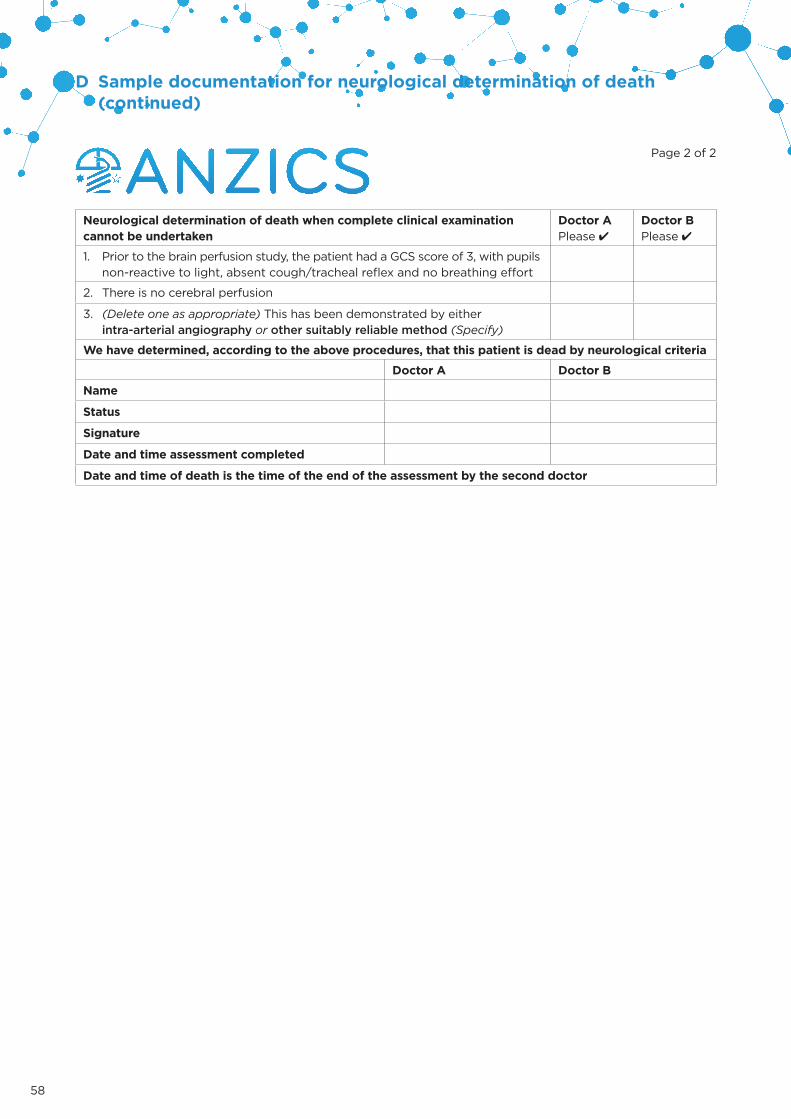
58
D Sample documentation for neurological determination of death (continued)
Page 2 of 2
Neurological determination of death when complete clinical examination cannot be undertaken
Doctor APlease 4
Doctor BPlease 4
1. Prior to the brain perfusion study, the patient had a GCS score of 3, with pupilsnon-reactive to light, absent cough/tracheal reflex and no breathing effort
2. There is no cerebral perfusion
3. (Delete one as appropriate) This has been demonstrated by eitherintra-arterial angiography or other suitably reliable method (Specify)
We have determined, according to the above procedures, that this patient is dead by neurological criteria
Doctor A Doctor B
Name
Status
Signature
Date and time assessment completed
Date and time of death is the time of the end of the assessment by the second doctor
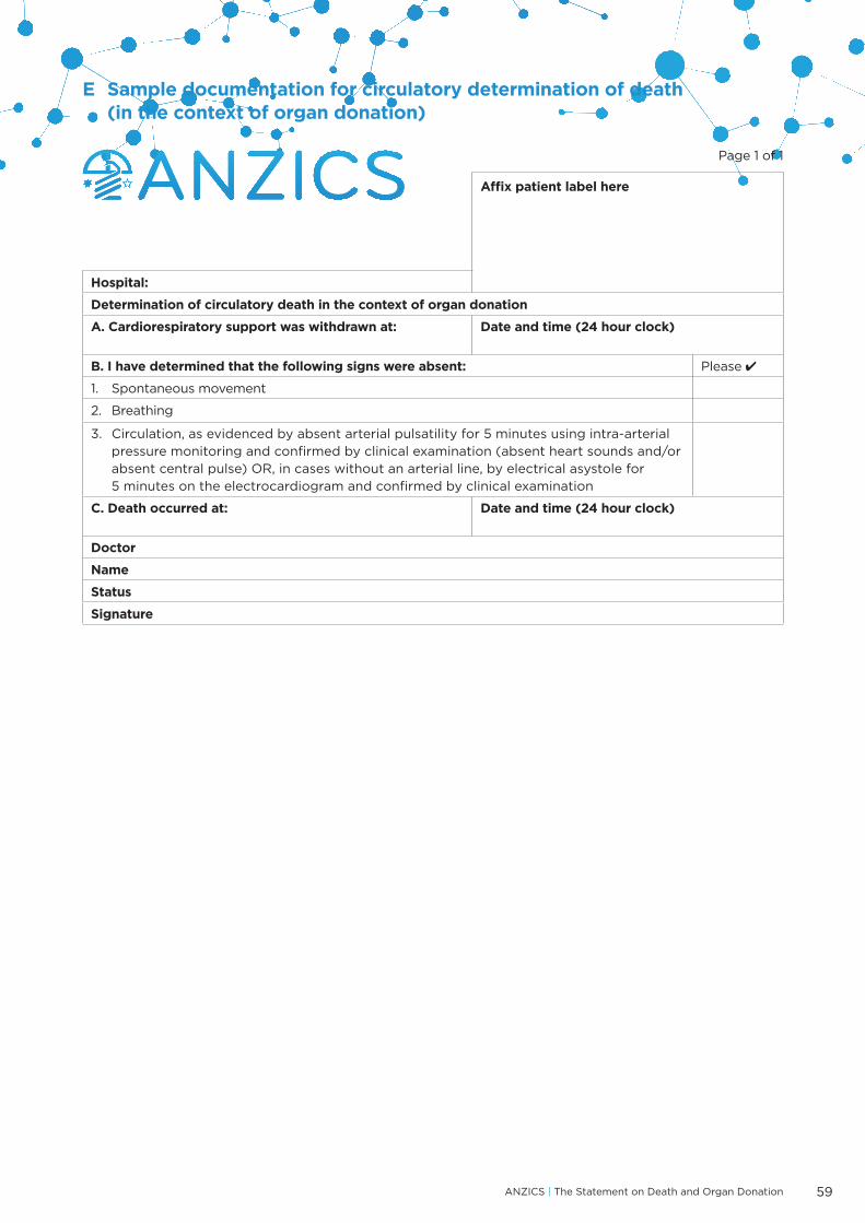
ANZICS | The Statement on Death and Organ Donation 59
E Sample documentation for circulatory determination of death (in the context of organ donation)
Page 1 of 1
Affix patient label here
Hospital:
Determination of circulatory death in the context of organ donation
A. Cardiorespiratory support was withdrawn at: Date and time (24 hour clock)
B. I have determined that the following signs were absent: Please 4
1. Spontaneous movement
2. Breathing
3. Circulation, as evidenced by absent arterial pulsatility for 5 minutes using intra-arterialpressure monitoring and confirmed by clinical examination (absent heart sounds and/orabsent central pulse) OR, in cases without an arterial line, by electrical asystole for5 minutes on the electrocardiogram and confirmed by clinical examination
C. Death occurred at: Date and time (24 hour clock)
Doctor
Name
Status
Signature

60
F Clinical and physiological support of a patient after neurological determination of death
Table F1: Checklist for clinical support of a patient with permanent loss of brain function
For full rationale of these recommendations see Section 2.3.1.
General care and Support
Instrumentation: endotracheal tube, nasogastric tube, urinary catheter, multilumen central line, arterial line, large peripheral intravenous line
Monitoring: ECG, pulse oximetry, intra-arterial pressure, core temperature, urine output (hourly)
Routine investigations: CXR, ECG, blood group, coag
Regular blood testing: FBC, ABG, U&Es, LFTs q6h and as necessary
Review all medications; cease unnecessary orders
Maintain core temperature 36–38°C
Eye care, mouth care and all other nursing cares
Respiratory support
Aim for SpO₂ 92–97% by FiO₂ and PEEP adjustment (minimum PEEP 5cm H₂O)
Aim for PaCO₂ 35–45 mmHg by ventilator adjustment (TV 6–8mL/kg ideal body weight, plateau pressure <30 mmHg)
Regular turns from side to side; elevate head of bed
Regular tracheal suctioning, chest physio and lung recruitment after suctioning; bronchoscopy as required
Cardiovascular support
Ensure patient is neither hypovolemic nor fluid overloaded
Aim for MAP 70–100 mmHg, good peripheral perfusion
Aim for urine output ~1 mL/kg/hr (range 0.5–3 mL/kg/hr)
Commence noradrenaline or vasopressin infusion for hypotension
If inotropic support, such as dobutamine, adrenalin or milrinome, is contemplated, consider further cardiac assessment or output monitoring
Fluids and metabolic management
Give maintenance fluid as free water e.g. glucose 5% 1mL/kg/hr, continue NG feeding or TPN, insulin infusion to keep blood glucose <15 mmol/L
Maintain urine output between 0.5 and 3 mL/kg/hr
If polyuric >300 mL/hr assume diabetes insipidus and immediately give desmopressin and/or commence vasopressin infusion
If Na >150 mmol/L give additional free water
Aim for Hb>70g/L

ANZICS | The Statement on Death and Organ Donation 61
References
1. ANZICS. ANZICS statement on care and decision-making at the end of life for the critically ill (Edition 1.0). Melbourne:Australian and New Zealand Inensive Care Society; 2014.
2. NHMRC. Ethical guidelines for organ transplantation from deceased donors. Canberra: National Health and Medical ResearchCouncil; 2016.
3. Harvey W. Exercitatio anatomica de motu cordis et sanguinis in animalibus [On the motion of the heart and blood in animals]. 1628.
4. Settergren G. Brain death: an important paradigm shift in the 20th century. Acta Anaesthesiol Scand. 2003; 47(9): 1053-8.
5. Machado C, Kerein J, Ferrer Y et al. The concept of brain death did not evolve to benefit organ transplants. J Med Ethics.2007; 33(4): 197-200.
6. Cushing H. Some experimental and clinical observations concerning states of increased intracranial tension. Am J Med Sci.1902; 124: 375–400.
7. Lassen HC. A preliminary report on the 1952 epidemic of poliomyelitis in Copenhagen with special reference to the treatmentof acute respiratory insufficiency. Lancet. 1953; 1(6749): 37-41.
8. Ibsen B. The anaesthetist’s viewpoint on the treatment of respiratory complications in poliomyelitis during the epidemic inCopenhagen, 1952. Proc R Soc Med. 1954; 47(1): 72-4.
9. Riishede J, Ethelberg S. Angiographic changes in sudden and severe herniation of brain stem through tentorial incisure; reportof five cases. AMA Arch Neurol Psychiatr. 1953; 70: 399–409.
10. Löfstedt S, Von Reis G. Intrakraniella lesioner med bilateralt upphävd kontrastpassage i a. carotis interna (Intracranial lesionswith abolished passage of x-ray contrast through the internal carotid arteries). Opusc Med. 1956; 1: 199–202.
11. Belkin GS. Brain death and the historical understanding of bioethics. J Hist Med Allied Sci. 2003; 58(3): 325-61.
12. Jeunet A. Premiѐres tentatives de respiration artificielle et de respiration assistée en neuro-chirurgie (First attempts atartificial respiration and mechanical ventilation in neurosurgery): Université de Lyon; 1957.
13. Wertheimer P, Jouvet M, Descotes J. A propos du diagnostic de la mort du système nerveux dans les comas avec arrêtrespiratoire traités par respiration artificielle (Concerning the diagnosis of death of the nervous system in coma withrespiratory arrest treated by ventilatory support). Presse Med. 1959; 67: 87–8.
14. Mollaret P, Goulon M. Le coma dépassé (mémoire préliminaire) (The depassed coma (preliminary report) [sic]). Rev Neurol(Paris). 1959: 1013–15.
15. A definition of irreversible coma. Report of the Ad Hoc Committee of the Harvard Medical School to Examine the Definition ofBrain Death. JAMA. 1968; 205: 337–40.
16. Gilder SSB. Twenty-second World Medical Assembly. Brit Med J. 1968; 3: 493-94.
17. World Medical Assembly. Declaration of Sydney on the determination of death and the recovery of organs 1983 Amended bythe 35th World Medical Assembly, Venice, Italy, October 1983.
18. Shemie SD, Hornby L, Baker A et al. International guideline development for the determination of death. Intensive Care Med.2014; 40(6): 788-97.
19. Beattie DS. Medico-legal aspects of organ transplantation in New Zealand. N Z Med J. 1972; 75(477): 70-4.
20. Australian Law Reform Commission. ALRC 7. Report of the Law Reform Commission on Human Tissue Transplants. Canberra:Australian Government Publishing Service; 1977.
21. Guidelines for the determination of death. report of the medical consultants on the diagnosis of death to the President’sCommission for the Study of Ethical Problems in Medicine and Biomedical and Behavioural Research. JAMA. 1981; 246(19): 2184–86.
22. President’s Commission for the Study of Ethical Problems in Medicine and Biomedical Behavioral Research. Defining death: areport on the medical, legal and ethical issues in the determination of death. Washington: The Commission; 1981.
23. Uniform Law Commission. Legislative fact sheet: Determination of Death Act 2017. Available from: http://www.uniformlaws.org/LegislativeFactSheet.aspx?title=Determination%20of%20Death%20Act. Accessed:
24. Medical Royal Colleges and their Faculties. Diagnosis of brain death. Statement issued by the honorary secretary of the Conferenceof Medical Royal Colleges and their Faculties in the United Kingdom on 11 October 1976. Br Med J. 1976; 2(6045): 1187–88.
25. Diagnosis of death. Memorandum issued by the honorary secretary of the Conference of Medical Royal Colleges and theirFaculties in the United Kingdom on 15 January 1979. Br Med J. 1979; 1(6159): 332.
26. Wijdicks EF. Brain death worldwide: accepted fact but no global consensus in diagnostic criteria. Neurology. 2002; 58: 20–5.
27. AOMRC. A Code of Practice on the Diagnosis and Confirmation of Death. London: Academy of Medical Royal Colleges; 2008.
28. Ogata J, Imakita M, Yutani C et al. Primary brain-stem death: a clinico-pathological study. J Neurol Neurosurg Psychiat. 1988;51: 646–50.
29. Cordato DJ, Herkes GK, Mather LE et al. Prolonged thiopentone infusion for neurosurgical emergencies: usefulness oftherapeutic drug monitoring. Anaesth Intensive Care. 2001; 29(4): 339-48.
30. O’Leary MJ, Cavazzoni E, Gomez MP. Ventilator auto-triggering in brain death: still a trap for the unwary? Crit Care Resusc.2015; 17(2): 145.
31. Rossetti AO, Oddo M, Logroscino G et al. Prognostication after cardiac arrest and hypothermia: a prospective study. AnnNeurol. 2010; 67(3): 301-7.
32. Samaniego EA, Mlynash M, Caulfield AF et al. Sedation confounds outcome prediction in cardiac arrest survivors treated withhypothermia. Neurocrit Care. 2011; 15(1): 113-9.
33. Roberts DJ, MacCulloch KA, Versnick EJ et al. Should ancillary brain blood flow analyses play a larger role in the neurologicaldetermination of death? Can J Anaesth. 2010; 57: 927–35.
34. Webb AC, Samuels OB. Reversible brain death after cardiopulmonary arrest and induced hypothermia. Crit Care Med. 2011;30(6): 1538-42.

62
35. Streat S. ‘Reversible brain death’--is it true, confounded, or ‘not proven’? Crit Care Med. 2011; 39(6): 1601-3.
36. Sagishima K, Kinoshita Y. Pupil diameter for confirmation of brain death in adult organ donors in Japan. Acute Med Surg. 2017;4(1): 19-24.
37. Olgun G, Newey CR, Ardelt A. Pupillometry in brain death: differences in pupillary diameter between paediatric and adultsubjects. Neurol Res. 2015; 37(11): 945-50.
38. Dominguez-Roldan JM, Barrera-Chacon JM, Murillo-Cabezas F et al. Clinical factors influencing the increment of blood carbondioxide during the apnea test for the diagnosis of brain death. Transplant Proc. 1999; 31(6): 2599-600.
39. Marks SJ, Zisfein J. Apneic oxygenation in apnea tests for brain death. A controlled trial. Arch Neurol. 1990; 47(10): 1066-8.
40. Van Norman GA. A matter of life and death: what every anesthesiologist should know about the medical, legal, and ethicalaspects of declaring brain death. Anesthesiology. 1999; 91: 275–87.
41. Olguner C, Koca U, Akan M et al. The safe limits of mechanical factors in the apnea testing for the diagnosis of brain death.Tohoku J Exp Med. 2007; 211(2): 115-20.
42. Tawil I, Brown LH, Comfort D et al. Family presence during brain death evaluation: a randomized controlled trial*. Crit CareMed. 2014; 42(4): 934-42.
43. Jain S, DeGeorgia M. Brain death-associated reflexes and automatisms. Neurocrit Care. 2005; 3(2): 122-6.
44. Saposnik G, Bueri JA, Maurino J et al. Spontaneous and reflex movements in brain death. Neurology. 2000; 54(1): 221-3.
45. Saposnik G, Maurino J, Saizar R et al. Undulating toe movements in brain death. Eur J Neurol. 2004; 11(11): 723-7.
46. Saposnik G, Maurino J, Saizar R et al. Spontaneous and reflex movements in 107 patients with brain death. Am J Med. 2005;118(3): 311-4.
47. Dosemeci L, Cengizm M, Yilmaz M et al. Frequency of spinal reflex movements in brain-dead patients. Transplant Proc. 2004;36: 17–9.
48. Saposnik G, Maurino J, Bueri J. Movements in brain death. Eur J Neurol. 2001; 8(3): 209-13.
49. Bueri JA, Saposnik G, Maurino J et al. Lazarus’ sign in brain death. Mov Disord. 2000; 15(3): 583-6.
50. Wu Y, Orizaola Balaguer P. Spontaneous and reflex head turning in brain death. Crit Care. 2013; 17(4): 440.
51. Langfitt TW, Kassell NF. Non-filling of cerebral vessels during angiography: correlation with intracranial pressure. ActaNeurochir (Wien). 1966; 14(1): 96-104.
52. Alvarez LA, Lipton RB, Hirschfeld A et al. Brain death determination by angiography in the setting of a skull defect. ArchNeurol. 1988; 45: 225–27.
53. Rosenklint A, Jorgensen PB. Evaluation of angiographic methods in the diagnosis of brain death. Correlation with local andsystemic arterial pressure and intracranial pressure. Neuroradiology. 1974; 7(4): 215-9.
54. Vatne K, Nakstad P, Lundar T. Digital subtraction angiography (DSA) in the evaluation of brain death. A comparison ofconventional cerebral angiography with intravenous and intraarterial DSA. Neuroradiology. 1985; 27(2): 155-7.
55. Laurin NR, Driedger AA, Hurwitz GA et al. Cerebral perfusion imaging with technetium-99m HM-PAO in brain death andsevere central nervous system injury. J Nucl Med. 1989; 30(10): 1627-35.
56. Spieth ME, Ansari AN, Kawada TK et al. Direct comparison of Tc-99m DTPA and Tc-99m HMPAO for evaluating brain death.Clin Nucl Med. 1994; 19(10): 867-72.
57. Keske U. Tc-99m-HMPAO single photon emission computed tomography (SPECT) as an ancillary test in the diagnosis of braindeath. Intensive Care Med. 1998; 24: 895–97.
58. Bonetti MG, Ciritella P, Valle G et al. 99mTc HM-PAO brain perfusion SPECT in brain death. Neuroradiology. 1995; 37(5): 365-9.
59. Munari M, Zucchetta P, Carollo C et al. Confirmatory tests in the diagnosis of brain death: comparison between SPECT andcontrast angiography. Crit Care Med. 2005; 33: 2068–73.
60. Okuyaz C, Gucuyener K, Karabacak NI et al. Tc-99m-HMPAO SPECT in the diagnosis of brain death in children. Pediatr Int.2004; 46(6): 711-4.
61. Frampas E, Videcoq M, de Kerviler E et al. CT angiography for brain death diagnosis. AJNR Am J Neuroradiol. 2009; 30(8):1566-70.
62. Brasil S, Bor-Seng-Shu E, de-Lima-Oliveira M et al. Role of computed tomography angiography and perfusion tomography indiagnosing brain death: A systematic review. J Neuroradiol. 2016; 43(2): 133-40.
63. Dupas B, Gayet-Delacroix M, Villers D et al. Diagnosis of brain death using two-phase spiral CT. AMJR Am J Neuroradiol. 1998;19(4): 796.
64. Lee DH, Nathanson JA, Fox AJ et al. Magnetic resonance imaging of brain death. Can Assoc Radiol J. 1995; 46(3): 174-8.
65. Jones KM, Barnes PD. MR diagnosis of brain death. AJNR Am J Neuroradiol. 1992; 13(1): 65-6.
66. Orrison WW, Champlin AM, Kesterson OL et al. MR ‘hot nose sign’ and ‘intravascular enhancement sign’ in brain death. Am JNeuroradiol. 1994; 15: 913–16.
67. Karantanas AH, Hadjigeorgiou GM, Paterakis K et al. Contribution of MRI and MR angiography in early diagnosis of braindeath. Eur Radiol. 2002; 12(11): 2710-6.
68. Dosemeci L, Dora B, Yilmaz M et al. Utility of transcranial doppler ultrasonography for confirmatory diagnosis of brain death:two sides of the coin. Transplantation. 2004; 77(1): 71-5.
69. Quality Standards Subcommittee of the American Academy of Neurology. Practice parameters for determining brain death inadults (summary statement). Neurology. 1995; 45(5): 1012-4.
70. Shemie SD, Doig C, Dickens B et al. Severe brain injury to neurological determination of death: Canadian forumrecommendations. CMAJ. 2006; 174(6): S1-13.
71. Nakagawa TA, Ashwal S, Mathur M et al. Guidelines for the determination of brain death in infants and children: an update ofthe 1987 Task Force recommendations. Crit Care Med. 2011; 39(9): 2139-55.
72. Royal College of Paediatrics and Child Health. The diagnosis of death by neurological criteria in infants less than two monthsold. London: Royal College of Paediatrics and Child Health; 2015.
73. Volpe JJ. Brain death determination in the newborn. Pediatrics. 1987; 80(2): 293-7.
74. Ashwal S. Brain death in the newborn. Current perspectives. Clin Perinatol. 1997; 24(4): 859-82.
75. Ashwal S. Clinical diagnosis and confirmatory testing of brain death in children. In: Widjicks E, editor. Brain death. Philadelphia:Lippincott Williams & Wilkins Inc; 2001. p. 91–114.

ANZICS | The Statement on Death and Organ Donation 63
76. Mata-Zubillaga D, Oulego-Erroz I. Persistent cerebral blood flow by transcranial Doppler ultrasonography in an asphyxiatednewborn meeting brain death diagnosis: case report and review of the literature. J Perinatol. 2012; 32(6): 473-5.
77. Health Council of the Netherlands. Brain death protocol. The Hague: Health Council of the Netherlands; 2006. Contract No.:Publication no. 2006/04.
78. Conference of Medical Royal Colleges and their Faculties in the United Kingdom. Diagnosis of brainstem death in infants andchildren. London: British Paediatric Association; 1991.
79. Volpe JJ. Neurologic examination: Normal and abnormal features. In: J.J. Volpe, editor. Neurology of the newborn.Philadelphia: WB Saunders; 2001. p. 103–33.
80. Rigatto H, Brady JP, de la Torre Verduzco R. Chemoreceptor reflexes in preterm infants: II. The effect of gestational andpostnatal age on the ventilatory response to inhaled carbon dioxide. Pediatrics. 1975; 55(5): 614-20.
81. Frantz ID, 3rd, Adler SM, Thach BT et al. Maturational effects on respiratory responses to carbon dioxide in premature infants.J Appl Physiol. 1976; 41(1): 41-5.
82. Pasternak JF, Volpe JJ. Full recovery from prolonged brainstem failure following intraventricular hemorrhage. J Pediatr. 1979;95(6): 1046-9.
83. Hornby L, Dhanani S, Shemie SD. Update of a systematic review of autoresuscitation after cardiac arrest. Crit Care Med. 2018;46(3): e268-e72.
84. Cook DA, Widdicombe N. Audit of ten years of donation after circulatory death experience in Queensland: observations ofagonal physiology following withdrawal of cardiorespiratory support. Anaesth Intensive Care. 2018; 46(4): 400-03.
85. Dhanani S, Hornby L, van Beinum A et al. Resumption of Cardiac Activity after Withdrawal of Life-Sustaining Measures. N EnglJ Med 2021;384:345-52.
86. Dhanani S, Hornby L, Ward R et al. Vital signs after cardiac arrest following withdrawal of life-sustaining therapy: a multicenterprospective observational study. Crit Care Med. 2014; 42(11): 2358-69.
87. Bernat JL, D’Alessandro AM, Port FK et al. Report of a national conference on donation after cardiac death. Am J Transplant.2006; 6(2): 281-91.
88. DeVita MA. The death watch: certifying death using cardiac criteria. Prog Transplant. 2000; 10: 2–10.
89. Ethics Committee ACoCCM, Society of Critical Care M. Recommendations for nonheartbeating organ donation. A positionpaper by the Ethics Committee, American College of Critical Care Medicine, Society of Critical Care Medicine. Crit Care Med.2001; 29(9): 1826-31.
90. DeVita MA, Snyder JV, Arnold RM et al. Observations of withdrawal of life-sustaining treatment from patients who becamenon-heart-beating organ donors. Crit Care Med. 2000; 28(6): 1709-12.
91. WHO. WHO guiding principles on human cell, tissue and organ transplantation. Transplantation. 2010; 90(3): 229–33.
92. Participants in the International Summit on Transplant Tourism and Organ Tracking. The declaration of Istanbul on organtracking and transplant tourism. Exp Clin Transplant. 2008; 6(3): 171–79.
93. Streat S. Standard Operating Procedure for Directed Donation from Deceased Donors. Wellington: Organ Donation NewZealand; 2018.
94. TSANZ. Clinical Guidelines for Organ Transplantation from Deceased Donors Version 1.1. Sydney: Transplantation Society ofAustralia and New Zealand; 2017.
95. Albertsen A. Deemed consent: assessing the new opt-out approach to organ procurement in Wales. J Med Ethics. 2018; 44(5):314-8.
96. Parsons JA. Welsh 2013 deemed consent legislation falls short of expectations. Health Policy. 2018; 122(9): 941-44.
97. Shepherd L, O’Carroll RE, Ferguson E. An international comparison of deceased and living organ donation/transplant rates inopt-in and opt-out systems: a panel study. BMC Med. 2014; 12: 131.
98. Bramhall S. Presumed consent for organ donation: a case against. Ann RC Surg Engl. 2011; 93(4): 270–72.
99. Fabre J, Murphy P, Matesanz R. Presumed consent: a distraction in the quest for increasing rates of organ donation. BMJ.2010; 341: c4973.
100. Boyarsky BJ, Hall EC, Deshpande NA et al. Potential limitations of presumed consent legislation. Transplantation. 2012; 93(2):136-40.
101. ABS. 2024.0 - Census of Population and Housing: Australia Revealed, 2016. Canberra: Australian Bureau of Statistics; 2017.
102. Statistics New Zealand. 2013 Census QuickStats about culture and identity. Wellington: Statistics New Zealand; 2014.
103. Tervalon M, Murray-Garcia J. Cultural humility versus cultural competence: a critical distinction in defining physician trainingoutcomes in multicultural education. J Health Care Poor Underserved. 1998; 9(2): 117-25.
104. Organ and Tissue Authority. Multicultural Resources 2014. Available from: http://www.donatelife.gov.au/for-the-community/multicultural-resources. Accessed: 27 November 2017.
105. ANZOD. ANZOD registry report 2016. Adelaide: Australia and New Zealand Organ Donation Registry 2016.
106. TSANZ. Clinical Guidelines for Organ Transplantation from Deceased Donors Version 1.0. Melbourne: Transplantation Societyof Australia and New Zealand; 2016.
107. Goldberg DS, Abt PL, Reese PP et al. Transplanting HCV-Infected Kidneys into Uninfected Recipients. N Engl J Med. 2017;377(11): 1105.
108. Hathorn E, Smit E, Elsharkawy AM et al. HIV-positive-to-HIV-positive liver transplantation. N Engl J Med. 2016; 375(18): 1807-9.
109. Muller E, Barday Z, Mendelson M et al. HIV-positive-to-HIV-positive kidney transplantation--results at 3 to 5 years. N Engl JMed. 2015; 372(7): 613-20.
110. Kootstra G, Daemen JH, Oomen AP. Categories of non-heart-beating donors. Transplant Proc. 1995; 27(5): 2893-4.
111. Sanchez-Fructuosos AI, Prats D, Torrente J et al. Renal transplantation from non-heart beating donors: a promising alternativeto enlarge the donor pool. J Am Soc Nephrol. 2000; 11(350-58).
112. Morrissey PE, Monaco AP. Donation after circulatory death: current practices, ongoing challenges, and potentialimprovements. Transplantation. 2014; 97(3): 258-64.
113. Callaghan CJ, Charman SC, Muiesan P et al. Outcomes of transplantation of livers from donation after circulatory deathdonors in the UK: a cohort study. BMJ Open. 2013; 3(9): e003287.
114. Zaroff JG, Rosengard BR, Armstrong WF et al. Consensus conference report: maximizing use of organs recovered from thecadaver donor: cardiac recommendations, March 28-29, 2001, Crystal City, Va. Circulation. 2002; 106(7): 836-41.

64
115. Novitzky D, Wicomb WN, Cooper DK et al. Prevention of myocardial injury during brain death by total cardiac sympathectomy in the Chacma baboon. Ann Thorac Surg. 1986; 41(5): 520-4.
116. Dujardin KS, McCully RB, Wijdicks EF et al. Myocardial dysfunction associated with brain death: clinical, echocardiographic, and pathologic features. J Heart Lung Transplant. 2001; 20(3): 350-7.
117. Vaghadia H. Atropine resistance in brain-dead organ donors. Anesthesiology. 1986; 65(6): 711-2.
118. Excell L, Hee K, Russ G. ANZOD registry report 2007. Adelaide: Australia and New Zealand Organ Donation Registry; 2007.
119. Schnuelle P, Gottmann U, Hoeger S et al. Effects of donor pretreatment with dopamine on graft function after kidney transplantation: a randomized controlled trial. JAMA. 2009; 302(10): 1067-75.
120. Solidoro P., Schreiber A, Boffini M et al. improving donor lung suitability: from protective strategies to ex-vivo reconditioning. Minerva Medica. 2016; 107(3 Suppl 1): 7-11.
121. Bansal R, Esan A, Hess D et al. Mechanical ventilatory support in potential lung donor patients. Chest. 2014; 146(1): 220-7.
122. Mascia L. Effect of a Lung Protective Strategy for Organ Donors on Eligibility and Availability of Lungs for Transplantation A Randomized Controlled Trial. JAMA 2010; 304(23): 2620-7.
123. Finfer S, Bohn D, Colpitts D et al. Intensive care management of paediatric organ donors and its effect on post-transplant organ function. Intensive Care Med. 1996; 22(12): 1424-32.
124. Totsuka E, Dodson F, Urakami A et al. Influence of high donor serum sodium levels on early postoperative graft function in human liver transplantation: effect of correction of donor hypernatremia. Liver Transpl Surg. 1999; 5(5): 421-8.
125. Kazemeyni SM, Esfahani F. Influence of hypernatremia and polyuria of brain-dead donors before organ procurement on kidney allograft function. Urol J. 2008; 5(3): 173-7.
126. Harris AS. Clinical experience with desmopressin: efficacy and safety in central diabetes insipidus and other conditions. J Pediatr. 1989; 114(4 Pt 2): 711-8.
127. Katz K, Lawler J, Wax J et al. Vasopressin pressor effects in critically ill children during evaluation for brain death and organ recovery. Resuscitation. 2000; 47(1): 33-40.
128. Wood KE, Becker BN, McCartney JG et al. Care of the potential organ donor. N Engl J Med. 2004; 351(26): 2730-9.
129. Ralston C, Butt W. Continuous vasopressin replacement in diabetes insipidus. Arch Dis Child. 1990; 65(8): 896-7.
130. Shann F. Drug doses. Melbourne: Collective Pty Ltd; 2005.
131. Reilly PM, Morgan L, Grossman MD et al. Lung procurement from solid organ donors: role of fluid resuscitation in procurement failures. Chest. 1996; 110: S222.
132. Novitzky D, Cooper DK, Reichart B. Hemodynamic and metabolic responses to hormonal therapy in brain-dead potential organ donors. Transplantation. 1987; 43(6): 852-4.
133. Phongsamran PV. Critical care pharmacy in donor management. Prog Transplant. 2004; 14(2): 105-11; quiz 12-3.
134. Macdonald PS, Aneman A, Bhonagiri D et al. A systematic review and meta-analysis of clinical trials of thyroid hormone administration to brain dead potential organ donors. Crit Care Med. 2012; 40(5): 1635-44.
135. Follette DM, Rudich SM, Babcock WD. Improved oxygenation and increased lung donor recovery with high-dose steroid administration after brain death. J Heart Lung Transplant. 1998; 17(4): 423-9.
136. Lutz-Dettinger N, de Jaeger A, Kerremans I. Care of the potential pediatric organ donor. Pediatr Clin North Am. 2001; 48(3): 715-49.
137. Shemie SD, Ross H, Pagliarello J et al. Organ donor management in Canada: recommendations of the forum on Medical Management to Optimize Donor Organ Potential. CMAJ. 2006; 174(6): S13-32.
138. Holmes CL, Patel BM, Russell JA et al. Physiology of vasopressin relevant to management of septic shock. Chest. 2001; 120(3): 989-1002.
139. Niemann CU, Feiner J, Swain S et al. Therapeutic hypothermia in deceased organ donors and kidney-graft function. N Engl J Med. 2015; 373(5): 405-14.
140. Schnuelle P, Benck U, Kramer BK et al. Impact of donor core body temperature on graft survival after heart transplantation. Transplantation. 2018; 102(11):1891-9.
141. Singer P, Cohen J, Cynober L. Effect of nutritional state of brain-dead organ donor on transplantation. Nutrition. 2001; 17(11-12): 948-52.
142. Singer P, Shapiro H, Cohen J. Brain death and organ damage: the modulating effects of nutrition. Transplantation. 2005; 80(10): 1363-8.
143. Gravel MT, Arenas JD, Chenault R, 2nd et al. Kidney transplantation from organ donors following cardiopulmonary death using extracorporeal membrane oxygenation support. Ann Transplant. 2004; 9(1): 57-8.
144. Yang HY, Lin CY, Tsai YT et al. Experience of heart transplantation from hemodynamically unstable brain-dead donors with extracorporeal support. Clin Transplant. 2012; 26(5): 792-6.
145. Isnardi DI, Olivero F, Lerda R et al. Extracorporeal membrane oxygenation as a bridge to organ donation: a case report. Transplant Proc. 2013; 45(7): 2619-20.
146. Dalle Ave AL, Gardiner D, Shaw DM. The ethics of extracorporeal membrane oxygenation in brain-dead potential organ donors. Transpl Int. 2016; 29(5): 612-8.
147. Young PJ, Matta BF. Anaesthesia for organ donation in the brain-stem dead – why bother? Anaesthesia. 2000; 55: 105–06.
148. Fitzgerald RD, Hieber C, Schweitzer E et al. Intraoperative catecholamine release in brain-dead organ donors is not suppressed by administration of fentanyl. Eur J Anaesthesiol. 2003; 20: 952–56.
149. ANZOD. ANZOD registry report 2017. Adelaide: Australian and New Zealand Organ Donation Registry; 2017.
150. IRODaT. International Registry in Organ Donation and Transplantation 2016. Available from: http://www.irodat.org/. Accessed:
151. Steinbrook R. Organ donation after cardiac death. N Engl J Med. 2007; 357(3): 209-13.
152. AOTDTA. National protocol for donation after cardiac death. Melbourne: Australian Organ and Tissue Donation and Transplantation Authority 2010.
153. Andrews PA, Burnapp L, Manas D. Summary of the British Transplantation Society guidelines for transplantation from donors after deceased circulatory death. Transplantation. 2014; 97(3): 265-70.
154. Citerio G, Cypel M, Dobb GJ et al. Organ donation in adults: a critical care perspective. Intensive Care Med. 2016; 42(3): 305-15.
155. Cooper JT, Chin LT, Krieger NR et al. Donation after cardiac death: the university of wisconsin experience with renal transplantation. Am J Transplant. 2004; 4(9): 1490-4.

ANZICS | The Statement on Death and Organ Donation 65
156. Metcalfe MS, Butterworth PC, White SA et al. A case-control comparison of the results of renal transplantation from heart-beating and non-heart-beating donors. Transplantation. 2001; 71(11): 1556-9.
157. Gamez P, Cordoba M, Ussetti P et al. Lung Transplant Group of the Puerta de Hierro Hospital. Lung transplantation from out-of-hospital non-heart-beating lung donors. one-year experience and results. J Heart Lung Transplant. 2005; 24: 1098–102.
158. Wigfield C. Donation after cardiac death for lung transplantation: a review of current clinical practice. Curr Opin Organ Transplant. 2014; 19(5): 455–59.
159. Reeb J, Keshavjee S, Cypel M. Expanding the lung donor pool: advancements and emerging pathways. Curr Opin Organ Transplant. 2015; 20(5): 498-505.
160. Elgharably H, Shafii AE, Mason DP. Expanding the donor pool: donation after cardiac death. Thorac Surg Clin. 2015; 25(1): 35-46.
161. Popov AF, Sabashnikov A, Patil NP et al. Ex vivo lung perfusion - state of the art in lung donor pool expansion. Med Sci Monit Basic Res. 2015; 21: 9-14.
162. Machuca TN, Cypel M. Ex vivo lung perfusion. J Thorac Dis. 2014; 6(8): 1054-62.
163. Trotter JF. Liver transplantation around the world. Curr Opin Organ Transplant. 2017; 22(2): 123-7.
164. Calne R, Loughridge L, MacGillivray J et al. Further observations on renal transplants in man from cadaveric donors. Br Med J. 1966; 2: 1345–51.
165. Sutherland AI, Oniscu GC. Challenges and advances in optimizing liver allografts from donation after circulatory death donors. J Nat Sci Biol Med. 2016; 7(1): 10-5.
166. Pavel MC, Fondevila Campo C, Calatayud Mizrahi D et al. Normothermic perfusion machine in liver transplant with cardiac death donor grafts. Cir Esp. 2015; 93(8): 485-91.
167. Dhital KK, Iyer A, Connellan M et al. Adult heart transplantation with distant procurement and ex-vivo preservation of donor hearts after circulatory death: a case series. Lancet. 2015; 385(9987): 2585-91.
168. Dhital KK, Chew HC, Macdonald PS. Donation after circulatory death heart transplantation. Curr Opin Organ Transplant. 2017; 22(3): 189-97.
169. Macdonald PS, Chew HC, Connellan M et al. Extracorporeal heart perfusion before heart transplantation: the heart in a box. Curr Opin Organ Transplant. 2016; 21(3): 336-42.
170. Macdonald P, Verran D, O’Leary M et al. Heart transplantation from donation after circulatory death donors. Transplantation. 2015; 99(6): 1101-2.
171. Tibballs J, Bhatia N. Transplantation of the heart after circulatory death of the donor: time for a change in law? Med J Aust. 2015; 203(6): 268-70 e1.
172. Nair-Collins M, Gerend MA. Moral evaluations of organ transplantation influence judgments of death and causation. Neuroethics. 2015; 8(3): 283-97.
173. Miller FG. Heart donation without the dead donor rule. Ann Thorac Surg. 2014; 97(4): 1133-4.
174. Zamperetti N, Bellomo R, Latronico N. Heart donation and transplantation after circulatory death: ethical issues after Europe’s first case. Intensive Care Med. 2016; 42(1): 93-5.
175. Berney T, Boffa C, Augustine T et al. Utilization of organs from donors after circulatory death for vascularized pancreas and islet of Langerhans transplantation: recommendations from an expert group. Transpl Int. 2016; 29(7): 798-806.
176. Shahrestani S, Webster AC, Lam VW et al. Outcomes from pancreatic transplantation in donation after cardiac death: A systematic review and meta-analysis. Transplantation. 2017; 101(1): 122-30.
177. Murphy P, Boffa C, Manara A et al. In-hospital logistics: what are the key aspects for succeeding in each of the steps of the process of controlled donation after circulatory death? Transpl Int. 2016; 29: 760–70.
178. Thuong M, Ruiz A, Evrard P et al. New classification of donation after circulatory death donors definitions and terminology. Transpl Int. 2016; 29(7): 749-59.
179. Krmpotic K, Payne C, Isenor C et al. Delayed referral results in missed opportunities for organ donation after circulatory death. Crit Care Med. 2017; 45(6): 989-92.
180. Jochmans I, Akhtar MZ, Nasralla D et al. Past, Present, and Future of Dynamic Kidney and Liver Preservation and Resuscitation. Am J Transplant. 2016; 16(9): 2545-55.
181. Liu S, Pang Q, Zhang J et al. Machine perfusion versus cold storage of livers: a meta-analysis. Front Med. 2016; 10(4): 451-64.
182. Brieva J, Coleman N, Lacey J et al. Prediction of death in less than 60 minutes after withdrawal of cardiorespiratory support in potential organ donors after circulatory death. Transplantation. 2014; 98(10): 1112-8.
183. Lewis J, Peltier J, Nelson H et al. Development of the University of Wisconsin donation After Cardiac Death Evaluation Tool. Prog Transplant. 2003; 13(4): 265-73.
184. DeVita MA, Brooks MM, Zawistowski C et al. Donors after cardiac death: validation of identification criteria (DVIC) study for predictors of rapid death. Am J Transplant. 2008; 8(2): 432-41.
185. Munshi L, Dhanani S, Shemie SD et al. Predicting time to death after withdrawal of life-sustaining therapy. Intensive Care Med. 2015; 41(6): 1014-28.
186. Sidiropoulos S, Treasure E, Silvester W et al. Organ donation after circulatory death in a university teaching hospital. Anaesth Intensive Care. 2016; 44(4): 477-83.
187. Pine JK, Goldsmith PJ, Ridgway DM et al. Predicting donor asystole following withdrawal of treatment in donation after cardiac death. Transplant Proc. 2010; 42(10): 3949-50.
188. de Groot YJ, Lingsma HF, Bakker J et al. External validation of a prognostic model predicting time of death after withdrawal of life support in neurocritical patients. Crit Care Med. 2012; 40(1): 233-8.
189. Shore PM, Huang R, Roy L et al. Development of a bedside tool to predict time to death after withdrawal of life-sustaining therapies in infants and children. Pediatr Crit Care Med. 2012; 13(4): 415-22.
190. Scalea JR, D’Alessandro AM, Fernandez LA. Predicting donor death: early changes in oxygen saturation after withdrawal of support predict successful donation during donation after circulatory death. Transplant Proc. 2016; 48(6): 1887-92.
191. Wind J, Snoeijs MG, Brugman CA et al. Prediction of time of death after withdrawal of life-sustaining treatment in potential donors after cardiac death*. Crit Care Med. 2012; 40(3): 766-9.
192. NHMRC. Organ and tissue donation, after death, for transplantation: Guidelines for ethical practice for health professionals. Canberra: National Health and Medical Research Council; 2007.
193. Boglione MM, Morandini MA, Barrenechea ME et al. Pre-arrest heparinization and ventilation during warm ischemia preserves lung function in non-heart-beating donors. J Pediatr Surg. 1999; 34(12): 1805-9.

66
194. Gok MA, Shenton BK, Peaston R et al. Improving the quality of kidneys from non-heart-beating donors, using streptokinase:an animal model. Transplantation. 2002; 73: 1869–74.
195. Bessems M, Doorschodt BM, Albers PS et al. Wash-out of the non-heart-beating donor liver: a matter of flush solution andtemperature? Liver Int. 2006; 26(7): 880-8.
196. NSW Health. Organ donation after circulatory death: NSW guidelines. Sydney: NSW Health; 2014.
197. Richards B, Rogers WA. Organ donation after cardiac death: legal and ethical justification for antemortem interventions. MedJAust. 2007; 187: 168–70.
198. Corkery-Lavender T, Millar J, Cavazzoni E et al. Patterns of organ donation in children in Australia and New Zealand. Crit CareResusc. 2017; 19(4): 296-302.
199. ANZICS. Report of the Australian and New Zealand Paediatric Intensive Care Registry 2016 Melbourne; 2016 2017.
200. Weiss MJ, Hornby L, Witteman W et al. Pediatric Donation After Circulatory Determination of Death: A Scoping Review.Pediatr Crit Care Med. 2016; 17(3): e87-e108.
201. Weiss MJ, Hornby L, Rochwerg B et al. Canadian Guidelines for Controlled Pediatric Donation After Circulatory Determinationof Death—Summary Report*. Pediatric Critical Care Medicine. 2017; 18(11): 1035-46.
202. Committee on Bioethics. Ethical controversies in organ donation after circulatory death. Pediatrics. 2013; 131(5): 1021-6.
203. Academy of Royal Medical Colleges Ethical issues in paediatric organ donation –a position paper by the UK Donation EthicsCommittee (UKDEC), London, June 2015.
204. Cauley RP, Suh MY, Kamin DS et al. Multivisceral transplantation using a 2.9 kg neonatal donor. Pediatr Transplant. 2012; 16(8):E379-82.
205. Hawkins KC, Scales A, Murphy P et al. Current status of paediatric and neonatal organ donation in the UK. Arch Dis Child.2018; 103(3): 210-5.
206. Charles E, Scales A, Brierley J. The potential for neonatal organ donation in a children’s hospital. Arch Dis Child Fetal NeonatalEd. 2014; 99(3): F225-9.
207. Labrecque M, Parad R, Gupta M et al. Donation after cardiac death: the potential contribution of an infant organ donorpopulation. J Pediatr. 2011; 158(1): 31-6.
208. Stiers J, Aguayo C, Siatta A et al. Potential and actual neonatal organ and tissue donation after circulatory determination ofdeath. JAMA Pediatr. 2015; 169(7): 639-45.
209. Saha S, Kent AL. Length of time from extubation to cardiorespiratory death in neonatal intensive care patients andassessment of suitability for organ donation. Arch Dis Child Fetal Neonatal Ed. 2014; 99(1): F59-63.
210. Atreja G, Godambe S. First neonatal organ donation in the UK. Arch Dis Child Fetal Neonatal Ed. 2015; 100(3): F276-F7.
211. Wijetunga I, Ecuyer C, Martinez-Lopez S et al. Neonatal organ donation for transplantation in the UK. Arch Dis Child FetalNeonatal Ed. 2015; 100(5): F469.
212. Dave RV, Hakeem AR, Dawrant MJ et al. Renal Transplantation From Pediatric Donors in the United Kingdom. Transplantation.2015; 99(9): 1968-75.
213. Balachandran VP, Aull MJ, Goris M et al. Successful transplantation of single kidneys from pediatric donors weighing less thanor equal to 10 kg into standard weight adult recipients. Transplantation. 2010; 90(5): 518-22.
214. Sharma A, Fisher RA, Cotterell AH et al. En bloc kidney transplantation from pediatric donors: comparable outcomes withliving donor kidney transplantation. Transplantation. 2011; 92(5): 564-9.
215. Sureshkumar KK, Marcus RJ. Expanding the donor pool: optimal utilization of pediatric donor kidneys. Transplantation. 2011;91(8): e56-7; author reply e7-8.
216. Smyth GP, Eng MP, Power RP et al. Long-term outcome of cadaveric pediatric en bloc transplantation--a 15-year experience.Transplant Proc. 2005; 37(10): 4228-9.
217. Organ and Tissue Authority. Australian donation and transplantation activity report 2016. Canberra: Organ and TissueAuthority; 2016.
218. Therapeutic Goods Order No. 88 – Standards for donor selection, testing and minimising infectious disease transmission viatherapeutic goods that are human blood and blood components, human tissues and human cellular therapy products., (2013).
219. Curtis JR, Engelberg RA, Wenrich MD et al. Missed opportunities during family conferences about end-of-life care in theintensive care unit. Am J Respir Crit Care Med. 2005; 171(8): 844–49.
220. Truog RD, Campbell ML, Curtis JR et al. American Academy of Critical Care Medicine. Recommendations for end-of-life carein the intensive care unit: a consensus statement by the American Academy of Critical Care Medicine. Crit Care Med. 2008;36(3): 953–63.
221. McDonagh JR, Elliott TB, Engelberg RA et al. Family satisfaction with family conferences about end-of-life care in theintensive care unit: increased proportion of family speech is associated with increased satisfaction. Crit Care Med. 2004;32(7): 1484–88.
222. Lautrette A, Darmon M, Megarbane B et al. A communication strategy and brochure for relatives of patients dying in the ICU.N Engl J Med. 2007; 356(5): 469-78.
223. Zib M, Saul P. A pilot audit of the process of end-of-life decision-making in the intensive care unit. Crit Care Resusc. 2007;9(2): 213-8.
224. Orr RD. Withdrawing the critical but not the care. JAMA. 1994; 272: 1326.
225. Faber-Langendoen K, Lanken PN. Dying patients in the intensive care unit: forgoing treatment, maintaining care. Ann InternMed. 2000; 133: 886–93.
226. Prendergast TJ, Puntillo KA. Withdrawal of life support: intensive caring at the end of life. JAMA. 2002; 288: 2732–40.
227. Kesselring A, Kainz M, Kiss A. Traumatic memories of relatives regarding brain death, request for organ donation andinteractions with professionals in the ICU. Am J Transplant. 2007; 7(1): 211–17.
228. Streat S. Clinical review: moral assumptions and the process of organ donation in the intensive care unit. Crit Care. 2004; 8(5):382-8.
229. Streat S. No more “harvesting” of organs. BMJ. 2014; 349: g6174.
230. AOTA, DonateLife. Best practice guideline for offering organ and tissue donation in Australia. Canberra: Commonwealth ofAustralia; 2017.
231. Simpkin AL, Robertson LC, Barber VS et al. Modifiable factors influencing relatives’ decision to offer organ donation:systematic review. BMJ. 2009; 338: b991.

ANZICS | The Statement on Death and Organ Donation 67
232. ACSQHC. The national consensus statement: essential elements for safe and high-quality end-of-life care. Canberra: AustralianCommission on Safety and Quality in Health Care; 2015.
233. Cuthbertson SJ, Margetts MA, Streat SJ. Bereavement follow-up after critical illness. Crit Care Med. 2000; 28(4): 1196-201.
234. Milberg A, Olsson EC, Jakobsson M et al. Family members’ perceived needs for bereavement follow-up. J Pain SymptomManage. 2008; 35(1): 58-69.
235. ABS. 3238.0.55.001 - Estimates of Aboriginal and Torres Strait Islander Australians, June 2016. Canberra: Australian Bureau ofStatistics; 2018.
236. AIATSIS. Indigenous Australian languages: Australian Institute of Aboriginal and Torres Strait Islander Studies; 2018. Availablefrom: https://aiatsis.gov.au/explore/articles/indigenous-australian-languages. Accessed: 18 July 2018.
237. ABS. 4714.0 - National Aboriginal and Torres Strait Islander Social Survey, 2014-15. Canberra: Australian Bureau of Statistics; 2016.
238. Stephens D. Exploring pathways to improve indigenous organ donation. Intern Med J. 2007; 37(10): 713-6.
239. Queensland Health. Sad News, Sorry Business. Guidelines for caring for Aboriginal and Torres Strait Islander people throughdeath and dying. Ver 2. Brisbane: State of Queensland; 2015.
240. Waran E, O’Connor N, Zubair MY et al. ‘Finishing up’ on country: challenges and compromises. Intern Med J. 2016; 46(9): 1108–11.
241. Mauriora Health Education Research. Foundation in Cultural Competency Course (Maori) undated. Available from: members.maoriora.co.nz. Accessed: 13 February 2018.
242. Reilly M. Whanaungatanga. In: al TMKae, editor. Ki te whaiao: an introduction to Maori culture and society. Auckland: PearsonLongman; 2004. p. 63-72.
243. Mead HM. Te Tapu o te Tangata — the Tapu of the person. In: Mead HM, editor. Ti Kanga Māori. Wellington: Huia Publishers;2003. p. 335-51.
244. Mead HM. Ngā Ahi E Ngiha Mai Nei — The fires that flare up. In: Mead HM, editor. Ti Kanga Māori. Wellington: Huia Publishers;2003. p. 335-51.
245. Lewis G, Pickering N. Māori spiritual beliefs and attitudes towards organ donation. N Z Bioeth. 2003; 4(1): 31-5.
246. Mead HM. Ngā Pūtake o te Tikanga — underlying principles and values. In: Mead HM, editor. Ti Kanga Māori. Wellington: HuiaPublishers; 2003. p. 25-34.
247. Mead HM. Tangihanga — Ceremonies of the Dead. In: Mead HM, editor. Ti Kanga Māori. Wellington: Huia Publishers; 2003. p. 133-50.
248. ABS. Media release: 2016 Census data reveals “no religion” is rising fast Canberra: Australian Bureau of Statistics; 2017.Available from: http://www.abs.gov.au/AUSSTATS/[email protected]/mediareleasesbyReleaseDate/7E65A144540551D7CA258148000E2B85?OpenDocument. Accessed: 14 March 2018.
249. Dominguez-Gil B, Murphy P, Procaccio F. Ten changes that could improve organ donation in the intensive care unit. IntensiveCare Med. 2016; 42(2): 264-7.
250. Department of Human Services. Australian Organ Donor Register (AODR) Statistics 2017. Available from: https://data.gov.au/dataset/australian-organ-donor-register-statistics. Accessed: 5 february 2018.
251. Aubert O, Kamar N, Vernerey D et al. Long term outcomes of transplantation using kidneys from expanded criteria donors:prospective, population based cohort study. BMJ. 2015; 351: h3557.
252. Giessing M, Fuller TF, Friedersdorff F et al. Outcomes of transplanting deceased-donor kidneys between elderly donors andrecipients. J Am Soc Nephrol. 2009; 20(1): 37-40.
253. Ma MK, Lim WH, Craig JC et al. Mortality among younger and older recipients of kidney transplants from expanded criteriadonors compared with standard criteria donors. Clin J Am Soc Nephrol. 2016; 11(1): 128-36.
254. Nunnink L, Cook DA. Palliative ICU beds for potential organ donors: an effective use of resources based on quality-adjustedlife-years gained. Crit Care Resusc. 2016; 18(1): 37-42.
255. First Nations Health Authority. Cultural humility West Vancouver, BC: First Nations Health Authority; 2018. Available from:http://www.fnha.ca/wellness/cultural-humility. Accessed: 2 September 2018.
256. NIH State-of-the-Science Conference Statement on improving end-of-life care. NIH Consens State Sci Statements. 2004; 21: 1-26.
257. Streat S. When do we stop? Crit Care Resusc. 2005; 7: 227–32.
258. NHMRC. Ethical guidelines for the care of people in post-coma unresponsiveness (vegetative state) or a minimally responsivestate. Canberra: National Health and Medical Research Council; 2008.
259. The Royal Childrens’ Hospital Melbourne. Paediatric intensive care guidelines 4th edition. Melbourne: The Royal Childrens’Hospital Melbourne; 2017.
260. Eytan D, Goodwin AJ, Greer R et al. Heart rate and blood pressure centile curves and distributions by age of hospitalizedcritically ill children. Front Pediatr. 2017; 5: 52.
