the structure & function of large biological macromolecules campbell and reece chapter 5
TRANSCRIPT
THE STRUCTURE & FUNCTION OF
LARGE BIOLOGICAL MACROMOLECULES
Campbell and Reece CHAPTER 5

Macromolecules are Polymers polymer: long molecule consisting
of many similar, sometimes identical, building blocks linked by covalent bonds
monomer: the smaller units that make up a polymer


Making Polymers
2 monomers joined by dehydration reaction

Disassembling Polymers
hydrolysis reaction breaks apart 2 monomers in a polymer

Diversity of Polymers
possible varieties of macromolecules infinite
only use 40 -50 monomers small molecules common to all
organisms are ordered into species unique macromolecules
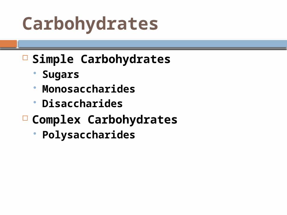
Carbohydrates
Simple Carbohydrates Sugars Monosaccharides Disaccharides
Complex Carbohydrates Polysaccharides

Monosaccharides
multiples of the unit CH2O glucose most common
monosaccharide
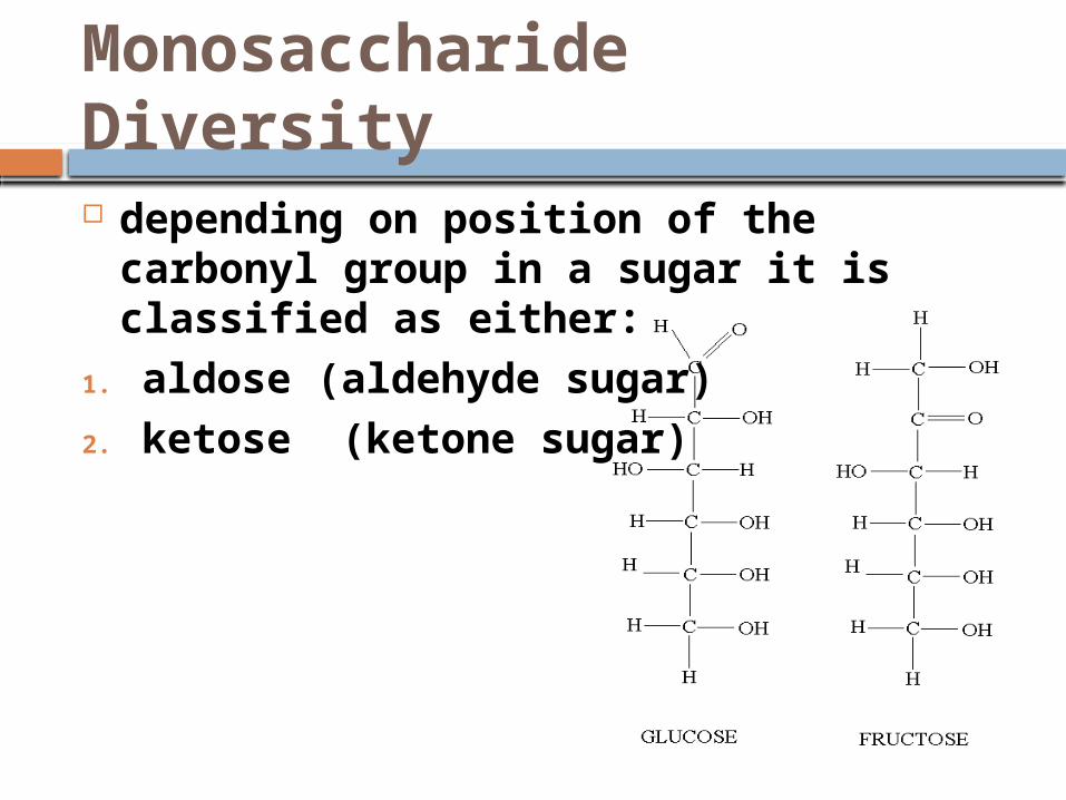
Monosaccharide Diversity depending on position of the carbonyl
group in a sugar it is classified as either:
1. aldose (aldehyde sugar)2. ketose (ketone sugar)

Monosaccharide Diversity 3 to 7 carbons hexose: 6 carbons long pentose: 5 carbons triose: 3 carbons

Monosaccharide Diversity most hexoses and pentoses form
rings in aqueous solutions used in cellular respiration
(especially glucose) serve as raw materials for synthesis
of amino acids and fatty acids
if not immediately used in these ways used to build disaccharides or polysaccharides
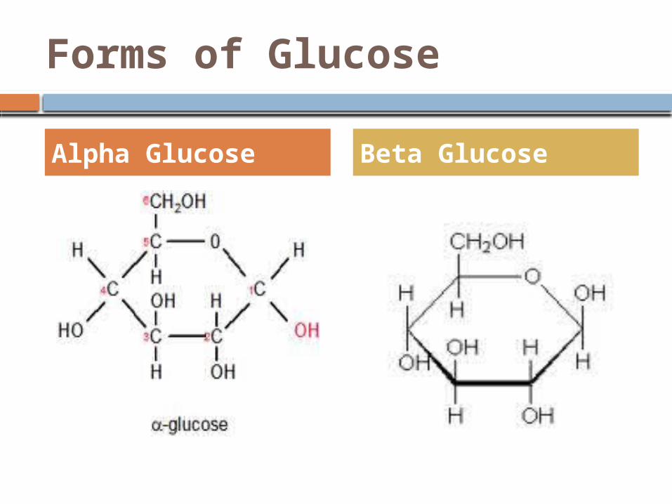
Forms of Glucose
Alpha Glucose Beta Glucose
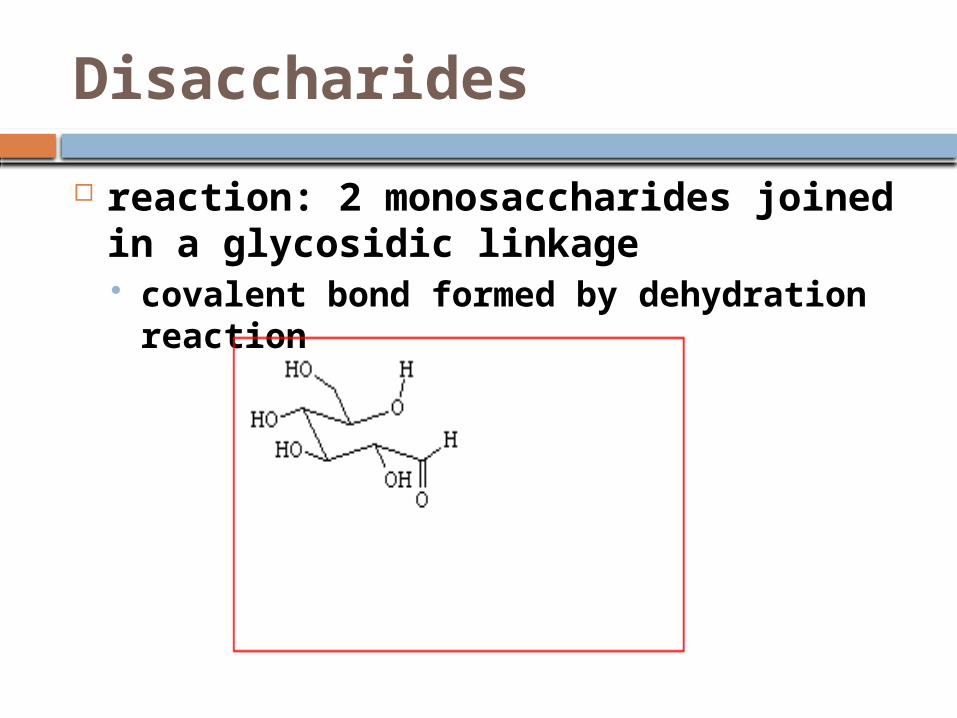
Disaccharides
reaction: 2 monosaccharides joined in a glycosidic linkage covalent bond formed by dehydration
reaction
Disaccharides
2 glucose = maltose (malt sugar) glucose + galactose glucose + fructose = sucrose (table
sugar)
sucrose: form plants use to transport sugars from leaves roots & other nonphotosynthetic parts of plant


Polysaccharides
polymers of hundreds to thousands of monosaccharides joined by glycosidic linkages
function determined by its sugar monomers & positions of glycosidic linkages
2 types:1. storage of monosaccharides to be
used for energy when needed2. building material

Storage Polysaccharides
Plants store glucose (the monomers)as starch (the polymer) represents stored energy

Starch
most is made of α glucose monomers joined in 1-4 linkages simplest form of starch (amylose) is
unbranched complex starch, amylopectin, has 1-
6 linkage

Storage Polysaccharides
Animals: store glucose (the monomers) as glycogen (the polymer) in 1-4 & 1-6 linkages stored mainly in liver & muscle cells humans store about 1 days supply of
glucose this way

Structural Polysaccharides Cellulose: most abundant organic
cpd on Earth is polymer of β glucose (makes
every monomer of glucose “upside down” from its neighbors)

Starch & Cellulose
many are mostly helical
digested by enzymes breaking its α linkages
never branched has –OH groups
available for H-bonds
digested by enzymes breaking its β linkages
Starch Cellulose
Cellulose
digested by very few organisms (don’t have enzymes to do it)
in humans: passes thru GI tract abrading walls & stimulating mucus secretion along the way smoother passage of food thru
not technically a nutrient but is important

“Insoluble Fiber” = Cellulose

Cellulose
Cows: have bacteria and protists in their guts that have enzymes that can digest cellulose nutrients that can be used by cow
Termites unable to digest cellulose in wood it eats have prokaryotes & protists to break it down and so termite can use nutrients
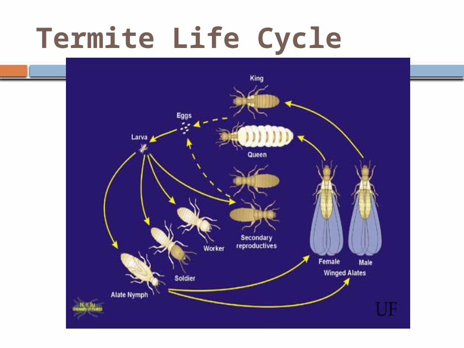
Termite Life Cycle

Termites
Chitin
another structural polysaccharide used by arthropods to build
exoskeletons exoskeletons: made of chitin +
calcium carbonate

Chitin
also in many fungi cell walls monomer has N group attached


Lipids
large group of hydrophobic molecules
do not have true monomers Includes:
Waxes Steroids Some Pigments Oils, Fats Phospholipids

Fats
large molecules assembled from smaller molecules by a dehydration reaction
2 parts:1. Glycerol2. Fatty Acid

Glycerol

Fatty Acids
long (16-18) chain of carbons (hydrophobic)
@ one end carboxyl group (hence fatty acid)

Triglyceride
3 fatty acids + glycerol
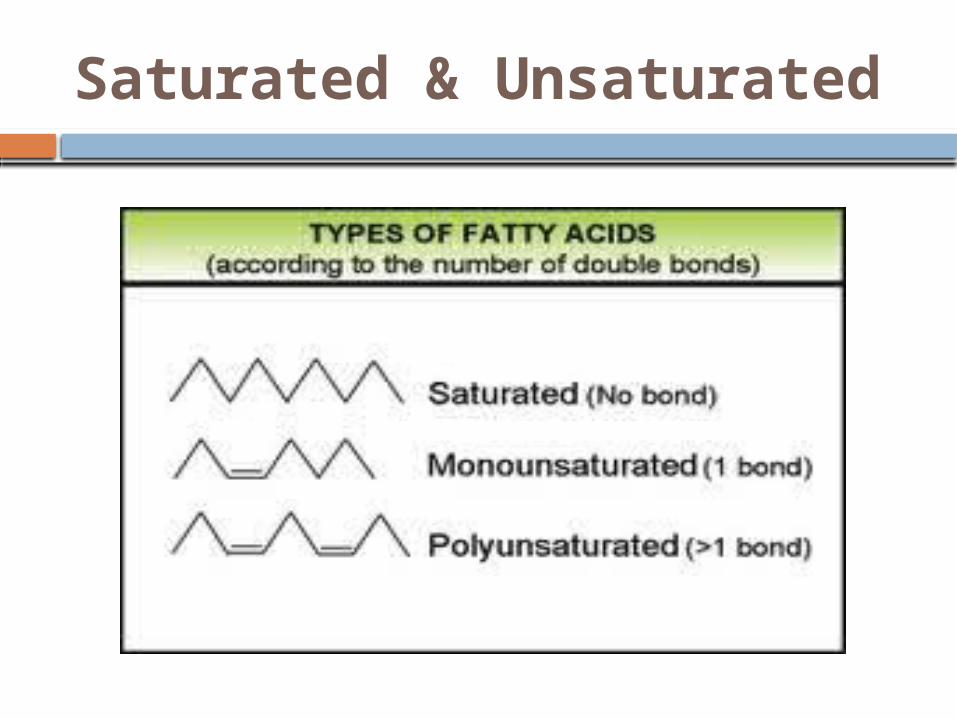
Saturated & Unsaturated

Saturated Fats
include most animal fats most are solids @ room
temperatures
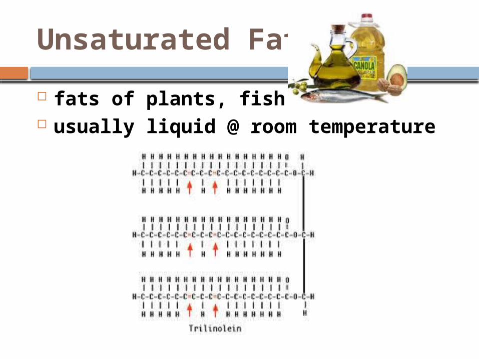
Unsaturated Fats
fats of plants, fish usually liquid @ room temperature
Hydrogenated Vegetable Oil seen on some food labels means that unsaturated fats have
been synthetically converted to saturated fats to keep from separating

Plaques
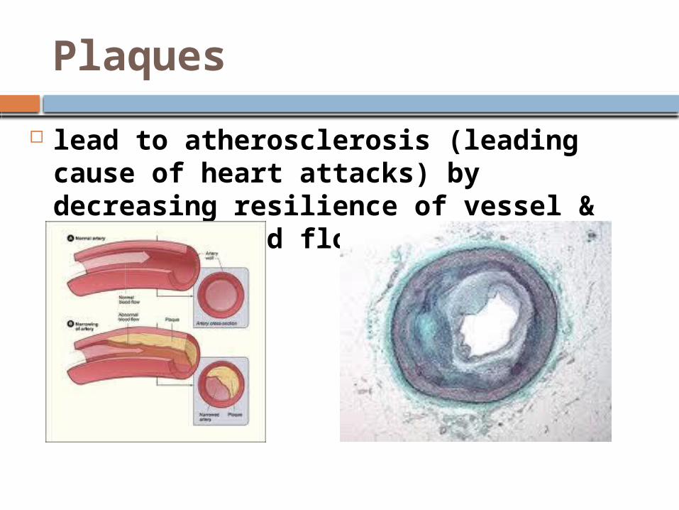
deposits of saturated & trans fats (hydrogenated vegetable oils with trans double bonds) in muscularis of arteries
Plaques
lead to atherosclerosis (leading cause of heart attacks) by decreasing resilience of vessel & impeding blood flow
Trans Fats
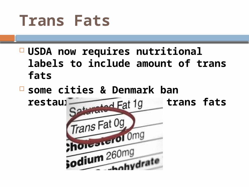
USDA now requires nutritional labels to include amount of trans fats
some cities & Denmark ban restaurants from using trans fats

Essential Fatty Acids
cannot be synthesized in body so must be included in diet
include: omega-3 fatty acids:required for normal growth in children
probably protect against cardiovascular disease in adults

Omega-3 Fatty Acids

Energy Storage
1 g fat has 2x chemical potential energy as 1 g of polysaccharide
plants (generally immobile) can store majority of their energy in polysaccharides except vegetable oils extracted from their seeds

Functions of Fat
Plants: storage of energy Animals: 1. storage of energy2. protect organs3. insulation


Phospholipids
essential component of cell membranes

Phospholipids
when added to water self-assemble into lipid bilayers

Steroids
lipids characterized by a carbon skeleton made of 4 fused rings
cholesterol & sex hormones have functional groups attached to these fused rings

Cholesterol in Animals
part of cell membranes precursor for other steroids vertebrates make it in liver +
dietary intake saturated fats & trans fats increase
cholesterol levels which is ass’c with atherosclerotic disease

In plant seeds, the inside of the seed is rich in lipids (oils). Describe & explain the form the membrane around a droplet of oil would need to take:

Proteins
word in Greek from “primary” account for >50% of dry mass of
most cells instrumental in almost everything
organisms do

Proteins are Worker Molecules

Proteins
humans have tens of thousands of proteins, each with specific structure & function
all made from 20 amino acids (a.a.)
Proteins are biologically functional molecules made of 1 or more polypeptides, each folded & coiled into a specific 3-D structure

Amino Acid Monomers
all a.a. share common structure:

Amino Acid Structure
alpha carbon: center asymmetric carbon
its 4 covalent bonds are with:1. amino group2. carboxyl group3. H atom4. R = variable group= side chain
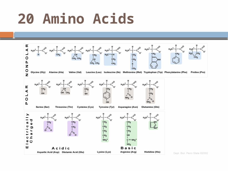
20 Amino Acids

R Groups
its physical & chemical properties determine the unique characteristics of a.a. so affect the physical & chemical properties of the polypeptide chain
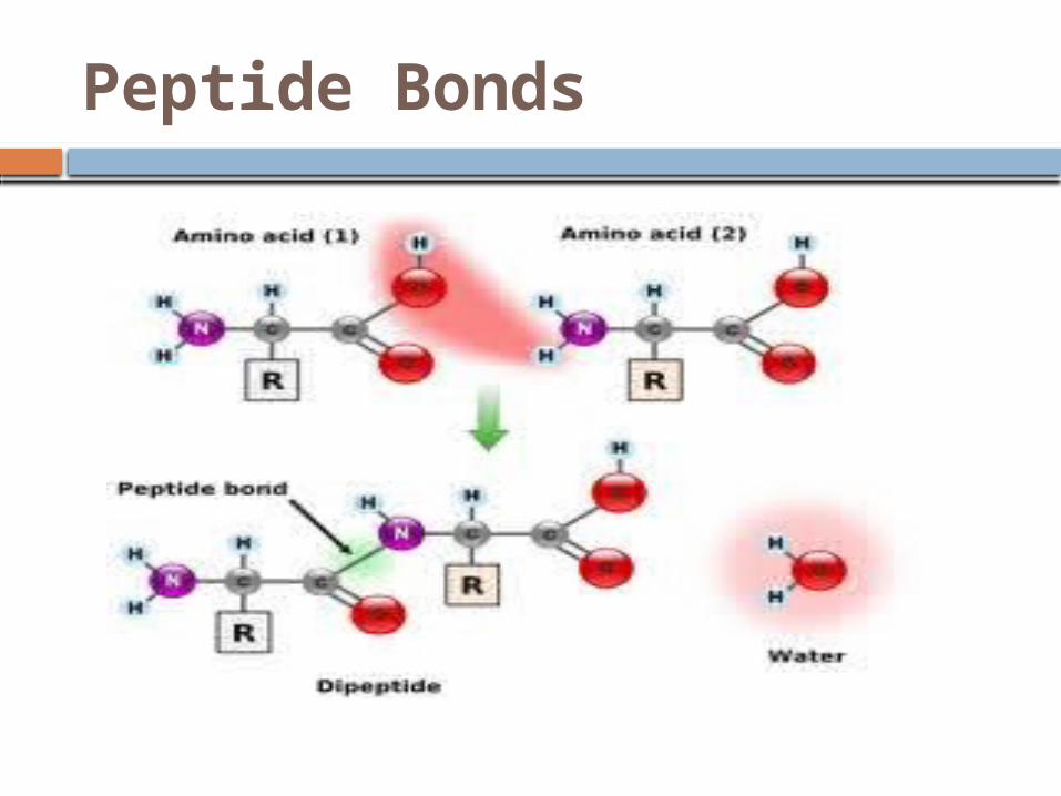
Peptide Bonds
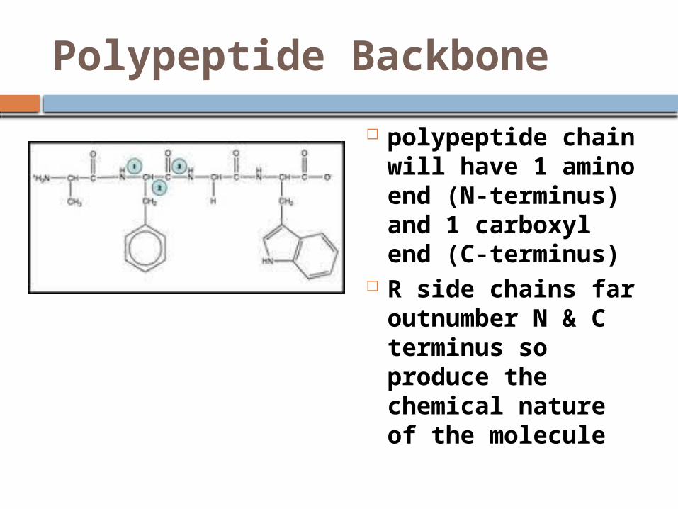
Polypeptide Backbone
polypeptide chain will have 1 amino end (N-terminus) and 1 carboxyl end (C-terminus)
R side chains far outnumber N & C terminus so produce the chemical nature of the molecule
Protein Structure & Function
polypeptide ≠ protein

Functional Protein
is not just a polypeptide chain but 1 or more polypeptides precisely twisted, folded, & coiled into a uniquely shaped molecule

Protein Shape
determined by a.a. sequence

Protein Shape
1. Globular Protein
roughly spherical
2. Fibrous Protein
long fibers
when polypeptide released from ribosome it will automatically assume the functional shape for that protein’s (due to its primary structure)

Name that Shape

Protein Structure
determines how it functions almost all proteins work by
recognizing & binding to some other molecule
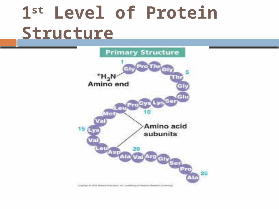
1st Level of Protein Structure

Secondary Structure
segments of each polypeptide chain that coil or fold in patterns
result of: H-bonds in polypeptide backbone α helix: every 4th a.a. held together by
H-bonds β pleated: 2 parallel β strands held
together by H-bonds (is what makes spider silk so strong)

Secondary Structure

Tertiary Structure
3-D shape stabilized by interactions between side-chains
1. hydrophobic interactions a.a. with nonpolar side chains usually
end up together at core of protein: result of exclusion of nonpolar parts by water
once nonpolar side chains away from water, van der Waals forces hold them together
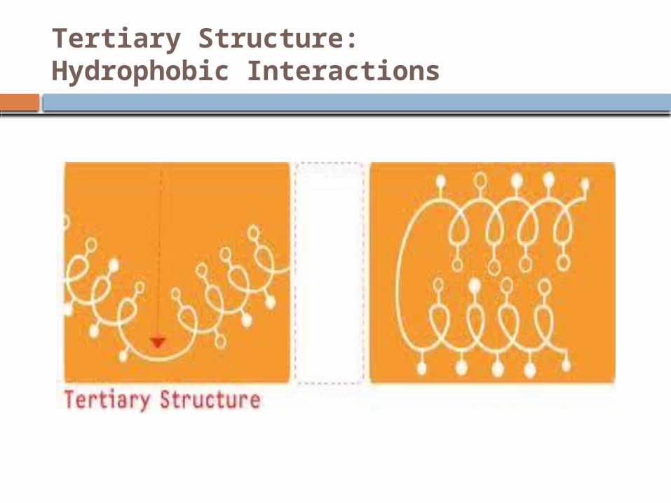
Tertiary Structure: Hydrophobic Interactions
Tertiary Structure
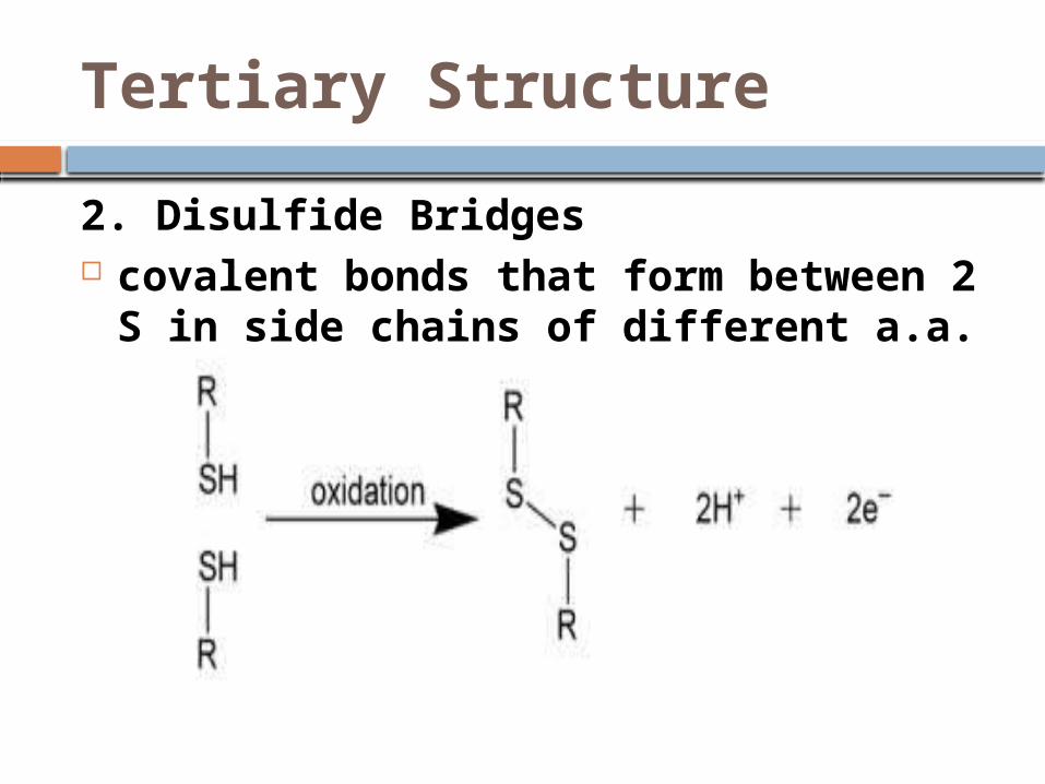
2. Disulfide Bridges covalent bonds that form between 2
S in side chains of different a.a.
Quarternary Structure
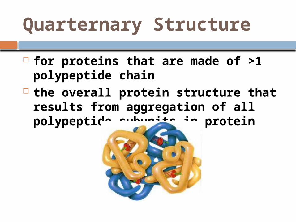
for proteins that are made of >1 polypeptide chain
the overall protein structure that results from aggregation of all polypeptide subunits in protein
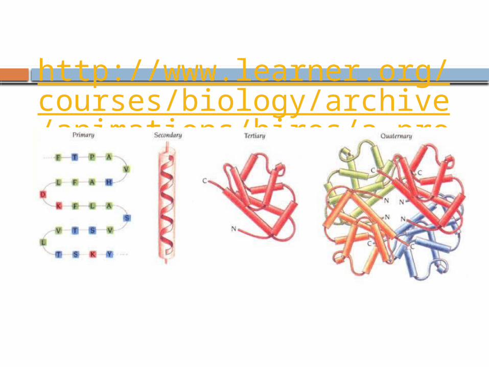
http://www.learner.org/courses/biology/archive/animations/hires/a_proteo1_h.html

Protein Structure
http://www.stolaf.edu/people/giannini/flashanimat/proteins/protein%20structure.swf
Collagen
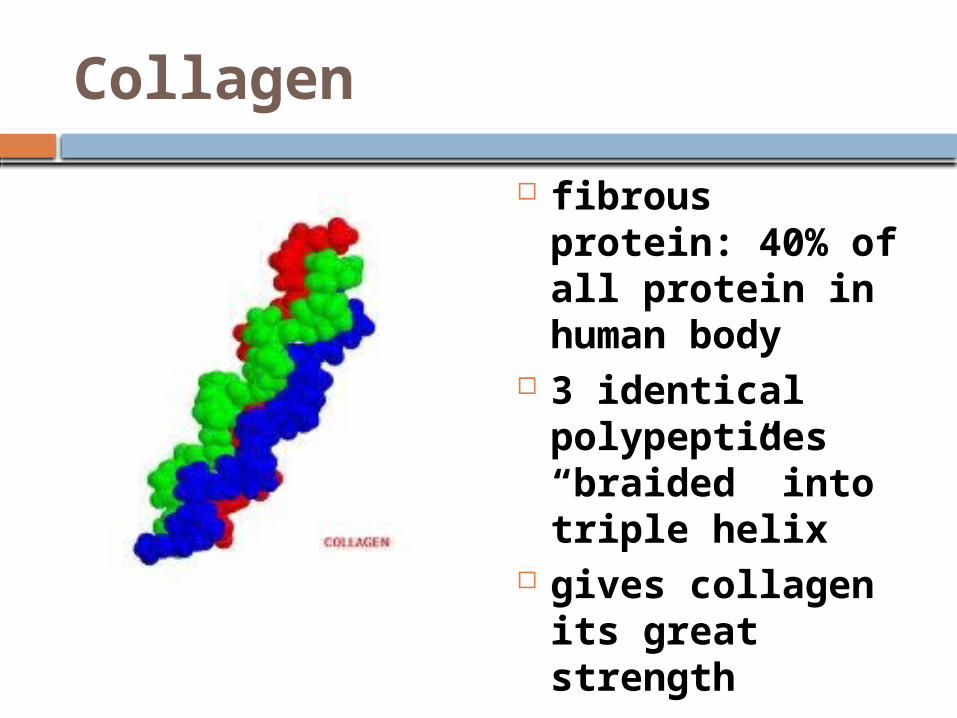
fibrous protein: 40% of all protein in human body
3 identical polypeptides “braided” into triple helix
gives collagen its great strength

Hemoglobin
globular protein made of 2 alpha & 2 beta subunits (polypeptides)
each has nonpolypeptide part = heme which has Fe to bind O2

Sickle Cell Disease
due to substitution of one a.a. (valine) for the normal one, glutamine
causes normal disc-shape of RBC to become sickle shaped because the abnormal hemoglobin crystallizes

Sickle Cell Disease
go thru periodic “sickle-cell crises” angular sickled cells clog small
blood vessels impedes blood flow causes pain

Protein Structure
also depends on physical & chemical environment protein is in:
1. pH2. salt concentration3. temperature
all of the above can change weak bonds & forces holding protein together

Denaturation
process in which a protein loses its native shape due to the disruption of weak chemical bonds & interactions
denatured protein becomes biologically inactive

Denaturation Agents
taking protein out of water nonpolar solvent: hydrophilic a.a that were on outer edge to core vise versa with hydrophobic a.a.
Protein Structure
most proteins probably go thru some intermediate shape stages b/4 achieving their stable shape
chaperonins: protein molecules that assist in the proper folding of other proteins

Chaperonins

Misfolded Proteins
ass‘c with: Alzheimer’s Mad Cow disease Parkinson’s Senile Dementia
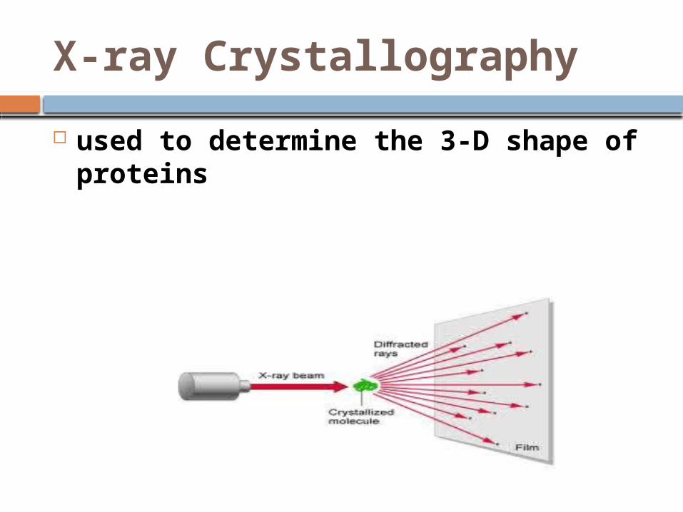
X-ray Crystallography
used to determine the 3-D shape of proteins
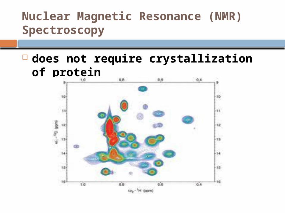
Nuclear Magnetic Resonance (NMR) Spectroscopy
does not require crystallization of protein
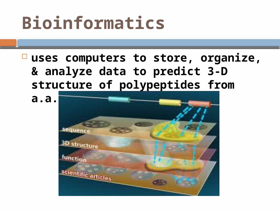
Bioinformatics
uses computers to store, organize, & analyze data to predict 3-D structure of polypeptides from a.a. sequences


NUCLEIC ACIDS
are polymers made of monomers called nucleotides
genes code for a.a. sequences in proteins
1. DNA deoxyribonucleic acid1. RNA ribonucleic acid

Nucleic Acid Roles
DNA:1. self-replication2. reproduction of organism3. flow of genetic information: DNA
RNA synthesis protein synthesis

Nucleic Acid Roles
RNA:1. mRNA
conveys genetic instructions for building proteins from DNA ribosomes
in eukaryotic cells means from nucleus cytoplasm
prokaryotic cells also use mRNA

Nucleic Acids
polymers of nucleotides (the monomers)

Nucleoside
portion of a nucleotide w/out any phosphate group(s)

Nitrogenous Bases
each has 1 or 2 rings that include N are bases because the N atoms can
take up H+ 2 families:1. Pyrimidines
(1) 6-sided ring made of C & N
2. Purines (1) 6-sided ring fused to a 5-sided
ring
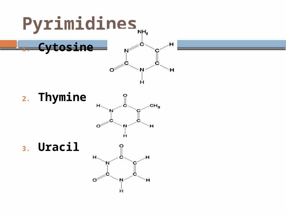
Pyrimidines 1. Cytosine
2. Thymine
3. Uracil

Purines
1. Adenine
2. Guanine

Sugars in Nucleic Acidsadded to
1. Deoxyribose
2. Ribose

Phosphate Group
added to 5’ C of the sugar (base was added to 1’ C)

Nucleotide Polymers
1 nucleotide added to next in phosphodiester linkages

Nucleic Acid Backbone
Phosphodiester linkages repeating pattern of phosphate – sugar – phosphate – sugar..
notice: phosphate end
is 5’ sugar end is 3’

Polynucleotides have built-in direction along their sugar-phosphate backbones

DNA bases held together by H-bonds with backbones going in opposite directions

Linear Order of Bases
specifies start, stop of transcription/translation and codons determine primary structure of proteins (which determines the 3-D structure of a protein which in turn determines the function of the protein)

DNA Molecules
dbl stranded (in opposite directions = antiparallel)
bases held together by H-Bonds most have thousands – millions base
pairs bases pair using complementary
base rules

Complimentary Bases

DNA Molecules
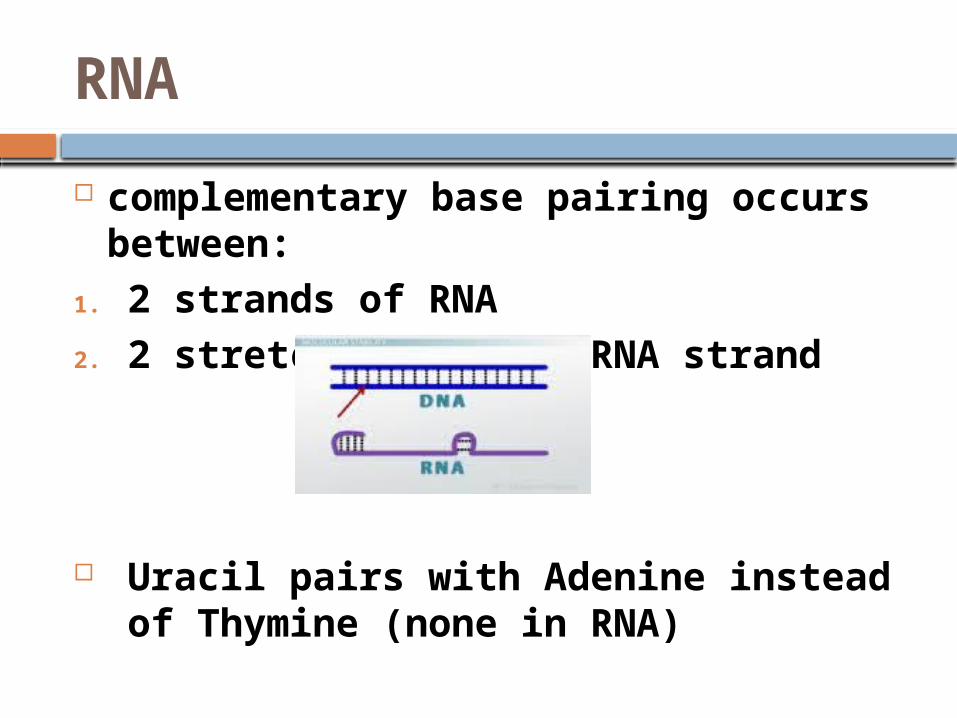
RNA
complementary base pairing occurs between:
1. 2 strands of RNA2. 2 stretches of same RNA strand
Uracil pairs with Adenine instead of Thymine (none in RNA)

DNA & Proteins
genes & their proteins document the hereditary background of an organism
able to expect 2 species that appear to be closely related based on fossil & anatomical evidence to also share a greater proportion of their DNA & protein sequences than do more distantly related species
Hemoglobin
human & gorilla hgb differ only by 1 a.a out of 46 in β chain
human & frog differ by 67 a.a.
