the surgical technologistconsistent with multiple aspiration events and esophageal pathologies...
TRANSCRIPT


JUNE 2010 | The Surgical Technologist | 257
Bronchoesophageal fistulae are a relatively uncommon clinical condition
that may present within a number of patient populations. Etiologies will
include congenital malformation, trauma, malignancies, or secondary-to-
inflammatory processes or infection.1 Diagnosis of bronchoesophageal fis-
tulae may be difficult due to its often-insidious nature of presentation.1 The
open thoracotomy and video-assisted thoracotomy are effective modalities
of surgical intervention.2
L E A R N I N G O B J E C T I V E S
▲ Review the relevant anatomy for
this procedure
▲ Examine the set-up and surgical
positioning for this procedure
▲ Compare and contrast the
osteoplasty-cam procedure
and the acetabuloplasty-pincer
procedure.
▲ Assess the indications for
femoroacetabular impingement
▲ Evaluate the recovery and rehabili-
tation process following FAI
T his article will examine the common approach to clini-cal assessment and diagnosis of this disease state, as well as detail surgical technique with an emphasis on the specific role of surgical technologists in the open-
thoracotomy-type procedure.
E T I O L O G Y
Fistulae may result from congenital malformations, often present-ing with esophageal atresia,3 a disordered formation of the esoph-agus in utero, which may cause disruption of the esophageal tissue and, in some cases, communication with the trachea and bronchi.4 Bronchoesophageal fistulae of a congenital nature are categorized into four typical presentations by Baimbridge and Keith.5
Type one fistulae occur as an esophageal diverticulum becomes subject to inflammatory processes, causing the endpoint to perforate the pleura to communicate with the bronchial lumen.
Type two fistulae are the most common,1 and are also known as “H-type” fistulae. This configuration will feature direct tissue communication between esophageal and tracheal lumen.
by Sophanna Pech and Grant Drake
The Open Thoracotomy Approach to Bronchoesophageal Fistulae Repair
an examination of diagnosis and surgical intervention in
| The Surgical Technologist | JUNE 2010 258
the carina, the bronchi separate into the right and left main-stem bronchi. As air moves distally throughout the bron-chi, the conductive airways become progressively smaller in diameter, contain a decreasing amount of cartilage, and
bifurcate numerous times, eventually reaching respira-tory bronchioles and finally the alveoli where the actual gas exchange occurs. The alveoli make up the majority of the parenchymal surface area of the lung along with the pulmonary capillaries.
The esophagus begins at the level of the epiglottis and continues posterior to the trachea which is typi-cally 10-11 cm long and typically will be 1.5-two cm in diameter in adults and is made up of semi-circular cartilaginous rings which are “c” shaped with no carti-lage over the area where tracheal smooth muscle sits in direct contact with the esophagus.10 The one exception to the “c” shaped cartilaginous rings is the cricoid car-tilage, which is a continuous cartilaginous ring and sits at the base of the larynx directly inferior to the thyroid cartilage. Patency of the alveoli is maintained by radial traction,12 the pressure of the alveolar units themselves creating negative pressure against the parietal pleura pulling the small airways open. The trachea and both mainstem bronchi contain smooth muscle, connective tissue and a luminal surface of ciliated columnal epi-
lthelium interspersed with mucous-producing goblet cells. The trachea, mainstem bronchi, and conducting airways are lumenally coated with a sol and gel type colloid,13 or accord-ing to Schürch, “an aqueous substrate which covers the sur-face of the conducting airways consist[ing] of water, ions, sugars, proteins, proteoglycans, glycoproteins and lipids,”14
which is moved upward in airway clearance by cilia and smooth muscle function as in a cough.
In the case of bronchoesophageal or tracheoesophageal fistula, the most common type of communicating fistulae will occur as this posterior noncartilagenous area of tra-chea or mainstem bronchus is perforated. In neoplastic disease states, such as a squamous cell carcinoma, neopla-sia within tracheal smooth muscle may result in exophytic or ulcerative lesions,8 causing erosion of the tracheal wall from within the epithelium moving outward, or beginning with disruption of the superficial layer of ciliated columnar epithelium, and leading to eventual erosion of connective tissues and basal cells until perforation occurs. Neoplasms may then extend extraluminally into the esophagus, eventu-ally creating a communication between trachea and esopha-gus as the neoplasm expands and invades esophageal tissue.
Type three consists of fistulous tissue connection between the esophagus and a lobar cyst, which in turn com-municates with the bronchus.
Type four involves fistulous tract connection with seques-tered parenchyma. Parenchy-mal sequestration occurs as a congenital malformation in which an area of parenchymal tissue is separated from the bronchial tree and is not vas-cularized by the pulmonary vasculature in the typical fash-ion,6 nor does it participate in oxygenation or ventilatory ac-tivity. This parenchymal lesion is instead supplied blood by the systemic circuit, according to Boetzkes et al, “[it] is sup-plied with blood from an aber-rant artery mostly originating in the thoracic aorta.”7
More commonly, broncho-esophageal fistula may pres-ent secondary to a variety of malignancies,8 including but not limited to Hodgkin’s lymphoma, esophageal carcinomas and bronchogenic carcinomas. Additionally, bronchoesoph-ageal fistula may present secondary to occupational expo-sure to respiratory irritants such as asbestos and silicone, or as a complication of infectious processes where cavitating lesions may occur as in the fungal infection histoplasmosis.8
P A T H O P H Y S I O L O G Y
Brochoesophageal fistulae may originate in either the tra-chea or esophagus but will eventually involve each tissue type. This article examines the anatomical makeup of each organ and discusses pathological changes that may occur as fistulae evolve from both conducting airways and the esophagus.
The respiratory system is subdivided into two main regions. The first is the upper respiratory tract, which con-sists of the nose, nasopharynx, mouth, and oropharynx, where air is drawn inward upon inhalation and released during exhalation. The second is the lower respiratory tract,10 which starts at the laryngopharynx and continues through the larynx (which includes the epiglottis), trachea, carina (where the trachea bifurcates), and the bronchi. At
An extra bump on the femoral head
can cause a deviation from the
normal sphericity of the femoral
head, leading to increa sed an
An extra bump on the femoral head
can cause a deviation from the
normal sphericity of the femoral
head, leading to increa sed an
An extra bump on the femoral head

JUNE 2010 | The Surgical Technologist | 259
Congenital type bronchoesophageal fistulae often occur in embryonic development. In gestational-week four, lungs begin to form as respiratory diverticuli emerge from the foregut eplithelium,15 and in subsequent weeks, a tracheoesphageal ridge and the tracheoesophgeal septum form to separate trachea from esopha-gus. In the case that this septum is not formed completely, bronchoesophageal fistula will result.
The esophagus connects the laryn-gopharynx with the stomach and digestive organs of the alimentary tract. It is composed of smooth muscles and is an average of 25 cm long and ter-minates at the lower esophageal sphincter (also called the cardiac sphincter),10 the entrance to the stomach. As the esophagus contains no cartilaginous surfaces, it is subject to constriction in areas where it meets with other anatomic surfaces. Narrowing occurs as the esophagus passes by the aortic arch, the left mainstem bronchus and the diaphragm. Histologically, the esophagus is composed of four tissue lay-ers, the luminal mucosa layer, submucosa, muscularis and an outer layer called the serosa or adventitia.10 The muco-sal layer is made up of stratified squamous epithelial cells, but also contains goblet cells, which produce mucous. The
submucosal layer also contains mucous-producing cells, as well as vasculature, nerve fibers and some smooth muscle
fibers. The muscle tissue contained in the third layer con-sists of longitudinal and circular smooth muscle tissue, which carries the major workload of peristaltic activity. The outer layer of the esophagus contains a thin, fascial layer of connective tissue housing vasculature and is considered an adventitial layer except when in contact with the trachea, when it is then considered the outer serosa.11
D I A G N O S I S
Diagnosis of bronchoesophageal fistula is considerably com-plex in that congenital-type fistulae may go undiagnosed for a number of years if the configuration is such that both the esophagus and conducting airways maintain relatively
Since most hip joint pathology is found within the intra-
articular region, distraction is necessary to achieve arthro-
scopiic access.11 DDetermiiniing thhe siigns andd symptoms thhat
suggest intra-articular pathology are essential.
The patient is prepped and draped in routine fashion. The skin incision and its rela-
tion to the scapula are marked with a sterile marking pen. The patient is prepped
and draped in routine fashion.
The skin is incised with a scalpel. Fat and fascia are divided with cautery as far as
the muscle fascia. The triangle of fascia between the trapezius and latissimus is
rasied, exposing serratus and the rhomboid muscles deeply
Imag
es
cou
rte
sy o
f K
iera
n M
cMan
us

| The Surgical Technologist | JUNE 2010 260
normal patency.1 In cases such as this, bronchoesophageal fistula may be considered when a patient has a significant history of pneumonia and respiratory infection, particular-ly if these are found to be associated with aspiration-type events. A patient may present with a chronic cough of mod-erate to paroxysmal characteristics. Paroxysmal cough is a very sudden and severe form of cough, a “coughing attack,” which involves extreme muscle stricture and spastic move-ment of airway musculature. Cough may present in either a productive or nonproductive nature, dysphagia and, less commonly, hemoptysis may also occur.1
Common diagnostic procedures for any of the malig-nant or congenital types include bronchoscopy, esophago-gastroduodenoscopy9 and contrast esophogram with bar-ium swallow or methylene blue instillation. Imaging may show chronic, inflammatory-type changes to the airway including bronchiectatic configurations of the bronchus,14 consistent with multiple aspiration events and esophageal pathologies including, but not limited to, diverticuli or esophagomalachia.
I N D I C A T I O N S F O R S U R G I C A L I N T E R V E N T I O N
Dysphagia is of particular concern in judging surgical can-didacy as this may lead to aspiration events, chronic cough, chronic infection and suppurative-type pulmonary lesion.
It has been recommended by Altorki and colleagues that all patients with thoracic esophageal diverticulae consider sur-gery as fistula may result.17 Additionally, surgical manage-ment in malignant-type cases is with precedent, and timely excision in some cases may be prophylactic to development of fistulae. In cases where recurrent infection and lack of maintainable patency in either airway or esophagus occur, surgical intervention is indicated.
S U R G I C A L T E C H N I Q U E
Bronchoesophageal fistulae are managed according to con-figuration and location of the fistula. Both open surgery and video-assisted thoracotomy are utilized and have proven to be effective in fistulae closure.
Preoperatively, the patient is positioned according to ease of access to the fistula. This will typically be lateral or supine positioning.18 The example used in this article shall assume lateral positioning and fistula between lower respi-ratory tract, for instance the right middle lobe segmental bronchi and esophagus.
Prior to positioning, the patient will be anaesthetized and intubated with a double lumen endotracheal tube in the supine position. If available, two compatible ventilators may be synchronized to allow for independent lung ventila-tion to anticipate upcoming deflation of the operative lung.
The serratus fascia is now incised as in a standard postero-lateral thoracotomy.The latissimus is freed from the underlying serratus. Care must be taken not
to damage the neurovascular supply to the serratus, which lies on the superficial
surface of the muscle. Similarly, the trapezius is freed from the underlying
rhomboids.

JUNE 2010 | The Surgical Technologist | 261
The nonsterile surgical team members then position the patient in the left lateral position. The patient will be posi-tioned so as their nonoperative side will contact the table. Sequential compression devices are applied to the legs to prevent embolic activity, then the leg proximal to the oper-
ating table will be slightly flexed at the knee and abducted toward the chest. A pillow is then placed under the knee. The leg will be padded so contact with the table will not disturb the peroneal nerve. Arms are placed and secured on padded armboards with the superior arm pronated and slightly flexed, and the inferior arm positioned so the wrist is supinated and exposed so radial pulse may be palpated if necessary, and to protect the ulnar nerve. Additionally, a small roll is placed under the patient just below the last rib along the axillary line to allow for chest expansion, and to
relieve pressure placed on the brachial plexus. Rolled blan-kets are placed under the scapula on the nonoperative side. Finally, three-inch safety straps are placed over the thigh and shoulder to ensure stability.
When positioning is complete, the patient is prepped for initial incision with application of top-ical anti-microbial preparatory agents to the area from the axilla to iliac crest, and bilaterally beyond the midline or to the table top. The incision site is then draped in a square configuration with towels, then a laparotomy drape is applied exposing only the incision site.
Various supplies are required to ensure proper patient and clinician safety in the thoracot-omy procedure. The required equipment may include Bair-hugger®, cell saver, electrosurgical generator, smoke evacua-tor, arm boards and connected and functional suction.
Instrument sets commonly utilized are the thoracotomy tray, minor orthopedic tray and major vascular set.18 Blades required are #10, #15, and electrosurgical unit. Thoracoto-my trays will typically contain large ratcheting, self-retain-ing retractors such as the Toufier and Reinhoff, which may be used to retract large areas of tissue, a Davidson scapula
During the procedure, an initial diagnostic arthroscopy is per-
formed and the structures of the hip joint are examined.1 The fem-
orall hheadd iis obbservedd ffor chhonddrall ddefformiitiies or ddamage, as wellll
aass tthhee ssuurrffaaccee ooff tthhee aacceettaabbuulluumm, tthhee ccoonnddiittiioonn ooff tthh
The serratus is further dissected off the underlying ribs The intercostal incision continues anteriorly.
Imag
es
cou
rte
sy o
f K
iera
n M
cMan
us

| The Surgical Technologist | JUNE 2010 262
retractor and various smaller handheld retractors.The thoracotomy tray may also include sternal needle
holders and Duvall lung-grasping forceps, which are impor-tant for manipulation of the lung. The minor orthopedic tray is also commonly used and contains a variety of avail-able retractors and forceps types as well as bone and soft tissue-cutting instruments, such as the Metzenbaum, which
may be utilized at the surgeon’s preference in place of the ESU or scalpel. Frasier suction tips are included for suction, and skin hooks for manipulation of distal tissues, the double prong hooks may be particularly useful in this respect.
The initial incision via #10 blade is made in the fourth or fifth intercostal space following the line of the rib.8 The incision runs laterally across the area of the fifth rib and is extended around the scapula cranially to the midline of the
scapula. Subcutaneous tissue is divided with an ESU. The latissumus dorsi is divided, and then deep to the latissimus dorsi, the serratus anterior is divided. The rhomboid muscle may be divided as well to allow retraction of the scapula.18
The surgical assistant may retract the musculature with a Richardson hand-held retractor to allow access to the inter-costal muscle. When the intercostal muscle is visualized, the
fascia and muscle are divided with an ESU. Care is taken to make the inci-sion as low in the intercostal space as possible to avoid the intercostal blood supply and innervation.
Next, a rib-spreader is placed, allowing for ease of visualization and access to a fistula between lower respi-ratory tract and esophagus. At this time, the surgeon may elect to partial-
ly dissect the laterally-running band of intercostal muscle from the posterior aspect of the muscle to later use as a pedicle upon fistula closure in the case of large fistula.8 The #10 blade, or ESU, continues to incise deeper until the lungs are visualized. The lung is deflated. Upon visualization of the lung, Duval clamps are used to manipulate the lung. The surgical assistant will assist the surgeon in holding the clamps to allow for visualization of the fistula. Upon visu-
In order to prevent a possible fracture, a resection of less
than 30 percent of the head-neck junction is recommended
bbecause thhiis hhas bbeen shhown not to allter thhe lloadd-bbeariing capac-
ity of the femoral neck.4,8
A second DeBakey rib spreader is placed at right angles to the first to retract the
latissimus and serratus forward and the trapezius backwards.
The upper blade of the retractor is inserted directly adjacent to the rib, apply-
ing no pressure to the intercostal nerve.
JUNE 2010 | The Surgical Technologist | 263
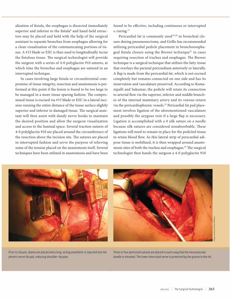
alization of fistula, the esophagus is dissected immediately superior and inferior to the fistula8 and hand-held retrac-tors may be placed and held with the help of the surgical assistant to separate bronchus from esophagus allowing for a clean visualization of the communicating portions of tis-sue. A #15 blade or ESU is then used to longitudinally incise the fistulous tissue. The surgical technologist will provide the surgeon with a series of 4-0 polyglactin 910 sutures, at which time the bronchus and esophagus are sutured in an interrupted technique.
In cases involving large fistula or circumferential com-promise of tissue integrity, resection and anastomosis is per-formed at this point if the lesion is found to be too large to be managed in a more tissue sparing fashion. The compro-mised tissue is excised via #15 blade or ESU in a lateral inci-sion running the entire distance of the tissue surface slightly superior and inferior to damaged tissue. The surgical assis-tant will then assist with dandy nerve hooks to maintain the desired position and allow the surgeon visualization and access to the luminal space. Several traction sutures of 4-0 polylglactin 910 are placed around the circumference of the resection above the incision site. The sutures are placed in interrupted fashion and serve the purpose of relieving some of the tension placed on the anastomosis itself. Several techniques have been utilized in anastomosis and have been
found to be effective, including continuous or interrupted suturing.17
Pericardial fat is commonly used19,20 in bronchial clo-sure during pneumonectomy, and Grillo has recommended utilizing pericardial pedicle placement in bronchoesopha-geal fistula closure using the Brewer technique21 in cases requiring resection of trachea and esophagus. The Brewer technique is a surgical technique that utilizes the fatty tissue that overlays the parietal pericardium anteriorly or laterally. A flap is made from the pericardial fat, which is not excised completely but remains connected on one side and has its innervation and vasculature preserved. According to Koma-nipalli and Sukumar, the pedicle will retain its connection to arterial flow via the superior, inferior and middle branch-es of the internal mammary artery and its venous return via the pericardiophrenic vessels.22 Pericardial fat pad place-ment involves ligation of the aforementioned vasculature and possibly the azygous vein if a large flap is necessary. Ligation is accomplished with a 0 silk suture on a needle because silk sutures are considered nonabsorbable. These ligations will need to remain in place for the pedicled tissue to retain blood flow. As this lateral strip of pericardial adi-pose tissue is mobilized, it is then wrapped around anasto-mosis sites of both the trachea and esophagus.23 The surgical technologist then hands the surgeon a 4-0 polyglactin 910
B
Prior to closure, drains are placed and a long-acting anesthetic is injected into the
phrenic nerve fat pad, reducing shoulder-tip pain.
Three or four pericostal sutures are placed in such a way that the neurovascular
bundle is elevated. The lower intercostal nerve is protected by the groove in the rib.
Imag
es
cou
rte
sy o
f K
iera
n M
cMan
us
| The Surgical Technologist | JUNE 2010 264
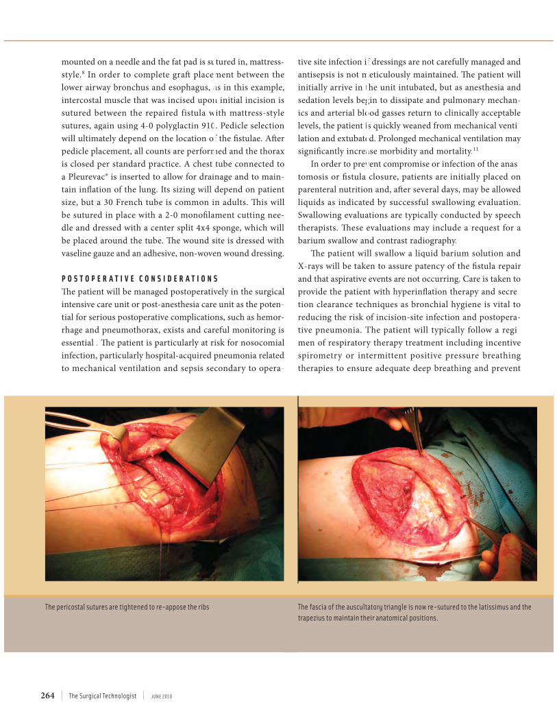
mounted on a needle and the fat pad is sutured in, mattress-style.8 In order to complete graft placement between thelower airway bronchus and esophagus, as in this example, intercostal muscle that was incised upon initial incision issutured between the repaired fistula with mattress-style sutures, again using 4-0 polyglactin 910. Pedicle selectionwill ultimately depend on the location of the fistulae. After pedicle placement, all counts are performed and the thorax is closed per standard practice. A chest tube connected to a Pleurevac® is inserted to allow for drainage and to main-tain inflation of the lung. Its sizing will depend on patient size, but a 30 French tube is common in adults. This willbe sutured in place with a 2-0 monofilament cutting nee-dle and dressed with a center split 4x4 sponge, which will be placed around the tube. The wound site is dressed with vaseline gauze and an adhesive, non-woven wound dressing.
P O S T O P E R A T I V E C O N S I D E R A T I O N S
The patient will be managed postoperatively in the surgical intensive care unit or post-anesthesia care unit as the poten-tial for serious postoperative complications, such as hemor-rhage and pneumothorax, exists and careful monitoring is essential . The patient is particularly at risk for nosocomial infection, particularly hospital-acquired pneumonia related to mechanical ventilation and sepsis secondary to opera-
tive site infection if dressings are not carefully managed and antisepsis is not meticulously maintained. The patient will initially arrive in the unit intubated, but as anesthesia andsedation levels begin to dissipate and pulmonary mechan-ics and arterial blood gasses return to clinically acceptable levels, the patient is quickly weaned from mechanical venti-lation and extubated. Prolonged mechanical ventilation may significantly increase morbidity and mortality.11
In order to prevent compromise or infection of the anas-tomosis or fistula closure, patients are initially placed on parenteral nutrition and, after several days, may be allowedliquids as indicated by successful swallowing evaluation.Swallowing evaluations are typically conducted by speech therapists. These evaluations may include a request for abarium swallow and contrast radiography.
The patient will swallow a liquid barium solution and X-rays will be taken to assure patency of the fistula repair and that aspirative events are not occurring. Care is taken toprovide the patient with hyperinflation therapy and secre-tion clearance techniques as bronchial hygiene is vital toreducing the risk of incision-site infection and postopera-tive pneumonia. The patient will typically follow a regi-men of respiratory therapy treatment including incentivespirometry or intermittent positive pressure breathingtherapies to ensure adequate deep breathing and prevent
The fascia of the auscultatory triangle is now re-sutured to the latissimus and the
trapezius to maintain their anatomical positions.
The pericostal sutures are tightened to re-appose the ribs

JUNE 2010 | The Surgical Technologist | 265
atelectasis. During incentive spirometry, the patient inhales through the incentive spirometer device, which will mea-sure the volume of air the patient is able to inspire. This is used to encourage deep breathing, provide the patient with a visualization of deep breathing goals and allows the respiratory therapist to measure the volume of gas inspired to assess lung expansion and respiratory effort. Therapies such as intermittent positive pressure breathing function in the same manner, but provide the patient assistance by delivering an additional degree of ventilation upon inspira-tory effort.
C O N C L U S I O N
Although tracheoesophageal fistula and bronchioesopha-geal fistula are relatively rare, understanding the pathology of their surgical technique will aid the surgical technologist in developing critical and anticipatory thinking in regards to instrument choice as well as gaining an understanding of thoracic and pulmonary anatomy and physiology. Surgical literature includes various additional techniques that may be utilized in these cases, including bronchial or esophageal stenting, video-assisted thorascopic approaches and various wound-sealing choices, including surgical gluing and endo-scopic clipping. These techniques may be used as an adjunct or alternative to the open technique examined herein.
A B O U T T H E A U T H O R S
Sophie Pech is currently employed as a surgical technolo-gist in the St Luke’s Regional Medical Center cardiovascu-lar surgery unit in Boise, Idaho. She is a recent graduate of The College of Western Idaho Surgical Technology Program and is currently awaiting the results of her credentialing examination.
Grant Drake is a recent graduate of the Boise State Uni-versity Respiratory Therapy Program. In August 2010, he will begin the post –Baccalaureate pre-Medical program at Duquesne University and hopes to attend medical school in the future.
After placing a stay suture at the midpoint of the fascial layer to ensure the
two sides are properly aligned, the subcutaneous fascia is closed with running
polyglactin 910. A routine subcuticular polyglactin 910 suture closes the skin.
The final result.
Imag
es
cou
rte
sy o
f K
iera
n M
cMan
us
| The Surgical Technologist | JUNE 2010 266
References1. Matilla S; Ramo O; Salo J. “Congenital bronchoesophageal fistula in the
adult.” Ann Thorac Surg. 1995. 59(6):887-890.2. Braghetto I; Cardemil G; Schwartz E; et al. “Videothoracoscopic manage-
ment of middle esophageal diverticulum with secondary bronchoesopha-geal fistula: report of a case.” Surg Today. 2008. 38(12):1124-8.
3. Aksoy T; Ceran C; Demircan M; Kafkasli A. “Tracheal agenesis and esoph-ageal atresia with proximal and distal bronchoesophageal fistulas.” J Ped-Surg. 2008. 43(8): e1-e3.
4. Spitz L. 2007. “Oesphageal atresia.” Orphanet J Rare Dis. 2:24. Accessed: November 12, 2009. Available at: http://www.ojrd.com/content/2/1/24.
5. Baimbridge M; Keith H. “Oesophago-bronchial fistula in the adult.Tho-rax.” 1965. 20:226-233.
6. De Boeck K; Devlieger H; Van Raemdonck D; et al. “Pulmonary seques-tration: a comparison between pediatric and adult patients.” Eur J Cardio-thorac Surg. 2001. 19;(4):388-95.
7. de Beukelaar T; Boetzkes S; Ramet J; Wojciechowski M; Van Schil P. “Intra-lobar bronchopulmonary sequestration with mixed venous drainage in a 10-year-old girl.” Acta Chir Belg. 2009. 109(4): 501-3.
8. Grillo H. Surgery of the Trachea and Bronchi. Hamilton, Ontario. 2004. 576-579, 592-594
9. Cooperman R; Paragi P; Shah S. “Adult congenital bronchoesophageal fis-tula.” Thorac Cardiovasc Surg. 2007. 55(5):334-6.
10. Bannister L; Berry M; Collins P; Dyson M; Dussek J; Ferguson M; Williams P. “Respiratory system.” Gray’s Anatomy 38th ed. New York. Pearson. 1995. 1648-1656.
11. King D. 2010. “Histology at Southern Illinois School of Medicine.” Acessed March 15, 2010. Available at: http://www.siumed.edu/~dking2/erg/giguide.htm.
12. Wilkins R; Stoller J; Kacmarek R et al. Egan’s Fundamentals of Respiratory Care 9th ed. Mosby. 2008.
13. Anderson J. Cardiopulmonary Renal Physiology Lecture. Boise State Uni-versity. September 2008.
14. Schürch S; Geiser M; Lee M; Gehr P. “Particles at the airway interfaces of the lung.” Colloids and Surfaces B: Biointerfaces. 1999. 15(3-4):339-353.
15. Langman T; Sadler J. Langman’s medical embryology 9th ed. Philadelphia. Lipincott&Williams. 2003. 278-285.
16. Iwazawa T; Imazato M; Ohnishi T; Kimura Y; Yano H; Monden T. “Double congenital bronchoesophageal fistulae in an adult.” Jpn J Thorac Cardiovasc Surg. 2004. 52(8):386-9.
17. Altorki N; Sunagawa M; Skinner D. “Thoracic esophageal diverticula. Why is operation necessary?” J Thorac Cardiovasc Surg. 1993. 105(2):260-4.
18. Frey K; Ross T. Surgical Technology for the Surgical Technologist: A Positive Care Approach 3rd ed. Clifton Park, NY. Dlelmar Cengage Learning. 2008. 942-945.
19. Durrleman N; Massard G. “Posterolateral thoracotomy.” MMCTS. 2006. (0810):1453.
20. Bardini R; Bonavina L; Asolati M; Ruol A; Castoro C; Tiso E. “Single-lay-ered cervical esophageal anastomoses: a prospective study of two suturing techniques.” Ann Thorac Surg. 1994. 58:1087-1089.
21. Taghavi S; Marta G; Lang G; et al. “Bronchial Stump Coverage with a Pedi-cled Pericardial Flap: An Effective Method for Prevention of Postpneumo-nectomy Bronchopleural Fistula.” Ann Thorac Surg. 2005. 79(1):284-288.
22. Anderson T; Miller J. “Surgical technique and application of pericardial fat pad and pericardiophrenic grafts.” Ann Thorac Surg. 1995. 59(6):1590-1591.
23. . Komanapalli C; Sukumar M. “Pericardial Fat Pad Flap Buttress.” The Car-diothoracic surgery network. Accessed March 27, 2010. Available at: http://www.ctsnet.org/sections/clinicalresources/thoracic/expert_tech-28.html.
24. Brewer L; King E; Lilly L; Bai A. “Bronchial closure in pulmonary resec-tion: a clinical and experimental study using pedicled pericardial fat graft reinforcement.” J Thorac Surg. 1953. 26(5):507–532.
25. Anderson T; Miller J. “Use of Pleura, Azygos Vein, Pericardium, and Mus-cle Flaps in Tracheobronchial Surgery.” Ann Thorac Surg. 1995. 60:729-733.
C E E X A M
JUNE 2010 | The Surgical Technologist | 267
Bronchoesophageal Fistula Repair318 J U N E 2 0 1 0 1 CE credit
O P E N T H O R A C T O M Y A P P R O A C H T O B R O C H O E S O P H E G E A L F I S T U L A R E P A I R 318 J U N E 2 0 1 0 1 CE credit
Earn CE Credits at Home
You will be awarded continuing education
(CE) credit(s) for recertification after read-
ing the designated article and completing the
exam with a score of 70% or better.
If you are a current AST member and are
certified, credit earned through completion
of the CE exam will automatically be recorded
in your file—you do not have to submit a CE
reporting form. A printout of all the CE credits
you have earned, including Journal CE cred-
its, will be mailed to you in the first quarter
following the end of the calendar year. You
may check the status of your CE record with
AST at any time.
If you are not an AST member or are not
certified, you will be notified by mail when
Journal credits are submitted, but your cred-
its will not be recorded in AST’s files.
Detach or photocopy the answer block,
include your check or money order made
payable to AST, and send it to Member Ser-
vices, AST, 6 West Dry Creek Circle, Suite 200,
Littleton, CO 80120-8031.
Members: $6, nonmembers: $10
a b c d a b c d
1 ■ ■ ■ ■ 6 ■ ■ ■ ■
2 ■ ■ ■ ■ 7 ■ ■ ■ ■
3 ■ ■ ■ ■ 8 ■ ■ ■ ■
4 ■ ■ ■ ■ 9 ■ ■ ■ ■
5 ■ ■ ■ ■ 10 ■ ■ ■ ■
Mark one box next to each number.
Only one correct or best answer can be selected for each question.
1. Bronchoesophageal fistulae are
categorized into ___________ typical
presentations.
a. 1 c. 3
b. 2 d. 4
2. During embryonic development, the lungs
begin to form during gestational week
_____________ .
a. 2 c. 4
b. 3 d. 5
3. Bronchoesophageal fistulae may present
secondary to __________ .
a. Hodgkin’s lymphoma
b. Certain respiratory irritants
c. Cavitating lesions
d. All of the above
4. The ___________ is made up of stratified
squamous epithelial cells.
a. Mucosal layer
b. Submucosa
c. Mainstem bronchus
d. Muscularis
5. The most common type of fistula is ______ .
a. Type 1
b. H-type
c. Sequestered parenchyma
d. None of the above
6. ___ has been proven as an effective
surgical method in fistula closure.
a. Endotracheotomy
b. Open thoracotomy
c. Video-assisted thoracotomy
d. b & c
7. According to ease of access to the fistula,
the typical patient is preoperatively
positioned in either _______ or _______
position.
a. Lateral/Trendelenburg
b. Supine/Fowler’s
c. Supine/Lateral
d. No proper combination
8. An “H-type fistula” refers to a direct
connection between the esophagus and
the ______________ .
a. Tracheal lumen c. Parenchymal tissue
b. Bronchus d. None of the above
9. Postoperative swallowing evaluations
may include ___________ .
a. Speech therapy c. Contrast radiography
b. Barium swallow d. b & c
10. Postoperative complications may include
______________ .
a. Hemorrhage
b. Pneumothorax
c. Nosocomial infection
d. All of the above
NBSTSA Certification No.
AST Member No.
■ My address has changed. The address below is the new address.
Name
Address
City State Zip
Telephone