the tamil nadu dr. m.g.r. medical university chennai...
TRANSCRIPT

DETERMINATION OF SENSITIVITY PATTERN AND
TREATMENT OUTCOME IN CULTURE POSITIVE
BACTERIAL KERATITIS
Dissertation submitted to The Tamil Nadu Dr. M.G.R. Medical
University in partial fulfilment of the requirements for the degree of
MS Ophthalmology
BRANCH - III
OPHTHALMOLOGY
THE TAMIL NADU
DR. M.G.R. MEDICAL UNIVERSITY
CHENNAI –600032
MAY 2018

DECLARATION
I, Dr.Abinaya.C hereby declare that this dissertation entitled
"DETERMINATION OF SENSITIVITY PATTERN AND
TREATMENT OUTCOME IN CULTURE POSITIVE
BACTERIAL KERATITIS" is being submitted in partial fulfilment for
the award of MS degree in Ophthalmology by The Tamilnadu Dr. M.G.R
Medical University in the examination to be held in May 2018. I declare
that this dissertation is my original words and has not formed the basis for
the award of any other degree or diploma awarded to me previously
Dr.Abinaya.C
Aravind eye hospital &
PG Institute of Ophthalmology,
Madurai, Tamilnadu.

CERTIFICATE
This is to certify that this dissertation entitled
"DETERMINATION OF SENSITIVITY PATTERN AND
TREATMENT OUTCOME IN CULTURE POSITIVE
BACTERIAL KERATITIS" is a bonafide work done by Dr. ABINAYA
.C under our guidance and supervision at Aravind Eye Hospital and PG
Institute of Ophthalmology, Madurai during the period of her Postgraduate
training in Ophthalmology for June 2015 to May 2018.
GUIDE:
Dr.Lalitha Prajna,MD.,DNB.,
Chief Microbiologist,
Department of Ocular microbiology,
Aravind Eye Hospital &
PG Institute of Ophthalmology,
Madurai.
Dr.N.Venkatesh Prajna DO, DNB, FRCOphth.,
Prof. & Head of the Department,
Aravind Eye Hospital &
PG Institute of Ophthalmology,
Madurai.
Dr.R.Rathinam, DO, DNB, Ph.D.,
Principal,
Aravind Eye Hospital &
PG Institute of Ophthalmology,
Madurai.

ACKNOWLEDGEMENT
First and foremost, I would like to thank God without him, I can do
nothing.
I take this opportunity to pay my respect and homage to
Dr.G.Venkatasamy, our founder and visionary whose dynamism had led
Aravind against all odd to its high scale of achievement.
I would like to Thank Dr.Venkatesh Prajna, Chief Cornea Clinic for
allowing me to work on this study, guiding me at every step and for being a
constant source of motivation and encouragement.
I would also like to thank Dr.Lalitha Prajna, Chief microbiologist
Dept. of ocular microbiology for her constant guidance and support.
I would also like to thank my co-guide Dr.Hemshah, Dr.Ashis
Ghosh for being a constant source of motivation and encouragement and for
helping me fine tune my thesis.
I am very grateful to Dr.R.D.Ravindran, Chairman of Aravind eye
hospital for having created an environment enriched with all the facilities
for learning and gaining knowledge. I am privileged to have on my side
Dr.P.Namperumalsamy, Chairman Emeritus Dr.G.Natchiar, Director
Emeritus, Dr.M.Srinivasan, Director Emeritus, Dr.S.Rathinam, Director
emeritus, Dr.S.R.Krishnadas, Director Human resources and other scholar
of ophthalmology at Aravind eye care system.

I am much graceful to Mr.G.Ramesh kumar microbiologist for his
support. My sincere thanks to Mrs.Kumaragurupari, Sr.Librarian,
Mr.R.Govindarajan Asst. Librarian for their prompt and efficient response
to my innumerable requests for articles and information.
I would also like to thank biostatistician Mrs.Iswaraya who has
helped with the task of compiling the statistics and enable in preparation of
results.
I would fail in my duty if I did not thank the countless patients who
have been the learning ground for my study and my residency.
Last but not the least I would like to thank my parents, my husband
Dr.S.Harikrishnan and my friends for their constant support and unfailing
love towards me.

CONTENTS
Sl.No. TITLE Page No.
1. INTRODUCTION 1
2. REVIEW OF LITERATURE 30
3. AIMS AND OBJECTIVES 34
4. METHODOLOGY 35
5. RESULTS 41
6. DISCUSSION 77
7. CONCLUSION 81
BIBLIOGRAPHY
ANNEXURES
URKUND PLAGIARISM SCREEN SHOT
DIGITAL RECEIPT
ETHICAL COMMITTEE APPROVAL ORDER
CONSENT FORM
PROFORMA
MASTERCHART

INTRODUCTION
1
INTRODUCTION
DEFINITION
Bacterial keratitis is defined as an inflammatory infiltrate of the corneal
stroma associated with an epithelial defect from which one or more bacterial
species were cultured.
CORNEAL ULCERATION AS A CAUSE OF BLINDNESS
1. Blindness continues to be one of the major public health problems in
developing countries1
2. According to World Health Organization, corneal diseases are among
the major causes of vision loss and blindness in the world today, after
cataract and glaucoma1
3. In India, it is estimated that there are approximately 6.8 million people
who have vision less than 6/60 in at least one eye due to corneal
diseases.
4. According to the National Programme for Control of Blindness (NPCB)
there are currently 1,20,000 corneal blind persons in the country.
According to this estimate there is addition of 25,000 -30,000 corneal
blindness cases every year in the country2.
5. It is expected that the number of individuals with unilateral corneal
blindness in India will increase to 10.6 million by 2020.

2
6. Corneal ulceration is a significant cause of corneal blindness .Corneal
ulceration has been recognized as a silent epidemic in developing
countries, especially the South-east Asian Region. The causal factors
responsible for corneal blindness vary with age.
7. Significant causes of corneal blindness in adults residing in countries
with less-developed economies are corneal scars (28.1%) and active
keratitis (12.2%)1.
8. In tropical countries like India Bacterial keratitis is the second most
important cause of microbial keratitis next to fungal keratitis.
REASON FOR THE STUDY:
1. Bacterial keratitis is a serious ocular infectious disease that can lead to
severe visual disability3.
2. Once a bacterial corneal ulcer has been diagnosed, scraping of the ulcer
for Gram stain evaluation, culture and sensitivity, and initiation of
antibiotic therapy should follow in rapid succession9.
3. The indiscriminate use of antibiotics has lead to the development of
bacterial strains resistant to many commonly used agents(4,5)
4. Over the last few years, there has been a major shift in the preferred
topical antibiotic therapy for bacterial keratitis6.
5. The spectrum of causative organisms and their susceptibility to
antimicrobials varies according to the latitude and the degree of
urbanization7

3
6. Periodic susceptability testing should be performed to ensure currently
available antimicrobial are providing good coverage against recent
clinical isolates of pathogenic bacteria5.
NORMAL CORNEAL DEFENCE MECHANISM
o There are several defense mechanisms to protect the surface of the eye
from many infectious agents .
o Eyelids serves as a physical barrier .
o Mechanical flushing action of the tears is an important defense
mechanism against infection due to presence of lysozyme, lactoferrin,
beta-lysin and IgA antibodies10
.
o The mucin layer present in the tear flim can trap and remove potentially
pathogenic organisms.
o The normal ocular flora helps in preventing overgrowth of indigenous
organisms or invasion of pathogens.
o The conjunctiva contains sub-epithelial mucosal associated lymphoid
tissue (MALT) with a collection of lymphoid cells having specific
defensive functions.
o The eye's acute non-specific inflammatory reaction to injury is mainly
through phagocytosis of the invading neutrophils and later by
macrophages, helping the immunocompetent host to control and destroy
invading organisms. Specific hormonal and cellular reactions also
countered opportunists.

4
RISK FACTORS
EXTRINSIC FACTORS:
Use of contact lenses , especially when associated with the following :
o Overnight wear and long term use11
.
o Inadequate disinfection of contact lenses
o Contamination of the contact lens storage container.
o Infected or contaminated contact lens solution
o Storage or rinsing contact lens in tapwater
Trauma , including chemical and thermal injuries , foreign bodies , and
local irradiation
Previous ocular and eyelid surgery ,mainly corneal surgery including
refractive surgery and penetrating keratoplasty.
Loose corneal sutures in keratoplasty
Medication related factors (e.g. contaminated ocular medications,
topical NSAIDS, anesthetics, corticosteroids, preservatives, glaucoma
medications.
Immunosuppression (local and systemic)
OCULAR SURFACE DISEASE
Tear flim deficiencies

5
Abnormalities of the eyelid anatomy and their function (entropion,
ectropion, blepharitis, trichiasis, lagophthalmos etc)
Adnexal infection/inflammation ( including gonococcal conjunctivitis,
canaliculitis, dacrocystitis)
CORNEAL EPITHELIAL ABNORMALITIES
Neurotrophic keratopathy
Disorders causing recurrent erosion of the cornea
Abrasion of cornea or epithelial defect
Viral keratitis
Corneal epithelial edema , especially bullous keratopathy
SYSTEMIC CONDITIONS
Diabetes mellitus
Debilitating illness especially malnourishment
Dermatological /mucous membrane disorders (e.g Steven- Johnson
syndrome, ocular mucous membrane pemphigoid)
Immunocompromised status of the host
ORGANISM FACTORS
Virulence factors:
Infection of ocular surface tissues requires the microorganisms to
attach, penetrate , invade, persist and replicate inspite of many protective

6
mechanisms in the host12
. The intact epithelial layer acts as a barrier that few
pathogens can overcome. The organisms that can penetrate intact epithelium
include Neisseria gonorrhoeae, Corynebacterium diptheriae, Haemophilus
influenza, Listeria and Acanthamoeba. Other ocular pathogens require
distruption of the epithelium for adherence and penetration. Virulence factors
that favours tissue invasion include microbial exotoxins and proteases that
destroy tissue cells , inflammatory cells and tissue matrix.
INNOCULUM SIZE AND ROUTE
The capability of the host's defenses determines the threshold of
innoculum size at which infection is inevitable. Break in the epithelium is the
most common route of entry

7
Table-1.1: CLASSIFICATION OF BACTERIAL OF IMPORTANCE IN
MICROBIAL KERATITIS
Gram-positive cocci(aerobic)
Streptococcus pneumoniae
CONS
Staphylococcus aureus
Staphylococcus epidermidis
Enterococcus
Micrococcus
α, β, and non hemolytic streptococci
Gram positive bacilli(aerobic) Bacillus spp
Corynebacterium spp
Listeria monocytogenes
Gram negative bacilli(aerobic)
Pseudomonas aeruginosa
Acinetobacter spp
Enterobacteriaceae
Klebsiella
Serratia
Proteus
Citrobacter
Escherichia
Gram negative diplococci(aerobic)
Neisseria
Gram negative diplobacilli(aerobic) Moraxella
Gram negative
coccobacilli(aerobic) Haemophilus
Gram positive cocci(anaerobic)
Peptococcus
Peptostreptococcus
Gram positive bacilli(anaerobic)
Propionibacterium acnes
Actinomyces
Clostridium(rare)
Gram negative bacilli(anaerobic)
Fusobacterium
Bacteroides
Gram negative cocci(anaerobic)
Veillonella
Spirochetes
Treponema
Borrelia
Leptospira
Gram positive filaments
Mycobacterium(non tuberculous)
Nocardia

8
Fig-1.1: PATHOGENIC MECHANISM OF KERATITIS
1
Breakdown of corneal defense
mechanism
Adherence, Replication and invasion of
microorganisms
Antigen
response
Compliment
activation
Antigen
response Release of enzymes
and exotoxins
Polymorpho Nuclear Leukocute
infiltration
Release of
lysosomal
enzymes
Oxidative burst
with toxic
metabolites
Activation of
corneal Lytic
enzymes
Suppurative corneal
ulceration

9
PATHOLOGY OF CORNEAL ULCER
The pathology can be divided into 3 stages:
1.Stage of infiltration
2.Stage of ulceration
3.Stage of healing
Stage of infiltration:
Tear flim and intact corneal epithelium act as effective barriers against
invasion by organisms, which penetrate intact corneal epithelium are
N.Gonorrhea, C.diptheria and H.influenza.
The most common corneal pathogens like staphylococcus aureus,
staphylococcus epidermidis, Pseudomonas aeruginosa are known to posses
adhesiveness to a breached epithelium. The glycocalyx helps in adhesion of
organisms to the epithelium13
.
Once the organisms gain entry via breached epithelium they proliferate
and invade stroma leading to swelling and necrosis which clinically cause a
white or yellow lesion in the cornea. Infiltrate of acute inflammatory cells
(mostly polymorphs) occurs following invasion of organism in the tissue.
Necrosis of the tissue occurs due to the toxins and enzymes liberated by
the organisms. Even after the death of microorganisms their residues release
endotoxins which can perpetuate inflammation. Toxins liberated by most of

10
the bacteriae and in case of pseudomonas having collagenolytic activity,
which produces a protease, which can destroy the collagen. Polymorpho
nuclear leukocytes liberate several toxins causing tissue damage. Chemotaxis
of leukocytes and their adherence and activation is by activation of
complement cascade
Stage of ulceration:
If infection is not controlled in infiltration stage deeper stromal
invasion occurs. There will be sloughing of epithelium and stroma leading to
tissue loss creating a crater. Margins of the crater are surrounded by edematous
corneal epithelium and acute and chronic inflammatory cells into the anterior
chamber(hypopyon)14
Stage of Repair:
In this stage both humoral and cellular immunity came in to action
results in neutralization of organism by phagocytosing them as well as the
celluler debris. The corneal epithelium grows over the crater of the ulcer from
its margin. A leash of blood vessels followed by fibroblasts and macrophages
encroach subepithelial space resulting in scar formation. Histopathologically
post inflammation is suggested by fibrocytes with characteristic deep staining
nuclei and cytoplasm with dense collagen fibers and absence of normal
lamellar clefts14
. Since Bowman's membrane is not capable of regeneration it is
replaced by fibrous tissue. The stage of healing is complicated by keratectasia
(forward protrusion of Descement's membrane)

11
Distinguishing features of specific bacteria:
Classic bacterial ulcer is usually , sharply demarcated with an
epithelial defect overlying a stromal infiltrate with possible mucopurulent
exudate , can be associated with surrounding edema and folds, endothelial
fibrin plaques, anterior chamber reaction , hypopyon . Symptoms are more than
signs15
.
Fig 1.2 : Showing diagrammatic representation of bacterial corneal ulcer
Staphylococcal ulcer:
Staphylococcal ulcer are more localized with minimal surrounding
epithelial edema and stromal infiltrates.
Pneumococcal ulcer:
Pneumococcal ulcer are deep oval central stromal ulcer with undermined
edges and associated hypopyon following trauma. Ulcer margin will have

12
serpiginous edge of activity thereby the ulcer progresses on one edge and heals
on the other edge(15,16)
.
Pseudomonas ulcer:
Is most pathognomonic. It progress more rapidly can leads to
perforation and loss of eye in a short span of time. Blue yellow green purulent
material may be adherent to ulcer surface. Diffuse epithelial graying
(inflammation and epithelial edema) is very characteristic16
.
Morexella ulcer:
Is paracentral or perilimbal. As the organism is less virulent
indolent ulcer, less destructive to corneal stroma and hence appearance is more
grey than white.
Gonococcal ulcer:
Gonococcal ulcer is associated with severe papillary conjunctivitis,
chemosis and copious purulent discharge in a child with bilateral ulcer.
Ring abscess of cornea is associated with Ps aeruginosa, Proteus,
B.cereus and Streptococcus. They most frequently occur following penetrating
injury at the limbus.
Streptococcus viridans causes an infectious crystalline keratopathy due
to sheets of bacteria infiltrating between corneal lamellae.

13
Fig - 1.3 Showing Pseudomonas keratitis with hypopyon.
Fig 1.4 Showing peripheral bacterial corneal ulcer with thinning.

14
Microbiological investigation:
Smears:
Grams stain
10% KOH
Media:
Blood agar
Chocolate agar
Sabouraud's Dextrose agar
Potato dextrose agar
Optional smears and cultures:
Giemsa stain
Acid fast stain
Lowenstein - jensen medium
Non nutrient agar
Collection and processing of samples:
Conjunctival and lid swabs
Corneal scrapings
Corneal biopsy
Anterior chamber paracentesis

15
Table-1.2 : Common media used in diagnosis of microbial keratisis
Routine culture media Purpose
Blood agar
Chocolate agar
Thioglycollate broth
Supplemental media
Lowenstein media
Thayer martin agar
Anaerobic blood agar
Aerobic and Facultative , anaerobic
bacteria including P.aeruginosa,
S.aureus, S.pneumoniae
Aerobic and facultative , anaerobic
bacteria including H.influenzae,
N.gonorrheae and Bartonella spp
Aerobic and facultative , anaerobic
bacteria.
Mycobacteria, Nocardia
Pathogenic Neisseria species
Propionibacterium.acnes,
Peptostreptococcus
Criteria for positive culture17
:
1. The growth of the same organism on two or more solid media on the C-
streak; semi-confluent growth at the site of inoculation on one solid
medium,
2. The same organism grown from repeated scrapings,
3. The growth consistent with clinical signs,
16
4. Microscopy consistent with cultures- associated with the identification
of the organism of appropriate morphology and staining characteristics
on Gram or Giemsa stained corneal smears.
Cultures for Staphylococcus epidermidis and Diphtheroids were
considered positive only if there was moderate growth on at least two solid
media. Liquid media are susceptible to contamination and so they could not be
relied upon for accurately identifying organisms.
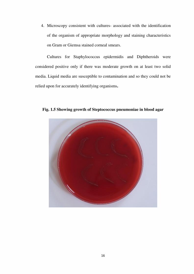
Fig. 1.5 Showing growth of Steptococcus pneumoniae in blood agar
17
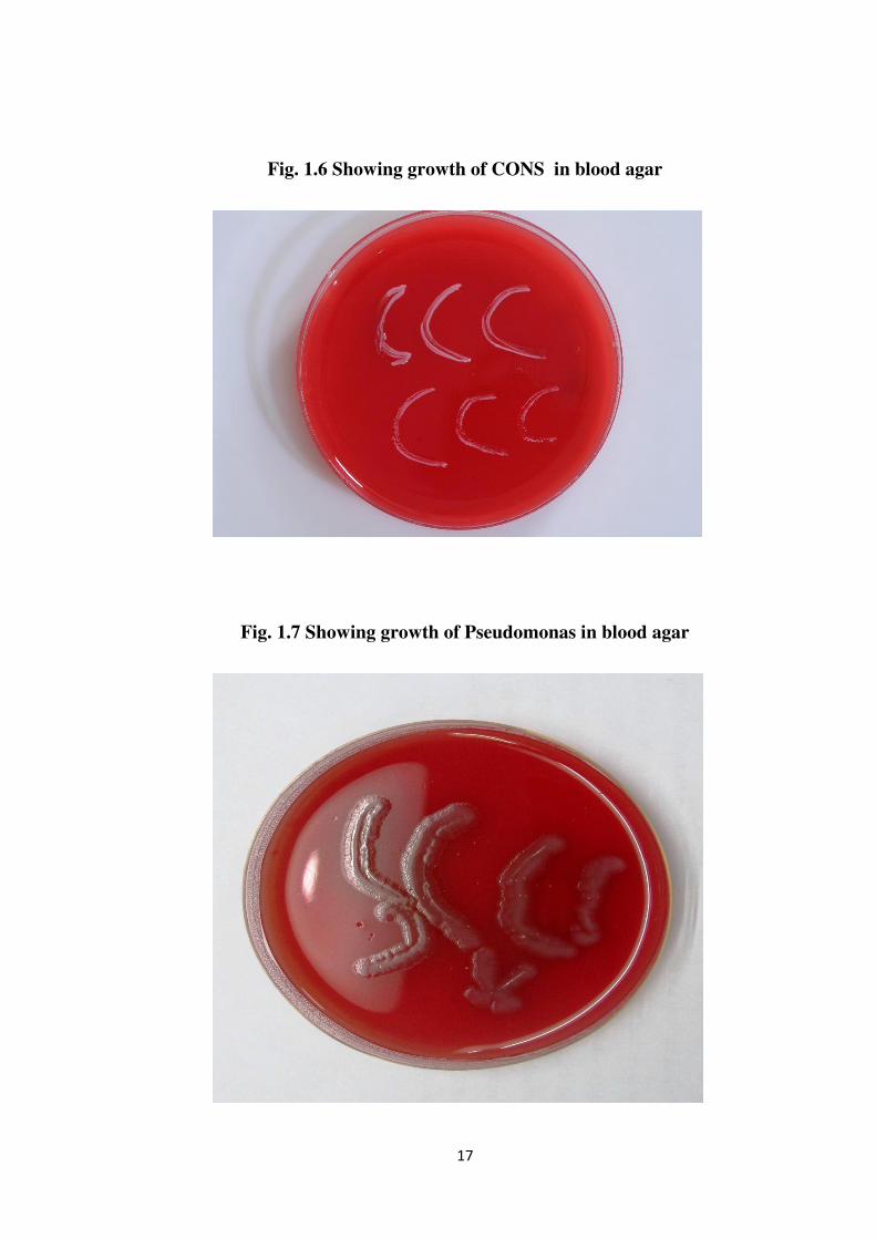
Fig. 1.6 Showing growth of CONS in blood agar
Fig. 1.7 Showing growth of Pseudomonas in blood agar

18
Determination of antimicrobial susceptibility:
Standard disc diffusion or microdilution techniques are used to
determine the antimicrobial susceptibility18
.
MIC and MPC in evaluation of invitro antimicrobial potency and
antibiotic resistance development18
Minimum inhibitory concentration(MIC):
MIC is an in vitro determination of the lowest concentration of a
specific drug that inhibits the growth of an organism at a defined inoculum of
bacteria ( usually 105
colony forming units/ml) 18
Mutant prevention concentration(MPC):
MPC defines the drug concentration required to block the growth of
organisms containing first step resistance mutations.
MPC testing is relevant only with organisms that are determined to be
susceptible to the antimicrobial agent by traditional susceptibility testing. 18
Determination of MICs:
The MIC of an antibiotic for a particular strain of bacteria is determined
by standard microbiological agar and broth tests.
MIC90 represents the antibiotic concentration that inhibits 90% of the
isolates tested and is calculated when there are at least 10 isolates of a
particular microorganism(19,20)
.

19
MIC50 represents the antibiotic concentration that inhibits 50% of the
isolates tested and is calculated when there are at least 5 isolates of a particular
microorganism21
.
In MIC tests surviving microorganisms are detected by their ability to
produce visible growth either on a series of agar plates (agar dilution or agar
diffusion methods) or in microtitre plate wells of broth (microbroth dilution
tests)
Agar dilution method:
In the agar dilution method, Petri dishes are filled with growth media
containing various concentrations of antibiotic and solidified with agar. A
defined amount of test organism is inoculated onto the top surface of the solid
medium. After incubation at the proper temperature and atmosphere for
approximately 18-24 hours the plates are screened for growth. The lowest drug
concetration preventing growth is the MIC18
.
Agar diffusion methods:
In Kirby-Bauer disk diffusion test an antibiotic impregnated paper disk
is placed on top of the agar that has been inoculated with bacteria. Alternatively
a paper strip containing increasing concentrations of antibiotic is placed on top
of the seeded agar(an E test)(17,18)
.
For both kirby - bauer and E tests , the agar plates are incubated
overnight to allow the bacterial inoculum to grow and form a continuous dense

20
film of growth on top of the agar except where growth of the bacterial isolate
is inhibited by the antibiotic.
This visible zone of inhibition around the disk is measured and the zone
size is used to estimate the relative susceptibility or resistance of the organism
to the antibiotic. The E test yields an actual MIC.
The bacterial strain is reported as sensitive , intermediate or resistant
depending on the zone size or the MIC

23
the penicillins and tetracyclines and most gram-negative facultative bacilli
were susceptible to the tetracyclines or streptomycin22
.
In the 1950s and early 1960s, early-generation penicillins and
streptomycin were popular choices for topical treatment of bacterial keratitis.
Antibiotics, including cephalothin, cephaloridine, neomycin, erythromycin,
lincomycin, colistin, and vancomycin, soon became available.
In the 1960s, as penicillin resistance became common among S. aureus
isolates, semisynthetic penicillins (e.g., oxacillin, nafcillin) and first-generation
cephalosporins, such as cephalothin and cefazolin, all of which were resistant
to staphylococcal beta- lactamases, became popular.
At the end of the 1960s, gentamicin, a potent new congener of the old
aminoglycoside, streptomycin, was introduced mainly for pseudomonas
aeruginosa24
.
Over the next decade, other aminoglycosides, tobramycin and amikacin,
were introduced. These aminoglycosides and first-generation cephalosporins,
began to be used by ophthalmologists for the treatment of bacterial corneal
ulcers.
Although aminoglycosides are only modestly active against
streptococci, including the pneumococcus, the high concentrations achieved in
the tear film were adequate to treat keratitis, the use of fortified drops and
periocular injections together with an agent, such as cefazolin, which is active
against gram-positive cocci, offered good activity22
.

21
Fig. 1.8 Showing disc diffusion in blood agar
Fig. 1.9 Showing disc diffusion in Muller Hinton agar

22
Fig 1.10 : Showing Kirby - bauer disc diffusion method for
culture sensitivity.
An organism is defined as susceptible to an antibiotic if its MIC is
below the serum break points.
Pathogen is considered resistant to the antibiotic if the MIC is at or
above the established breakpoint.
Organisms demonstrate intermediate resistance if the MIC falls between
the susceptible and resistant breakpoints.
Evolution of antibiotics for bacterial keratitis:
Sulfonamides were introduced in the 1930s and 1940s. Two of the
congeners, sulfacetamide and trimethoprim are still used today23
.
In the 1940s and 1950s, penicillin, streptomycin, and tetracyclines,
became commercially available, followed by chloramphenicol, polymyxin, and
bacitracin. Most cocci (gram-positive and gram-negative) were susceptible to

24
In the 1970s and 1980s, beta-lactams third generation cephalosporins
were introduced to overcome the problems of resistant gram- negative bacterial
infections and of aminoglycoside toxicity25
.
Some of these agents were used as “fortified” drops for the treatment of
microbial keratitis .In the late 1980s manipulating nalidixic acid ,like
introduction of fluorine atoms on the molecules, led to drugs with much
broader spectrums. An early congener, ciprofloxacin26
, had great potency
against gram-negative bacilli and H. influenzae but only moderate activity
against S. aureus and modest activity against streptococci and pneumococci.
The newest fluoroquinolones third generation such as levo- floxacin,
fourth generation such as moxifloxacin and gatifloxacin have better potency
than the older agents against gram positive cocci, pneumococci and are active
against penicillin-resistant pneumococci.
25
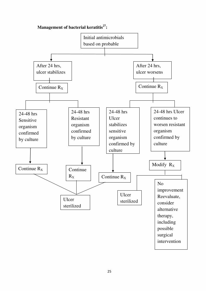
Management of bacterial keratitis27
:
Initial antimicrobials
based on probable
After 24 hrs,
ulcer stabilizes
After 24 hrs,
ulcer worsens
Continue RX Continue RX
24-48 hrs
Sensitive
organism
confirmed
by culture
24-48 hrs
Resistant
organism
confirmed
by culture
24-48 hrs
Ulcer
stabilizes
sensitive
organism
confirmed by
culture
24-48 hrs Ulcer
continues to
worsen resistant
organism
confirmed by
culture
Continue RX Continue
RX Continue RX
Modify RX
Ulcer
sterilized
Ulcer
sterilized
No
improvement
Reevaluate,
consider
alternative
therapy,
including
possible
surgical
intervention

26
In two phases
Sterilisation phase,
Healing phase .
Sterilisation phase:
Initial treatment should be intensive with hourly application of antibiotic
so that the corneal tissue is rapidly saturated with high antibiotic
concentration. A high concentration (usually exceeding the minimum
inhibitory concentration (MIC) by a number of log units) can be achieved
within a few hours, so that 48 hours of sustained high concentrations is usually
enough to eliminate most bacterial infections28
.
Sustained intensive treatment day and night for the first 48 hours, hourly
would allow for more than an adequate chance of sterilising the corneal ulcer.
Healing phase:
Following the initial treatment phase the antibiotic application is
reduced to four times a day to allow for healing of the epithelial defect.
Tapering the initial therapy offers no clinical advantage and is only
likely to increase the likelihood of toxicity28
.
The healing phase may be prolonged for large culture positive ulcers,
especially in the elderly who may also have ocular surface disease.

27
Common antibiotics active against gram positive bacteria:
Cephazolin(50mg/ml)
Chloramphenicol(5-10mg/ml)
Moxifloxacin 0.3%
Vancomycin(15-50 mg/ml)
Common antibiotics active against gram negative bacteria:
Tobramycin(3-14 mg/ml)
Gentamicin(3-14mg/ml)
Amikacin(20mg in 0.5ml)
Ceftazidime
Ciprofloxacin(3mg/ml)
Levofloxacin(3mg/ml)
Ofloxacin(3mg/ml)
Nocardia:
Amikacin 2%
Trimethoprim+sulfamethoxazole
Atypical mycobacteria:
Amikacin 2%
Clarithromycin 1%

28
Fortification of antibiotics:
Commonly fortified antibiotics:
Cefazolin:
Add 5ml to 10ml distilled water or sterile saline to 500mg vial of
cefazoline to obtain 10% or 5% solution.
Vancomycin:
Add 10ml of distilled water or saline to 500mg vial of vancomycin and
obtain a 5% solution.
Amikacin:
Add 10ml of distilled water to 100mg of amikacin to get 1% solution.
Gentamicin:
Add 2ml of injectable gentamycin to 5ml of commercial topical
preparation to get 13.5mg/ml of fortified gentamicin.
Adjuvant treatment:
Mydriatics and cycloplegics are used to prevent posterior synechiae.
Steroids in severe inflammatory keratitis.
Systemic antibiotics:
Chance of Potential for systemic involvement( In N.meningitis, H.
influenzae, N.gonorrhoeae infection)
Severe corneal thinning and scleral involvement.
29
Signs of clinical improvement:
Size and density of infiltrate and edema decreases, edema of the ulcer
get blunted, anterior chamber reaction comes down and epithelilal and stromal
healing begins. At this stage pupil dilates and hypopyon disappears.
Other modes of treatment:
Cyanoacrylate Glue:
In sealing of small perforations and thin decemetoceles
Conjunctival flap:
Used for peripheral ulcers and also non healing central ulcers Excisional
keratoplasty ,penetrating or deep lamellar keratoplasty:
Non healing corneal ulcer in spite of appropriate and adequate
antimicrobial therapy.
Impending perforation,
Perforated corneal ulcer.
Complications of corneal ulcer:
Descemetocele
Perforation
Anterior synechiae
Secondary glaucoma
Cataract
Purulent iridocyclitis
Endophthalmitis

REVIEW OF
LITERATURE

30
REVIEW OF LITERATURE
1. Bourcier et al conducted a retrospective analysis during 1998-1999 at
France with 300 cases of bacterial keratitis. Potential predisposing factors,
usually multiple, were identified in 90.6% of cases. Contact lens wear was
the main risk factor (50.3%). Trauma or a history of keratopathy was found
in 15% and 21% of cases, respectively and they concluded contact lens
wear and trauma are the most important risk factors for bacterial keratitis29
.
2. Kaye et al in 2006 investigate the relationship between susceptibility of
bacteria to topical antimicrobials and clinical outcome. MIC data were
available for 421 patients, Sixteen (4%) patients required enucleation and
23 (5%) surgical treatment; in 382 (91%) the ulcer healed with intensive
topical antimicrobial therapy. There were significant linear associations
between clinical outcome and MIC for Pseudomonas spp. (P - 0.047),
Staphylococcus aureus (P - 0.04), and Enterobacteriaceae (P - 0.045), but
not for Streptococcus spp. (P - 0.85) and coagulase-negative staphylococci
(CNS) (P - 0.88)30
3. Jayahar et al analyse the invitro efficacy of commonly used antimicrobials
against bacterial pathogens from corneal ulcers. They evaluated 596
patients over 18 months period September 1999 through March 2001.
Antibacterial susceptibility of isolated bacteria were determined by the
Kirby-Bauer disc-diffusion method 626 bacterial pathogens were isolated
from 2596 corneal ulcer cases. 411(65.65%) were gram positive cocci.
Streptococcus pneumoniae (41.85%) was the predominant bacterial species.

31
The antibacterial susceptibility was 451(72.04%) to cefazolin, 471(75.24%)
to chloramphenicol, 321(51.28%) to cephaloridine, 430(68.69%) to
vancomycin,564(90.09%) to ciprofloxacin; 429(68.53%) to norfloxacin;
464(74.12%) to gentamicin and 202(32.27%) to cotrimoxazole. This study
provides information on the efficacy of ocular antibacterials commonly
used against bacterial pathogens of keratitis31
.
4. Srinivasan et al conducted randomized placebo controlled double masked
clinical trial called SCUT (Steroids for corneal ulcer trial).This multicenter
trial compared 1.0% prednisolone sodium phosphate to placebo in the
treatment of bacterial keratitis among 500 patients with culture positive
ulcers receiving 48 hours of moxifloxacin before randomization. The
primary end point was 3 months from enrollment and 399 patients were
evaluated at 12 months. The outcomes examined were best spectacle
corrected visual acuity(BSCVA) and scar size at 12 months. They
concluded that adjunctive corticosteroid therapy may be associated with
improved long term clinical outcomes in bacterial corneal ulcers not caused
by nocardia species32
5. Chen et al conducted a retrospective analysis using samples and data
collected in a pilot study conducted in preparation for the SCUT, to
determine whether clinical outcomes in bacterial keratitis are associated
with antibiotic susceptibility. They observed MIC was associated with three
month infiltrate/scar size each two fold increase in MIC was associated with
a 0.33mm average diameter increase in scar size (P=.01). MIC was not

32
associated with three month BSCVA (P=.71) or time to reepithelialization
(P=.35) and they concluded that MIC was associated with infiltrate / scar
size in bacterial keratitis33
.
6. Srinivasan et al in1994 conducted a clinical trial to determine the
epidemiological characteristics and risk factors predisposing to corneal
ulceration in Madurai to identify the specific pathogenic organisms
responsible for infection. In 3 month period 434 patients with central
corneal ulceration were evaluated. A history of previous corneal injury was
present in 284 patients (65.4%). Cornea cultures were positive in 297
patients (68.4%). Of those individuals with positive cultures 140 (47.1%)
had pure bacterial infections, The most common bacterial pathogen isolated
was Streptococcus pneumoniae, representing 44.3% of all positive bacterial
cultures, followed by Pseudomonas spp (14.4%).They concluded central
corneal ulceration is a common problem in south India and most often
occurs after a superficial corneal injury with organic material and most
common organism causing bacterial keratitis is streptococcus
pneumoniae34
.
7. Bharathi et al in 2007 conducted a retrospective analysis to identify the
etiology , incidence and prevalence of ocular bacterial infection and to
assess the ocular bacterial isolates to commonly used antibiotics. The most
common bacterial species isolated were staphylococcus aureus,
streptococcus pneumoniae , pseudomonas aeruginosa. According to this
study gram positive cocci were the most frequent bacteria isolated from

33
ocular infections and were sensitive to moxifloxacin and vancomycin while
gram negative isolates were more sensitive amikacin and gatifloxacin35
8. Prajna Lalitha et al conducted a clinical trial to determine the emerging
moxifloxacin resistance in pseudomonas aeruginosa isolates in South India
during 2007, 2008 and 2009. They found a sharp increase in the proportion
of resistant isolates from 19% in 2007 to 52% in 2009. They found a sharp
increase in the proportion of isolates that were resistant to moxifloxacin
from 2007 to 200936

AIMS AND OBJECTIVES

34
AIMS AND OBJECTIVES
1. To determine the bacteriological profile and sensitivity pattern of culture
positive bacterial corneal ulcers in a tertiary care centre in a developing
country
2. To analyze the various risk factors and co morbidities associated with it
3. To study the treatment outcome of culture positive bacterial keratitis

METHODOLOGY

35
METHODOLOGY
STUDY DESIGN:
This is a prospective observational study where in all corneal ulcer
patients attending the cornea clinic with culture proven bacterial keratitis were
included according to inclusion and exclusion criteria
DURATION:
One year of recruitment and three months of follow up,
1st December 2015 to 28
th February 2017
SAMPLE SIZE:
The sample size of minimum 92 patients will be need to include in the
study with assumed percentage of success is 60% as per the clinical experience,
and also assuming 10% precision error and 95% confidence interval.
Formula:
P : Sensitivity of the new test
d : Precision
Z1-α/2 : Desired confidence level

36
INFORMED CONSENT:
outcome of the treatment including various complications were
explained to the patients in their own language and informed consent is
obtained. patients were informed about the need for follow ups involved in the
study.
INCLUSION CRITERIA:
All culture positive bacterial keratitis and who are willing to participate
in the study will be included
EXCLUSION CRITERIA:
• Viral, fungal, acanthamoeba and mixed keratitis.
• Culture negative keratitis.
• Mooren’s ulcers, marginal ulcers, interstitial keratitis, sterile
neurotrophic ulcers, and any ulcers associated with autoimmune
conditions.
• Bacterial keratitis with impending perforation.
• Perforated bacterial keratitis
• Pre existing posterior segment pathology.
• Patients not willing to participate in the study
37
HISTORY:
When included in the study all patient underwent a thorough workup.
Patients 's age, gender and residence were recorded. A detailed history
regarding the causative factor, mode of injury , systemic predisposing factor if
any should be asked and recorded . An enquiry regarding duration of
compliant, co-morbidity (Diabetes mellitus) , prior treatment or native
medication if any should be done
VISUAL ACUITY:
Unaided visual acuity was recorded by means of snellens chart kept at 6
meter distance. In children following methods were used to determine the
vision
<2 years- Picking up cake decoration
2-5 years-Sheridan Gardener chart
In illiterate children and adults E chart is used. During every visit UCVA was
tested and recorded
CLINICAL PROCEDURES:
All patients with corneal ulcer attending the cornea clinic were
examined under slit lamp biomicroscopy by trained ophthalmologist .
Complete ocular examination were made with special attention to ulcer site
(Entirely in periphery, Overlapping the corneal 4mm circle and periphery
without filling the center, Entirely in the central 4mm circle, Completely filling
4mm circle extending to periphery)37
is diagrammatically representated

38
according to the optscore, size (2mm,2-6mm, >6mm)17
, depth of infiltrate (0-
33%,33-67%,67-100%)38
,margin, edge and represented in diagram for further
followup. Presence of hypopyon if any should be noted .Nasolacrimal duct
patency and random blood sugar is also checked. After complete ocular
examination and verbal consent from the patient, corneal scrapings were
obtained for microscopic analysis and culture under aseptic precaution.
Following instillation of 4% lignocaine corneal scrapings were obtained
from the leading edge and base of the ulcer using heat steriled kimura spatula
and was applied in even manner for 10%KOH and gram's stain in labelled glass
slides, then for culture they were inoculated directly into blood agar in C
shaped streaks and into Sabouraud's dextrose agar. 10% KOH wet mount
preparation was examined immediately under microscopy mainly to identify
the fungus. Other slide was stained using gram's iodine and examined under oil
immersion lenses using 40X and 100X magnification and organisms were
classified into
Gram positive cocci
Gram positive bacilli
Gram negative cocci
Gram negative bacilli
Fungi
No organisms

39
LABORATORY PROCEDURES:
All inoculated media were incubated at 37 degree celsius aerobically.
These plates were examined daily for the growth. Criteria for culture positivity:
1. Growth of same organisms on two or more solid media, semi confluent
growth at the site of inoculum on one solid media
2. Same organisms grown from repeated scrapings
3. Growth is consistent with clinical signs
4. Microscopy findings consistent with culture and morphology
5. Others are considered negative and are exculded from the study
SENSITIVITY PATTERN:
Positive bacterial cultures were sub cultured into Muller hinton agar for
determination of sensitivity pattern. Sensitivity pattern were determined using
disc diffusion method by measuring the zone of inhibition around the antibiotic
disc and were interpreted as sensitive, intermediate and resistant according to
Clinical and laboratory standards institute guidelines.
MANAGEMENT:
Initial antibiotic therapy were started empirically according to gram
stain report in addition to that topical cycloplegics and oral analgesics were
also given if needed. Predisposing conditions if any like blockage in
nasolacrimal duct, high blood sugar lagophthalmos, lid abnormalities etc
should be corrected.

40
FOLLOWUP:
Patients willing to participate in the study with regular followups were
followed on regular intervals depending on the severity . On each followup
UCVA with complete ocular examination done . Ulcer were studied for
features of healing or worsening. During followup change in antibiotics if
needed can be done according to sensitivity pattern and also by clinical
response . Patients were followed in 3 weeks and at 3 months or till it heals
whichever is earlier. ulcer is considered as improved when there is a blunting
of edges, decreased density of stromal infiltrate, decrease in stromal edema
and endothelial plaque, decrease in anterior chamber reaction, reduction in size
of hypopyon, re-epithelization , cessation in corneal thinning.

RESULTS

41
RESULTS
During the study period 2130 corneal ulcer patients attended cornea
clinic, Out of which 969 patients were culture positive in which 26.4% (n=256)
patients were bacterial culture positive. Out of 256 bacterial culture positive
patients 55.4% (n=142) were gram positive organisms and 44.6% (n=114) were
gram negative organisms.
Table-4.1. Showing the analysis of corneal ulcer in
2015-2016
Total No. Of Corneal Ulcers 2130
Total Number Of Culture Positive Corneal Ulcer 969
Bacterial 256
Fungal 685
Total Number Of Gram Positive Organisms 142
Total Number Of Gram Negative Organisms 114
MICROBIOLOGICAL ANALYSIS :
In our study out of 256 cases of bacterial culture positive keratitis about
174 patients were enrolled for studying treatment outcome after exclusion by
exclusion criteria.
Among 142 gram positive organisms, 59% (n=84) were Streptococcus ,
8.4% (n=12) Staphylococcus , 16 were Corynebacterium , 9 were Coagulase
negative Staphylococcus, 6 were Nocardia and 15 comprise of other organisms

42
Among 114 gram negative organisms grown in total bacterial culture
(n=256), 69.2% (n=79) were pseudomonas, 11.2% (n=8) were moraxella.,
1.7% (n=2) were klebsiella, 3.3% (n=4) were acinetobacter.
Table –4.2. Showing microbiological profile of culture positive
bacterial keratitis
GRAM POSITIVE GRAM NEGATIVE
1. Streptococcus sp ( n=84 )
2. Corynebacterium ( n=16)
3. Staphylococcus (n=12)
4. CONS (n=9)
5. Nocardia (n=6)
6. Others (n=15)
1. Pseudomonas (n=79)
2. Moraxella (n=8)
3. Acinetobacter (n-4)
4. Klebsiella (n=2)
5. Others (n=21)
Fig-4.1. Showing gram positive organisms in total culture positive
bacterial keratitis in our study period
84
12 9 16
6 15
0
20
40
60
80
100
GRAM POSITIVE ORGANISMS
STREPTOCOCCUS STAPHYLOCOCCUS CONS
CORYNEBACTERIUM NOCARDIA OTHERS

43
Fig-4.2. Showing gram negative organisms in total culture positive
bacterial keratitis in our study period
79
2 8
4
21
0
10
20
30
40
50
60
70
80
90
GRAM NEGATIVE ORGANISMS
PSEUDOMONAS KLEBSIELLA MORAXELLA
ACINETOBACTER SP. OTHERS

44
SENSITIVITY AND RESISTANT PATTERN OF MOST COMMON
ORGANISMS:
Sensitivity and resistant pattern among organisms grown in culture of
corneal scrapings were studied . Streptococcus species were most resistant to
ciprofoloxcin and ofloxcin. most sensitive to 3rd generation fluroquinolones,
cefazolin and vancomycin
Fig-4.3: Showing sensitivity and resistant pattern of streptococcus species.
75%
80%
85%
90%
95%
100%
RESISTANT
SENSITIVE

45
Table-4.3. Showing sensitivity and resistant pattern of
streptococcus species.
ANTIBIOTICS SENSITIVE RESISTANT
LEVOFLOX 82 1
GATIFLOX 72 11
MOXIFLOX 80 3
CEPHOTAXIM 79 4
CIPROFLOX 70 13
OFLOXCIN 71 12
CHLORAMPHENICOL 79 4
CEFAZOLIN 79 4
VANCOMYCIN 83 0
TETRACYCLIN 75 8

46
Among 114 gram negative organisms grown in total bacterial culture
(n=256), 69.2% (n=79) were pseudomonas . Pseudomonas species were most
resistant to cephotaxim. Most sensitive to fluroquinolones.
Fig-4.4: Showing sensitivity and resistant pattern of Pseudomonas species.
0%
10%
20%
30%
40%
50%
60%
70%
80%
90%
100%
SENSITIVE RESISTANT

47
Table-4.4: Showing sensitivity and resistant pattern of
Pseudomonas species.
ANTIBIOTICS SENSITIVE RESISTANT
LEVOFLOX 69 10
GATIFLOX 68 11
MOXIFLOX 69 10
GENTAMYCIN 58 21
TOBRAMYCIN 64 15
CEPHOTAXIM 42 37
CIPROFLOX 71 8
OFLOXCIN 69 10
AMIKACIN 62 17
POLYMYXIN 69 10

48
DEMOGRAPHIC ANALYSIS:
In our study out of 256 cases of bacterial culture positive keratitis about
174 patients were enrolled for studying treatment outcome after exclusion by
exclusion criteria. In our study mean age of the patients were 54.55years
Regarding sex ratio among 174 patients 109 were males and 65 were
females
Table-4.6 : Gender distribution among study population
Sex n (%)
Male 109 (62.64)
Female 65 (37.36)
Total 174 (100)
Table -4.5 : Age distribution among study population
Age (Years)
n Mean (SD) Min - Max
174 54.55 (19.04) 1 - 90

49
Fig- 4.5 : Gender distribution among study population
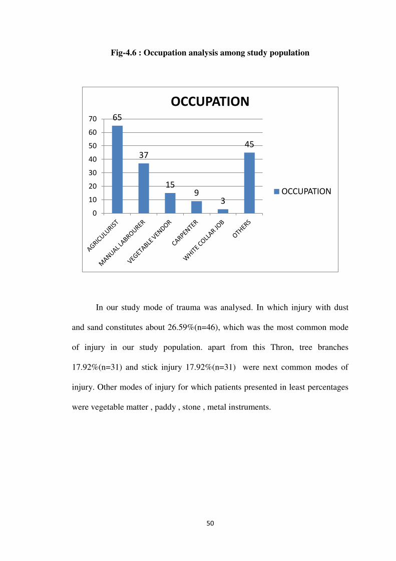
Among 174 patients, categorised based on occupation. About 65
patients were farmers and 37 were manual labourers. These two categories of
patients itself form above 60% of study population. Apart from this vegetable
vendor ,carpenters were affected from bacterial culture positive keratitis in our
study.
Table-4.7 : Occupation analysis among study population
Occupation n (%)
Agriculture 65 (37.36)
Manual Labourer 37 (21.26)
Vegetable Vendor 15 (8.62)
Carpenter 9 (5.17)
White collar job 3 (1.72)
Others 45 (25.86)
Total 174 (100)
109
65
0
50
100
150
MALE FEMALE
SEX
SEX
50
Fig-4.6 : Occupation analysis among study population
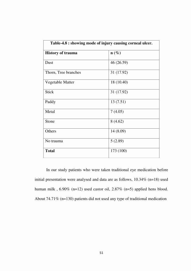
In our study mode of trauma was analysed. In which injury with dust
and sand constitutes about 26.59%(n=46), which was the most common mode
of injury in our study population. apart from this Thron, tree branches
17.92%(n=31) and stick injury 17.92%(n=31) were next common modes of
injury. Other modes of injury for which patients presented in least percentages
were vegetable matter , paddy , stone , metal instruments.
65
37
15 9
3
45
0
10
20
30
40
50
60
70
OCCUPATION
OCCUPATION
51
In our study patients who were taken traditional eye medication before
initial presentation were analysed and data are as follows, 10.34% (n=18) used
human milk , 6.90% (n=12) used castor oil, 2.87% (n=5) applied hens blood.
About 74.71% (n=130) patients did not used any type of traditional medication
Table-4.8 : showing mode of injury causing corneal ulcer.
History of trauma n (%)
Dust 46 (26.59)
Thorn, Tree branches 31 (17.92)
Vegetable Matter 18 (10.40)
Stick 31 (17.92)
Paddy 13 (7.51)
Metal 7 (4.05)
Stone 8 (4.62)
Others 14 (8.09)
No trauma 5 (2.89)
Total 173 (100)

52
Table -4.9 : Traditional eye medication
Traditional eye medicine n (%)
Human milk 18 (10.34)
Castor Oil 12 (6.90)
Hens blood 5(2.87)
Others 9(5.17)
No traditional medication 130 (74.71)
Total 174 (100)
Fig-4.7 : Traditional eye medication
Human milk
10%
Castor Oil
7%
Hens blood
3%
Others
5%
No traditional
medication
75%

53
SYMPTOM ANALYSIS:
In our study population most common presenting symptoms were
studied among 174 patients. In which photophobia (n=130) ,redness (n=129)
,blurring of vision (n=126) were most common symptoms followed by
pain(n=121) and watering of eye(n=116). Most of the patients were presented
with two or more above described symptoms.
Table-4.10 : symptom analysis among study population.
Symptoms Redness
n (%)
Photophobia
n (%)
Pain
n (%)
Watering
n (%)
Blurring of
eyes
n (%)
Yes 129
(74.14)
130
(74.71)
121
(69.54)
116
(66.67)
126
(72.41)
No 45
(25.86)
44
(25.29)
53
(30.46)
58
(33.33)
48
(27.59)
Fig -4.8 : Symptom analysis among study population.
129 130 121 116 126
0% 10% 20% 30% 40% 50% 60% 70% 80% 90%
100%
NO
YES
54
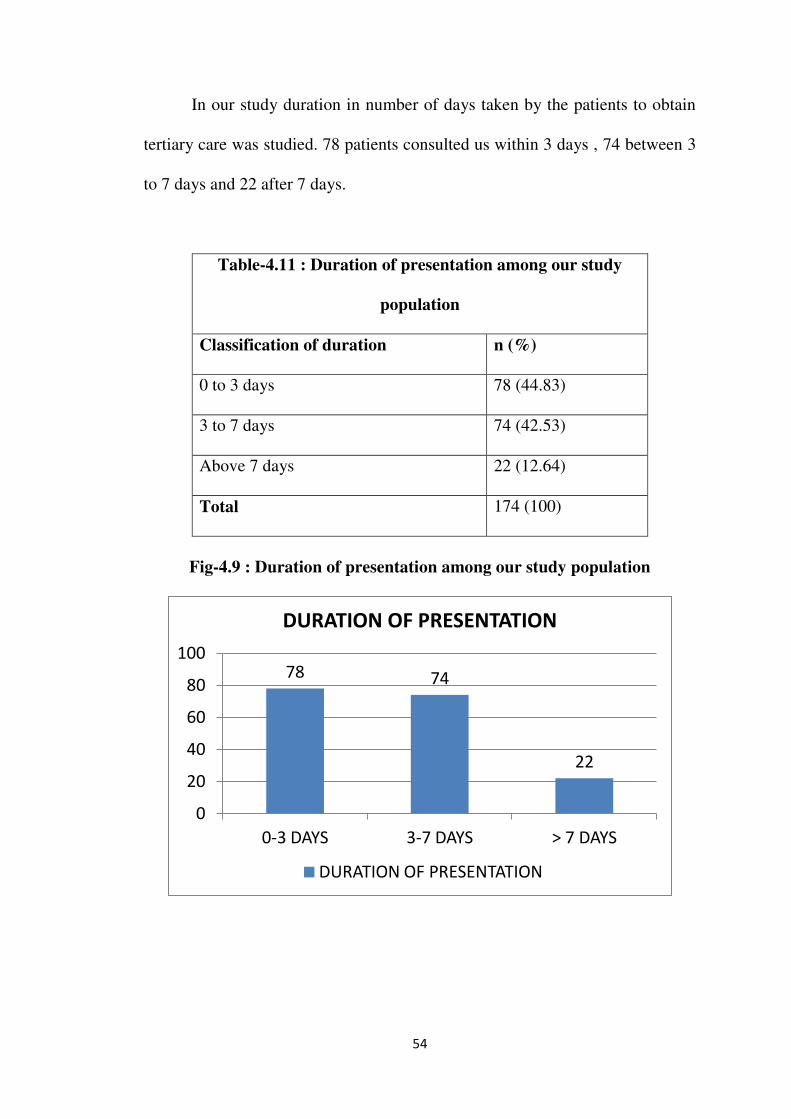
In our study duration in number of days taken by the patients to obtain
tertiary care was studied. 78 patients consulted us within 3 days , 74 between 3
to 7 days and 22 after 7 days.
Table-4.11 : Duration of presentation among our study
population
Classification of duration n (%)
0 to 3 days 78 (44.83)
3 to 7 days 74 (42.53)
Above 7 days 22 (12.64)
Total 174 (100)
Fig-4.9 : Duration of presentation among our study population
78 74
22
0
20
40
60
80
100
0-3 DAYS 3-7 DAYS > 7 DAYS
DURATION OF PRESENTATION
DURATION OF PRESENTATION

55
DIABETIC STATUS OF STUDY POPULATION:
Number of patients with history of diabetes were analysed as poor
glycemic control will hamper the corneal ulcer healing. In our study out of 174
patients studied for treatment outcome 75 patients were diabetics and 99 were
non diabetics. Also their current blood glucose levels were measured in terms
of random blood sugar levels. Random blood sugar taken as high if the patients
have value more than 200mg/dl. Out of 174 patients studied 40.80%(n=71)
patients had high sugar levels and 59.20%(n=103) patients had normal levels
Table-4.12 : Incidence of diabetes mellitus in our study
population
Diabetes n (%)
Yes 75 (43.10)
No 99 (56.90)
Total 174 (100)
Fig-4.10 : Showing incidence of diabetes mellitus in our study population
DIABETIC MELLITUS
YES NO

56
Table-4.13: Glycemic status at the time of initial presentation
Random Blood Sugar n (%)
High 71 (40.80)
Normal 103 (59.20)
Total 174 (100)
Fig-4.11 : Showing glycemic status at the time of initial presentation
CLINICAL ANALYSIS:
In our study corneal ulcer site at the time of initial presentation was
analysed. The site of corneal ulcer was categorised as follows with observed
data .
Patients presented with corneal ulcer entirely in the periphery were
24.71% (n=43) , patients with ulcer overlapping the corneal 4mm circle and
periphery without filling the center were 39.66%(n=69) . there are
23.56%(n=41) of patients presented with ulcer entirely in the central 4mm
71
103
RANDOM BLOOD SUGAR
HIGH
NORMAL

57
circle and 12.07% (n=21) were had ulcer Completely filling 4mm circle
extending to periphery.
Table-4.14: Distribution of corneal ulcer
Corneal Ulcer site n (%)
Entirely in periphery 43 (24.71)
Overlapping the corneal 4mm circle and periphery
without filling the center
69 (39.66)
Entirely in the central 4mm circle 41 (23.56)
Completely filling 4mm circle extending to periphery 21 (12.07)
Total 174 (100)
Fig -4.12 : Showing Distribution of corneal ulcer
43
69
41
21
CORNEAL ULCER SITE
Entirely in periphery
Overlapping the corneal
4mm circle and periphery
without filling the center
Entirely in the central
4mm circle
Completely filling 4mm
circle extending to
periphery

58
On analysing the size of the culture positive bacterial corneal ulcer
among study population, 16.09% (n=28) had ulcer size of less than 2mm ,
64.94% (n=113) had size of 2mm-6mm and 18.96%(n=33) of patients had
ulcer size above 6mm.
Table 4.15 : Size of culture positive bacterial corneal ulcer
Ulcer size n(%)
<2mm 28 (16.09%)
2mm-6mm 113 (64.94%)
>6mm 33 (18.96%)
Fig- 4.13: Size of culture positive bacterial corneal ulcer
28
113
23
corneal ulcer size
<2mm
2-6mm
>6mm
59
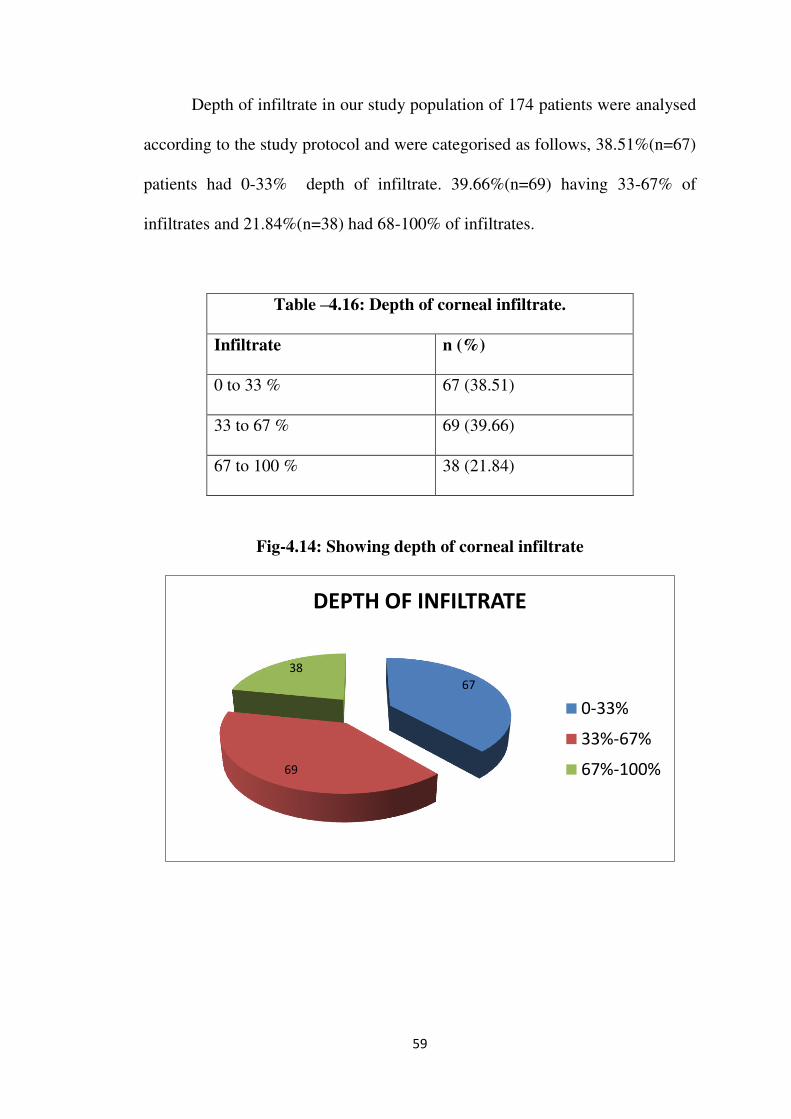
Depth of infiltrate in our study population of 174 patients were analysed
according to the study protocol and were categorised as follows, 38.51%(n=67)
patients had 0-33% depth of infiltrate. 39.66%(n=69) having 33-67% of
infiltrates and 21.84%(n=38) had 68-100% of infiltrates.
Table –4.16: Depth of corneal infiltrate.
Infiltrate n (%)
0 to 33 % 67 (38.51)
33 to 67 % 69 (39.66)
67 to 100 % 38 (21.84)
Fig-4.14: Showing depth of corneal infiltrate
67
69
38
DEPTH OF INFILTRATE
0-33%
33%-67%
67%-100%

60
In our study 13.29% of patients had hypopyon associated with bacterial
culture positive keratitis.
Table-4.17: Corneal ulcer associated with hypopyon
Hypopyon n (%)
Yes 24 (13.79)
No 150 (86.21)
Fig-4.15 : Showing corneal ulcer associated with hypopyon.
MANAGEMENT OF BACTERIAL KERATITIS:
After gram stain report patients who are willing for giving consent and
matching the inclusion and exclusion criteria were started on antibiotics based
on clinical features and gram staining results. Data were as follows,
0%
10%
20%
30%
40%
50%
60%
70%
80%
90%
100%
HYPOPYON
NO
YES

61
Table-4.18: Initial antimicrobials based on gram staining.
Initial Antimicrobial n (%)
Cefazolin 32 (18.39)
Moxifloxcin 24 (13.79)
Amikacin 5 (2.87)
Tobramycin 9 (5.17)
Gatifloxcin 3 (1.72)
Chloramphenicol and polymixcin b 4 (2.30)
Cefazolin + chloramphenicol +
polymixcin b 44 (25.29)
Moxifloxcin+ chloramphenicol +
polymixcin b 26 (14.94)
Amikacin + chloramphenicol +
polymixcin b 8 (4.60)
Cefazolin + moxifloxcin 15 (8.62)
Amikacin + moxifloxcin 1 (0.57)
Gatifloxcin + Chloramphenicol +
Polymixcin B 3 (1.72)
Total 174 (100)
After initiation of empirical antibiotics based on gram stain reports
patients were evaluated by clinical resolution of ulcer and culture sensitivity
reports. Based on above evaluation change in antimicrobial therapy may be

62
instituted for certain number of patients. By which 31.03% (n=54) of patients
needed change in antimicrobial therapy
Table-4.19: Change in antibiotic according to culture and
sensitivity pattern
Change in antibiotic n (%)
Yes 54 (31.03)
No 120 (68.97)
Total 174 (100)
Fig-4.16 : Showing change in antibiotic according to culture and sensitivity
pattern.
54
120
CHANGE IN ANTIBIOTIC
YES
NO

63
TREATMENT OUTCOME ANALYSIS:
After 3 weeks of treatment, patients were evaluated for progress in the
ulcer healing when compared to the initial presentation. 48.85% (n=85) of
patients had improved outcome at 3rd week itself. 34.48% (n=60) had stable
ulcer healing and 16.67% (n=29) had worsening of ulcer clinically.
Table-4.20 : Ulcer healing at 3rd
week
Ulcer Healing at 3rd
week n (%)
Improved 85 (48.85)
Stable 60 (34.48)
Worsened 29 (16.67)
Total 174 (100)
Fig-4.17 : Showing Ulcer healing at 3rd
week.
0
20
40
60
80
100
120
140
160
180
200
ULCER HEALING AT 3RD WEEK
WORSENED
STABLE
IMPROVED

64
Treatment outcome is measured at the end of 3 months or complete resolution
of corneal ulcer whichever is earlier. 79.89% (n=139) had favourable clinical
outcome in means of ulcer healing and 20.11% (n=35) were not improved by
means of ulcer healing.
Table-4.21 : Ulcer healing at final follow up
Ulcer healing at final follow up n (%)
Improved 139 (79.89)
Not Improved 35 (20.11)
Total 174 (100)
Fig-4.18 : Showing ulcer healing at final follow up.
139
35
ULCER HEALING AT FINAL FOLLOWUP
IMPROVED
NOT IMPROVED

65
ASSOCIATION BETWEEN DURATION OF PRESENTION AND
ULCER HEALING:
As duration of symptoms at time of initial presentation was analysed
earlier in our study population it was compared with ulcer healing. Results
were as follows – 82.05% (n=64) patients presented within 3 days of symptoms
had good clinical outcome and 86.94% ( n=64 ) of patients presented between
3 to 7 days were improved. Whereas only 50%(n=11) of patients presented
after 7 days were improved clinical outcome
Table-4.22 : Duration of initial presentation VS Ulcer healing at final
followup
Duration
classification
Ulcer healing at final follow up
P value f Improved
n (%)
Not Improved
n (%)
Total
0 to 3 days 64 (82.05) 14 (17.95) 78 (100)
0.002 3 to 7 days 64 (86.49) 10 (13.51) 74 (100)
Above 7 days 11 (50.00) 11 (50.00) 22 (100)
Total 139 (79.89) 35 (20.11) 174 (100)

66
Fig-4.19 : Duration of initial presentation VS ulcer healing at final
followup
ASSOCIATION BETWEEN DIABETICS AND ULCER HEALING:
In our study ulcer healing in known diabetic individuals were analysed.
In which, out of 75 diabetics 28% (n=21) patients corneal ulcer were not
improved as per study criteria. Out of 99 diabetics 85.8% (n=85) were
improved and 14.2% (n=14) were not improved as per study criteria.
0
10
20
30
40
50
60
70
0 to 3 days 3 to 7 days Above 7 days
IMPROVED
NOT IMPROVED

67
Table-4.23: Diabetes mellitus Vs Ulcer healing at final followup.
Diabetes
Ulcer healing at final follow up
P value c Improved
n (%)
Not Improved
n (%)
Total
Yes 54 (72.00) 21 (28.00) 75 (100)
0.024 No 85 (85.86) 14 (14.14) 99 (100)
Total 139 (79.89) 35 (20.11) 174 (100)
C - Chi Square test
The above p value 0.024 (<0.05) shows that there is an association
between diabetes and ulcer healing at the final follow up.
Fig-4.20: Showing association between diabetes and ulcer healing
at final followup.
0
10
20
30
40
50
60
70
80
90
DIABETIC NON DIABETIC
IMPROVED
NOT IMPROVED

68
ASSOCIATION BETWEEN ULCER SIZE AND ULCER HEALING AT
FINAL FOLLOW UP:
Among 28 patients with less than 2mm of ulcer size, 85.71% (n=24)of
patients had improved ulcer healing and 14.28% (n=4) of patient had no
improvement in ulcer healing. Among 113 patients with ulcer size between
2mm and 6mm, 88.49% (n=100) shows improvement to the treatment and
11.5% (n=13) shows no improvement. Among patients with ulcer size above
6mm, 45.45% (n=15) shows improvement with treatment and 54.54% (n=18)
shows no improvement at final followup.
Table-4.24 : Ulcer size vs Ulcer healing at final follow up
Ulcer size
Ulcer healing at final follow up
P value c
Improved
n (%)
Not
Improved
n (%)
Total
<2mm 24 (89.55) 4(14.28) 28 (100)
< 0.001 2mm-6mm 100 (88.49) 13(11.5) 113(100)
>6mm 15 (45.45) 18 (54.54) 33 (100)
Total 139 (79.89) 35 (20.11) 174 (100)

69
Fig-4.21: Ulcer size vs Ulcer healing at final follow up
ASSOCIATION BETWEEN DEPTH OF INFILTRATE AND ULCER
HEALING.
As infiltrate size having major impact over final treatment outcome in
cases of corneal ulcer we compared infiltrate size as per study definition to the
final outcome in ulcer healing.
Out of 67 patients having 0-33% of corneal infiltrates , 89.5% (n=60)
had improved ulcer healing and 10.5% (n=7) were not improved in ulcer
healing clinically. In 69 patients with 33-67% of infiltrates, 84.5% (n=58) were
improved and 15.5% (n=11) were not improved. In 38 patients with extensive
infiltrates 67-100% , 55%(n=21) were improved and 45% (n=17) were not
improved.
0%
10%
20%
30%
40%
50%
60%
70%
80%
90%
100%
<2mm 2mm-6mm >6mm
NOT IMPROVED
IMPROVED

70
Table-4.25 : Depth of infiltrate vs Ulcer healing at final follow up
Infiltrate
Ulcer healing at final follow up
P valuec Improved
n (%)
Not Improved
n (%)
Total
0 to 33 % 60 (89.55) 7 (10.45) 67 (100)
< 0.001 33 to 67 % 58 (84.06) 11 (15.94) 69 (100)
67 to 100 % 21 (55.26) 17 (44.74) 38 (100)
Total 139 (79.89) 35 (20.11) 174 (100)
Fig-4.22: Showing association between depth of infiltrate and ulcer healing
at final follow up
0
10
20
30
40
50
60
70
0-33% 33-67% 67-100%
IMPROVED
NOT IMPROVED

71
ASSOCIATION OF ULCER SITE AND UCVA AT FINAL FOLLOWUP:
The association between the corneal ulcer site and UCVA at final follow
up was analysed and results are as follows, in our study population of 174
patients, 43 patients had ulcer at entirely in periphery, in which 40 patients had
improved UCVA and only 3 patient’s UCVA was not improved.
About 69 patients had ulcer Overlapping the corneal 4mm circle and
periphery without filling the center ,56 patients had improved UCVA, 9
patients had stable UCVA and 4 patients had worsened UCVA at final
followup.
In 41 patients with ulcer at Entirely in the central 4mm circle,35 patients
had improved UCVA, 4 patients had stable UCVA and 2 had worsened UCVA.
In about 21 patients who had ulcer Completely filling 4mm circle extending to
periphery,13 patients had improved UCVA, 4 patients had stable UCVA and 4
had worsened UCVA.

72
Table-4.26: Ulcer site vs UCVA
Corneal
ulcer 1 line 2 lines 3 lines 4 lines 5lines Stable Worsened Total
1* 34
(79.07)
6
(13.95)
0
(0)
0
(0)
0
(0)
1
(2.33)
2
(4.65)
43
(100)
2@ 32
(46.38)
12
(17.39)
8
(11.59)
2
(2.90)
2
(2.90)
9
(13.04)
4
(5.80)
69
(100)
3# 10
(24.39)
10
(24.39)
10
(24.39)
2
(4.88)
3
(7.32)
4
(9.76)
2
(4.88)
41
(100)
4$ 2
(9.52)
4
(19.05)
1
(4.76)
6
28.57)
0
(0)
4
(19.05)
4
(19.05)
21
(100)
Total 78
(44.83)
32
(18.39)
19
(10.92)
10
(5.75)
5
(2.87)
18
(10.34)
12
(6.90)
174
(100)
* ulcer at entirely in periphery
@ ulcer overlapping the corneal 4mm circle and periphery without filling the center
# ulcer at entirely in the central 4mm circle
$ ulcer completely filling 4mm circle extending to periphery
DIFFERENCE IN ULCER HEALING BETWEEN 3RD
WEEK AND
FINAL FOLLOWUP:
Clinical outcome in ulcer healing between 3rd week presentation and 3rd
month presentation were analysed. This analysis gives us the data regarding
late response to the treatment due to change in antibiotics. None of patients
who were improved during 3rd week had worsened healing. Out of 60 patients
who had stable ulcer healing at the time of 3rd week, 65% (n=39) were
improved and 35% (n=21) were not improved. Out of 29 patients who already
had worsened clinical scenario at 3rd week , 51% (n=15) were improved and
48.28% (n=14) were not improved.

73
Table-4.27 : Difference in ulcer healing between 3rd
week and final
followup
Ulcer healing
at 3rd
week
Ulcer healing at final follow up
P value c
Improved
n (%)
Not Improved
n (%) Total
Improved 85 (100) - 85 (100)
< 0.001 Stable 39 (65.00) 21 (35.00) 60 (100)
Worsened 15 (51.72) 14 (48.28) 29 (100)
Total 139 (79.89) 35 (20.11) 174 (100)
Fig-4.23: Difference in ulcer healing between 3rd
week and final followup
0
20
40
60
80
100
IMPROVED STABLE WORSENED
IMPROVED
NOT IMPROVED

74
ASSOCIATION BETWEEN UCVA AND ULCER HEALING AT END
OF THE STUDY:
At end point of the study, patients UCVA and ulcer healing were studied
and results are as follows,
In our study 139 patients were improved in corneal ulcer healing , in
which 129 patients had 1 to 5 lines improvement in UCVA , 8 patients had no
change in UCVA and only 2 patients had decrement in UCVA. But in cases in
which ulcer is not healed, only 15 patients had improved UCVA of about 1-5
lines and 10 patients had stable UCVA and 10 had worsened UCVA.
Table -4.28 : UCVA vs ulcer healing at final followup.
Ulcer
healing at
final
follow up
1
line
2
lines
3
lines
4
lines
5
lines Stable Worsened Total
Improved 48.92
(68)
21.5
8
(30)
12.23
(17)
6.47
(9)
3.60
(5)
5.76
(8)
1.44
(2)
100
(139)
Not
improved
28.57
(10)
5.71
(2)
5.71
(2)
2.86
(1)
0
(0)
28.57
(10)
28.57
(10)
100
(35)

75
Fig-4.24: UCVA vs ulcer healing at final followup.
ANALYSIS OF UCVA AT INITIAL VISIT AND AT FINAL
FOLLOW UP:
Visual acuity was recorded by snellens chart in all patients at initial and
final followup. Snellen logMAR UCVA at final followup shows 0.92± 0.51(
mean±SD) which was significantly better than Snellen logMAR UCVA at
initial followup 1.12 ±0.43. The above p value <0.01 shows that there is a
significant difference between Initial VA and UCVA at final follow up.
Table- 4.29:Analysis of UCVA at initial visit and at final follow up
Parameter n Mean (SD) Min – Max P value^
Initial VA 174 1.12 (0.43) 0.18 – 2.6
< 0.01 UCVA at final follow up 174 0.92 (0.51) 0.18 – 2.9
0
50
100
150
IMPROVED NOT
IMPROVED
1-5 LINE
IMPROVEMENT
STABLE UCVA
WORSENED
UCVA

76
EFFECT OF CHANGE IN ANTIBIOTICS AND ULCER HEALING:
The effect of change in antibiotics over ulcer healing was analysed. In
which out of 54 patients who had change of antibiotics during follow-ups
64.81% (n=35) were improved and 35.19% (n=19) were not improved.
Whereas out of 120 patients who did not had change of antibiotics during
follow-ups, 86.67% (n=104) were improved and 13.33% (n=16) were not
improved.
Table-4.30 :Effect of change in antibiotics and ulcer healing
Change in
Antibiotic
Ulcer healing at final follow up
P value c Improved
n (%)
Not Improved
n (%)
Total
Yes 35 (64.81) 19 (35.19) 54 (100)
0.001 No 104 (86.67) 16 (13.33) 120 (100)
Total 139 (79.89) 35 (20.11) 174 (100)
Fig-4.25 : Effect of change in antibiotics and ulcer healing
0
20
40
60
80
100
120
YES NO
IMPROVED
NOT IMPROVED

DISCUSSION

77
DISCUSSION
The aim of our study is mainly to determine the bacterial profile,
demographic characteristics and culture and sensitivity pattern and treatment
outcome in bacterial keratitis in a tertiary care centre.
Spectrum of causative organism and their susceptibility varies according
to latitude. Indiscriminate use of antimicrobials had lead to emergence of many
strains resistance to commonly used antimicrobials.
Hence periodic susceptibility testing should be performed to ensure
currently available antibiotics are providing good coverage against recent
clinical isolate of pathogenic bacteria.
Our study is a prospective observational study which include 174
patients with culture positive bacterial keratitis
MICROBILOGICAL ANALYSIS:
In our study with population of 174 patient with culture positive
bacterial keratitis, gram positive organisms(n=142) were more than the gram
negative(n=114).
In gram positive organisms, streptococcus (59%) is the most common
organism and Pseudomonas (69%) is the most common among gram negative
This is in consistent with the study done by Jayahar et. al 31
Streptococcus species were more sensitive to 3rd
generation
cephalosporins, 3rd
generation fluroquinolones and vancomycin and most

78
resistant to old generation fluroquinolone (ciprofloxacin, ofloxcin).
Pseudomonas species were most sensitive to fluroquinolones like moxifloxcin
and most resistant to cephotaxim. This is in accordance with the study done by
Gangopadhyay et.al39
Almost all organisms both gram positive and gram negative are sensitive
to vancomycin except Nocardia. Out of 6 isolates, four were resistant even to
vancomycin but these are found to be more sensitive to aminoglycosides
mainly amikacin.
Methicillin resistance were prevalent among staphylococcus isolates,
with many strains demonstrating multidrug resistance, most of them were
found to be sensitive to vancomycin.
DEMOGRAPHIC ANALYSIS:
In our study population which comprise of 174 patients, bacterial
keratitis is more common among male than females. The mean age of
presentation in our study is about 54.5 which comprise mainly of working
population. This shows occupational exposure is one of the leading cause of
bacterial keratitis.
Most common mode of injury in our study population is exposure to
dust 26.59% (n=46) followed by thorn and vegetable matter. This is in relevant
to the study done by Srinivasan et. al34
Among 75 diabetics, 28% (n=21) did not improved at final followup
whereas out of 99 non diabetics only 14.14% (n=12) shows no improvement at

79
final followup. This shows that diabetes mellitus is one of the confounding
factors between the bacterial keratitis and its treatment outcome. Among 174
patients 74% of patients did not had any type of traditional eye medications.
Among 26% of traditional medication users human milk and castor oil user
were majority in number after which comes hens blood users.
In our study among 152 patients who presented with in a week, 80-86%
showed good improvement in ulcer healing at final followup whereas only 50%
of 22 patients who presented late after a week showed improvement to the
treatment at final followup. This concludes patients who seeks early
consultation at tertiary center have a better outcome when compared to others.
This is in consistence with bourcier et al3
CLINICAL PROFILE AND TREATMENT OUTCOME:
In our study the impact of clinical profile over the treatment outcome
were analysed and are statistically significant with p value <0.05. Regarding
ulcer size, only 10-15% had got poor treatment outcome among 141 patients
presented with <6mm ulcer size. Whereas 54.54% of patient had poor outcome
when ulcer size is >6mm. This shows there is a linear relationship between the
ulcer size and the treatment outcome. This is already stated by Stephen kaye et.
al. Like this patients with deep stromal infiltrate had poor treatment outcome in
our study. Patients with deeper infiltrates of >67% only 55% of patients had
improved clinical outcome.
When site of corneal ulcer and UCVA at final follow up was analysed,
patients with peripheral ulcer had good improvement in UCVA. And patients

80
who had ulcer more in the center had poor improvement in UCVA. This
analysis is statistically significant with P value of <0.01 (Fisher’s exact test).
Association among patients who had change in antibiotics during follow
up and their treatment outcome in terms of ulcer healing were analyzed.
Among 54 patients who had change in antibiotics during their treatment
35.19% had poor prognosis whereas patients who responded to initial
antibiotics had only 13.33% of poor treatment outcome. This analysis is
statistically significant with P value of <0.001.
In our study, patients who improved before 3 weeks shows good
treatment outcome at final followup. whereas 30% of patients who had stable
or worsened ulcer healing at 3rd
week showed poor treatment outcome at final
follow up.

CONCLUSION

81
CONCLUSION
In our study population with male predominance , Streptococcus is most
common among gram positive which is most sensitive to cephalosporins
and 3rd generation fluroquinolones . Pseudomonas is the most common
gram negative organism which is most sensitive to fluroquinolones.
Hence from our study patients with corneal ulcer if gram's stain results in
Gram positive most suitable antibiotic will be cephalosporins and 3rd
generation fluroquinolones and in cases of Gram negative patients should
be started on empirical antibiotics which includes 3rd generation
fluroquinolones.
Initial empirical antibiotics has important role in defining the final
treatment outcome. In our study patients who had change in antibiotics
which is not sensitive to initial one had increased risk of poor treatment
outcome.
Among co-morbidities , Patients who had diabetes mellitus shown to have
poor treatment outcome when compared to non diabetic individual.
From our study it is studied that the treatment outcome is also well
influenced by the corneal ulcer characters at the time presentation. Corneal
ulcer with following characters had poor outcome in our study
o central ulcer had poor outcome in UCVA

82
o larger ulcer with size >6mm and infiltrate depth >67% had poor
ulcer healing.
Following were the predictors of poor outcome of culture positive
bacterial keratitis
Patients who presented late ( >7 days )
Patients who had high / uncontrolled glycemic levels
Patients with traditional eye medication
Patients who needs change in antibiotics either for clinical
deterioration or according to sensitivity results
Patients with central, larger and deeper corneal ulcer.
Hence patients with above clinical characteristics should be followed up
regularly. By doing which the morbidity and preventable blindness can be
decreased.

BIBLIOGRAPHY

1
BIBLIOGRAPHY
1. Noopur Gupta, Radhika Tandon, Sanjeev K Gupta, V Sreenivas et al ;
Corneal Blindness in India ; Indian J Community Med. 2013 Oct-Dec;
38(4): 198–206. doi: 10.4103/0970-0218.120153 PMCID:
PMC3831688
2. Ramesh Verma, Pardeep Khanna, Varun Arora et al The National
Programme for Control of Blindness in India, Australas Med J. 2011;
4(1): 1–3. Published online 2011 Jan 31. doi: 10.4066/AMJ.2011.505
,PMCID: PMC3562965
3. T Bourcier, F Thomas, V Borderie, C Chaumeil, and L Laroche ,
Bacterial keratitis: predisposing factors, clinical and microbiological
review of 300 cases ; Br J Ophthalmol. 2003 Jul; 87(7): 834–838.
PMCID: PMC1771775
4. Kunimoto DY, Sharma S, Garg P, et al. In vitro susceptibility of
bacterial keratitis pathogens to ciprofloxacin. Ophthalmology
1999;106:80–85.
5. Chalita MR, Höfling-Lima AL, Paranhos A Jr et al. Shifting trends in in
vitro antibiotic susceptibilities for common ocular isolates during a
period of 15 years. Am J Ophthalmol. 2004 Jan;137(1):43-51.
6. Chidambaram JD, Recent advances in the diagnosis and management of
bacterial keratitis. International opthalmology clinics, 2007
Summer;47(3):1-6.

2
7. Stephen kaye, stephen Tuft,Timothy Neal et al. Bacterial susceptibility
to topical antimicrobials and clinical outcome in bacterial keratitis.
Invest ophthalmol Vis Sci. 2010;51:362-368.
8. Goldstein MH, Kowalski RP, Gordon YJ. Emerging fluoro- quinolone
resistance in bacterial keratitis. Ophthalmology 1999;106:1313–1318.
9. Baum JL. Initial therapy of suspected microbial corneal ulcers. I. Broad
antibiotic therapy based on prevalence of organisms. Surv opthal 1999
Sep-Oct;24(2):97-105.
10. Rodrigo Bolaños-Jiménez, Alejandro Navas, Erika Paulina López-
Lizárraga, Francesc March de Ribot,et al ; Ocular Surface as Barrier of
Innate ImmunityOpen Ophthalmol J. 2015; 9: 49–55 Published online
2015 May 15. doi: 10.2174/1874364101509010049 PMCID:
PMC4484240
11. F Stapleton and N Carnt ; Contact lens-related microbial keratitis: how
have epidemiology and genetics helped us with pathogenesis and
prophylaxis Eye (Lond). 2012 Feb; 26(2): 185–193. PMCID:
PMC3272197
12. Eric Pearlman, Yan Sun, Sanhita Roy, Mausita Karmakar, Amy G. Hise,
Loretta Szczotka-Flynn., et al ; Host Defense at the Ocular Surface Int
Rev Immunol. 2013 Feb; 32(1): 4–18. PMCID: PMC3750950
13. Vinay Agrawal, Jyotirmay Biswas, HN Madhavan, Gurmeet Mangat, M
Srinivasan et al., Current perspectives in infectious keratitis CURRENT

3
OPHTHALMOLOGY Year : 1994 Volume : 42 Issue : 4 Page : 171-
192
14. Geeta K. Vemuganti, Somasheila I. Murthy, and Sujata Das Update on
Pathologic Diagnosis of Corneal Infections and Inflammations Middle
East Afr J Ophthalmol. 2011 Oct-Dec; 18(4): 277–284. PMCID:
PMC3249812
15. M Srinivasan, MS, Jeena Mascarenhas, MS, DNB, and CN Prashanth,
MS Distinguishing infective versus noninfective keratitis indian J
Ophthalmol. 2008 May-Jun; 56(3): 203–207. PMCID: PMC2636110
16. Prashant Garg, MS and Gullapalli N Rao, MD Corneal Ulcer:
Diagnosis and Management Community Eye Health. 1999; 12(30): 21–
23. PMCID: PMC1706003
17. M J Bharathi, R Ramakrishnan, R Meenakshi et al Microbiological
diagnosis of infective keratitis : comparative evaluation of direct
microscopy and culture results BJO ophthalmology 2006;90:1271-1276
18. Barry A. Schlech and Eduardo alfonso , overview of the potency of
moxiflocin opthalmic solution 0.5% , survey of opthalmology , Volume
50 , supplement -1 , november 2005.
19. Blondeau JM, Hansen G, et al: Mutant prevention concentrations of
fluoroquinolones for clinical isolates of Streptococcus pneumoniae.
Antimicrob Agents Chemotheraphy 45:433-8,2001
20. Metzler K, Hansen GM, Hedlin P,et al : Comparison of minimal
inhibitory and mutant prevention drug concentrations of

4
fluoroquinolones against clinical isolates of methicillin susceptible and
resistant Staphylococcus aureus. Int J Antimicrobial agents 24:162-7,
2004.
21. Mather R, Karenchak LM, Romanowski EG, et al: Fourth generation
fluoroquinolones: new weapons in the arsenal of ophthalmic antibiotics.
Am J Ophthalmol 133:463–6, 2002
22. Jules Baum, M.D., and Michael Barza, M.D . The Evolution of
Antibiotic Therapy for Bacterial Conjunctivitis and Keratitis: 1970–
2000. Cornea 19(5): 659–672, 2000.
23. Thygeson P, Spenser WH et al. The changing character of corneal
infectious disease. Trans Am Ophthalmol Soc 1973;LXXI:246–53.
24. Furgiuele FP, Kiesel ML. et al Pseudomonas infections of the rabbit
cornea treated with gentamicin. Am J Ophthalmol 1965;60:818–22.
25. Galentine PG, Cohen EJ, Laibson PR, et al. Corneal ulcers associated
with contact lens wear. Arch Ophthalmol 1984;102:891–4.
26. O’Brien TP, Sawusch MR, Dick JD, et al. Topical ciprofloxacin
treatment of pseudomonas keratitis in rabbits. Arch Ophthalmol
1988;106:1444–52.
27. George 0. Waring, Initial Therapy Of Suspected Microbial Cornea1
Ulcers. Survey Of Ophthalmology Volume 24 L Number 2. September-
October 1979.
28. N Morlet, M Daniell et al ; Empirical fluoroquinolone therapy is
sufficient initial treatment Br J Ophthalmol 2003;0:1169–1172

5
29. T Bourcier, F Thomas, V Borderie, C Chaumeil, and L Laroche ,
Bacterial keratitis: predisposing factors, clinical and microbiological
review of 300 cases ; Br J Ophthalmol. 2003 Jul; 87(7): 834–838.
PMCID: PMC1771775
30. Kaye S, Tuft S, Neal T, Tole D, Leeming J, Figueiredo F, Armstrong M,
McDonnell P, Tullo A, Parry C. Bacterial susceptibility to topical
antimicrobials and clinical outcome in bacterial keratitis. Invest
Ophthalmol Vis Sci. 2010 Jan;51(1):362-8. doi: 10.1167/iovs.09-3933.
Epub 2009 Aug 13.
31. M Jayahar Bharathi, R Ramakrishnan, S Vasu, R Meenakshi,et al., In-
vitro efficacy of antibacterials against bacterial isolates from corneal
ulcers .IJO year : 2002 , Volume : 50, Issue : 2, Page : 109-114
32. Muthiah Srinivasan,
Meenakshi Ravindran,
Prajna Lalitha et al,The
Steroids for Corneal Ulcers Trial (SCUT): secondary 12-month clinical
outcomes of a randomized controlled trial, Am J Ophthalmol. 2014 Feb;
157(2): 327–333.e3.
33. Aiyin Chen, Lalitha Prajna, Muthiah Srinivasan, et al., Does in vitro
susceptibility predict clinical outcome in bacterial keratitis? Am J
Ophthalmol. 2008 Mar; 145(3): 409–412.
34. Srinivasan M, Gonzales CA, George C, Cevallos V, et al., Epidemiology
and aetiological diagnosis of corneal ulceration in Madurai, south India,
Br J Ophthalmol. 1997 Nov;81(11):965-71.

6
35. M Jayahar Bharathi, R Ramakrishnan, C Shivakumar,et al, Etiology and
antibacterial susceptibility pattern of community-acquired bacterial
ocular infections in a tertiary eye care hospital in south India., Year :
2010 ; Volume : 58 ; Issue : 6 ; Page : 497-507
36. Catherine E. Oldenburg, Prajna Lalitha, Muthiah Srinivasan et al.,
Emerging moxifloxacin resistance in Pseudomonas aeruginosa keratitis
isolates in South India, Ophthalmic Epidemiol. 2013 Jun; 20(3): 155–
158.
37. N Venkatesh Prajna, T Krishnan, J Mascarenhas, M Srinivasan et al,
Predictors of outcome in fungal keratitis, Eye (Lond). 2012 Sep; 26(9):
1226–1231.
38. Muthiah Srinivasan, MD,Revathi Rajaraman, MD, Meenakshi
Ravindran, MD, Prajna Lalitha, MD et al., Corticosteroids for Bacterial
Keratitis, The Steroids for Corneal Ulcers Trial (SCUT) Arch
Ophthalmol. 2012 Feb; 130(2).
39. Nibaran Gangopadhyay, Mark Daniell, LeAnn Weih et al.,
Fluoroquinolone and fortified antibiotics for treating bacterial corneal
ulcers., bjo 2000;84;378-384
40. Dhakhwa K, Sharma MK, Bajimaya S, Dwivedi AK, Rai (KC) S.,
Causative organisms in microbial keratitis, their sensitivity pattern and
treatment outcome in western Nepal., Nepal J opthamol 2012;4(7);119-
127.


7
PLIAGRISM SCREEN SHOT

Urkund Analysis Result Analysed Document: full thesis 25.pdf (D30902851)Submitted: 9/29/2017 6:33:00 PM Submitted By: [email protected] Significance: 6 %
Sources included in the report:
https://www.yumpu.com/en/document/view/34375482/119-original-articles-causative-organisms-in-microbial-keratitis-their-/2 http://bjo.bmj.com/content/81/11/965
Instances where selected sources appear:
16
U R K N DU

8
CONSENT FORM
I confirm that I have understood the information about the
study, procedures and treatments for the above study and have
had the opportunity to ask questions and I received satisfactory
answers to all of my questions. I have been given a copy of
the informed consent form to take home
[ ]
I understand that my participation in the study is voluntary and
that I am free to withdraw at any time, without giving any
reason, without my medical care or legal rights being affected.
However, this is may not be possible for certain surgical
procedures
[ ]
I understand that the Investigator of the study to access my
health records for the research purpose. However, I understand
that my identity will not be revealed in any information
released to third parties or published.
[ ]
Informed Consent form to participate in a clinical trial
Study Title- sensitivity pattern and treatment outcome in
culture positive bacterial keratitis
Protocol Number:
Subject’s Name: _______________Subject’s Initials: _______________
Subject ID No: _________________
Date of Birth / Age: _________________

9
I agree not to restrict the use of any data or results that arise
from this study provided such a use is only for scientific
purpose(s)
[ ]
Signature (or Thumb impression) of the Subject:
_____________________________
Date: _____/_____/______
Subject’s Name: ___________________________________________
Signature (or Thumb impression) of Legally Acceptable Representative (LAR):
_________________________________ Date: ____________
Signature of the Investigator: ___________________________
Date: _____/_____/______
Investigator’s Name:
__________________________________________________
Signature of the Witness ______________________
Date:_____/_____/_______
Name of the Witness:
_______________________________________________________

10

EVALUATION FORM
PATIENT DETAILS: Date:
Name:
Age:
Gender: 1.Male 2.Female
Address and contact no:
MR number:
Study sample number:
Occupation:
1. Agriculturist
2. Manual labourer
3. Vegetable vendor
4. Carpenter
5. White collar job
6. House wife
7. others
PRESENT HISTORY:
Eye involved: RIGHT LEFT
H/o trauma: YES NO
Mode of injury:
1. Dust
2. Thorn
3. Vegetable matter
4. Stick
5. Paddy
6. Metal
7. Stone
8. Others
9. none

Duration in days (from onset to day of presentation):
Any H/o Traditional eye medicine :
1. Human milk
2. Hens blood
3. Castor oil
4. Vegetable juice
5. others
Any H/o previous ocular medications:
1. Antibiotics
2. Antifungals
3. Antivirals
4. Steroids
5. Others
H/o contact lens usage: 1.Yes 2.No
Previous ophthalmic history:
1. Corneal disease ………………………………………………
2. Uveitis ………………………………………………
3. Dacrocytitis ………………………………………………
4. Eye surgeries ………………………………………………
5. Others ………………………………………………
SYSTEMIC HISTORY:
1. Diabetes 6. Others
2. Malnutrition 7. None
3. Immune-suppression
4. leprosy
5. Neurological disorders

OCULAR EXAMINATION:
EXAMINATION RIGHT EYE LEFT EYE
SNELLEN’S
CHART
UCVA
BCVA
LIDS
1. Normal 1. Normal
2. Edema 2. Edema
3. Blepharitis 3. Blepharitis
4. Trichiasis 4. Trichiasis
5. Dry eye 5. Dry eye
6. Lagophthalmos 6. Lagophthalmos
7. Entropion 7. Entropion
8. Ectropion 8. Ectropion
9. Others 9. Others
CONJUNCTIVA
1. Congestion 1. Congestion
2. Edema 2. Edema
3. Discharge 3. Discharge
4. Others 4. Others

ANTERIOR
CHAMBER
1. Quiet Yes No Yes No
2. AC cells 0 0.5+ 1+ 2+ 3+ 4+ 0 0.5+ 1+ 2+ 3+ 4+
3. AC flare 0 1+ 2+ 3+ 4+ 0 1+ 2+ 3+ 4+
4. Hypopyon
Yes
Vertical
height
………mm
No
Yes
Vertical
height
………mm
No
IRIS IRITIS Yes No Yes No
PUPIL
Shape : Shape :
Size : Size :
1. RTL 1. RTL
2. SRTL 2. SRTL
3. NRTL 3. NRTL
4. RAPD 4. RAPD
LENS
1. Normal 1. Normal
2. Abnormal
…………………………….
2. Abnormal
…………………………..
FUNDUS
1. Deferred
2. Normal
3. Abnormal
.…………………………
1. Deferred
2. Normal
3. Abnormal
………………………

DESCRIPTION OF CORNEAL ULCER:
1. Site of the corneal ulcer: ……….mm from the limbus
2. Size of the corneal ulcer: ……..mm
3. Epithelial defect: ……..mm
4. Infiltrate: 1.YES ………………mm 2. NO
5. Depth of ulcer:
1. Superficial
2. Deep
3. Impending perforation
4. Perforated
6. Margins of ulcer:
7. Stromal edema 1. YES ………………..mm 2. NO
8. DM fold 1. YES 2. NO
7. Corneal sensation: 1. NORMAL 2.DIMINISED 3.ABSENT
8. IOP by digital tonometry : 1. NORMAL 2.INCREASED 3.DECREASED
9. Duct of the affected eye:
1. Free
2. Not free with clear fluid
3. Not free with pus
4. None

LABORATORY RESULT
1.SMEAR/ GRAM STAIN:
1. GPC
2. GPB
3. GNC
4. GNB
5. FUNGI
6. NONE
2.10% KOH
1. POSITIVE
2. NEGATIVE
3.CULTURE AND SENSITIVITY REPORT
1. Date of culture performed
2. Were organism grown in the culture
3. If yes 1. Mild 2. Moderate 3. Heavy 4.None
4. Organism : ……………………………………………………..

CULTURE AND SENSITIVITY REPORT
Sl.No
Gram Positive Cocci in pairs
(Streptococcus species)
Gram positive cocci in groups
(Staphylococcus species)
Gram Positive Bacilli
(Nocardia, Bacillus spp
& Corynebacterium spp)
1 Chloramphenicol Chloramphenicol Chloramphenicol
2 Cefazoline Cefazoline Cefazoline
3 Cephotaxime Cephotaxime Cephotaxime
4 Ciprofloxacin Ciprofloxacin Ciprofloxacin
5 Cefuroxime Ofloxacin Ofloxacin
6 Gatifloxacin Gatifloxacin Gatifloxacin
7 Levofloxacin Levofloxacin Levofloxacin
8 Moxifloxacin Moxifloxacin Moxifloxacin
9 Ofloxacin Vancomycin Vancomycin
10 Oxacillin - for Pneumococci Gentamycin Gentamycin
11 Optochin / Bacitracin Tobramycin Tobramycin
12 Penicillin Tetracycline Nocardia spp - Ceftazidime
13 Piperacillin/Tazobactam Oxacillin Nocardia spp - Amikacin
14 Tetracycline Cefoxitin Nocardia spp - Co-trimoxazole
15 Vancomycin Penicillin
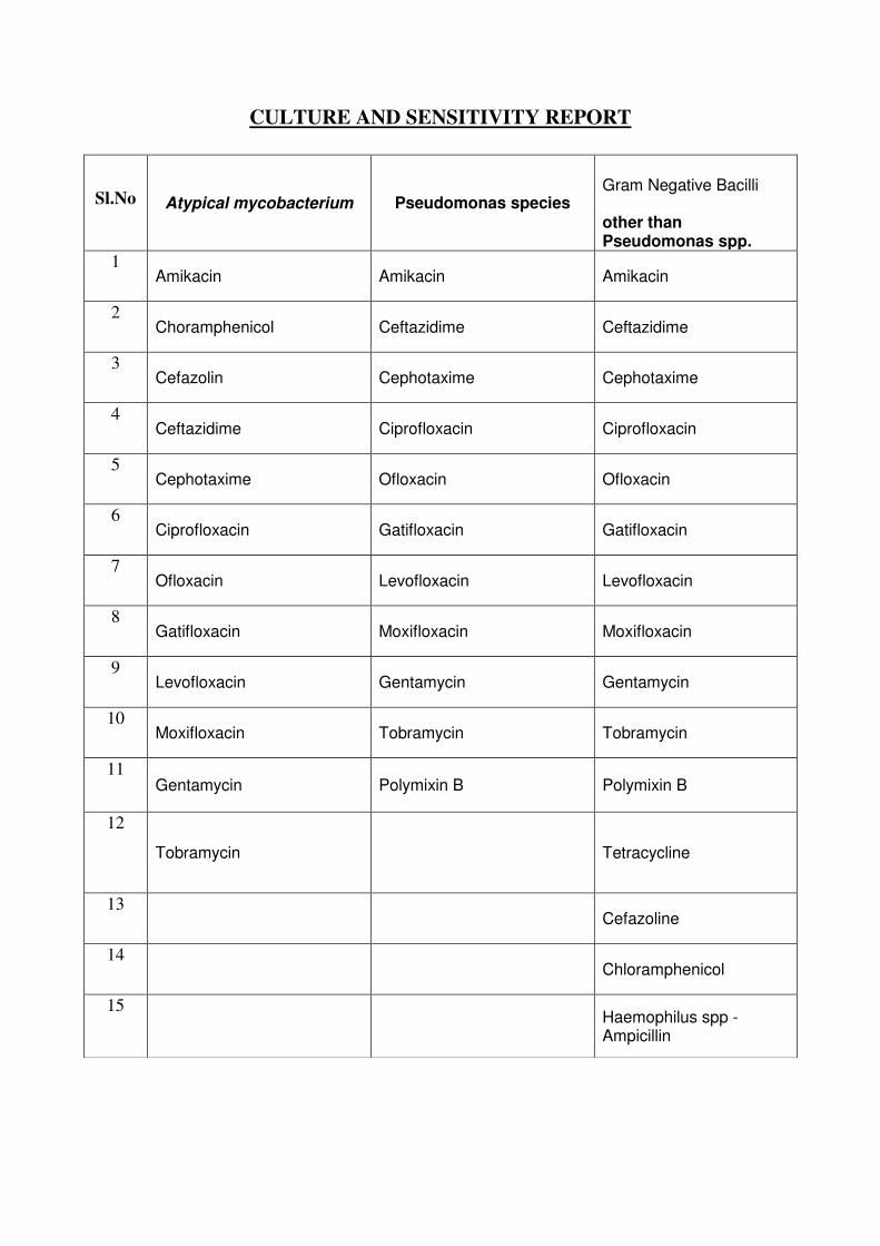
CULTURE AND SENSITIVITY REPORT
Sl.No Atypical mycobacterium Pseudomonas species
Gram Negative Bacilli other than Pseudomonas spp.
1 Amikacin Amikacin Amikacin
2 Choramphenicol Ceftazidime Ceftazidime
3 Cefazolin Cephotaxime Cephotaxime
4 Ceftazidime Ciprofloxacin Ciprofloxacin
5 Cephotaxime Ofloxacin Ofloxacin
6 Ciprofloxacin Gatifloxacin Gatifloxacin
7 Ofloxacin Levofloxacin Levofloxacin
8 Gatifloxacin Moxifloxacin Moxifloxacin
9 Levofloxacin Gentamycin Gentamycin
10 Moxifloxacin Tobramycin Tobramycin
11 Gentamycin Polymixin B Polymixin B
12
Tobramycin Tetracycline
13 Cefazoline
14 Chloramphenicol
15 Haemophilus spp -
Ampicillin

TREATMENT
1. Empirical antibiotic:………………………………………………………………………
2. Dosage of empirical antibiotic:……………………………………………………………
3. No. of days given:………………………………………………………………………….
4. Change of antibiotic according to sensitivity pattern:………………………………………
5. Duration of sensitive antibiotic given:……………………………………………………..
6. Doasage sensitive antibiotic:……………………………………………………………….
7. Other ocular medications if any: ………………………………………………………….

FOLLOWUP:
DESCRIPTION OF ULCER:
VISUAL ACUITY
(snellen’s chart)
1st MONTH 3
rd MONTH
Right
Eye
Left
Eye
Right
Eye
Left
Eye
UCVA
BCVA
DURATION 1st MONTH 3
rd MONTH
Size of ulcer
Size of infiltrate or scar
Margins of ulcer
Depth of the ulcer
AC reaction
Size of hypopyon

IMPRESSION:
1. Time taken for ulcer to heal ( in days) :
2. State of ulcer after change of medicine at the end of the study:
1. Healed
2. Healing
3. Perforated
4. Remained the same
3. Final visual acuity:
1.Improved
2.Remained the same
3. Worsened

CODING SHEET AGE SEX MALE 1 FEMALE 2 EYE INVLOVED RIGHT 1 LEFT 2 OCCUPATION AGRICULURIST 1 MANUAL LABROURER 2 VEGETABLE VENDOR 3 CARPENTER 4 WHITE COLLAR JOB 5 OTHERS 6 PRESENTING HISTORY SYMPTOMS REDNESS 1 PHOTOPHOBIA 2 WATERING 3 PAIN 4 BLURRING OF VISION 5 IRRITATION 6 OTHERS 7 NO SYMPTOMS 8 DURATION HISTORY OF TRAUMA DUST 1 THORN, THREE BRANCH 2 VEGETABLE MATTER 3 STICK 4 PADDY 5 METAL 6 STONE 7 OTHERS 8 NO TRAUMA 9 HISTORY OF CONTACT LENS USAGE
YES 1 NO 2 TRADITIONAL EYE MEDICINE
HUMAN MILK 1

CASTOR OIL 2 VEGETABLE JUICE 3 OTHERS 4 NO TRADITIONAL
MEDICATION 5
PREVIOUS OCULAR MEDICATION
ANTIBIOTICS 1 ANTIFUNGAL 2 ANTIVIRUS 3 STEROIDS 4 OTHERS 5 NO PREVIOUS
MEDICATION 6
PAST OCUALR HISTORY CORNEAL DISEASE 1 UVEITIS 2 DACROCYSTITIS 3 GLAUCOMA 4 EYE SURGERIES 5 OTHERS 6 NIL 7 SYSTEMIC HISTORY DIABETES 1 MALNUTRITION 2 IMMUNE SUPPPRESSION 3 LEPROSY 4 NEUROLOGICAL
DISORDERS 5
OTHERS 6 NONE 7 RANDOM BLOOD SUGAR VISUAL ACQUITY NO PL 1 PL 2 HM 3 1/60 4 2/60 5 3/60 6 4/60 7 5/60 8 6/60 9 6/36 10 6/18 11 6/12 12

6/9 13 6/6 14 LIDS NORMAL 1 EDEMA 2 BLEPHARITIS 3 TRICHIASIS 4 DRY EYE 5 LAGOPHTHALMOS 6 ENTROPION 7 ECTROPION 8 OTHERS 9 CONJUNCTIVA NORMAL 1 CONGESTION 2 DISCHARGE 3 OTHERS 4 ANTERIOR CHAMBER QUIET YES 1 NO 2 AC CELLS 0 1 0.5+ 2 1+ 3 2+ 4 3+ 5 4+ 6 AC FLARE 0 1 1+ 2 2+ 3 3+ 4 4+ 5 HYPOPYON YES 1 NO 2 PUPIL NORMAL 1 ABNORMAL 2 LENS NORMAL 1 ABNORMAL 2 NO VIEW 3 FUNDUS DEFERRED 1

NORMAL 2 ABNORMAL 3 CORNEAL ULCER SITE ENTIRELY IN PERIPHERY 1 OVERLAPPING THE
CORNEAL 4MM CIRCLE AND PERIPHERY WITHOUT FILLING THE CENTER
2
ENTIRELY IN THE CENTRAL 4MM CIRCLE
3
COMPLETELY FILLING 4MM CIRCLE EXTENDING TO PERIPHERY
4
SIZE - ED INFILTRATE 0-33% 1 33-67% 2 67-100% 3 NO 4 MARGINS OF ULCER REGULAR 1 IRREGULAR 2 STROMAL EDEMA YES 1 NO 2 DM FOLD YES 1 NO 2 CORNEAL SENSATION NORMAL 1 DIMINISHED 2 ABSENT 3 IOP BY DIGITAL TONOMETRY
NORMAL 1 INCREASED 2 DECREASED 3 DUCT OF AFFECTED EYE FREE 1 WITH CLEAR FLUIDS 2 WITH PUS 3 GRAM STAIN GRAM NEGATIVE BACILLI 1 GRAM POSITIVE COCCI 2 GRAM POSITIVE BACILLI 3
NO ORGANISM WITH PUS / POLYMORPHS
4
FUNGI 5 NOCARDIA 6 10% KOH POSITIVE 1 NEGATIVE 2 CULTURE REPORT PSEUDOMONAS
AERUGINOSA 1
STREPTOCOCCUS PNEUMONIAE
2
STAPHYLOCOCCUS AERUS 3 MRSA 4 CONS 5 STREPTOCOCCUS
PYOGENES 6
PSEUDOMONAS SP 7 STREPTOCOCCUS
VIRIDIANS 8
CORNYBACTERIUM 9 MOREXELLA SP 10 KLEBSIELLA SP 11 ACINECTOBACTER SP 12 CITROBACTER SP 13 HAEMOPHILUS
INFLUENZA 14
AEROMONAS SP 15 PASTURELLA SP 16 ENTEROBACTER SP 17 BACILLUS SP 18 PROPIONIBACTERIUM 19 ALCALIGENE 20 NOCARDIA 21 PROTEUS 22 STREPTOCOCCUS + MRSA 23 ANTIBIOTIC SENSITIVITY Levofloxacin S 1 I 2 R 3 ND 4 gatifloxacin S 1 I 2 R 3

ND 4 moxifloxacin S 1 I 2 R 3 ND 4 cephotaxime S 1 I 2 R 3 ND 4 Gentamicin S 1 I 2 R 3 ND 4 Tobramycin S 1 I 2 R 3 ND 4 Ciprofloxacin S 1 I 2 R 3 ND 4 Amikacin S 1 I 2 R 3 ND 4 Ofloxacin S 1 I 2 R 3 ND 4 chloramphenicol S 1 I 2 R 3 ND 4 cefazolin S 1 I 2 R 3 ND 4

ceftazidime S 1 I 2 R 3 ND 4 Cotrimaxazole S 1 I 2 R 3 ND 4 Vancomycin S 1 I 2 R 3 ND 4 Norfloxacin S 1 I 2 R 3 ND 4 Polymixin B S 1 I 2 R 3 ND 4 Tetracyclin S 1 I 2 R 3 ND 4 Piperacillin/Tazobactam S 1 I 2 R 3 ND 4 Cefuroxime S 1 I 2 R 3 ND 4 TREATMENT INITIAL ANTIMICROBIAL CEFAZOLIN 1 MOXIFLOXCIN 2 AMIKACIN 3 TOBRAMYCIN 4

GATIFLOXCIN 5
CHLOROMPHENICOL AND POLYMIXCIN B
6
CIPROFLOXCIN 7
CEFAZOLIN + CHLORAMPHENICOL + POLYMIXCIN B
8
MOXIFLOXCIN+ CHLORAMPHENICOL + POLYMIXCIN B
9
AMIKACIN + CHLORAMPHENICOL + POLYMIXCIN B
10
CEFAZOLIN + MOXIFLOXCIN
11
AMIKACIN + MOXIFLOXCIN
12
GATIFLOXCIN + CHLORAMPHENICOL + POLYMIXCIN B
13
MYDRIATICS YES 1 NO 2 ULCER HEALING IMPROVED 1 STABLE 2 WORSENED 3

S1.N
o
Order_ID date mr
no
patientname sex Age E
y
e
Oc
cu
pa
tio
n
Re
dn
ess
phot
opho
bia
pai
n
water
ing
blurri
ng of
vision
Sympt
oms
Dura
tion
in
days
Inju
ry
Typ
e
Conta
ct
lens
use
Traditi
onal
eye
treatm
ent
Pre
viou
s
ocul
ar
med
icati
on
Diab
etes
RBS Visua
l
acuit
y
LI
DS
Conj
uncti
va
Ant
erio
r
cha
mbe
r
quie
t
AC
cells
Hy
pop
yon
pupi
l
lens fund
us
cor
nea
l
ulc
er
site
infilt
rate
margi
ns of
ulcer
Strom
al
edema
DM
fold
Duct
of
affec
ted
eye
Initia
l
antib
iotics
Gra
m
stai
n
K
O
H
cult
ure
rep
ort
Lev
oflo
xaci
n
gatifl
oxac
in
mox
iflo
xaci
n
cep
hot
axi
me
Genta
micin
To
br
am
yci
n
Cip
rofl
oxa
cin
Ami
kaci
n
Ofl
ox
aci
n
chlo
ram
phen
icol
cefa
zolin
ceft
azi
dim
e
Cot
rim
axa
zol
e
Va
nc
om
yci
n
Po
ly
mi
xi
n
B
Tet
rac
ycli
n
Cefur
oxime
chan
ge in
antib
iotics
Ulc
er
hea
ling
at 3
wee
k
UC
VA
afte
r 3
wee
ks
Ulce
r
heal
ing
at
final
follo
wup
UC
VA
at
final
follo
w upUC
VA
im
pro
ve
me
nt
1 M19881/2015 01-12-2015 P4
17
ALANTHAMMA
L
2 68 2 1 1 1 1 1 1 1 1 3 2 5 1 2 1 8 1 2 1 2 2 1 1 1 2 1 2 2 2 1 1 4 2 1 1 1 2 3 1 2 1 1 1 4 4 1 4 4 1 4 4 2 2 8 1 9 1
2 M19928/2015 01-12-2015 P4
17
SANTHANAM C. 1 61 2 1 1 2 1 1 1 4 8 2 2 5 1 1 1 4 1 3 1 3 2 1 1 1 4 1 2 1 1 3 2 1 2 1 1 1 1 1 1 1 1 1 1 4 4 1 4 4 1 4 4 1 2 4 2 6 2
3 M19967/2015 02-12-2015 F1
63
DEVARAJ M. 1 66 1 2 2 2 2 2 2 3 1 1 2 2 3 2 1 8 1 2 1 3 2 1 1 1 2 2 1 1 1 1 8 4 2 1 1 1 1 2 1 1 1 1 1 4 4 1 4 4 1 4 4 1 2 8 1 9 1
4 M19988/2015 02-12-2015 P4
17
KALYANI M. 2 48 1 3 1 2 1 1 1 2 5 3 2 5 3 1 2 8 1 1 2 5 1 1 1 1 2 1 1 2 2 1 9 4 2 1 1 1 1 1 1 1 1 1 1 4 4 1 4 4 1 4 4 1 1 9 1 10 1
5 M19993/2015 02-12-2015 P4
17
MUTHALAGAR R. 1 29 1 6 1 1 1 2 1 4 4 8 1 5 3 2 2 4 2 2 1 4 2 1 1 1 2 3 2 1 1 1 1 4 2 1 1 1 1 1 1 1 1 1 1 4 4 1 4 4 1 4 4 1 2 6 2 7 1
6 M20008/2015 02-12-2015 F1
63
MARIYAYEE 2 63 2 1 2 1 2 2 2 6 13 1 2 2 1 1 1 4 1 1 1 5 2 1 1 1 4 3 2 1 1 1 9 1 2 1 1 1 1 2 2 1 1 1 1 4 4 1 4 4 1 4 4 2 3 3 3 3 6
7 M20010/2015 03-12-2015 P2
91
KANDASAMY A. 1 76 1 6 1 2 2 1 1 1 5 1 2 5 2 2 2 10 1 1 1 4 2 1 1 1 2 2 1 1 1 2 1 2 2 2 1 1 1 1 4 4 1 4 1 3 1 4 4 1 4 2 1 1 2 10 1 11 1
8 M20095/2015 04-12-2015 P4
17
PAKKIRI 1 61 2 1 1 2 1 1 1 1 2 2 2 5 3 2 2 11 1 2 1 2 2 1 1 1 1 1 2 2 2 1 2 4 2 5 1 1 1 1 1 1 1 4 1 1 1 4 4 1 4 1 4 1 3 8 1 10 2
9 M20129/2015 05-12-2015 P4
17
PALANISAMY P. 1 45 1 6 1 1 1 1 1 4 5 8 2 1 3 2 2 8 1 1 1 2 2 1 1 1 1 1 1 2 2 2 10 1 2 1 1 1 1 1 1 1 1 1 1 4 4 1 4 4 1 4 4 2 3 5 1 9 4
10 M20182/2015 07-12-2015 F1
50
MURUGANDI M. 1 57 2 1 2 2 2 2 2 1 2 5 2 2 3 2 2 12 1 2 1 2 2 1 1 1 1 1 1 2 2 1 8 2 2 2 1 1 1 1 3 4 1 4 1 1 1 4 4 1 4 1 4 1 3 9 2 11 2
11 M20409/2015 10-12-2015 F1
63
MANIMEGALAI T. 2 51 1 6 1 1 2 2 1 4 5 2 5 1 2 2 8 1 1 1 2 2 1 1 1 1 1 1 2 2 1 8 4 2 6 1 1 1 1 4 4 1 4 1 1 1 4 4 1 4 3 4 2 2 8 1 10 2
12 M20537/2015 12-12-2015 F1
63
SANJEEVI 1 71 1 1 2 2 2 2 2 4 1 4 2 5 3 1 2 9 1 2 1 2 2 1 1 1 2 1 1 2 2 1 11 3 2 1 1 1 1 2 1 1 1 1 1 4 4 1 4 4 1 4 4 1 1 10 1 10 6
13 M21029/2015 22-12-2015 P4
15
GANESAN P. 1 51 1 1 1 1 1 1 1 3 8 2 2 5 1 2 2 4 1 1 1 3 2 1 1 1 3 2 2 1 1 1 2 1 2 1 1 1 1 2 2 1 1 1 1 3 1 4 4 4 1 3 4 2 2 4 2 7 3
14 M21322/2015 26-12-2015 P4
18
ANJAMMAL R. 2 56 2 1 1 1 1 1 1 3 3 5 2 2 3 2 2 5 1 2 1 3 2 1 1 1 2 3 1 1 1 1 6 2 2 2 1 1 1 1 4 4 2 4 2 1 1 4 4 1 4 1 4 2 2 7 1 9 2
15 M21406/2015 28-12-2015 P4
18
VIRUMANDI V. 1 65 1 1 1 1 1 2 1 1 2 4 2 5 2 2 2 5 1 2 1 2 2 1 1 1 2 1 2 2 2 1 9 1 2 1 1 1 2 1 1 2 1 3 1 4 4 1 4 4 1 4 4 1 2 7 1 10 3
16 M21433/2015 28-12-2015 F1
56
ERUKALAIAH 1 56 1 1 2 1 2 2 2 2 6 3 2 5 1 1 1 3 1 2 1 4 2 1 1 1 3 3 2 1 1 1 3 1 2 1 3 3 3 3 3 4 3 3 3 4 4 3 4 4 1 4 4 2 3 7 2 8 1
17 M21513/2015 29-12-2015 P4
18
SABUR NISHA
M.
2 51 2 3 1 1 2 1 1 4 4 8 2 5 3 2 2 7 1 2 1 4 2 1 1 1 2 2 1 1 1 1 1 2 2 5 1 1 1 2 3 3 2 4 1 1 2 4 4 1 4 1 4 1 1 8 1 13 5
18 M126/2016 04-01-2016 F1
63
VIRAMALAI R. 1 80 1 1 1 1 1 1 1 6 2 2 2 5 3 2 2 8 1 2 1 2 2 1 1 1 2 1 1 2 2 1 8 2 2 11 1 1 1 1 1 1 1 1 1 1 2 1 4 4 1 1 4 2 3 7 1 10 3
19 M1274/2016 25-01-2016 P4
20
SAGUNTHALA
K.
2 59 2 6 1 1 1 1 1 2 5 8 2 2 1 1 1 5 1 2 1 5 2 1 1 1 3 3 2 1 1 1 3 5 2 21 3 3 3 1 1 1 3 1 3 1 3 1 3 3 4 4 4 1 2 6 2 8 2
20 M1317/2016 25-01-2016 P4
20
THANSIRA
BANU
2 38 2 2 1 1 2 2 1 3 5 3 2 5 3 1 1 10 1 2 1 3 2 1 1 1 1 2 1 1 1 1 1 4 2 5 1 1 1 1 1 2 1 1 10 1 11 1
21 M1333/2016 25-01-2016 F1
64
MARUTHAMUT
HU V.
1 40 2 1 1 1 1 1 1 1 2 2 2 5 3 2 2 6 1 2 1 3 2 1 1 1 3 2 1 2 2 1 2 1 2 10 1 1 1 1 1 1 1 1 1 1 1 1 4 4 1 1 4 2 3 4 1 9 5
22 M155/2016 04-01-2016 P4
19
NAGAMMAL 2 55 1 1 1 1 1 1 1 1 6 1 2 5 2 2 2 11 1 2 1 3 2 1 1 1 1 1 1 2 2 1 11 2 2 2 1 1 1 1 4 4 1 4 1 1 1 4 4 1 4 1 4 1 1 12 1 12 6
23 M1685/2016 30-01-2016 P4
20
PERIYASAMY A. 1 63 1 2 1 1 1 1 1 1 10 1 2 5 1 2 1 5 1 3 2 5 1 1 1 1 4 2 1 1 1 3 8 4 2 2 1 1 1 1 4 4 1 4 1 1 1 4 4 1 4 1 4 1 2 5 3 3 7
24 M1686/2016 30-01-2016 P4
19
PAASAM RAVI 1 47 1 1 2 2 2 2 2 4 9 2 2 2 3 1 2 5 1 1 1 3 2 1 1 1 3 1 2 1 1 2 1 4 2 9 3 3 3 1 3 3 3 4 3 1 1 4 4 1 4 3 4 2 2 6 3 5 7
25 M1708/2016 30-01-2016 P4
20
ALAGARSAMY A. 1 65 1 1 1 1 1 2 1 3 3 2 2 2 3 2 2 8 1 2 1 3 2 1 1 1 3 1 1 2 2 1 8 4 2 2 1 1 1 1 4 4 1 4 1 1 1 4 4 1 4 1 4 1 1 9 1 10 1
26 M1711/2016 30-01-2016 P4
20
SAIVARAJ R. 1 56 1 1 2 1 2 2 2 1 1 3 2 5 3 2 2 6 1 2 1 4 2 1 1 1 3 3 2 1 1 1 8 4 2 2 1 1 1 1 4 4 1 4 1 1 1 4 4 1 4 1 4 2 2 6 3 6 6
27 M423/2016 08-01-2016 F1
63
RAMAN M. 1 80 2 4 2 2 2 2 2 4 3 1 2 5 3 1 2 6 1 2 1 4 2 1 1 1 2 2 2 1 1 1 1 3 2 1 1 1 1 2 2 1 1 1 1 4 4 1 4 4 1 4 4 2 3 6 2 8 2
28 M462/2016 09-01-2016 P3
22
RAMARAJ K. 1 68 1 2 1 1 2 1 1 1 3 1 2 5 3 2 1 8 1 1 1 3 2 1 1 1 2 3 1 1 1 1 9 1 2 1 1 1 1 1 1 1 1 1 1 4 4 1 4 4 1 4 4 1 1 9 1 11 2
29 M561/2016 11-01-2016 P3
48
KARUPPASAMY N. 1 25 1 6 2 1 2 2 2 3 4 1 1 2 1 2 2 11 1 2 1 2 2 1 1 1 2 1 2 2 2 1 2 1 2 10 1 1 1 1 1 1 1 1 1 1 1 1 4 4 1 1 4 1 2 12 1 12 6
30 M709/2016 14-01-2016 P4
19
SELVAM S. 1 27 2 6 1 1 1 1 1 4 5 1 2 1 3 2 2 11 1 1 1 3 2 1 1 1 1 2 2 1 1 1 4 1 2 1 1 1 1 3 1 1 1 1 1 4 4 1 4 4 1 4 4 1 1 12 1 12 6
31 M714/2016 14-01-2016 P1
73
APPANA SAMY
S.
1 62 2 1 1 1 1 1 1 2 1 2 2 5 3 2 2 8 1 2 1 3 2 1 1 1 1 1 1 2 2 1 1 4 2 9 1 1 1 1 1 1 2 2 8 1 9 1
32 M76/2016 02-01-2016 F1
63
MAHA KAVI P. 1 32 1 1 1 1 1 2 1 6 4 2 2 5 3 2 2 5 1 2 2 5 1 1 1 1 4 3 1 1 1 1 8 2 2 2 1 1 1 1 4 4 1 4 1 1 1 4 4 1 4 3 4 2 2 5 2 7 2
33 M786/2016 18-01-2016 F1
64
MARIAPPAN S. 1 55 1 2 1 1 2 1 1 4 5 1 2 5 3 1 2 4 1 1 1 3 2 1 1 1 3 2 1 1 1 2 8 4 2 1 1 1 1 2 1 1 1 1 3 4 4 1 4 4 1 3 4 1 2 5 2 8 3
34 M789/2016 18-01-2016 P4
20
RAMANAMMA
M.
2 48 2 6 1 1 1 1 1 1 6 4 2 5 3 2 2 11 1 2 1 4 2 1 1 1 1 2 1 1 1 1 9 1 2 13 1 1 1 1 1 1 1 1 1 1 2 1 4 4 1 1 4 1 1 12 1 12 6
35 M877/2016 19-01-2016 P4
20
NAIR K.G. 1 77 2 6 1 1 1 2 1 4 3 1 2 2 3 2 2 5 1 2 1 3 2 1 1 1 3 3 1 1 1 1 8 4 2 5 3 3 3 1 1 1 3 4 3 1 1 4 4 1 4 3 4 2 2 7 1 10 3
36 M965/2016 20-01-2016 P4
19
ANNAMMA
THANAKACHAN
2 70 1 6 1 1 1 1 1 3 4 8 2 5 2 2 2 9 1 2 1 3 2 1 1 1 2 2 2 2 2 1 8 2 2 1 1 1 1 2 1 1 1 1 1 4 4 1 4 4 1 4 4 1 1 10 1 10 6
37 M1760/2016 01-02-2016 F1
64
CHENGAL REDDY
B.
1 75 1 1 1 1 2 2 1 3 5 8 2 5 3 2 2 8 1 1 1 3 2 1 1 1 2 1 1 2 2 3 2 4 2 2 1 1 1 1 4 4 1 4 1 1 1 4 4 1 4 3 4 1 1 9 1 10 1
38 M1761/2016 01-02-2016 P4
21
MUTHULAKSHMI
M.
2 54 1 6 1 1 1 1 1 4 5 4 2 5 3 2 2 12 1 2 1 3 2 1 1 1 1 2 1 1 1 1 1 2 2 2 1 1 1 1 4 4 1 4 1 1 1 4 4 1 4 1 4 1 1 12 1 13 1
39 M1778/2016 01-02-2016 P4
21
ALAGAN T. 1 85 2 6 1 1 1 1 1 1 6 2 2 1 2 1 2 6 1 2 1 3 2 1 1 1 3 2 1 1 1 1 11 2 2 2 1 1 1 2 4 4 1 4 1 1 1 4 4 1 4 3 4 2 3 6 2 7 1
40 M1833/2016 01-02-2016 F1
46
INRANI M. 2 52 1 2 1 1 2 2 1 1 1 6 2 5 1 1 2 7 2 2 2 5 1 1 1 1 3 1 1 1 1 1 2 1 2 10 1 1 1 1 1 1 1 1 1 1 1 1 4 4 1 4 4 1 3 6 3 5 7
41 M1934/2016 03-02-2016 P4
21
NATCHAMMAL N. 2 55 1 1 1 1 1 1 1 6 4 2 2 5 3 2 2 8 1 2 1 2 2 1 1 1 3 1 2 2 2 1 8 2 2 2 1 1 1 1 1 1 1 1 9 1 10 1
42 M2034/2016 04-02-2016 P2
91
VELUSAMY C. 1 45 1 2 2 2 2 2 2 4 7 1 2 5 1 1 1 6 1 2 2 5 1 1 1 1 3 3 1 1 1 1 10 1 2 14 3 3 3 1 3 3 3 1 3 3 3 1 4 4 1 4 4 2 2 6 2 6 6
43 M2118/2016 05-02-2016 P3
68
VENKATESH V. 1 48 1 2 2 1 2 2 2 1 5 1 2 5 3 2 1 9 1 2 1 3 2 1 1 1 1 1 1 2 2 1 2 2 2 5 1 1 1 3 3 3 1 4 1 1 1 4 4 1 4 1 4 1 1 9 1 10 1
44 M2192/2016 06-02-2016 P4
21
RAKKAMMAL 2 45 1 3 1 1 1 2 1 2 5 2 2 5 2 2 2 8 1 2 1 4 2 1 1 1 1 2 1 1 1 1 1 2 2 2 1 1 1 1 4 4 1 4 1 1 1 4 4 1 4 1 4 1 1 9 1 9 6
45 M2213/2016 08-02-2016 P4
21
MARAPPAN K. 1 68 2 4 1 1 1 1 1 1 3 1 2 5 3 1 1 3 1 1 1 3 2 1 1 1 4 2 2 1 1 1 1 4 2 1 1 1 1 1 1 1 1 1 1 4 4 1 4 4 1 4 4 2 3 2 3 2 6
46 M2716/2016 15-02-2016 F1
64
VELLAIKANNU
P.
1 69 2 6 1 1 1 1 1 1 5 2 2 5 1 1 1 7 1 2 1 4 2 1 1 1 3 2 1 1 1 1 4 4 2 2 1 1 1 1 4 4 1 4 1 1 1 4 4 1 4 1 4 1 2 7 1 9 2
47 M2717/2016 15-02-2016 F1
65
PERIYAKARUPP
AN
1 65 2 1 1 1 1 2 1 4 6 1 2 5 3 1 2 5 1 2 2 5 1 1 1 1 3 3 1 1 1 1 8 2 2 2 1 1 1 1 4 4 1 4 1 1 1 4 4 1 4 1 4 2 2 6 2 8 2
48 M2829/2016 17-02-2016 P4
04
NANDHINI D.S. 2 18 1 6 1 1 1 1 1 3 5 1 2 5 1 2 2 12 1 1 1 3 2 1 1 1 1 1 2 2 2 1 13 1 2 1 1 1 1 1 1 1 1 1 1 4 4 1 4 4 1 4 4 1 1 13 1 13 6
49 M2843/2016 17-02-2016 F1
65
SHINNAIAH M. 1 55 1 2 1 1 1 1 1 6 6 1 2 5 3 1 1 4 1 3 1 3 2 1 1 1 3 1 1 1 1 2 1 4 2 12 1 1 1 1 1 1 1 1 1 1 1 1 4 4 1 1 4 1 3 4 3 3 7
50 M3299/2016 24-02-2016 F1
65
THANGARAJ.T 1 69 1 2 2 2 2 2 2 3 4 1 2 5 3 1 1 5 1 2 2 5 1 1 1 1 3 1 2 2 2 1 11 2 2 2 1 1 1 1 4 4 1 4 1 1 1 4 4 1 4 1 4 2 2 6 1 10 4
51 M3391/2016 26-02-2016 P3
36
MUNIAMMAL
M.
2 37 2 1 1 1 1 2 1 2 2 4 2 5 3 2 2 7 1 2 1 3 2 1 1 1 2 2 1 1 1 1 2 1 2 1 3 3 3 3 3 3 3 3 3 4 4 3 4 4 1 4 4 1 3 6 2 8 2
52 M3418/2016 26-02-2016 F1
65
ARUMUGAM C. 1 85 1 1 2 1 2 2 2 4 4 2 2 5 3 1 1 8 1 2 1 3 2 1 1 1 2 2 1 2 2 1 8 2 2 2 1 1 1 1 4 4 1 4 1 1 1 4 4 1 4 1 4 1 1 10 1 10 6
53 M3501/2016 29-02-2016 P4
19
KARUPPU V. 1 41 1 6 1 1 1 1 1 1 3 1 2 5 3 2 2 10 1 2 1 4 2 1 1 1 1 2 1 1 1 1 8 4 2 2 1 1 1 1 4 4 1 4 1 1 1 4 4 1 4 1 4 1 1 11 1 11 6
54 M3630/2016 01-03-2016 F1
65
SUBBAIAH P. 1 50 2 4 1 1 1 1 1 2 4 1 2 5 3 1 1 6 1 1 2 5 1 1 1 1 4 1 2 2 2 1 1 4 2 1 1 1 1 1 1 1 1 1 1 4 4 1 4 4 1 4 4 2 2 7 2 8 1
55 M3739/2016 03-03-2016 P4
23
ESWARAN 1 60 1 2 1 1 1 2 1 4 3 5 2 5 3 1 2 5 1 2 1 4 2 1 1 1 4 2 2 1 1 1 10 4 2 21 3 3 3 1 1 1 3 1 3 1 1 3 3 1 4 4 4 2 3 7 2 9 2
56 M3870/2016 04-03-2016 P4
23
NALLUSAMY 1 65 1 2 1 2 1 1 1 1 8 1 2 5 1 1 1 9 1 1 1 4 2 1 1 1 3 2 1 1 1 3 2 2 2 1 1 1 1 4 4 1 4 1 1 1 4 4 1 4 1 4 1 1 10 1 10 6
57 M4127/2016 09-03-2016 P3
96
SEKAR R. 1 51 2 1 1 1 1 1 1 3 14 9 2 1 3 1 1 6 1 3 1 4 2 1 1 1 2 2 2 1 1 3 8 4 2 2 1 1 1 1 4 4 1 4 1 1 1 4 4 1 4 1 4 1 2 7 2 7 6
58 M4206/2016 10-03-2016 P4
23
GURUVAMMAL
R.
2 55 2 1 1 2 1 1 1 6 3 4 2 5 1 2 2 8 1 2 2 5 1 1 1 1 2 1 1 2 2 1 8 4 2 3 1 1 1 1 1 1 1 4 1 1 1 4 4 1 4 1 4 2 3 7 1 9 2

59 M4242/2016 10-03-2016 P3
94
SUBBAIAH K. 1 78 1 6 1 2 1 1 1 4 5 1 2 2 2 1 2 9 1 1 1 3 2 1 1 1 2 2 1 2 2 1 9 4 2 10 1 1 1 1 1 1 1 1 1 1 2 1 4 4 1 1 4 1 1 9 1 9 6
60 M4617/2016 15-03-2016 P4
24
KARTHICK A. 1 14 2 6 1 2 1 1 1 1 15 3 2 5 1 2 2 5 1 1 1 4 2 1 1 1 2 2 2 1 1 1 9 4 2 1 1 1 1 1 1 1 1 1 1 4 4 1 4 4 1 4 4 2 3 5 3 5 6
61 M4731/2016 16-03-2016 F1
66
VELLAIAMMAL
M.
2 70 2 6 2 1 2 2 2 4 3 2 2 5 1 2 2 9 1 2 1 4 2 1 1 1 2 2 2 2 2 1 1 4 2 2 1 1 1 1 4 4 1 4 1 1 1 4 4 1 4 3 4 1 1 9 1 9 6
62 M5205/2016 24-03-2016 P2
58
ANILKUMAR V. 1 56 1 1 1 1 1 1 1 6 5 1 2 5 3 1 1 4 1 1 1 4 2 1 1 1 3 3 1 1 1 1 9 4 2 1 1 1 1 3 1 1 1 1 1 4 4 1 4 4 1 4 4 1 3 4 2 7 3
63 M5263/2016 25-03-2016 P4
24
VEERASAMY V. 1 60 1 4 2 2 2 2 2 1 5 1 2 5 3 1 1 8 1 1 1 2 2 1 1 1 1 1 1 2 2 3 1 4 2 2 1 1 1 1 4 4 1 4 1 1 1 4 4 1 4 1 4 1 1 9 1 9 6
64 M5396/2016 28-03-2016 P4
24
RADHA S. 2 51 1 2 1 2 1 1 1 2 3 3 2 2 3 2 2 6 1 2 1 4 2 1 1 1 3 3 1 1 1 1 4 4 2 3 1 1 1 1 3 3 1 4 1 1 1 4 4 1 4 1 4 2 2 6 2 7 1
65 M5421/2016 28-03-2016 F1
66
KARMEGAM A. 1 65 1 2 2 1 2 2 2 3 10 9 2 5 1 1 2 5 1 2 2 5 1 1 1 1 3 3 2 1 1 1 8 4 2 2 1 1 1 1 4 4 1 4 1 1 1 4 4 1 4 1 4 2 2 7 3 7 6
66 M5489/2016 29-03-2016 F1
64
GANESAN.P 1 50 1 1 2 1 2 2 2 1 5 1 2 5 3 1 1 4 1 2 1 3 2 1 1 1 4 3 1 1 1 1 9 4 2 21 3 3 3 3 1 1 3 1 3 3 3 3 3 3 4 4 4 1 3 3 3 3 6
67 M6054/2016 06-04-2016 P4
25
SYED IBRAHIM 1 65 2 2 1 2 1 1 1 4 3 4 2 5 3 1 2 10 1 2 2 5 1 1 1 1 2 1 1 2 2 1 12 4 2 21 3 1 1 1 3 3 2 1 3 1 3 3 4 3 4 4 4 1 2 11 1 11 6
68 M6103/2016 07-04-2016 P4
19
PREETHIKA M. 2 8 1 6 1 2 1 1 1 1 2 3 2 5 3 2 2 9 1 2 1 4 2 1 1 1 2 2 1 1 1 1 6 2 2 2 1 1 1 1 4 4 1 4 1 1 1 4 4 1 4 1 4 1 1 9 1 9 6
69 M6114/2016 07-04-2016 F1
66
MUNIYANDI 1 68 1 1 1 2 1 1 1 3 5 1 2 2 1 1 1 9 1 1 1 3 2 1 1 1 2 2 2 2 2 1 2 4 2 1 1 1 1 1 1 1 1 1 1 4 4 1 4 4 1 4 4 2 3 7 1 10 3
70 M6191/2016 08-04-2016 P4
25
PANDI M. 1 42 2 6 1 1 1 1 1 4 1 4 2 5 3 2 2 8 1 2 1 4 2 1 1 1 1 2 1 2 2 1 5 1 2 10 1 1 1 1 1 1 1 1 1 1 1 1 4 4 1 1 4 1 1 10 1 10 6
71 M6319/2016 11-04-2016 P4
25
PONNUTHAI 2 55 1 1 1 2 1 1 1 1 1 2 2 5 1 1 1 6 1 2 1 5 2 1 1 1 2 3 2 1 1 1 1 2 2 2 1 1 1 1 4 4 1 4 1 1 1 4 4 1 4 3 4 2 2 7 2 7 6
72 M6330/2016 11-04-2016 F1
66
ARUMUGAM 1 70 1 2 1 2 1 1 1 6 4 3 2 1 3 1 1 10 1 2 1 3 2 1 1 1 2 2 1 1 1 1 8 2 2 2 1 1 1 1 4 4 1 4 1 1 1 4 4 1 4 1 4 1 1 10 1 13 3
73 M6358/2016 12-04-2016 P4
25
VELLAIAN P. 1 69 2 1 2 1 2 2 2 1 1 2 2 5 3 1 1 4 1 2 2 5 1 1 1 1 3 2 1 1 1 1 11 2 2 2 1 4 1 1 1 4 1 4 1 1 1 4 4 1 4 1 4 1 3 5 2 6 1
74 M6492/2016 14-04-2016 P4
25
MARIAPPAN P. 1 33 1 5 1 1 1 1 1 4 5 1 2 2 1 2 2 12 1 2 1 3 2 1 1 1 1 2 1 1 1 1 9 1 2 1 1 1 1 1 1 1 1 1 1 4 4 1 4 4 1 4 4 1 1 13 1 13 6
75 M6496/2016 14-04-2016 F1
62
DHAKSHINAMOO
RTHI P.
1 64 1 2 1 1 1 1 1 3 2 4 2 1 3 1 2 5 1 2 1 3 2 1 1 1 4 1 2 1 1 1 2 1 2 14 1 1 1 3 1 1 1 1 1 4 3 3 4 4 1 3 4 2 2 8 1 9 1
76 M6596/2016 16-04-2016 P4
26
BIJU P.S. 1 35 2 5 1 1 1 1 1 3 2 7 2 5 3 2 2 9 2 2 1 3 2 1 1 1 2 1 1 2 2 1 8 4 2 4 1 1 1 2 3 3 2 4 2 1 2 4 4 1 4 1 4 1 1 10 1 10 6
77 M6622/2016 16-04-2016 F1
66
MAHA LINGAM
M.
1 49 1 3 1 1 1 1 1 1 7 5 2 5 2 1 1 8 1 2 2 5 1 1 1 1 1 2 1 1 1 1 1 5 2 15 1 1 1 1 1 1 1 2 1 1 3 1 4 4 1 1 4 1 3 7 1 10 3
78 M6816/2016 20-04-2016 F1
66
SUNDARAMOORT
HI S.
1 70 1 6 1 1 1 1 1 4 1 4 2 5 3 1 1 10 1 2 1 2 2 1 1 1 1 1 1 2 2 1 8 3 2 9 1 1 1 1 1 1 1 4 1 1 1 4 4 1 4 4 4 1 1 11 1 11 6
79 M6968/2016 22-04-2016 F1
66
RANJITHA R. 2 11 2 6 1 1 1 1 1 1 3 3 2 5 2 2 2 7 1 2 1 3 2 1 1 1 2 2 1 1 1 1 4 1 2 1 1 1 1 2 1 2 1 3 1 4 2 4 4 4 1 4 4 1 1 8 1 10 2
80 M6988/2016 22-04-2016 P4
26
DURAIRAJ . B 1 6 2 6 1 1 1 1 1 1 6 4 2 5 1 2 2 8 1 2 1 3 2 1 1 1 1 2 1 2 2 1 9 1 2 16 1 1 1 1 1 1 1 1 1 1 1 1 4 4 1 1 4 1 1 10 1 10 6
81 M8202/2016 11-05-2016 P4
28
NITHIN KUMAR .A 1 3 1 6 1 1 1 1 1 4 8 4 2 2 3 2 2 10 1 3 1 4 2 1 1 1 2 3 2 1 1 2 9 1 2 1 1 1 1 1 1 1 1 1 1 4 4 1 4 4 1 4 4 2 2 10 2 10 6
82 M8211/2016 11-05-2016 P4
28
ALAGAMMAL P. 2 70 1 1 1 1 1 1 1 3 4 4 2 5 3 1 2 6 1 2 2 5 1 1 1 1 2 3 2 1 1 1 9 4 2 1 1 1 1 1 1 1 1 1 1 4 4 1 4 4 1 4 4 1 3 6 2 9 3
83 M8401/2016 14-05-2016 P1
71
SINGARAVEL
A.K.A
1 90 2 1 1 1 1 1 1 1 3 4 2 5 3 2 2 6 1 2 1 3 2 1 1 1 3 1 1 2 2 1 1 2 2 2 1 1 1 1 4 4 1 4 1 1 1 4 4 1 4 1 4 1 1 8 1 9 1
84 M8408/2016 14-05-2016 P4
14
KUNJARAM 2 71 2 6 1 1 1 1 1 6 4 2 2 5 3 2 2 8 1 2 1 3 2 1 1 1 2 2 1 1 1 1 8 4 2 1 1 1 1 1 1 1 1 1 1 4 4 1 4 4 1 1 4 1 1 9 1 10 1
85 M8446/2016 17-05-2016 P4
28
RAJAMMA N. 2 71 1 1 1 1 1 1 1 3 2 5 2 5 3 2 2 11 1 2 1 3 2 1 1 1 2 1 1 2 2 1 2 1 2 13 1 1 1 1 1 1 1 1 1 1 1 1 4 4 1 1 4 1 1 12 1 12 6
86 M9017/2016 25-05-2016 P4
29
RAMAYA NAIDU
R.
1 82 1 1 1 1 1 1 1 2 8 5 2 5 1 1 1 5 1 3 1 4 2 1 1 1 3 2 2 1 1 3 1 4 2 22 3 3 3 3 3 3 3 3 3 3 3 3 4 4 3 3 4 2 2 7 2 8 1
87 M9076/2016 26-05-2016 P4
26
MARIMUTHU R. 1 40 1 1 1 1 1 1 1 4 3 3 2 5 3 2 2 8 1 2 1 3 2 1 1 1 1 2 2 1 1 1 3 1 2 1 1 1 1 1 1 1 1 1 1 4 4 1 4 4 1 4 4 1 3 6 1 9 3
88 M9142/2016 27-05-2016 P4
29
LAKSHMANAN
C.
1 74 2 1 1 1 1 1 1 1 1 4 2 2 3 2 2 6 1 2 1 3 2 1 1 1 2 3 2 1 1 1 9 1 2 1 1 1 1 1 1 1 1 1 1 4 4 1 4 4 1 4 4 1 1 7 1 10 3
89 M9150/2016 27-05-2016 P4
29
SRINIVASAN V. 1 55 2 2 1 1 1 1 1 3 1 6 2 5 1 2 2 9 1 2 1 3 2 1 1 1 2 2 2 1 1 1 9 4 2 3 1 1 1 1 3 3 1 4 1 1 1 4 4 1 4 1 4 1 1 9 1 9 6
90 M9152/2016 27-05-2016 P4
29
PARVATHI N. 2 83 1 6 1 1 1 1 1 4 3 4 2 5 3 2 2 6 1 2 1 5 2 1 1 1 3 3 1 1 1 1 10 1 2 1 1 1 1 1 1 1 1 1 1 4 4 1 4 4 1 4 4 2 3 5 2 8 3
91 M9155/2016 27-05-2016 P4
29
PARVATHI N. 2 83 2 6 1 1 1 1 1 2 3 1 2 2 3 2 2 11 1 2 1 4 2 1 1 1 1 2 1 2 2 1 2 2 1 2 1 1 1 1 1 1 1 12 1 12 6
92 M9209/2016 28-05-2016 P4
29
SATHASRI S. 2 1 1 6 1 2 1 1 1 3 2 5 2 5 3 2 2 8 1 2 1 3 2 1 1 1 2 1 1 2 2 1 2 1 2 17 3 3 3 1 1 1 3 3 3 1 1 1 4 4 1 3 4 2 2 9 1 10 1
93 M9212/2016 28-05-2016 F1
68
VALLI P. 2 50 2 1 2 1 2 2 2 3 2 4 2 5 1 1 2 5 1 2 1 3 2 1 1 1 2 3 1 1 1 1 2 2 2 2 1 1 1 1 4 4 1 4 1 1 1 4 4 1 4 1 4 1 3 3 1 3 6
94 M9300/2016 30-05-2016 F1
68
SUBBAMMAL C. 2 50 1 2 2 1 2 2 2 1 2 4 2 5 2 1 1 7 1 1 2 5 1 1 1 1 3 2 2 1 1 1 1 2 5 3 3 3 1 3 3 3 4 3 1 1 4 4 1 4 1 4 2 2 7 2 8 1
95 M9361/2016 30-05-2016 P4
29
ALAGARSAMY R. 1 65 1 3 1 2 1 1 1 4 9 1 2 5 3 1 2 6 1 2 1 5 2 1 1 1 3 3 2 1 1 1 8 2 2 2 1 1 1 1 4 4 1 4 1 1 1 4 4 1 4 3 4 1 2 7 2 8 1
96 M9416/2016 31-05-2016 P4
29
RAMAKRISHNAN
V.
1 75 1 4 2 1 2 2 2 2 12 6 2 2 3 2 2 10 1 1 1 3 2 1 1 1 2 2 1 1 1 2 8 3 2 18 1 1 1 3 1 1 3 4 1 1 3 4 4 1 4 4 4 2 2 10 1 13 3
97 M10512/2016 17-06-2016 F1
69
KAMUTHAI K. 2 75 1 1 1 1 1 1 1 2 5 2 2 5 3 1 2 12 1 1 1 2 2 1 1 1 1 1 1 2 2 1 9 4 2 9 1 1 1 1 1 1 1 4 1 1 1 4 4 1 4 4 4 1 1 13 1 13 6
98 M10927/2016 23-06-2016 P4
31
NAGARATHINAM
M.
1 75 1 2 2 1 2 2 2 3 8 9 2 5 1 2 2 5 1 3 1 4 2 1 1 1 4 3 2 1 1 2 5 1 2 1 1 1 1 2 1 1 1 1 1 4 1 2 4 4 1 4 4 1 2 6 2 6 6
99 M10936/2016 23-06-2016 P4
31
KARUPPAYEE
R.
2 64 2 3 1 2 1 1 1 1 3 8 2 5 2 2 2 13 1 1 1 3 2 1 1 1 2 1 1 1 1 3 1 2 2 2 1 1 1 1 4 4 1 4 1 3 1 4 4 1 4 1 4 1 1 13 1 13 6
100 M10953/2016 24-06-2016 P4
31
THIRUPPATHY RAJ
K.
1 10 1 6 1 2 1 1 1 6 2 1 2 2 3 2 2 8 1 2 1 3 2 1 1 1 1 2 1 2 2 1 11 2 2 3 1 1 1 1 3 3 1 4 1 1 1 1 4 1 4 1 4 1 1 9 1 10 1
101 M10997/2016 24-06-2016 F1
69
PANDI V. 1 45 1 6 2 1 2 2 2 4 9 1 2 5 3 2 2 7 1 1 2 5 1 1 1 1 2 3 2 1 1 2 8 2 2 2 1 1 1 1 4 4 1 4 1 1 1 4 4 1 4 1 4 1 3 6 3 6 6
102 M11062/2016 25-06-2016 P4
31
MEENAKSHI M. 2 31 1 6 1 1 1 1 1 2 3 8 1 1 3 2 2 11 1 2 1 3 2 1 1 1 2 1 2 2 2 1 4 1 2 1 1 1 1 2 1 1 1 1 1 4 4 1 4 4 1 4 4 1 1 12 1 12 6
103 M11063/2016 25-06-2016 P4
31
BHARATHA K. 2 40 2 2 2 1 2 2 2 4 2 3 2 5 1 2 2 12 2 2 1 4 2 1 1 1 1 2 1 1 1 1 1 2 2 2 1 1 1 1 4 4 1 4 3 1 1 4 4 1 4 1 4 1 1 13 1 13 6
104 M11985/2016 08-07-2016 P4
29
IMRANKHAN S. 1 18 1 6 1 1 1 1 1 3 2 8 2 5 2 2 1 9 1 3 1 2 2 1 1 1 1 1 1 2 2 3 4 4 2 1 1 1 1 1 1 1 1 1 1 1 4 1 4 4 1 4 4 1 1 10 1 10 6
105 M12171/2016 11-07-2016 P4
32
NATCHAMMAL
K.
2 70 2 6 2 1 2 2 2 1 5 1 2 5 3 1 2 8 1 2 2 5 1 1 1 1 3 2 1 1 1 1 11 2 2 2 1 1 1 1 4 4 1 4 1 1 1 4 4 1 4 3 4 2 2 8 2 8 6
106 M12331/2016 13-07-2016 P4
33
THANGA
VELAMMAL K.
2 60 2 2 2 2 2 2 2 1 1 7 2 5 3 2 2 10 1 2 1 3 2 1 1 1 2 2 1 2 2 1 8 2 2 2 1 1 1 1 4 4 1 4 1 1 1 4 4 1 4 1 4 1 1 11 1 11 6
107 M12332/2016 13-07-2016 F1
70
SUDALI MUTHU
N.
1 82 1 1 1 1 1 1 1 2 5 9 2 5 3 2 2 8 1 1 1 3 2 1 1 1 2 1 1 2 2 3 11 2 2 2 1 1 1 1 4 4 1 4 1 1 1 4 4 1 4 3 4 1 1 9 1 9 6
108 M12734/2016 19-07-2016 P4
14
VAIYAPURI M. 1 66 1 1 1 1 1 1 1 1 5 4 2 5 3 1 1 7 1 2 1 4 2 1 1 1 3 3 2 1 1 1 1 3 2 9 3 3 3 1 3 3 3 4 3 3 1 4 4 1 4 3 4 1 2 7 3 6 1
109 M12929/2016 21-07-2016 P4
33
POUNAMBAL S. 2 80 1 1 2 2 2 2 2 2 6 1 2 5 2 2 2 7 1 2 1 3 2 1 1 1 2 3 2 1 1 1 9 1 2 1 1 1 1 2 1 1 1 1 1 4 4 1 4 4 1 1 4 1 1 9 1 10 1
110 M13256/2016 26-07-2016 P4
33
THRESIAMMA
JOHN
2 75 2 6 1 1 1 1 1 3 7 8 2 5 3 2 2 10 1 2 1 3 2 1 1 1 2 1 1 1 1 1 8 3 2 9 3 3 3 1 3 3 3 4 3 1 3 4 4 1 4 4 4 1 1 10 1 12 2
111 M13355/2016 27-07-2016 P4
34
KARUPAYEE K. 2 55 1 1 2 1 2 2 2 1 10 2 2 5 3 2 2 12 1 2 1 2 2 1 1 1 1 1 1 2 2 1 8 2 2 2 1 1 1 1 4 4 1 4 1 1 1 4 4 1 4 1 1 1 1 13 1 13 6
112 M13410/2016 27-07-2016 P4
33
BALUPILLAI R. 1 65 2 1 2 2 2 2 2 1 4 4 2 5 3 1 1 8 1 2 1 3 2 1 1 1 1 1 1 2 2 1 2 2 2 8 1 1 1 1 4 4 1 4 1 1 1 4 4 1 4 3 4 1 1 10 1 10 6
113 M14342/2016 08-08-2016 F1
71
ALURI PEDA
MASTHANAIAH
1 71 1 1 1 1 1 1 1 4 1 4 2 5 3 1 2 5 1 2 2 5 1 1 1 1 3 1 1 1 1 1 1 2 2 6 1 1 1 1 4 4 3 4 3 1 1 4 4 1 4 3 4 2 2 5 1 8 3
114 M14055/2016 03-08-2016 P4
34
ARUMUGAM S. 1 80 1 1 1 1 1 1 1 1 5 3 2 1 3 2 2 9 1 1 1 3 2 1 1 1 1 2 1 1 1 1 2 1 2 1 3 3 3 3 3 3 3 3 3 4 3 3 4 4 4 3 4 2 2 9 2 9 6
115 M14925/2016 16-08-2016 P3
50
GNANAGURUSAM
Y K.
1 71 1 1 2 2 2 2 2 1 1 4 2 5 1 2 2 8 1 2 1 4 2 1 1 1 2 2 1 1 1 1 9 4 2 5 1 1 1 1 1 1 1 4 1 1 1 4 4 1 4 1 4 1 1 9 1 10 1
116 M15459/2016 23-08-2016 P4
36
GOMATHI M. 2 6 1 6 2 2 2 2 2 2 7 1 2 5 3 2 2 9 1 2 1 3 2 1 1 1 1 1 2 2 2 1 9 1 2 1 1 1 1 2 1 1 1 1 1 4 4 1 4 4 3 3 4 1 1 10 1 10 6
117 M14520/2016 10-08-2016 P4
35
KUBENDRAN K. 1 8 2 6 1 1 1 1 1 6 5 5 2 5 3 2 2 13 1 1 1 2 2 1 1 1 2 1 1 2 2 1 8 2 2 8 1 1 1 1 4 4 1 4 1 1 1 4 4 1 4 3 4 1 1 13 1 13 6
118 M15801/2016 29-08-2016 F1
59
MALLIKA 2 59 1 6 1 1 1 1 1 4 5 8 2 5 3 2 2 8 1 2 1 3 2 1 1 1 2 3 1 1 1 1 13 1 2 1 3 3 3 3 1 1 3 1 3 4 4 1 4 4 1 4 4 1 1 9 1 9 6
119 M14615/2016 11-08-2016 P4
35
MARIAPPAN M. 1 21 2 4 1 1 1 1 1 1 1 8 1 5 3 2 2 6 2 2 1 4 2 1 1 1 3 2 1 1 1 1 11 3 2 19 1 1 1 1 1 1 1 4 1 1 1 4 4 1 4 4 4 1 1 6 1 9 3
120 M14149/2016 04-08-2016 P4
34
MARIMUTHU V. 1 55 1 3 1 1 1 1 1 3 3 3 2 5 1 2 2 9 1 2 1 3 2 1 1 1 2 1 2 2 2 1 8 4 2 1 1 1 1 3 1 1 1 1 1 4 3 1 4 4 1 3 4 1 1 10 1 10 6
121 M15399/2016 23-08-2016 P4
36
MUTHUVELAMM
AL
1 86 1 6 1 1 1 1 1 3 1 7 2 5 3 2 2 6 2 2 1 4 2 1 1 1 2 3 1 1 1 1 2 1 2 12 2 3 3 2 1 3 2 2 6 2 6 6
122 M15730/2016 26-08-2016 P4
36
PALANIYAYEE 2 60 1 1 1 1 1 1 1 1 3 5 2 5 3 2 2 10 1 2 1 3 2 1 1 1 2 1 1 2 2 1 8 2 2 2 1 1 1 1 4 4 1 4 1 1 1 4 4 1 4 1 4 1 1 11 1 11 6
123 M14417/2016 09-08-2016 P4
29
PANJAVARNAM P. 2 56 1 2 2 1 2 2 2 2 1 6 2 5 2 2 2 8 2 2 1 3 2 1 1 1 2 2 1 1 1 1 1 2 2 6 1 2 1 1 4 4 3 4 1 1 1 4 4 1 4 3 4 1 1 9 1 9 6
124 M14743/2016 13-08-2016 F1
71
PONNAMMAL L. 2 65 1 2 1 1 1 1 1 4 4 1 2 4 3 2 2 12 1 2 1 2 2 1 1 1 1 1 1 1 1 1 9 1 2 1 1 1 1 2 1 1 1 1 1 4 4 1 4 4 1 4 4 1 1 13 1 13 6
125 M14690/2016 12-08-2016 P4
35
VANITHA S. 2 13 1 6 1 1 1 1 1 1 1 4 2 5 3 2 2 11 1 2 1 3 2 1 1 1 2 1 2 2 2 1 6 3 2 9 3 3 3 1 3 3 3 4 3 1 1 4 4 1 4 4 4 1 1 12 1 12 6

126 M14710/2016 12-08-2016 P4
35
VEERANNA
THEVAR
1 60 2 3 1 1 1 1 1 2 8 2 2 5 3 2 2 5 1 2 1 3 2 1 1 1 4 2 2 1 1 1 8 2 2 1 1 1 1 1 1 1 1 1 1 4 4 1 4 4 1 3 4 2 2 9 2 9 6
127 M16127/2016 01-09-2016 P4
36
RAJESWARI P. 2 50 2 2 2 2 2 2 2 3 2 6 2 1 3 2 2 8 2 2 1 2 2 1 1 1 1 1 2 2 2 1 8 4 2 8 1 1 1 2 4 4 3 4 3 1 1 4 4 1 4 1 4 1 1 9 1 9 6
128 M16210/2016 03-09-2016 P4
36
MURUGAN V. 1 49 1 5 1 1 1 1 1 4 7 5 2 5 3 2 2 4 1 1 1 3 2 1 2 1 1 2 2 1 1 1 3 1 2 1 1 1 1 2 1 1 1 1 1 4 4 1 4 4 1 4 4 2 2 4 2 5 1
129 M16287/2016 05-09-2016 P4
37
LAKSHMI P. 2 48 2 2 1 1 1 1 1 2 5 4 2 5 3 1 1 6 1 2 1 5 2 1 1 1 2 3 1 1 1 1 1 3 2 18 2 3 2 3 1 1 2 4 2 1 3 4 4 3 4 4 4 2 3 5 3 4 7
130 M16752/2016 13-09-2016 P4
37
LAKSHMANAN
G.
1 77 2 3 1 1 1 1 1 1 4 1 2 5 2 2 2 11 1 1 1 3 2 1 1 1 1 1 1 2 2 3 11 2 2 2 1 1 1 1 4 4 1 4 1 1 1 4 4 1 4 1 4 1 1 12 1 12 6
131 M16772/2016 14-09-2016 P4
34
KANNAN V. 1 41 1 6 1 1 1 1 1 4 8 1 2 4 3 2 2 11 1 1 1 2 2 1 1 1 1 1 1 2 2 1 8 3 2 9 1 1 1 1 1 1 1 4 1 1 1 4 4 1 4 1 4 1 1 12 1 12 6
132 M16778/2016 14-09-2016 P3
66
THIRUPATAIAH S. 1 51 1 2 1 1 1 1 1 2 2 7 2 5 3 1 1 5 2 2 1 4 2 1 1 1 3 3 1 1 1 1 11 3 2 2 1 2 1 1 4 4 2 4 2 1 1 4 4 1 4 1 4 1 3 9 1 10 1
133 M17035/2016 19-09-2016 P4
38
LOGAMBAL 2 55 1 2 2 2 2 2 2 1 4 6 2 5 3 1 1 10 1 2 2 5 1 1 1 1 2 1 1 1 1 1 8 2 2 2 1 3 1 1 4 4 3 4 3 1 1 4 4 1 4 1 4 1 1 11 1 11 6
134 M17036/2016 19-09-2016 P4
37
KASI K. 1 58 1 2 1 1 1 1 1 3 6 1 2 5 1 2 2 8 1 1 1 3 2 1 1 1 3 2 2 1 1 1 1 4 2 12 1 1 1 2 1 1 1 1 1 3 3 1 4 4 1 1 4 1 1 10 1 10 6
135 M17185/2016 21-09-2016 P4
38
RAMANAIAH U. 1 65 2 2 1 1 1 1 1 6 4 1 2 4 3 2 2 11 1 2 1 3 2 1 1 1 1 2 1 1 1 1 10 1 2 1 1 1 1 1 1 1 1 1 1 4 4 1 4 4 3 4 4 1 1 12 1 12 6
136 M17530/2016 26-09-2016 P3
30
PERUMAL K. 1 34 1 4 2 2 2 2 2 1 3 4 2 5 3 2 2 9 1 2 1 4 2 1 1 1 2 2 1 1 1 1 1 2 2 2 1 1 1 1 4 4 1 4 1 1 1 4 4 1 4 1 4 1 1 9 1 10 1
137 M18467/2016 07-10-2016 F1
73
YAMA DURGA
REEDY
1 34 1 1 1 1 1 1 1 1 5 4 2 5 3 2 2 11 1 2 1 3 2 1 1 1 2 1 2 2 2 1 11 3 2 9 1 1 1 1 1 1 1 4 1 1 1 4 4 1 4 4 4 1 1 12 1 12 6
138 M18491/2016 07-10-2016 F1
64
MOOKKAIAH K. 1 45 1 1 1 1 1 1 1 2 2 2 2 1 3 2 2 6 1 2 1 4 2 1 1 1 4 3 1 1 1 1 8 3 2 22 3 3 2 1 1 1 3 4 3 3 1 4 4 1 4 4 4 2 2 6 2 6 6
139 M18539/2016 08-10-2016 P2
80
PANDI S. 1 56 1 1 1 1 1 1 1 4 8 1 2 4 1 2 1 7 1 3 1 2 2 1 1 1 3 1 1 2 2 1 2 1 2 7 1 1 1 1 1 1 1 1 1 4 4 1 4 4 1 4 4 2 2 7 2 8 1
140 M18575/2016 08-10-2016 F1
55
CHENNIAPPA
GOUNDER
1 48 2 1 1 1 1 1 1 3 7 2 2 5 3 2 2 11 1 1 2 5 1 1 1 1 1 1 1 2 2 2 11 2 2 2 1 2 1 1 4 4 2 4 2 1 1 4 4 1 4 1 4 1 1 12 1 12 6
141 M18602/2016 10-10-2016 P4
39
VASANTHA R. 2 45 1 3 2 1 2 2 2 1 5 2 2 5 3 1 2 5 1 2 1 3 2 1 1 1 4 1 1 2 2 1 8 2 2 2 1 3 1 1 4 4 2 4 3 1 1 4 4 1 4 1 4 2 2 5 1 9 4
142 M18677/2016 11-10-2016 P2
59
ROOBA V. 2 47 1 3 2 2 2 2 2 1 3 8 2 4 3 1 2 6 1 2 1 4 2 1 1 1 3 2 2 1 1 1 9 1 2 1 3 3 3 3 3 3 3 3 3 4 4 2 4 4 3 4 4 2 3 5 3 5 6
143 M18775/2016 12-10-2016 P4
39
CHINNU 1 43 2 1 1 1 1 1 1 2 7 1 2 5 3 2 2 8 1 1 1 3 2 1 1 1 2 2 2 2 2 1 9 1 2 23 3 3 3 2 3 2 3 4 3 1 1 4 4 2 4 1 4 1 1 9 1 9 6
144 M18988/2016 15-10-2016 P4
11
SAVARI DOSS
C.
1 44 2 1 1 1 1 1 1 4 1 3 2 5 3 2 2 7 1 2 1 3 2 1 1 1 3 3 1 1 1 1 8 4 2 4 3 3 3 2 2 3 3 4 3 1 2 4 4 2 4 1 4 1 2 8 2 8 6
145 M19062/2016 17-10-2016 P4
40
MANIVEL M. 1 34 1 1 2 2 2 2 2 1 3 3 2 4 1 2 2 10 1 1 1 3 2 1 1 1 1 1 1 2 2 1 1 2 2 4 1 2 1 1 1 4 3 4 3 1 1 4 4 3 4 3 4 1 1 11 1 11 6
146 M19132/2016 18-10-2016 P3
84
MANESH M. 1 58 1 1 1 1 1 1 1 3 5 2 2 5 3 2 2 5 1 2 1 3 2 1 1 1 3 2 1 1 1 1 8 2 2 2 1 2 1 1 4 4 2 4 2 1 4 4 4 1 4 3 4 2 2 5 2 6 1
147 M19157/2016 18-10-2016 F1
73
KADHAR BEEVI
A.
2 32 2 2 1 1 1 1 1 1 1 6 2 5 3 1 1 4 1 2 1 3 2 1 1 1 2 2 1 1 1 1 2 2 2 2 1 1 1 1 4 4 2 4 1 1 4 4 4 1 4 1 4 1 2 5 2 5 6
148 M19243/2016 19-10-2016 P4
38
PONNAMMAL
R.
2 65 2 1 1 1 1 1 1 3 8 2 2 5 1 1 2 10 1 2 2 5 1 1 1 1 2 1 2 1 1 1 2 1 2 1 1 1 1 1 1 1 1 2 1 4 1 4 4 4 1 4 4 1 1 11 1 11 6
149 M20118/2016 04-11-2016 P4
41
MUNIYANDI K. 1 70 2 6 2 1 2 2 2 4 1 2 2 4 3 2 2 5 1 2 1 3 2 1 1 1 2 2 2 1 1 1 4 1 2 1 3 3 3 2 3 3 3 3 3 4 4 1 4 4 1 4 4 2 2 5 2 6 1
150 M20288/2016 07-11-2016 P4
29
PALANISAMY C. 1 57 1 2 1 1 1 1 1 4 7 9 2 5 3 2 2 8 1 2 1 2 2 1 1 1 2 1 1 2 2 1 4 1 2 7 1 1 1 1 3 2 1 2 1 4 4 2 4 4 4 1 4 1 1 10 1 10 6
151 M20660/2016 14-11-2016 F1
74
SUBRAMANI 1 61 1 6 2 2 2 2 2 2 15 1 2 5 3 2 2 5 1 2 1 3 2 1 1 1 4 3 1 1 1 1 8 2 2 2 1 1 1 1 4 4 1 4 1 1 1 4 4 1 4 1 4 1 3 5 3 4 7
152 M20738/2016 15-11-2016 P4
34
KARUPPAIAH 1 67 2 3 1 2 1 1 1 6 5 1 2 1 3 2 2 9 1 1 1 3 2 1 1 1 2 2 1 1 1 1 5 1 2 1 1 1 1 2 3 3 1 2 1 4 4 1 4 4 3 4 4 1 1 9 1 10 1
153 M20851/2016 16-11-2016 F1
74
CHINNA ALAGI S. 2 70 1 1 2 1 2 2 2 3 2 2 2 4 3 2 1 6 1 2 1 3 2 1 1 1 2 3 1 1 1 1 11 2 2 2 1 1 1 1 4 4 1 4 1 1 1 4 4 4 4 1 4 2 2 6 3 5 7
154 M21173/2016 21-11-2016 P4
42
ABIRAMI S. 2 23 1 6 1 2 1 1 1 4 3 1 1 5 3 2 2 5 1 2 1 3 2 1 1 1 4 2 2 1 1 1 3 1 2 13 1 1 1 1 1 1 1 1 1 1 3 3 4 4 3 3 4 1 1 9 1 12 3
155 M21180/2016 21-11-2016 P4
42
ALAGAN P. 1 68 2 3 1 2 1 1 1 1 5 5 2 5 1 2 2 6 1 1 1 3 2 1 1 1 3 1 1 2 2 1 2 3 2 20 1 1 1 1 1 1 1 1 1 1 1 1 4 4 4 1 4 1 1 6 1 8 2
156 M21328/2016 23-11-2016 P4
42
PERIAKARPAN 1 55 2 4 1 1 1 1 1 4 1 7 2 4 3 2 2 8 2 2 1 3 2 1 1 1 2 1 1 2 2 1 1 4 2 2 1 2 1 1 4 4 2 4 1 1 1 4 4 1 4 1 4 1 1 10 1 10 6
157 M21363/2016 23-11-2016 P2
66
MOHAMED
SALEEM T.
1 20 1 6 2 2 2 2 2 2 3 4 2 5 3 2 2 9 1 2 1 4 2 1 1 1 1 2 1 2 2 1 10 1 2 1 1 1 1 1 1 1 1 1 1 4 4 1 4 4 1 4 4 1 1 10 1 10 6
158 M21374/2016 23-11-2016 P4
42
HEMA M. 2 42 2 1 1 2 1 1 1 4 4 8 2 5 1 1 1 7 1 2 1 2 2 1 1 1 3 1 2 1 1 1 2 1 2 1 1 1 1 2 1 1 1 1 1 4 4 1 4 4 1 4 4 1 2 7 1 9 2
159 M21440/2016 24-11-2016 P2
93
SAVITHRIAMMAL
M.
2 71 1 1 1 1 1 1 1 2 5 3 2 1 3 2 2 8 1 2 1 3 2 1 1 1 4 1 2 1 1 1 9 1 2 1 1 1 1 1 1 1 1 1 1 4 4 1 4 4 1 4 4 2 2 8 2 8 6
160 M21466/2016 24-11-2016 F1
74
ARUMUGAM 1 39 2 6 2 1 2 2 2 1 2 7 2 4 3 1 1 12 1 2 1 3 2 1 1 1 1 1 1 2 2 1 9 1 2 7 1 1 1 1 2 2 1 2 1 4 4 3 4 4 1 4 4 1 1 13 1 13 6
161 M21639/2016 26-11-2016 F1
62
SORNAM A. 2 63 2 3 1 1 1 1 1 3 6 2 2 5 3 1 1 7 1 1 2 5 1 1 1 1 4 3 2 1 1 1 1 3 2 9 1 1 1 1 1 1 1 4 1 1 1 4 4 1 4 4 4 1 2 9 2 9 6
162 M21684/2016 28-11-2016 F1
65
JAYAKRISHNA
N P.
1 57 2 1 1 1 1 1 1 3 2 2 2 4 1 2 2 10 1 2 1 4 2 1 1 1 1 2 1 1 1 1 8 3 2 9 2 3 2 1 1 1 3 4 3 1 1 4 4 1 4 4 4 1 1 11 1 11 6
163 M21792/2016 29-11-2016 F1
61
KARUPPAYEE
R.
2 56 2 2 2 1 2 2 2 1 4 7 2 5 2 2 2 6 2 1 1 4 2 1 1 1 3 2 2 1 1 1 10 6 2 21 3 3 3 3 3 3 2 1 3 2 3 3 3 1 4 4 4 2 2 6 2 7 1
164 M22554/2016 14-12-2016 P4
41
DHANALAKSHMI
T.
2 74 1 2 1 1 1 1 1 4 2 4 2 5 3 2 2 10 1 1 2 5 1 1 1 1 1 2 1 1 1 1 13 1 2 1 1 1 1 1 1 1 1 1 1 4 4 1 4 4 1 4 4 1 1 11 1 11 6
165 M22576/2016 14-12-2016 P4
13
PUSHPAM D. 2 71 1 2 1 1 1 1 1 1 1 4 2 4 3 2 2 9 2 2 1 3 2 1 1 1 2 2 1 2 2 1 6 2 2 2 1 1 1 1 4 4 1 4 1 1 1 4 4 1 4 1 4 1 1 9 1 10 1
166 M22891/2016 19-12-2016 P4
44
SUBA LAKSHMI 2 9 1 1 1 1 1 1 1 2 4 1 2 5 2 2 2 9 1 2 1 3 2 1 1 1 1 1 1 2 2 1 9 1 2 10 1 1 1 1 1 1 1 1 1 1 1 1 4 4 3 1 4 1 1 10 1 10 6
167 M23058/2016 21-12-2016 F1
75
ANTONI SAMY
A.
1 65 2 1 1 2 1 1 1 4 2 4 2 5 1 2 2 8 1 2 1 2 2 1 1 1 2 1 1 2 2 1 8 2 2 2 3 3 3 1 4 4 3 4 3 1 1 4 4 1 4 1 4 1 1 9 1 9 6
168 M24199/2016 23-12-2016 P4
44
NALLAPRAVI 1 45 1 1 1 2 1 1 1 1 3 5 2 5 2 2 2 7 1 3 1 4 2 1 1 1 2 3 2 1 1 3 4 1 2 1 1 1 1 3 3 2 1 2 1 4 4 1 4 4 3 4 4 1 1 8 1 10 2
169 M24341/2016 26-12-2016 F1
75
KALIMUTHAN U. 1 58 1 1 2 1 2 2 2 3 2 4 2 5 3 2 2 10 1 2 1 3 2 1 1 1 2 1 2 2 2 1 11 3 2 9 1 1 1 1 1 1 1 4 1 1 1 4 4 1 4 4 4 1 1 11 1 11 6
170 M24390/2016 26-12-2016 P4
44
MUTHAIAH V. 1 62 1 4 1 1 1 1 1 4 8 1 2 4 1 2 2 5 1 1 1 3 2 1 1 1 4 3 1 1 1 2 1 2 2 2 3 3 2 3 3 1 1 3 3 2 3 2 3 1 1 3 3 2 2 7 2 8 1
171 M24474/2016 27-12-2016 P4
44
LEELAMMA
JOSEPH
2 67 1 2 1 1 1 1 1 6 2 7 2 5 3 2 2 4 1 2 1 4 2 1 1 1 4 3 1 1 1 1 10 1 2 2 3 3 3 3 3 1 2 2 3 1 1 1 1 2 2 2 3 1 2 4 3 4 6
172 M24485/2016 27-12-2016 P4
44
MARIYA THEPPAS
S.
1 67 1 3 1 1 1 1 1 3 4 5 2 5 2 2 2 6 2 2 1 2 2 1 1 1 4 1 1 2 2 1 8 4 2 1 3 3 2 1 1 1 3 3 3 3 3 2 1 2 1 3 2 1 1 7 1 10 3
173 M24496/2016 27-12-2016 F1
75
VISALATCHI Y. 2 62 1 2 1 1 1 1 1 1 2 3 2 5 3 1 1 3 1 2 2 5 1 1 1 1 4 2 1 1 1 1 8 3 2 1 1 3 2 2 3 2 2 3 1 2 3 2 2 2 1 3 1 2 2 3 2 3 6
174 M21911/2016 01-12-2016 P4
43
KAMALAM.R 2 70 2 1 1 1 1 1 1 4 5 2 2 4 3 2 2 4 1 2 1 4 2 1 1 1 3 2 1 1 1 1 9 1 2 1 1 1 1 2 1 1 1 1 1 4 4 1 4 4 1 4 4 2 2 5 2 7 2