the taxonomic status of myotis levis levis and myotis ... · the taxonomic status of myotis levis...
TRANSCRIPT

ZOOLOGIA 30 (5): 513–518, October, 2013http://dx.doi.org/10.1590/S1984-46702013000500007
© 2013 Sociedade Brasileira de Zoologia | www.sbzoologia.org.br | All rights reserved.
Myotis Kaup, 1829 is the largest genus of Vespertilionidae(LAVAL 1973, SIMMONS 2005). There are 17 species in the Neo-tropical region, five of which are polytypic, including the Myotislevis complex (SIMMONS 2005, STADELMANN et al. 2007, GARDNER
2008, MORATELLI & WILSON 2011, MORATELLI et al. 2011). AlthoughMyotis levis levis (I. Geoffroy, 1824) and Myotis levis dinellii Tho-mas, 1902 were described as species, both have commonly beentreated as subspecies (MILLER & ALLEN 1928, LAVAL 1973, SIMMONS
2005, GARDNER 2008). LAVAL (1973) proposed the designationof M. levis dinellii as a subspecies of M. levis due to their similar-ity and their allopatric distributions. However, because only afew specimens were available in collections until recently, ques-tions existed regarding the taxonomic status of these two taxa,which were discriminated mostly based on coloration and size(LAVAL 1973, BARQUEZ et al. 1999). With improved field sam-pling, M. levis levis and M. levis dinellii began to be recognizedas parapatric populations (ANDERSON 1997, BARQUEZ et al. 1999).Recently, both taxa have been observed in the same locationin Argentina (BARQUEZ 2006) and, since then, some authors haveconsidered M. levis dinellii as a species (BARQUEZ 2006, PASSOS etal. 2010, BARQUEZ et al. 2011, PAGLIA et al. 2012), promoting moreconfusion on the specific differentiation and taxonomic sta-
tus of these taxa (WILSON 2008, STEVENS et al. 2010, PERACCHI etal. 2012). In this study, we used multivariate approaches toassess the morphometric variation and taxonomic status ofthe Myotis levis complex.
MATERIAL AND METHODS
In order to assess variation in qualitative and quantita-tive characters among Myotis levis senso lato populations, 52adult specimens (with closed epiphyses) were examined (Ap-pendix 1). All specimens were preserved in 70% alcohol andtheir skulls were removed and cleaned. All measurements weremade by the same person (JMDM). These specimens have beenpreviously assigned to Myotis levis based on the following setof traits, listed as diagnostic by LAVAL (1973), BARQUEZ et al.(1999), LÓPEZ-GONZÁLEZ et al. (2001), WILSON (2008), and MIRANDA
et al. (2011): 1) sagittal crest usually absent, but if present,poorly developed; 2) fringe of hairs on the border of theuropatagium; 3) uropatagium with pale border; and 4) dorsalhairs bicolored. The following diagnostic traits were consid-ered in the sub-specific differentiation: 1) forearm longer than38 mm in M. levis levis and shorter than 38 mm in M. levis
The taxonomic status of Myotis levis levis and Myotis levis dinellii(Mammalia: Chiroptera: Vespertilionidae)
João M. D. Miranda1,5, Itiberê P. Bernardi2, Jonas Sponchiado3 & Fernando C. Passos4
1 Departamento de Biologia, Universidade Estadual do Centro-Oeste do Paraná. Rua Fagundes Varela de Sá 03,85040-080 Guarapuava, PR, Brazil.2 Programa de Pós-Graduação em Ecologia e Conservação, Setor de Ciências Biológicas, Universidade Federal do Paraná.Caixa Postal 19031, 81531-990 Curitiba, PR, Brazil.3 Programa de Pós-Graduação em Biodiversidade Animal, Centro de Ciências Naturais e Exatas, Universidade Federal deSanta Maria. Cidade Universitária, prédio 17, 97105-900 Santa Maria, RS, Brazil.4 Departamento de Zoologia, Setor de Ciências Biológicas, Universidade Federal do Paraná. Caixa Postal 19020,81531-9080 Curitiba, PR, Brazil.5 Corresponding author. E-mail: [email protected]
ABSTRACT. Investigating the Myotis levis complex is important for understanding the taxonomic status of the two
subspecies currently recognized in it: Myotis levis levis (I. Geoffroy, 1824) and M. levis dinellii Thomas, 1902. Both M. levis
levis and M. levis dinellii have been recently observed in sympatry in Argentina. This finding suggests that these popula-
tions might in fact correspond to distinct species rather than subspecies, as they have traditionally been designated. By
using a multivariate morphometric approach, we demonstrate that M. l. levis has secondary sexual dimorphism in
several measurements, with females being larger than males; sexual dimorphism was not detected in M. levis dinellii.
However, we found morphometric differences between the two taxa. These differences exceeded those documented
for other Neotropical Myotis. Based on their sympatry, morphological, and morphometrical differences, we propose a
change in the status of both subspecies to M. levis and M. dinellii.
KEY WORDS. Morphometric; multivariate analysis; Neotropical Myotis; southern South America.
514 J.M.D. Miranda et al.
ZOOLOGIA 30 (5): 513–518, October, 2013
dinellii; 2) greatest length of skull longer than 14.4 mm in M.levis levis and smaller than 14.25 mm in M. levis dinellii; 3)dorsal hair slightly bicolored, with dark brown bases and lightbrown tips in M. levis levis and dorsal hair bicolored and stronglycontrasting in M. levis dinellii, with dark brown bases and yel-low tips; and 4) ventral hairs with dark brown at the base andwhite tips with frosted appearance in M. levis levis and darkbrown at the base with light brown tips in M. levis dinellii(BARQUEZ et al. 1999, WILSON 2008, MIRANDA et al. 2011).
Twenty-two specimens were assigned to M. levis dinellii(12 Males, 10 Females) and 32 specimens were assigned to M.levis levis (9M, 23F). For a final comparative analysis, samplesof four additional Brazilian species of Myotis were included: 4specimens of Myotis albescens (É. Geoffroy, 1806) (3M, 1F); 37Myotis nigricans (Schinz, 1821) (16M, 21F); 14 Myotis ripariusHandley, 1960 (3M, 11F); and 38 Myotis ruber (É. Geoffroy, 1806)(25M, 13F). For the last analysis, characters associated withsecondary sexual dimorphism were not considered (each spe-cies was considered as a unique group). All specimens are de-posited in the following scientific collections and are listed inAppendix 1 (followed by their abbreviations in parentheses):the Coleção Científica de Mastozoologia do Departamento deZoologia da Universidade Federal do Paraná (DZUP) and theMuseu de Zoologia da Universidade de São Paulo (MZUSP).
Using a Mitutoyo® digital caliper with a precision of0.01 mm, 15 metric characters (four external and 11 craniodentalmetrics) were measured: total length (TL) – the longest bodylength, from the snout to the tip of the tail; tail length (TaL) –the distance from the most posterior region of the pelvis to thetip of the tail; ear length (EL) – the distance from the lowermostportion of the ear aperture to the tip of the ear; forearm length(FA) – the longest length of the forearm, including the carpus;condylobasal length (CBL) – the distance from the most poste-rior region of the occipital condyle to the proximal region ofthe central incisors; interorbital breadth (IB) – the smallest dis-tance between orbital constrictions; greatest skull length (GSL)– the distance between the most posterior region of the occiputto the proximal region of the central incisors; postorbital con-striction (POC) – the smallest width of the post-orbital constric-tion; breadth of braincase (BBC) – the longest length of thecranium; maxillar toothrow length (C-M) – the length from themost posterior region of the last molar to the anterior face ofthe canine of the corresponding side; mastoidal breadth (MB) –the longest breadth of the mastoidal processes; mandibletoothrow length (c-m) – the length from the most posterior re-gion of the last molar to the anterior face of the canine of thecorresponding side (on the mandible); mandible length (ML) –the distance from the anterior region of the anterior incisive tothe condylar process of the corresponding side; breadth acrosscanines (C-C) – the longest distance through the cingulum ofthe upper canines; and breadth across molars (M-M) – the long-est distance through the vestibular edges of the upper molars.Missing values (4.6%) were estimated by the Principal Compo-
nent Method (STRAUSS et al. 2003). Measures were normalized bylog-transforming the original morphometric dataset.
Secondary sexual dimorphism was evaluated for bothtaxa using a multivariate analysis of variance (MANOVA) withWilks’ lambda as the statistic test. The MANOVAs included all15 morphometric traits. In instances in which MANOVAsshowed significant differences between the sexes, univariateanalysis of variance (ANOVA) of each of the characters wasused to detect which traits differed statistically. Once dimor-phism was detected in a subspecies, the subsequent analysistreated these taxa as two groups (males and females).
In order to compare morphometric traits among threegroups (M. levis dinellii, M. levis levis females, and M. levis levismales), a Principal Component Analysis (PCA) was carried out.PCAs were based on the correlation matrix and were employedto assess patterns of morphometric variation. PCA was used tosummarize the general trends of size and shape variation withinall morphometric traits. In order to confirm that the differencesbetween taxa were not random, we used the MANOVA test.When the MANOVA with Wilks’ lambda showed significant dif-ferences among these taxa, we performed univariate analyses ofvariance (one-way ANOVAs) for each of the characters, to de-tect which traits differed statistically between subspecies.
In order to assess the magnitude of the differences be-tween M. levis levis and M. levis dinellii compared with otherBrazilian Myotis, four other Brazilian species were added to themorphometric analysis (M. albescens, M. nigricans, M. riparius,and M. ruber – see Appendix 1). A new PCA was carried outwith all six groups. All the analyses were carried out using thesoftware PAST® version 2.14 (HAMMER 2012), and for all statisti-cal analyses, findings were considered significant if p < 0.05.
RESULTS
Myotis levis levis – MANOVAs indicated the presence ofsignificant dimorphism (F15,16 = 3.345, p = 0.0109) in M. levislevis. Variable-by-variable comparisons showed that the sexesdiffered significantly in five variables: TL, TaL, FA, CBL, andML (Table I). The five variables were, on average, greater infemales than in males. Therefore, in later analyses, M. levis le-vis was considered as being composed of two groups (malesand females).
Myotis levis dinellii – MANOVAs indicated no dimorphism(F15,6 = 3.355, p = 0.07166) in M. levis dinellii. Therefore, in sub-sequent analyses, M. levis dinellii was evaluated as a single group.
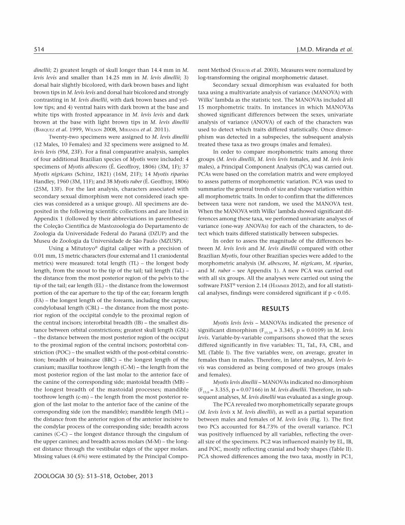
The PCA revealed two morphometrically separate groups(M. levis levis x M. levis dinellii), as well as a partial separationbetween males and females of M. levis levis (Fig. 1). The firsttwo PCs accounted for 84.73% of the overall variance. PC1was positively influenced by all variables, reflecting the over-all size of the specimens. PC2 was influenced mainly by EL, IB,and POC, mostly reflecting cranial and body shapes (Table II).PCA showed differences among the two taxa, mostly in PC1,
515The taxonomic status of Myotis levis levis and Myotis levis dinellii
ZOOLOGIA 30 (5): 513–518, October, 2013
i.e., size differences. The MANOVA tests confirmed the resultsof the PCA, indicating that both taxa were distinct accordingto their variables, and the sexes were distinct in M. levis levis(MANOVA, F30,76 = 12.63, p < 0.0001). All of the averages ofthese measures were significantly greater in M. levis levis thanin M. levis dinellii (Table I).
Table I. One-way ANOVAs of 15 log-transformed morphometrictraits of the Myotis levis levis and M. levis dinellii. Numbers in boldindicate significant differences.
Variable
M. levis levis males vs.M. levis levis females
M. levis levis vs.M. levis dinellii
F p F p
Total length 24.8900 <0.00010 90.08 <0.0001
Tail length 6.08200 0.019590 156.1 <0.0001
Ear length 1.50700 0.229200 96.29 <0.0001
Forearm length 4.48000 0.042700 252.3 <0.0001
Condylobasal length 6.89500 0.013480 195.9 <0.0001
Interorbital breadth 0.02960 0.864500 64.28 <0.0001
Greatest skull length 0.76240 0.389500 234.4 <0.0001
Postorobital constriction 2.74200 0.108200 14.93 0.00030
Breadth of braincase 0.04386 0.835500 54.35 <0.0001
Maxillar toothrow length 2.20500 0.148000 315.8 <0.0001
Mastoidal breadth 1.24300 0.273700 161.8 <0.0001
Mandibular toothrow length 0.88780 0.353600 430.5 <0.0001
Mandibular length 9.49900 0.004379 179.3 <0.0001
Breadth across canines 1.53400 0.225100 122.1 <0.0001
Breadth across molars 3.35700 0.076850 240.0 <0.0001
Figure 1. PCA scatter plot representing the projection of the threegroups ( )Myotis levis levis males, ( ) M. levis levis females and ( )Myotis levis dinellii, in the multivariate space formed by PC1 and PC2.
Table II. Loadings of 15 log-transformed morphometric traits andprincipal components (PC1 and PC2) for Myotis levis levis (malesand females) and Myotis levis dinellii.
Variable PC1 PC2
Total length 0.89850 0.023660
Tail length 0.09753 0.097530
Ear length 0.71840 -0.209500
Forearm length 0.94420 -0.041540
Condylobasal length 0.96140 -0.111600
Interorbital breadth 0.82800 0.261000
Greatest skull length 0.96270 -0.038250
Postorobital constriction 0.53250 0.814700
Breadth of braincase 0.78610 0.171800
Maxillar toothrow length 0.94330 -0.122900
Mastoidal breadth 0.93960 -0.004275
Mandibular toothrow length 0.96220 -0.140800
Mandibular length 0.95820 -0.137700
Breadth across canines 0.89520 -0.118300
Breadth across molars 0.93590 -0.074520
Percentage of variance (%) 78.7100 6.020000
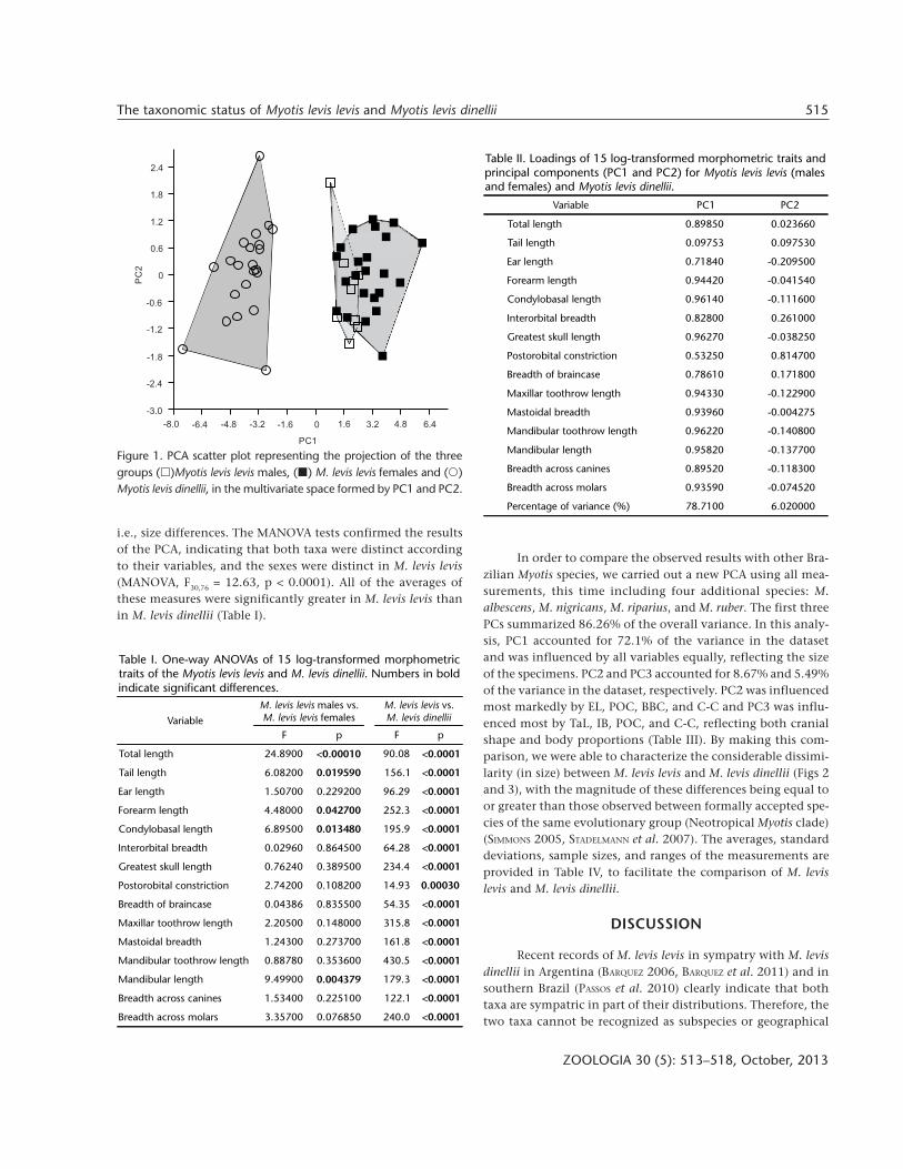
In order to compare the observed results with other Bra-zilian Myotis species, we carried out a new PCA using all mea-surements, this time including four additional species: M.albescens, M. nigricans, M. riparius, and M. ruber. The first threePCs summarized 86.26% of the overall variance. In this analy-sis, PC1 accounted for 72.1% of the variance in the datasetand was influenced by all variables equally, reflecting the sizeof the specimens. PC2 and PC3 accounted for 8.67% and 5.49%of the variance in the dataset, respectively. PC2 was influencedmost markedly by EL, POC, BBC, and C-C and PC3 was influ-enced most by TaL, IB, POC, and C-C, reflecting both cranialshape and body proportions (Table III). By making this com-parison, we were able to characterize the considerable dissimi-larity (in size) between M. levis levis and M. levis dinellii (Figs 2and 3), with the magnitude of these differences being equal toor greater than those observed between formally accepted spe-cies of the same evolutionary group (Neotropical Myotis clade)(SIMMONS 2005, STADELMANN et al. 2007). The averages, standarddeviations, sample sizes, and ranges of the measurements areprovided in Table IV, to facilitate the comparison of M. levislevis and M. levis dinellii.
DISCUSSION
Recent records of M. levis levis in sympatry with M. levisdinellii in Argentina (BARQUEZ 2006, BARQUEZ et al. 2011) and insouthern Brazil (PASSOS et al. 2010) clearly indicate that bothtaxa are sympatric in part of their distributions. Therefore, thetwo taxa cannot be recognized as subspecies or geographical

516 J.M.D. Miranda et al.
ZOOLOGIA 30 (5): 513–518, October, 2013
Figures 2-3. PCA scatter plot representing the projection of the six examined taxa ( ) Myotis levis levis, ( ) Myotis levis dinellii, ( )Myotis ruber, ( ) Myotis nigricans, ( ) Myotis riparius, and ( ) Myotis albescens, in the multivariate space formed by PC1 and PC2 (2)and PC1 and PC3 (3).
races of the same species (MAYR 1969, 1970, 1998), as tradition-ally accepted (MILLER & ALLEN 1928, LAVAL 1973, BARQUEZ et al.1999, BIANCONI & PEDRO 2007, WILSON 2008, STEVENS et al. 2010,PERACCHI et al. 2012).
The differences between M. levis levis and M. levis dinelliifound in the present study revealed considerable divergence,
2 3
Table III. Loadings of 15 log-transformed morphometric traits andprincipal components (PC1, PC2, and PC3) for Myotis levis levisand Myotis levis dinellii, Myotis albescens, Myotis ruber, Myotisriparius, and Myotis nigricans samples.
Variable PC1 PC2 PC3
Total length 0.8210 0.28530 -0.24710
Tail length 0.8083 -0.07001 -0.37190
Ear length 0.7158 0.33690 -0.25460
Forearm length 0.9484 -0.03826 -0.09477
Condylobasal length 0.9675 0.09750 -0.03321
Interorbital breadth 0.7045 0.05386 0.42230
Greatest skull length 0.9576 -0.03178 -0.09712
Postorobital constriction 0.6102 0.65080 0.36690
Breadth of braincase 0.7514 0.53620 -0.04146
Maxillar toothrow length 0.9246 -0.23680 -0.08447
Mastoidal breadth 0.8875 0.10090 0.15000
Mandibular toothrow length 0.9391 -0.24060 -0.08357
Mandibular length 0.9414 0.24090 0.01408
Breadth across canines 0.7700 -0.33320 0.42070
Breadth across molars 0.8869 -0.28010 0.10910
Percentage of variance (%) 72.100 8.67000 5.49000
Table IV. External and cranial measurements of Myotis levis levis andMyotis levis dinellii. Averages ± one standard deviation, followed bythe sample size in parenthesis and the range within each popula-tion and species.
Variable M. levis levis M. levis dinellii
Total length 92.94 ± 4.80 (n = 29)84.3-0102.20
82.74 ± 2.71 (n = 21)74.46-86.92
Tail length 45.16 ± 3.08 (n = 30)40.05-52.00
36.53 ± 2.13 (n = 21)29.48-40.28
Ear length 16.35 ± 1.55 (n = 30)12.45-20.45
13.71 ± 0.62 (n = 21)12.28-14.90
Forearm length 40.82 ± 1.43 (n = 34)38.15-43.40
35.87 ± 0.79 (n = 22)34.25-37.25
Condylobasal length 14.54 ± 0.42 (n = 33)13.85-15.40
13.05 ± 0.31 (n = 20)12.30-13.55
Interorbital breadth 4.91 ± 0.17 (n = 32)4.65-5.25
4.46 ± 0.18 (n = 22)4.15-4.75
Greatest skull length 15.39 ± 0.44 (n = 31)14.40-16.75
13.82 ± 0.30 (n = 21)13.00-14.25
Postorobital constriction 3.91 ± 0.11 (n = 34)3.70-4.10
3.81 ± 0.10 (n = 22)3.61-4.06
Breadth of braincase 7.38 ± 0.19 (n = 32)7.00-7.75
7.02 ± 0.16 (n = 21)6.70-7.36
Maxillar toothrow length 5.98 ± 0.20 (n = 34)5.65-6.80
5.18 ± 0.14 (n = 22)4.90-5.65
Mastoidal breadth 7.76 ± 0.15 (n = 32)7.45-8.05
7.27 ± 0.13 (n = 22)6.98-7.47
Mandibular toothrowlength
6.24 ± 0.17 (n = 33)5.90-6.60
5.40 ± 0.15 (n = 22)5.13-5.90
Mandibular length 11.22 ± 0.47 (n = 33)9.80-12.15
9.84 ± 0.25 (n = 22)9.23-10.30
Breadth across canines 3.82 ± 0.14 (n = 31)3.40-4.05
3.49 ± 0.09 (n = 22)3.29-3.64
Breadth across molars 6.04 ± 0.12 (n = 33)5.75-6.25
5.39 ± 0.19 (n = 22)5.05-5.90

517The taxonomic status of Myotis levis levis and Myotis levis dinellii
ZOOLOGIA 30 (5): 513–518, October, 2013
mostly in size. These differences indicate that distinct evolu-tionary pressures have affected their evolution. Size may be aline of least evolutionary resistance, driving the evolutionarydirection (MARROIG & CHEVERUD 2005). In fact, size tends to ac-count for most of the interspecific variation within the genus(see the main steps in the key of identifications of species, e.g.,BARQUEZ et al. 1999, GARDNER 2008). Variation in body size amongbat species changes the echolocation frequencies and breadthof feeding niches (SIMMONS & CONWAY 2003). The PCAs indi-cated that the main difference was in size (PC1), whereas theMANOVAs clearly separated both taxa. LAVAL (1973) had al-ready pointed out the size difference between the two taxa, yetunderscored the great morphological similarity between them.Our approach, based on multivariate morphometrics, indicatedthat M. levis levis and M. levis dinellii are distinct. This findingled us to the following more conceptual questions: 1) What isthe magnitude of the differences between M. levis levis and M.levis dinellii? 2) How many of these differences reflect taxo-nomic differences when compared with other species of theevolutionary group of Neotropical Myotis? 3) To what taxo-nomic hierarchical level should these two taxa be assigned?
The last PCA of M. levis levis, M. levis dinellii, M. albescens,M. nigricans, M. riparius, and M. ruber showed that the mor-phometric differences between M. levis levis and M. levis dinelliiwere as great as or greater than those observed among the otherspecies analyzed. Myotis levis levis was more similar in size toM. ruber than to M. levis dinellii, and the latter, in turn, wasmore similar in size to M. albescens and M. nigricans than to M.levis levis. Myotis levis levis was clearly distinct from M. ruber inthe color of the fur. Myotis levis dinellii was clearly distinct fromM. nigricans in fur color and in the presence of a fringe of hairin the border of the uropatagium, and from M. albescens in thecolor of the fur and a had a smaller POC (BARQUEZ et al. 1999,WILSON 2008, MIRANDA et al. 2011).
Further, secondary sexual dimorphism was observed inspecimens of M. levis levis, but not in M. levis dinellii. Femaleswere larger than males in M. levis levis. These findings are simi-lar to the sexual dimorphism observed in M. nigricans (MORATELLI
et al. 2011) and in other vespertilionid bats (MYERS 1978), pos-sibly a response to selective pressures relating to gestation,maternal care, and flight.
Therefore, our combined evidence supports the existenceof two distinct taxa, given that they are: 1) sympatric; 2) mor-phologically distinct (in fur coloration); 3) morphometricallydifferent; and 4) morphometrically different from one anotherto an extent that is similar to or greater than the differencesamong other species in their evolutionary group. Therefore,we agree with BARQUEZ (2006), PASSOS et al. (2010), BARQUEZ et al.(2011), and PAGLIA et al. (2012) and consider M. levis and M.dinellii to be separate species.
Despite new molecular data, uncertainties regardingMyotis evolutionary relationships still exist (RUEDI & MAYER 2001,STADELMANN et al. 2007). Increasing field efforts and also other
types of analysis such as tooth morphology and molecular data,which we consider better tools than the purely morphometricanalysis in the present study, will allow a more comprehensiveunderstanding of the evolutionary relationships relating to thediversification of this cosmopolitan genus.
ACKNOWLEDGMENTS
We thank CNPq for the productivity scholarship grantedto FCP (303757/2012-4), and REUNI-UFPR and CAPES for thescholarships of IPB and JS. We thank Mário de Vivo for access tospecimens from MZUSP. We also thank Marcio R. Pie, James J.Roper, and Diego Astúa de Moraes who provided useful commentsand suggestions on the manuscript and the English version of it.
LITERATURE CITED
ANDERSON, S. 1997. Mammals of Bolivia, taxonomy and distribution.Bulletin of the American Museum of Natural History 231:1-652.
BARQUEZ, R.M. 2006. Orden Chiroptera, p. 56-86. In: R.M.BARQUEZ; M. DÍAZ & R.A. OJEDA (Eds). Mamíferos de Argen-tina, Sistemática y Distribución. Tucumán, SAREM, 359p.
BARQUEZ, R.M.; M.A. MARES & J.K. BRAUN. 1999. The Bats of Ar-gentina. Special Publications. Museum of Texas TechUniversity 42: 1-275.
BARQUEZ, R.M.; M.S. SÁNCHEZ & M.L. SANDOVAL. 2011. Nuevos re-gistros de murciélagos (Chiroptera) en el norte de Argenti-na. Mastozoologia Neotropical 18 (1): 11-24.
BIANCONI, G.V. & W.A. PEDRO. 2007. Família Vespertilionidae, p.167-195. In: N.R. REIS; A.L. PERACCHI; W.A. PEDRO & I.P. LIMA
(Eds). Morcegos do Brasil. Londrina, Nélio R. dos Reis, 253p.GARDNER, A.L. 2008. Mammals of South America: volume 1:
marsupials, xenarthrans, shrews and bats. Chicago, TheUniversity of Chicago Press, 669p.
HAMMER, Ø. 2012. PAST. Paleontological statistic. Version 2.14.Reference manual. Oslo, Natural History Museum,University of Oslo, 225p.
LAVAL, R. 1973. A revision of the Neotropical bats of the genusMyotis. Natural History Museum Los Angeles County 15: 1-54.
LÓPEZ-GONZÁLEZ, C.; S.J. PRESLEY; R.D. OWEN & M.R. WILLIG. 2001. Thetaxonomic status of Myotis (Chiroptera: Vespertilionidae) inParaguay. Journal of Mammalogy 82: 138-160.
MARROIG, G. & J.M. CHEVERUD. 2005. Size as a line of leastevolutionary resistance: diet and adaptative morphologicalradiation in New World Monkeys. Evolution 59: 1128-1142.
MAYR, E. 1969. Principles of systematic zoology. New York,McGraw-Hill, 434p.
MAYR, E. 1970. Populations, species and evolution. Cambridge,Harvard University Press, 453p.
MAYR, E. 1998. O desenvolvimento do pensamento biológi-co: diversidade, evolução e herança. Brasília, Editora Uni-versidade de Brasília, 1107p.

518 J.M.D. Miranda et al.
ZOOLOGIA 30 (5): 513–518, October, 2013
MILLER, G.S. & G.M. ALLEN. 1928. The American bats of thegenera Myotis and Pizonyx. United States National MuseumBulletin 144: 1-218.
MIRANDA, J.M.D.; I.P. BERNARDI & F.C. PASSOS. 2011. Chave ilus-trada para a determinação dos morcegos da Região Suldo Brasil. Curitiba, João M.D. Miranda, 51p.
MORATELLI, R. & D.E. WILSON. 2011. A new species of Myotis Kaup,1829 (Chiroptera, Vespertilionidae) from Ecuador.Mammalian Biology 76: 608-614.
MORATELLI, R.; A.L. PERACCHI; D. DIAS & J.A. OLIVEIRA. 2011. Geographicvariation in South American populations of Myotis nigricans(Schinz, 1821) (Chiroptera, Vespertilionidae), with the descriptionof two new species. Mammalian Biology 76: 592-607.
MYERS, P. 1978. Sexual dimorphism in size of vespertilionid bats.American Naturalist 112 (986): 701-711.
PAGLIA, A.P.; G.A.B. FONSECA; A.B. RYLANDS; G. HERRMANN; L.M.S.AGUIAR; A.G. CHIARELLO; Y.L.R. LEITE; L.P. COSTA; S. SICILIANO; M.C.KIERULFF; S.L. MENDES; V.C. TAVARES; R.A. MITTERMEIER & J.L. PATTON.2012. Annotated checklist of Brazilian Mammals, 2nd Edition.Occasional Papers in Conservation Biology 6: 1-82.
PASSOS, F.C.; J.M.D. MIRANDA; I.P. BERNARDI; N.Y. KAKU-OLIVEIRA &L.C. MUNSTER. 2010. Morcegos da Região Sul do Brasil: aná-lise comparativa da riqueza de espécies, novos registros eatualizações nomenclaturais (Mammalia, Chiroptera).Iheringia 100 (1): 25-34.
PERACCHI, A.L.; I.P. LIMA; N.R. REIS; M.R. NOGUEIRA & H. ORTÊNCIO-FILHO. 2012. Ordem Chiroptera, p. 155-234. In: N.R. REIS;A.L. PERACCHI; W.A. PEDRO & I.P. LIMA (Eds). Mamíferos do
Brasil. Londrina, Nélio Roberto dos Reis, 2nd ed., 439p.RUEDI, M. & F. MAYER. 2001. Molecular systematics of bats of
the genus Myotis (Vespertilionidae) suggests deterministicecomorphological convergences. Molecular Phylogeneticsand Evolution 21: 436-448.
SIMMONS, N.B. 2005. Order Chiroptera, p. 312-529. In: D.E. Wil-son & D.M. Reeder (Eds). Mammal species of the world: ataxonomic and geographic reference. Baltimore, JohnsHopkins University Press, vol. 1, 2142p.
SIMMONS, N.B. & T.M. CONWAY. 2003. Evolution of ecologicaldiversity in bats, p. 493-535. In: T.H. KUNZ & M.B. FENTON
(Eds). Bat ecology. Chicago, University of Chicago Press,798p.
STADELMANN, B.; L.K. LIN; T.H. KUNZ & M. RUEDI. 2007. Molecularphylogeny of New World Myotis (Chiroptera, Vespertilionidae)inferred from mitochondrial and nuclear DNA genes.Molecular Phylogenetics and Evolution 43: 32-48.
STEVENS, R.D.; C. LÓPEZ-GONZÁLEZ; E.S. MCCULLOCH; F. NETTO & M.L.ORTIZ. 2010. Myotis levis (Geoffroy Saint-Hilaire) indeed occursin Paraguay. Mastozoologia Neotropical 17 (1): 195-200.
STRAUSS, R.E.; M.N. ATANASSOV & J.A. OLIVEIRA. 2003. Evaluationof the Principal-Component and Expectation-Maximizationmethods for estimating missing data in morphometricstudies. Journal of Vertebrate Paleontology 23: 284-296.
WILSON, D.E. 2008. Genus Myotis Kaup 1829, p. 468-481. In: A.L.GARDNER (Ed.). Mammals of South America: marsupials, xenar-thrans, shrews and bats. Chicago, The University of ChicagoPress, vol. 1, 669p.
Appendix 1. Specimens examined, museums and acronyms: mammalogy collections of the Universidade Federal do Paraná (DZUP), ZoologyMuseum of the Universidade de São Paulo (MZUSP), and Mammal Collection at Universidade Federal Rural do Rio de Janeiro (ALP).
Myotis albescens: BRAZIL, Rio Grande do Sul: Frederico Westphalen Municipality (DZUP 574, 575, 576); Paraná: Porto Rico, MutumIsland (DZUP 226).
Myotis levis dinellii: BRAZIL, Rio Grande do Sul: Derrubadas, Parque Estadual do Turvo (8) (DZUP 701, 703, 704, 705, 706, 707, 708,709, 710, 711); Santa Catarina: Ponte Alta do Norte (7) (DZUP 867-877); ARGENTINA: Tucumán: Las Vasquez Municipality (9)(MZUSP 2055).
Myotis levis levis: BRAZIL, Rio de Janeiro: Nova Friburgo (1) (MZUSP 2799); São Paulo: Cananéia, Parque Estadual Ilha do Cardoso (2)(MZUSP 27680, 27976); Paraná: Curitiba, Parque Municipal da Barreirinha (3) (DZUP 333, 334, 335, 336); Palmas (4) (DZUP216, 217, 218, 369, 371, 372, 373, 376, 377, 380, 381, 382, 384, 385, 386, 387, 388, 389, 391, 392, 393, 394, 395, 396); RioGrande do Sul: Cacequi (5) (MZUSP 3167).
Myotis nigricans: BRAZIL, Paraná: Cerro Azul, State Park of Campinhos (DZUP 056, 057,058, 059, 061, 062, 063, 064, 065, 066, 086,087, 088, 090, 092, 093, 096, 097, 099, 105, 106, 107, 108, 109, 110, 111, 112); Matinhos, State Park of Rio da Onça (DZUP141, 144); São José dos Pinhais (DZUP 235, 236); Balsa Nova, São Luiz do Purunã locality (DZUP 410, 419, 420, 421, 422).
Myotis riparius: BRAZIL, São Paulo: Juquitiba (MZUSP 32966); Piedade (MZUSP 32963, 32964); Paraná: Maringá (DZUP 494, 495,499, 500); Rio Grande do Sul: Frederico Westphalen (DZUP 577, 578).
Myotis ruber: BRAZIL, São Paulo: Buri (MZUSP 32968, 32971, 32972, 32974, 32975); São Paulo, State Park of Serra da Cantareira(MZUSP 31466, 31470, 31471, 31472, 31473); Paraná: Balsa Nova, Bugre (DZUP 191, 192, 193, 194, 206, 337); São Luiz doPurunã (DZUP 231, 232, 474, 475, 476, 477); Palmas (DZUP 213, 214); Santa Catarina: Passos Maia (DZUP 397, 478, 498);Urussanga (DZUP 580); Rio Grande do Sul: Frederico Westphalen (DZUP 582).
Submitted: 10.XII.2012; Accepted: 25.III.2013.Editorial responsibility: Diego Astúa de Moraes