the ultrastructure of the accessory sex organs of the male rat
TRANSCRIPT

Cell Tiss. Res. 171,285-296 (1976) Cell and Tissue Research ~ by Springer-Verlag 1976
The Ultrastructure of the Accessory Sex Organs of the Male Rat
XI. Nuclear Alterations of Prostatic Epithelial Cells Induced by Castration
Erik Dahl University of Oslo, Departments of Anatomy, Dental Faculty, Blindern. Oslo 3, Norway
Summary. The fine structure of the nuclei of epithelial cells of the dorsal lobe of the rat prostate were studied 2, 3, 5, 7 and 21 days after castration. The nucleolus appears to undergo a progressive disorganisation with partial fragmentation and dispersion of its normal components,
Changes in the nucleoplasm were primarily reflected by a condensation of chromatin, particularly along the nuclear membrane and adjacent to the nucleolus. Later, different types of intranuclear inclusions were observed.
After 21 days, the nuclei were characterized by an irregular outline with large indentation. Within the nucleoplasm aggregates of coarse granular chromatin were found. No cell necrosis was observed, indicating that andro- gen deprivation results in a remodeling of the cell to a less active state with marked cellular alterations and cessation of secretion, but apparently with some of their basic functions still intact.
Injections of testosterone completely reverse the castrated-induced alter- ations.
The changes observed are assumed to be due to the withdrawal of the androgenic stimulus, with a direct influence on the secretory function of the cell. The findings support the view that the stimulating secretory effect of androgen is mediated via an intrant, clear androgen receptor, probably located in the nucleolus-associated-chromatin. It is also proposed that the secretory function of the epithelial cells of the prostatic complex, initiated by androgens, may be regulated by an intranuclear secretory center.
Key words: Prostate - Rat - Castration - Nuclear alterations - Electron microscopy.
Introduction
Ultrastructural studies of the effect of castration on the epithelial cells of the rat prostate have previously been concentrated upon cytoplasmic alterations
Send q[]))rint requests to: Erik Dahl, University of Oslo, Department of Anatomy, Dental Faculty, P.O.B. 1052, Blindern, Oslo 3, Norway
286 E. Dahl
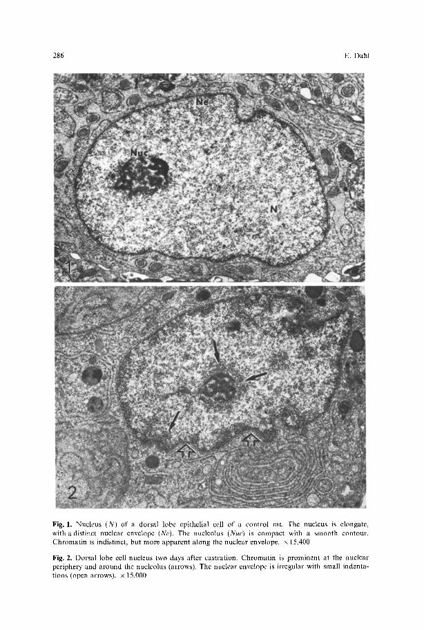
Fig. 1. Nucleus (N) of a dorsal lobe epithelial cell of a control rat. The nucleus is elongate, with a distinct nuclear envelope (Ne). The nucleolus (Nuc) is compact with a smooth contour. Chromat in is indistinct, but more apparent along the nuclear envelope, x 15,400
Fig. 2. Dorsal lobe cell nucleus two days after castration. Chromat in is prominent at the nuclear periphery and around the nucleolus (arrows). The nuclear envelope is irregular with small indenta- tions (open arrows), x 15,000

The Ultrastructure of the Accessory Sex Organs 287
(Brandes, Gy6rkey and Groth, 1962; Helminen and Ericsson, 1971; Dahl and Kjaerheim, 1973 ; Dahl and Tveter, 1973).
In the present investigation, further fine structural studies of the metabolic effect of castration on the rat prostate were carried out, and have revealed that in the dorsal lobe, the nucleus, as well as the cytoplasm of the epithelial cell, undergoes marked changes. The description of the nuclear alterations fol- lowing castration, and the apparent return to normal after administration of testosterone is the subject of this paper.
Material and Methods
Twenty-four albino male rats (derived from the Charles River CDF strain) 4 6 months old were used in this study. Eighteen rats were castrated through a scrotal incision 2, 3, 5, 7 and 21 days prior to examination. The remaining six were normal animals, used as controls. Each group consisted of 2-6 animals. During the last seven days of the experimental period, three of the 21 day group of castrates received injections of 15 mg testosterone every second day, i.e. a total of 45 mg testosterone. The animals were sacrificed two days after the last injection. Under ether anesthesia, the aorta was cannulated, and intraaortic perfusion of fixative was carried out as described by Dahl, Kjaerheim and Tveter (1973). The dissection of the prostate, the preparation of the samples and the electron microscopic techniques followed those of Dahl et al. (1973).
Observations
Controls: The nucleus of the epithelial cell of the dorsal lobe is roughly elongated, and shows a distinct, rather even nuclear envelope (Fig. 1). The nucleolus is compact with a smooth contour. Chromatin is indistinct, is evenly dispersed throughout the nucleus, and shows no focal aggregations. Usually chromatin is only faintly visible along the nuclear envelope.
Changes Following Castration. Within two days, definite changes were observed in the chromatin pattern, with distinct margination around the nuclear periphery and in the area adjacent to the nucleolus (Fig. 2). Some irregularity of the nuclear membrane was also seen. After three days, this was more prominent, and indentations were now consistently found (Fig. 3). Within the nucleoplasm, areas of fibrillar material were observed (Fig. 3), as were different types of inclusion (Figs. 4, 5). It was noteworthy, that the areas of fibrillar material
Fig. 3. Dorsal lobe cell nucleus (N) three days after castration. The nucleolus (Nuc) is slightly disorganized with partial confluence of perinucleolar and nucleolar chromatin. Small areas of fibrillar material (FM) are seen centrally. The indentations are now more frequent and deeper (arrows). x 24,000
Fig. 4. Intranuclear inclusion of dorsal lobe nucleus containing a rather coarse fibrillar material. Note the indentation (ld) and the membrane-l ike appearance of fuzzy material (arrows) partly sur- rounding the inclusion, x 30,000
Fig. 5. Complex intranuclear inclusions adjacent to the nuclear envelope. Part of the nucleoplasm in the process of being incorporated into the inclusion is arrowed, x 17,600

288 E. Dahl

The Ultrastructure of the Accessory Sex Organs 289
Fig. 6. Dorsal lobe cell nuclei (N) seven days after castration. To the left a common finding is demonstrated: fibrillar material (FM) located between an indentation (Id) and the nucleolus (Nut). In the right nucleus a pale area containing fibrillar-like material is arrowed. • 15,300
Figs. 7 and 8. Serial sections of the fibrillar area seen in Fig. 6, demonstrating the close relationship between the indentation (Id) and the fibrillar materials (arrows) adjacent to the nucleolus (Nuc). • 30,000

Fig. 9. Dorsal lobe cell nucleus seven days after castration with several typical pseudo-inclusions (PI) containing cytoplasmic material (indentations in transverse section) surrounded by heterochro- matin. Alterations presumed to originate de novo within the nucleus are arrowed. x 30,000 Fig. 10. Dorsal lobe cell nucleus with two nucleoli (NW), seven days after castration. Note that the lower nucleolus is partly fragmented and surrounded by a halo of fuzzy material (Figs. 4, 11 and 13) or a pseudo-membrane (arrows). x 24,000
Fig. 11. Nuclear body-like inclusion adjacent to the nucleolus with a granular content similar to the nucleolus, surrounded by fibrillar material (arrows). Note the irregularity of the nucleolus (NW) compared with the normal in Figure 1. x 120,000

Fig. 12. Dorsal lobe nucleus (N) twenty-one days after castration with deep indentations (Id) and prominent margination of chromatin (arrows). Note that the nucleoplasm appears pale and " e m p t y " except for the amount of chromatin, x 30,000
Fig. 13. Nuclear body-like inclusion similar to that in Figure 11. Note the differences in the fibrillar material surrounding the granular component which now is less distinct, x 60,000
Fig. 14. Part of a dorsal lobe cell nucleus twenty-one days after castration. A large intranuclear inclusion containing fibrillar material, is surrounded by heterochromatin, x 22,500

292 E. Dahl
Fig. 15. Dorsal lobe cell nuclei (N) after administration of testosterone. The micrograph demon- strates a cell presumed to be in late telophase (binucleate) with two almost mature nuclei. The chromatin is still more prominent than in the normal (Fig. 1). Nuc nucleolus, x 12,000
were very often localized between the nucleolus and the inden ta t ions (Fig. 6). Serial sect ions (Figs. 7, 8) revealed tha t the indenta t ions , when they enc roached deeper into the nucleus, came into close re la t ion with the areas o f f ibr i l lar mater ia l . Pale areas, which were relat ively sharp ly demarca t ed f rom the sur- round ing chromat in , were now regular ly encounte red within the nuc leop lasm (Fig. 6). In some sections, the inden ta t ions were cut t ransversal ly , and a p p e a r e d as pseudoinc lus ions (Fig. 9), su r rounded by he t e roch roma t in and in t ranuc lea r

The Ultrastructure of the Accessory Sex Organs 293
alterations similar to those described above. Occasionally nuclei with two distinct nucleoli were found. In one specimen (Fig. 10) one of the nucleoli was partly fragmented and surrounded by a halo of fuzzy, fibrillar material. In addition, nuclear body-like inclusions surrounded by the same fibrillar material were commonly seen (Fig. 11). These nuclear body-like inclusions were always found adjacent to the nucleolus.
After twenty-one days, the nucleus was very irregular, with deep indentations. The amount of heterochromatin was greatly increased, whereas the nucleoplasm appeared paler and more empty than normal (Fig. 12). Different types of alter- ation were still encountered, but now more infrequently. The fuzzy fibrillar material which surrounded the granular core of the nuclear body-like inclusion were broader and less distinct (Fig. 13) than was the case after one week, and the content of the large inclusions was more homogeneous and rather pale (Fig. 14). Fibrillar areas (as seen in Figs. 3, 6 8) were not observed in this group.
After administration of testosterone, the morphology of the nucleus was similar to that of the normal animals (Fig. 15).
Discussion
Autoradiographic studies have demonstrated that all parts of the rat prostate possess the ability to concentrate and retain androgen against a marked concentra- tion gradient with blood (Tveter, 1969a). Moseback et al. (1967) have suggested that the preferential accumulation of a hormone in a target organ might either be due to rapid metabolism of the hormone, or to the presence of specific hormone receptors in the target organ. It is now believed that the selective concentration of hormone in the prostate is due to an association of androgen (5-dihydrotestosterone) with two specific receptor or hormonal binding sites in the cells (Unhjem, 1970; Unhjem and Tveter, 1969; Fang, Anderson and Liao, 1969). One receptor, associated with a macromolecule, is localized in the cytoplasm, the other can be extracted from the prostatic nuclei. It has been suggested that the interaction of androgen with the prostate tissue involves a two stages mechanism. The hormone associates primarily with the extranuclear receptor to form a complex, by which androgen is transported to the nucleus. The earliest detectable biochemical events observed after stimulation with andro- gens are related to ribonucleic acid biosynthesis (Liao and Stumpf, 1968). The hormone may also influence the nuclear control to cytoplasmic protein synthesis, since it appears to bind to the nucleus in vivo (Stumpf, 1969; Tveter, 1969b; Tveter and Attramadal, 1969; Sar, Liao and Stumpf, 1970).
Castration results in a rapid decrease in the amount of androgen within the whole prostatic complex associated with reduced protein synthesis. This is reflected by a number of morphological changes, including a reduction in the size of the cells, and quantitative and qualitative cytoplasmic alterations involving a reduction in the amount of rough endoplasmic reticulum and the Golgi apparatus. This in turn is followed by a reduction in the amount of secretory material (Dahl and Kjaerheim, 1973; Dahl and Tveter, 1973).

294 E. Dahl
In the present study it has been demonstrated that after castration, marked alterations occur in the nuclei of the epithelial cells of the dorsal lobe. The nucleolus undergoes a progressive disorganization with some fragmentation and dispersion of its normal components. Changes in the nuclear envelope, the nucleoplasm and the chromatin pattern also occur. These alterations are revers- ible when the basic androgen deficiency is counteracted by administration of testosterone. From the biochemical and autoradiographic data available (Tveter, 1969a) the structural alterations appear to start concomitantly with the reduced concentration of the hormone in the target organ.
The nuclear changes appear to be of different origins. Obviously some of them represent portions of the cytoplasm which have been incorporated into the nucleus as pseudoinclusions, whereas others are formed de novo within the nucleus (Dahl and Kjaerheim, 1974). It should be emphazised, however, that the various nuclear alterations, including the large indentations of the nuclear envelope (pseudoinclusions), the changes in the nucleoplasm (fibrillar areas) and the alterations of the nucleolus, were very often encountered within the same section. This may indicate that the nuclear alterations occur in the same compartment of the cell, and represent a dysfunction of integrated bio- chemical events within this compartment. The findings also suggest that the effect of androgen may involve some form of interaction between the nuclear envelope, the nucleoplasm and the chromatin within specific, restricted areas of the cell.
Barton and Liao (1967) found that the nucleolus is functionally connected with special regions of the chromatin adjacent to the nucleolus, called nucleolus- associated chromatin. They suggested that enhancement of RNA synthesis after the administration of estrogen or testosterone to the appropriate target organs must preferentially involve certain RNA molecules. They concluded that this RNA fraction is nuclear RNA, probably a precursor of ribosomal RNA. When their results are correlated with the nuclear alterations observed in our investiga- tion, it seems likely that the morphological changes represent the direct effect of the deprivation of androgens from specific regions of the cell, namely the site containing the androgen receptors. This means, that even though there are both cytoplasmic and nuclear receptors, neither are necessarily evenly dispersed throughout the cytoplasm or the nucleoplasm, but may be confined to specific areas, at least within the nucleus.
Although marked degenerative changes of the cells were regularly found three weeks after castration, there was no cell necrosis. The intranuclear alter- ations were also less prominent at this time, with the exception of the increased amount of heterochromatin. This leads one to speculate that, while there may be specific areas within the nucleus which regulate the secretory function initiated by androgen, the rest of the cell is dependent on more general stimulating agents for its basic functions. In our study, this results in a remodeling of the cell appropriate to a lower degree of activity, with marked cytoplasmic alterations and cessation of secretion, but no cell necrosis (Dahl and Kjaerheim, 1973; Dahl et al., 1973). However, our investigation can not, with certainty, exclude the possibility that small amounts of androgen from other sources may be of some importance in this cellular remodeling.

The Ultrastructure of the Accessory Sex Organs 295
As to the progressive increase in the amount of heterochromatin over the experimental period, previous investigations have documented that margination of chromatin is one of the earliest changes occurring in cellular degenerative processes (Trump, Goldblatt and Stowell, 1965). Furthermore, there is evidence (e.g. Novikoff and Holtzman, 1970) to support the tentative conclusion that chromatin in the diffuse euchromatic state such as is seen in our controls, is active, but ceases to be so when converted into the condensed heterochromatic state, as seen in our non-secreting experimental animals.
In conclusion, the intranuclear morphological alterations found in the present study are obviously induced by the androgen deprivation following castration. The findings support the view that the stimulating secretory effect of androgen on the prostate is mediated via an intranuclear androgen receptor, most probably located in the nucleolus associated chromatin. A cytoplasmic receptor, necessary to carry the androgen to the nucleus, would also be expected. In addition, there seems to be some evidence that the intranuclear receptors are confined to specific regions within the nucleolus associated chromatin. Because of this, the initial steps of the secretory process are probably restricted to the specific compartments of the nucleus where the receptors are located. This leads to the proposal that the secretory function of the epithelial cells of the prostatic complex may be regulated by an intranuclear "secretory center".
The reversibility of the cellular remodeling to a less active state lends support to this proposal. This indicates that only specific functions, viz. those of "the secretory center" will be affected in androgen deprivation. When the basic androgen deficiency is counteracted by administration of testosterone, a complete restoration of the nucleus occurs, including the nuclear envelope, the nucleoplasm, the nucleolus and the chromatin pattern.
Finally it should also be emphasized that the nuclear changes observed are confined to the epithelial cells of the dorsal lobe. Nuclear alterations were not observed consistently in the other lobes. Autoradiographic studies (Tveter, 1969a; Tveter and Attramadal, 1969) have demonstrated that about 10% of the total radioactivity in the dorsal lobe is represented by conjugated compounds, unlike the ventral and lateral prostate and the seminal vesicles in which the radioactivity represents nonconjugated compounds. When the nuclear alter- ations observed in this study are correlated with the autoradiographic findings, it seems likely that although the entire prostatic complex is androgen-dependent, there are differences in the effect of testosterone on the different lobes. In the dorsal lobe, this may be due to differences in the metabolic transformation of androgens compared with the other lobes of the prostate.
References
Barton, R.W., Liao, S.: A similarity in the effect of estrogen and androgen on the synthesis of ribonucleic acid in the cell nuclei of gonadohormone-sensi t ive tissues. Endocrinology 81, 4 0 9 4 1 2 (1967)
Brandes, D., Gydrkey, F., Groth, D.: Fine structural and histochemical study of the effect of castration on the rat prostatic complex. Lab. Invest. 11, 339-349 (1962)
Dahl, E., Kjaerheim, A.: The ultrastructure of the accessory sex organs of the male rat. II. The postcastration of the ventral, lateral and the dorsal prostate. Z. Zellforsch. 144, 167 178 (1973)

296 E. Dahl
Dahl, E., Kjaerheim, A.: The ultrastructure of the accessory sex organs of the male rat. VI. The effect of cyproterone acetate on the dorsal lobe and the coagulating gland. Cell Tiss. Res. 148, 57-67 (1974)
Dahl, E., Kjaerheim, A., Tveter, K.J. : The ultrastructure of the accessory sex organs of the male rat. I. Normal structure. Z. Zellforsch. 137, 345 359 (1973)
Dahl, E., Tveter, K.J.: The ultrastructure of the accessory sex organs of the male rat. II1. The postcastration involution of the coagulating gland and the seminal vesicle. Z. Zellforsch. 144, 179 189 (1973)
Fang, S., Anderson, K.M., Liao, S.: Receptor proteins for androgens. On the role of specific proteins in selective retention of 17B-hydroxy-5a-androstan-3-one-by rat ventral prostate in vivo and in vitro. J. biol. Chem: 244, 6584 (1969)
Helminen, H.J., Ericsson, J.L.E. : Ultrastructural studies on prostatic involution in the rat. Mechanism of autophagy in epithelial cells, with special reference to the rough-surfaced endoplasmic reticu- lum. J. Ultrastruct. Res. 36, 708 (1971)
Liao, S., Stumpf, W.F. : Autoradiographic evidence for the selective enhancement of nuclear ribonu- cleic acid synthesis in prostatic nuclei by testosterone. Endocrinology 83, 629~32 (1968)
Mosebach, K.-O., Jfihe, H., Dirscherl, W.: Initiale Verteilung von Radio-C in unreifen Ratten nach Infusion physiologischer Mengen von Testosteron-414C. Acta endocr. (Kbh.) 54, 557 567 (1967)
Novikoff, A.B., Holtzman, E. : Cells and organelles~ New York: Holt, Rinehart/Winston Inc. 1970 Sar, M., Liao, S., Stumpf, W.E.: Nuclear concentration of androgens in rat seminal vesicles and
prostate demonstrated by dry-mount autoradiography. Endocrinology 86, 1008 1011 (1970) Stumpf, W.: Nuclear concentration of 3H-estradiol in target tissues, dry-mount autoradiography
of vagina, oviduct, ovary, testis, mammary tumor, liver and adrenal. Endocrinology 85, 31 37 (1969)
Trump, B.F., Goldblatt, P.J., Stowell, R.E. : Studies of mouse liver necrosis in vitro. Ultrastructural and cytochemical alterations in hepatic parenchymal cell nuclei. Lab. Invest. 14, 1969 2028 (1965)
Tveter, K.J. : Distribution of radioactivity in the different lobes of the rat prostate following adminis- tration of testosterone-l,2-aH. Acta endocr. (Kbh.) 60, 60~68 (1969a)
Tveter, K.J. : Subcellular localization of androgen in the rat ventral prostate in vivo. Endocrinology 85, 597~500 (1969b)
Tveter, K.J., Attramadal, A. : Autoradiographic localization of androgen in the rat ventral prostate. Endocrinology 85, 350-354 (1969)
Unhjem, O.: Binding of androgen to components of rat ventral prostate nuclei in vitro. Acta endocr. (Kbh.) 63, 69 78 (1970)
Unhjem, O., Tveter, K.J.: Localization of an androgen binding substance from the rat ventral prostate. Acta endocr. (Kbh.) 60, 571 578 (1969)
Received February 5, 1976 / in ji'nal form accepted April 13, 1976