the ultrastructure of the epicuticular …jeb.biologists.org/content/jexbio/117/1/87.full.pdfthe...
TRANSCRIPT
J. exp. Biol 117, 87-110 (1985) 8 7Printed in Great Britain © The Company of Biologists Limited 1985
THE ULTRASTRUCTURE OF THE EPICUTICULARINTERFERENCE REFLECTORS OF TIGER BEETLES
(CICINDELA)
BY T. D. SCHULTZ AND M. A. RANKINDepartment of Zoology, University of Texas, Austin, Texas 78712, U.SA.
Accepted 4 December 1984
SUMMARY
Tiger beetles of the genus Cicindela exhibit iridescent structural color-ation due to the presence of a non-ideal multilayer interference reflectorlocated in the outermost 2 ̂ m of the integument. The reflector is composedof alternating layers of electron-lucent and electron-dense material. Thisseries of layers was distinguished from chitinous procuticle by its position,ultrastructure and solubility in dilute KOH. The reflector appearshomologous with the inner epicuticle of current models. Measurements ofsurface reflectance, refractive index and the dimensions of the alternatinglayers suggests that the dense layer has a refractive index (RI) near 2-0 andmay be a melanoprotein.
INTRODUCTION
The widespread occurrence of structural coloration in insect cuticle receivedvigorous attention in the early part of this century (Biederman, 1914; Onslow, 1920;Mason, 1927). These researchers ascribed the physical colours of the integument tothe constructive interference of light reflected from thin layers within the exoskeleton.However, the exact anatomy of the reflector remained obscure, and proposals ofmorphologies varied widely. The advent of the electron microscope finally allowedultrastructural studies of cuticular reflectors. The bulk of this work focused on inter-ference reflectance by specialized structures such as insect cornea and lepidopteranwing scales (Bernard & Miller, 1968; Ghiradella et al. 1972). However, the mostcomplete study of interference reflectance by whole cuticle was Neville & Caveney's(1969) work on scarab beetles. While that work elegantly described the exocuticularreflectors within that family, the morphologies of cuticular reflectors in other metallic-coloured insects have remained undescribed.
Coincidental with the early work on insect coloration, several authors described theelytral structure of several groups of beetles which exhibit interference reflectance(Buprestidae; Hass, 1916; Cicindelidae: Stegemann, 1930; Carabidae: Sprung,1931). Stegemann (1930) described a superficial layer, the 'Sekretschicht', in tigerbeetles (Cicindelidae) which dissolves in dilute KOH at 60 °C and pours from secret-ory glands after ecdysis. Mandl (1931) later ascribed the iridescence of Cicindela to
Key words: Interference colour, epicuticle, Cicindela.

88 T . D . SCHULTZ AND M. A. RANKIN
this 'Sekretschicht'. Shelford (1917) independently suggested that a surface filmreflects interference colours in Cicindela.
Many of the elytral structures described by early histological and morphologicalstudies have not been re-examined rigorously under current concepts of cuticle struc-ture. However, homologies have been proposed for these structures based upon morerecent research involving other cuticles. Wigglesworth (1972) suggested that the'Sekretschicht' may be analogous to the cement layer of other insects. Richards (1951)preferred the term 'tectocuticle' for this outermost layer presumed to be secreted bythe dermal glands. A recent electron microscope study by Mossakowski (1980) iden-tified an interference reflector in the outermost layers of cuticle in Cicindelacampestris. He identified the reflective cuticle as exocuticle.
The following study attempts to locate the multilayer reflective system of cicindelidcuticle, to describe its ultrastructure, and to determine to what extent this cuticleconforms to the current models of insect morphology.
MATERIALS AND METHODS
Reflection from elytral surfaces was measured using a Cary 17D spectro-photometer. Elytra for reflectance measurements were taken from four species of tigerbeetles, Cicindelaformosa Say, C. splendida Hentz, C. scutellaris rugata Dejean andC. repanda Dejean. Adults of all four species were collected at Bastrop State Park,Bastrop County, Texas.
Samples of adult cuticle were cut or torn from the elytra and thoraces, and subjectedto a variety of chemical treatments. They were then prepared for either scanning ortransmission electron microscopy.
Chemical treatments of cuticle
In order to differentiate the epicuticle from the exocuticle, fragments of cuticlewere immersed in 8 % KOH at 60 °C for periods of 4, 9, 12, 24, 48 h and 6 days. Oneset of samples was treated with concentrated KOH for 15 min at 160 °C. A second setwas treated similarly, but followed with a 17-h immersion in 8 % KOH. Treatmentwith hot concentrated alkali is the initial step in the chitosan method of Campbell(1929) for detection of chitinous cuticle. All samples were rinsed with distilled water,dried, and any colour or gross textural change observed with a dissecting microscope.The fragments were cut to smaller size and prepared for either scanning or trans-mission electron microscopy.
In order to remove procuticular layers, elytra were immersed in concentrated H2SO4for 1,12, 24, 48 and 72 h. The elytra were rinsed with distilled water, dried, and anycolour changes noted. The elytral fragments were then prepared for scanning electronmicroscopy (SEM).
Since H2O2 will bleach melanin and alter the interference colours if melanin is onecomponent of the interference reflector, the role of melanin in cicindelid colorationwas investigated by treating C. formosa and C. scutellaris elytral fragments for 6 dayswith 10% H2O2 at 26 °C. When no colour change was observed, the samples weretreated for a seventh day at 60 °C. Samples were dried, observed and prepared fortransmission electron microscopy.

Tiger beetle interference reflector 89
Preparation of cuticle for scanning or transmission electron microscopy
Dried strips of treated or untreated cuticle were mounted on edge, sputter-coatedwith 2-5 nm of gold-palladium, and examined with a Hitachi scanning electronmicroscope. This orientation provided a view of the torn edge perpendicular to theelytral surface with each cuticle layer exposed in relief.
For transmission electron microscopy, treated and untreated samples of cuticlewere cut into extremely fine slivers (0*3 X 1 mm) with a tapering edge. Cuts werepositioned between elytral tracheae to avoid trapped air bubbles. The cuticle wasfixed in 2-5% gluteraldehyde and post-fixed in 2% OsCU for 2h, dehydrated in astandard ethanol and propylene oxide series, and infiltrated in a long series ofpropylene oxide/Epon-Araldite mixes. Each infiltration step required agitation, andthe final two immersions in pure Epon-Araldite were evacuated completely.
Thin sections were cut perpendicular to the elytral surface. Some sections fromeach treatment group were post-stained for 5 min in 1 % uranyl acetate in 50 %ethanol, followed by 5 min in lead citrate. Portions of the dorsal surface of C.scutellaris were also sectioned parallel to the plane of the elytral surface. All sectionswere examined under a Zeiss 10CA transmission electron miscroscope.
Thin sections of samples from each treatment group were mounted and reservedfor interference and polarized light microscopy.
RESULTS
Elytral reflectance
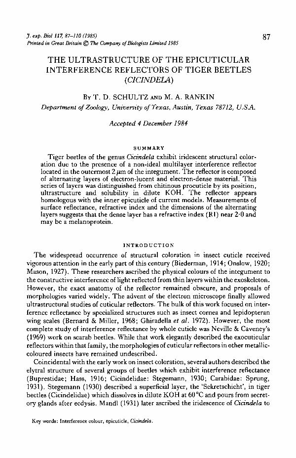
The elytra and thorax of C.formosa are deep metallic red with white maculations—peak reflectance occurring at 655 nm, with a secondary peak near 360 nm (Fig. 1A).The Bastrop population of C. formosa exhibits the elytral coloration of the race C.formosa pigmentosignata Horn. While the maculae of Bastrop individuals are rarelyreduced, many individuals exhibit a distal deep purple coloration on the elytron,which is characteristic of C. formosa pigmentosignata (Gaumer, 1977). For reflec-tance measurements and electron microscope studies, elytral fragments were selectedfrom elytral areas which were homogeneously red.
The elytra of C. splendida are metallic brick red with a metallic green border. Anelytron with the border removed shows maximal reflectance near 635 nm, with a verysmall secondary peak at 410nm (Fig. IB).
C. scutellaris rugata elytra reflect maximally at 499 nm in a relatively narrow band(Fig. 1C). C. scutellaris varies greatly in colour among its races, with rugata display-ing an iridescent blue-green coloration. Both the intensity and the purity of thisreflected colour exceed those of C.formosa and C. splendida.
The brown C. repanda elytron has a very different reflectance curve (Fig. ID).Instead of a sharp peak in the visible wavelengths, the reflectance increases steadilytowards the near-infrared.
The colour of tiger beetle elytra, like all interference colours, changes with the angleof viewing. As the angle of reflectance departs from the angle of incidence, thereflected wavelengths become shorter. The elytra of Cicindela are alveolate (Fig. 2)
90 T. D. SCHULTZ AND M. A. RANKIN
r B
325 405 485 565 645 725 325 405
Wavelength (nm)
485 565 645 725
Fig. 1. Reflectance curves of four species of Cicindela. (A) C. formosa pigmentosignata, (B) C.scutellaris rugata, (C) C. splendida and (D) C. repanda.
with small (10 X 10 /im) hexagonal depressions. The alveoli are superimposed uponlarger irregularities in the upper portion of the elytron. Light reflected from thissurface is a composite of wavelengths reflected from a variety of angles. Not onlywould the total intensity of reflection be diminished, but the peak reflectance wouldbe less sharply defined. Of the four species, C. scutellaris displays the most regularelytral surface with smooth, shallow alveoli. C. repanda and C. splendida possess thedeepest alveoli and most acute ridges.
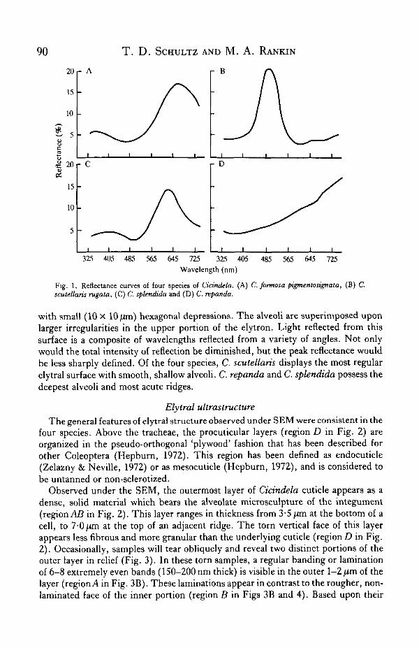
Elytral ultrastructureThe general features of elytral structure observed under SEM were consistent in the
four species. Above the tracheae, the procuticular layers (region D in Fig. 2) areorganized in the pseudo-orthogonal 'plywood' fashion that has been described forother Coleoptera (Hepburn, 1972). This region has been defined as endocuticle(Zelazny & Neville, 1972) or as mesocuticle (Hepburn, 1972), and is considered tobe untanned or non-sclerotized.
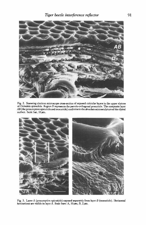
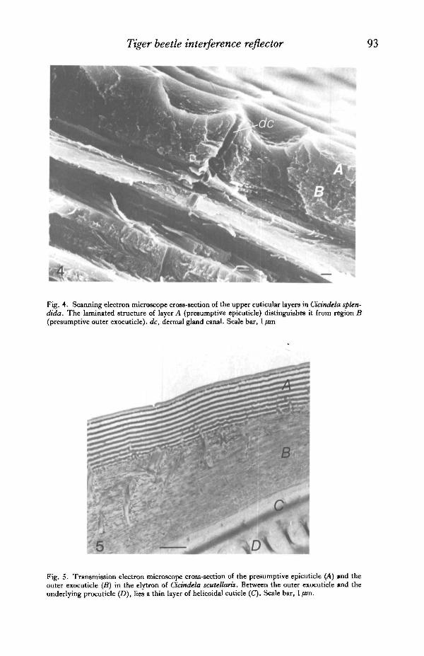
Observed under the SEM, the outermost layer of Cicindela cuticle appears as adense, solid material which bears the alveolate microsculpture of the integument(region AB in Fig. 2). This layer ranges in thickness from 3-5 /im at the bottom of acell, to 7-0/im at the top of an adjacent ridge. The torn vertical face of this layerappears less fibrous and more granular than the underlying cuticle (region D in Fig.2). Occasionally, samples will tear obliquely and reveal two distinct portions of theouter layer in relief (Fig. 3). In these torn samples, a regular banding or laminationof 6-8 extremely even bands (150-200 nm thick) is visible in the outer 1-2 /im of thelayer (regionA in Fig. 3B). These laminations appear in contrast to the rougher, non-laminated face of the inner portion (region B in Figs 3B and 4). Based upon their
Tiger beetle interference reflector
Fig. 2. Scanning electron microscope cross-section of exposed cuticular layers in the upper elytronof Cicindela splendida. RegionD represents the pseudo-orthogonal procuticle. The composite layerAB (the presumptive epicuticle and exocuticle) conforms to the alveolate microsculpture of the elytralsurface. Scale bar, 10 /an.
Fig. 3. Layer A (presumptive epicuticle) exposed separately from layer B (exocuticle). Horizontallaminations are visible in layer A. Scale bars: A, lOjon; B, 2ftra.
92 T . D. SCHULTZ AND M. A. RANKIN
ultrastructure, location, development and reaction to solvents, the outer and innerportions of the outer cuticle (layers A and B in Fig. 4) are here designated the epi-cuticle and outer exocuticle, respectively, and will be referred to as such throughoutthis report.
Dermal gland canals measure 0-8 /im in diameter and their orifices are distributedregularly over the surface of the elytron (Fig. 4, dc). The pattern of lamination is notdisrupted around the canal opening. Epicuticular thickness is enhanced on the ridges,compared to the basins of the alveoli, but is not greater at the dermal gland openingthan on adjacent ridges.
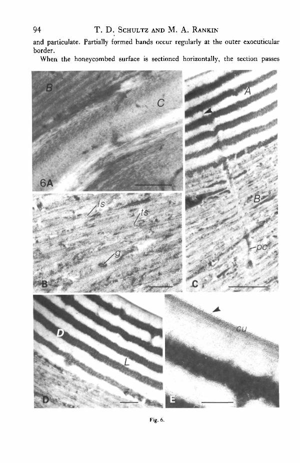
Transmission electron microscopy (TEM) reveals that the alveolate superficiallayer of the scanning electron micrograph is indeed a composite of two complex layers(layersA andB in Fig. 5). Immediately below this composite, lie 2-3 thin exocuticularlayers (layer C in Figs. 5, 6A) which exhibit the distinct arciform appearance ofhelocoidal cuticle as described by Bouligand (1972).
The outer exocuticle (layer B in Fig. 5) appears more electron-dense than thehelicoidal or preferred layers below. This layer consists of 25-35 densely packedlamellae ranging in width from 0-055 to 0-1/im. In some areas, electron-densegranules lie between or within lamellae. In post-stained sections, the horizontallamellae are resolved into alternate patterns of microfibrils viewed in perpendicularand transverse orientations (Fig. 6B). This pattern closely resembles the descriptionof the non-perfect helicoidal outer exocuticle of Tenebrio given by Filshie (1982).
Pore canals, 50-70nm in diameter, traverse the outer exocuticle (Fig. 6C, pc).Within the exocuticle, they appear as segments of tubes containing material muchdenser than the surrounding fibrils. The pore canals extend into the epicuticle butappear much narrower in diameter (Fig. 6C, arrow). This pattern is similar to thebranching of epicuticular filaments described in other insect cuticles (Filshie, 1982).
The epicuticle (Fig. 6D) is composed of a series of extremely dense bands (D) be-tween 0-03 and 0-1/mi thick, regularly spaced by electron-lucent bands (L) of0-06-0-125 /im. Each pair of electron-dense and electron-lucent layers corresponds toa single band observed in relief in the scanning electron micrographs. The number ofbands varies between four dense layers at the base of a depression to nine layers withinan adjacent ridge. Under high magnification, electron-dense layers appear to becomposed of vertically aligned granules (Fig. 6D). Post-staining enhances this ap-pearance. The electron-lucent bands exhibit no apparent ultrastructure. The outer-most layer is always electron-lucent and 20-30 nm thicker than the other light bands.The outer 20-30 nm appears as a faint shadow of higher density, possibly representinga cuticulin layer (Fig. 6E, cu). Mossakowski (1980) identified the outer series ofelectron-dense and electron-lucent bands as exocuticle. He concluded that the ban-ding was a diffraction pattern resulting from the structural organization of exo-cuticular fibrils.
Although there are occasional distortions, the organization of the epicuticularlayers is exceedingly regular. One individual L or D layer may vary in thicknesshorizontally within a section. This is usually an artifact resulting from the sectionpassing obliquely through the layer as it conforms to the alveolate surface. Each Lor D layer may differ slightly in thickness from other L/D layers in that region ofthe stack. In C. scutellaris, the peripheral dense band is typically fragmented
Tiger beetle interference reflector
Fig. 4. Scanning electron microscope cross-section of the upper cuticular layers in Cidndela splen-dida. The laminated structure of layer A (presumptive epicuticle) distinguishes it from region B(presumptive outer exocuticle). dc, dermal gland canal. Scale bar, 1 fun
Fig. 5. Transmission electron microscope cross-section of the presumptive epicuticle (A) and theouter exocuticle (B) in the elytron of Cidndela scutellaris. Between the outer exocuticle and theunderlying procuticle (D), lies a thin layer of helicoidal cuticle (C). Scale bar, 1 /an.
94 T. D . SCHULTZ AND M. A. RANKIN
and particulate. Partially formed bands occur regularly at the outer exocuticularborder.
When the honeycombed surface is sectioned horizontally, the section passes
Fig. 6.
Tiger beetle interference reflector
D
\ \
\\
i d .
\ \
v s.
NX\
^ ;
V\
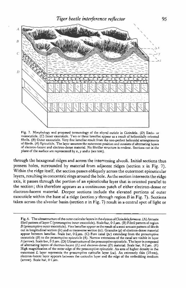
Fig. 7. Morphology and proposed terminology of the eltyral cuticle in Cicindela. (D) Endo- ormesocuticle. (C) Inner exocuticle. Two or three lamellae appear as a result of helicoidally orientedfibrils. (B) Outer exocuticle. Very fine lamellae result from the non-perfect helicoidal arrangementsof fibnis. (A) Epicuticle. The layer assumes the outermost position and consists of alternating layersof electron-lucent and electron-dense material. No fibrillar structure is evident. Sections cut in theplane of the surface are represented by x, y and z (see text).
through the hexagonal ridges and across the intervening alveoli. Initial sections thuspossess holes, surrounded by material from adjacent ridges (section x in Fig. 7).Within the ridge itself, the section passes obliquely across the outermost epicuticularlayers, resulting in concentric rings around the hole. As the section intersects the ridgeaxis, it passes through the portion of an epicuticular layer that is oriented parallel tothe section; this therefore appears as a continuous patch of either electron-dense orelectron-lucent material. Deeper sections include the elevated portions of outerexocuticle within the base of a ridge (section^ through region B in Fig. 7). Sectionstaken across the alveolar basin (section z in Fig. 7) result in a central spot of light or
Fig. 6. The ultrastructure of the outer cuticular layers in the elytron of Cicindela fomtosa. (A) Arcuatefibril pattern of layer C (presumptive inner exocuticle). Scale bar, 0-5 fan. (B) Fibril pattern of regionB (presumptive outer exocuticle) .Fine lamellae appear as the result of a semi-arcuate pattern of fibniscut in longitudinal section (Is) and in transverse section (ts). Granules ig) of electron-dense materialappear between lamellae. Scale bar, 0-2/an. (C) Pore canal {pc) extending from the presumptiveexocuticle (B) to the presumptive epicuticle (A). Narrow extensions of the canal are visible in layerA (arrow). Scale bar, OS fim. (D) Ultrastructure of the presumptive epicuticle. The layer is composedof alternating layers of electron-lucent (L) and electron-dense (D) material. Scale bar, 0-2 ftn\. (E)High magnification of the outer edge of the presumptive epicuticle. An area of higher density in theoutermost L layer represents the presumptive cuticulin layer (cu). An extremely thin (10nm),electron-lucent layer appears between the cuticulin layer and the edge of the embedding medium(arrow). Scale bar, 0-1 fun.
96 T. D. SCHULTZ AND M. A. RANKIN
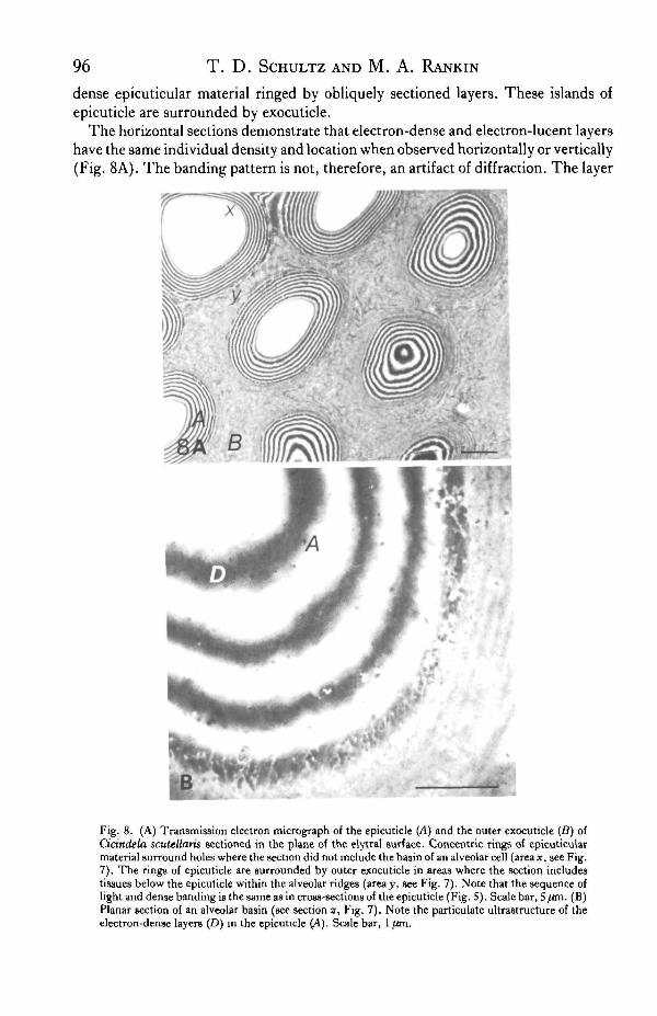
dense epicuticular material ringed by obliquely sectioned layers. These islands ofepicuticle are surrounded by exocuticle.
The horizontal sections demonstrate that electron-dense and electron-lucent layershave the same individual density and location when observed horizontally or vertically(Fig. 8A). The banding pattern is not, therefore, an artifact of diffraction. The layer
f e M l
Fig. 8. (A) Transmission electron micrograph of the epicuticle (A) and the outer exocuticle (B) ofCicindela scutellaris sectioned in the plane of the elytral surface. Concentric rings of epicuticularmaterial surround holes where the section did not include the basin of an alveolar cell (area*, see Fig.7). The rings of epicuticle are surrounded by outer exocuticle in areas where the section includestissues below the epicuticle within the alveolar ridges (areay, see Fig. 7). Note that the sequence oflight and dense banding is the same as in cross-sections of the epicuticle (Fig. S). Scale bar, 5 /an. (B)Planar section of an alveolar basin (see section z, Fig. 7). Note the particulate ultrastructure of theelectron-dense layers (D) in the epicuticle (A). Scale bar, 1 /an.
Tiger beetle interference reflector 97
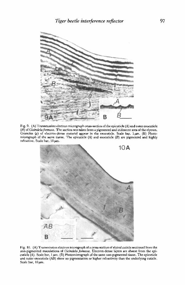
Fig. 9. (A) Transmission electron micrograph cross-section of the epicuticle (A) and outer exocuticle(B) of Cidndelaformosa. The section was taken from a pigmented and iridescent area of the elytron.Granules (?) of electron-dense material appear in the exocuticle. Scale bar, 1 /an. (B) Photo-micrograph of the same tissue. The epicuticle (A) and exocuticle (B) are pigmented and highlyrefractive. Scale bar, 10 jjtm.
10A
B
Fig. 10. (A) Transmission electron micrograph of a cross-section of elytral cuticle sectioned from thenon-pigmented maculations of Cicindela formosa. Electron-dense layers are absent from the epi-cuticle (A). Scale bar, 1 fim. (B) Photomicrograph of the same non-pigmented tissue. The epicuticleand outer exocuticle (AS) show no pigmentation or higher refractivity than the underlying cuticle.Scale bar,

98 T . D . SCHULTZ AND M. A. RANKIN
that borders the holes in horizontal sections is the outermost layer of the epicuticle,and is electron-lucent in both orientations. Furthermore, horizontal sections withina single epicuticular layer show continuous patches of electron-dense material.This material is very finely particulate when viewed perpendicular to the layer(Fig. 8B).
When thin sections are viewed through an optical microscope, the outer exocuticleand the epicuticle appear dark brown (Fig. 9B). This apparent pigmentation is con-fined to the outer 5 (Urn of the elytral cuticle. It is assumed that the dark brownappearance is due to the presence of melanin in this region, and that it is responsiblefor the dark opaque appearance of the elytron in transmitted light. The epicuticle andouter exocuticle display a greater retardation of light than the inner layers of procuticleunder interference microscopy.
Sections of elytral cuticle were also cut from the white maculations of C.formosa.The white areas show no trace of either iridescent colour or brown or black pigmenta-tion (Fig. 10B). Under interference microscopy, the sections of white cuticle show auniformity in refractive index throughout the elytron. The epicuticle shows a refrac-tive index of approximately 1-5 by the Becke line test (Neville, 1980).
Electron micrographs of this non-pigmented cuticle show an epicuticle which lacksthe electron-dense bands (layer A in Fig. 10A). In general, this homogeneous epi-cuticle is thinner and lower in contrast than the laminated epicuticle of pigmentedareas (layer A in Fig. 9A). SEM confirmed the absence of the epicuticular laminationsin the region of the elytral maculations. The outer exocuticle of these sections lacksthe electron-dense granules that were evident in the exocuticle of pigmented areas(layers B in Figs 9A and 10A).
Analysis of cuticle structure by chemical treatmentTiger beetle cuticle treated with dilute KOH displayed a gradual loss of structural
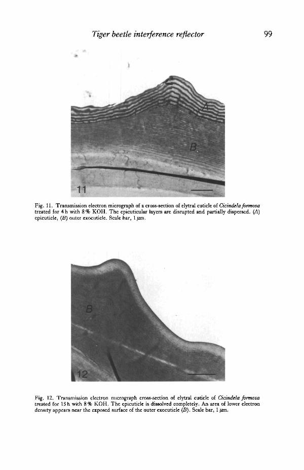
coloration, as well as pigmentation. Immersion in ethanol, xylene, acetone, sulphuricor nitric acid failed to produce any change in structural colour. Initially, structuralcolours became longer in wavelength as the solvent temporarily swelled the reflec-tance layers. After 4h, however, the maroon portion of the elytron of C.formosaappeared blue-green. The anterior portion, originally red, became ochreous withslight orange-green reflections. The overall iridescence was reduced to a dull sheen.Scanning electron micrographs of the treated elytra revealed a distorted and heavilyeroded surface microsculpture. Under transmission electron microscopy, the regularlaminations at the periphery of the cuticle were disrupted and often stripped from thesample (Fig. 11).
With the exception of a few blue-green or violet reflections in the basins ofpunctures, elytra treated with KOH for 12-15 h were virtually devoid of structuralcolours. The surface had a flat, brown, darker appearance at the elytral base, whichis assumed to be due to the residual presence of melanin in the exocuticle. The deephexagonal depressions of the surface microsculpture were eroded to a shallow outlineof the surface pattern. TEM micrographs revealed that the laminated epicuticle wascompletely removed from the surface of the outer exocuticle (Fig. 12). A light band,0-29 /xm thick, remained at the border of the exocuticle. The exocuticular laminationalso became more obscure and fragmented.
Tiger beetle interference reflector
11Fig. 11. Transmission electron micrograph of a cross-section of elytra] cuticle of Cicindela formosatreated for 4h with 8% KOH. The epicuticular layers are disrupted and partially dispersed. (A)epicuticle, (5) outer exocuticle. Scale bar, 1 /an.
Fig. 12. Transmission electron micrograph cross-section of elytral cuticle of Cicindela formosatreated for 15 h with 8% KOH. The epicuticle is dissolved completely. An area of lower electrondensity appears near the exposed surface of the outer exocuticle (B). Scale bar, 1 fan.
T. D. SCHULTZ AND M. A. RANKIN
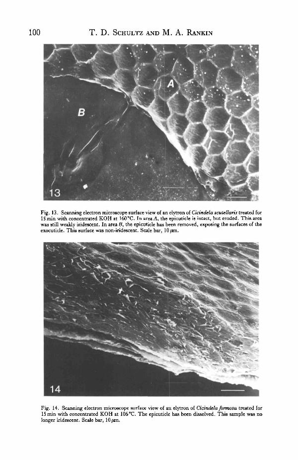
Fig. 13. Scanning electron microscope surface view of an elytron of Cidndela scutellaris treated forISmin with concentrated KOH at 160 °C. In area A, the epicuticle is intact, but eroded. This areawas still weakly iridescent. In area B, the epicuticle has been removed, exposing the surfaces of theexocuticle. This surface was non-iridescent. Scale bar, 10/im.
Fig. 14. Scanning electron microscope surface view of an elytron of Cidndela formosa treated forIS min with concentrated KOH at 106°C. The epicuticle has been dissolved. This sample was nolonger iridescent. Scale bar, 10/an.
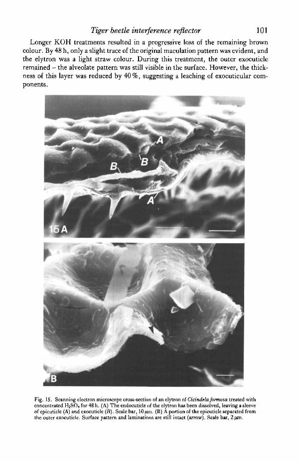
Tiger beetle interference reflector 101Longer KOH treatments resulted in a progressive loss of the remaining brown
colour. By 48 h, only a slight trace of the original maculation pattern was evident, andthe elytron was a light straw colour. During this treatment, the outer exocuticleremained - the alveolate pattern was still visible in the surface. However, the thick-ness of this layer was reduced by 40%, suggesting a leaching of exocuticular com-ponents.
Fig. IS. Scanning electron microscope cross-section of an elytron of Cidndela formosa treated withconcentrated H2SO4 for 48 h. (A) The endocuticle of the elytron has been dissolved, leaving a sleeveof epicuticle (A) and exocuticle (B). Scale bar, 10/an. (B) A portion of the epicuticle separated fromthe outer exocuticle. Surface pattern and laminations are still intact (arrow). Scale bar, 2/an.
102 T. D. SCHULTZ AND M. A. RANKIN
The chitosan method of Campbell (1929) and van Wisselingh (1914) has long beenused to distinguish chitinous from non-chitinous cuticle, although this technique isnot conclusive (Richards, 1951). It requires the conversion of chitin to chitosan bytreatment with concentrated KOH at 160 °C for 15 min. It is assumed that the non-chitinous elements of the exoskeleton (epicuticular components) are dissolved under
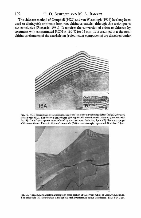
16AFig. 16. (A) Transmission electron microscope cross-section of pigmented cuticle of Ciandelajbrmosatreated with H2O2. The electron-dense layers of the epicuticle are reduced in thickness (compare withFig. 9). Outer layers appear most reduced by the treatment. Scale bar, 1 ftm. (B) Photomicrographof the same tissue. The epicuticle and exocuticle (AB) are not strongly pigmented. Scale bar, 10/im.
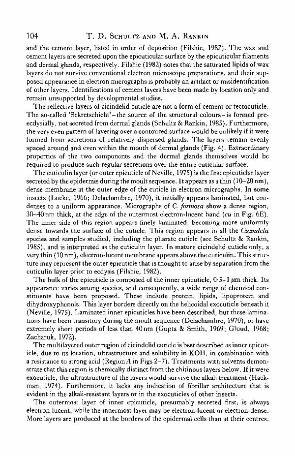
Fig. 17. Transmission electron micrograph cross-section of the elytral cuticle of Cicindela repanda.The epicuticle (A) is laminated, although no peak interference colour is reflected. Scale bar, 2fan.

Tiger beetle interference reflector 103
this alkali treatment. Samples of tiger beetle cuticle subjected to the chitosan testshowed a severe break-up of the presumptive epicuticle. In C. scutellaris, an erodedepicuticle (areaA in Fig. 13) was evident in some areas where a feebly iridescent sheenstill appeared. If these treated samples were immersed in 8 % KOH for 18 h at roomtemperature, all traces of the presumptive epicuticle were removed, as well as theremaining pigmentation. In C.formosa, the epicuticle and iridescence were complete-ly removed by the chitosan treatment (Fig. 14).
In contrast to the results of the alkali treatments, endocuticle is dissolved rapidlyin concentrated sulphuric acid (Sprung, 1931; Wigglesworth, 1933; Hackman, 1974),whereas sclerotized exocuticle and epicuticle are said to remain intact. Tiger beetleelytra immersed in sulphuric acid showed a rapid dissolution of inner 'plywood'mesocuticle, leaving a thin, flexible sleeve of putative epicuticle and outer exocuticle,while the structural coloration of the elytron remained unaltered (Fig. 15A). SEMexaminations of the treated cuticle revealed that the outer laminated layer (layer A)retained its ultrastructure intact (Fig. 15B). The outer exocuticle was also intact andpigmented through the first 48 h of treatment. The residual cuticle still appeared darkbrown in transmitted light. Portions of the helicoidal exocuticle below could be seenadhering to the lower side of the outer exocuticle. Frequently, areas were observedin which the epicuticle had separated from the outer exocuticle, and these fragmentswere found floating on the surface of the acid. Even these isolated fragments main-tained a laminated appearance under SEM, as well as an iridescence.
Treatments of elytral cuticle with H2O2 significantly altered the cuticle coloration.When observed in transmitted light, the brown coloration became more translucent.The outer exocuticle and epicuticle appeared either light brown or clear of pigmentin microscopic sections (Fig. 16B). The structural colour of the cuticle surfacebecame duller and shorter in principal wavelength. Similar results were obtainedwhen samples were treated with 20% H2O2 at 26 °C for 24-48 h.
TEM micrographs revealed a 40 % reduction in thickness of dense epicuticularlayers in the H2O2-treated samples (Fig. 16A). The bands were progressively thinnertowards the outer surface. The overall thickness of the epicuticle was not diminishedsignificantly.
DISCUSSION
Cuticle histology
Probably because it is extremely thin, the epicuticle is the least understood portionof the insect integument. Its chemistry, structure and the terminology of epicuticularcomponents have been controversial. Only a few, diverse arthropods have beenstudied intensively, and very little work has focused on the epicuticle of hard, adultexoskeletons (Delachambre, 1970). This is not surprising since hard cuticle is dif-ficult to prepare and section for transmission electron microscopy.
The epicuticle, by definition, is non-chitinous or reacts negatively to tests for chitin(chitosan method). It is the first layer formed during the moult sequence and assumesthe outermost position in the strata of the exoskeleton, bearing the surface micro-sculpture. The general model of epicuticle structure recognizes five components.These are the cuticulin layer, the inner epicuticle, the outer epicuticle, the wax layer
104 T. D. SCHULTZ AND M. A. RANKIN
and the cement layer, listed in order of deposition (Filshie, 1982). The wax andcement layers are secreted upon the epicuticular surface by the epicuticular filamentsand dermal glands, respectively. Filshie (1982) notes that the saturated lipids of waxlayers do not survive conventional electron microscope preparations, and their sup-posed appearance in electron micrographs is probably an artifact or misidentificationof other layers. Identifications of cement layers have been made by location only andremain unsupported by developmental studies.
The reflective layers of cicindelid cuticle are not a form of cement or tectocuticle.The so-called 'Sekretschicht'-the source of the structural colours-is formed pre-ecdysially, not secreted from dermal glands (Schultz & Rankin, 1985). Furthermore,the very even pattern of layering over a contoured surface would be unlikely if it wereformed from secretions of relatively dispersed glands. The layers remain evenlyspaced around and even within the mouth of dermal glands (Fig. 4). Extraordinaryproperties of the two components and the dermal glands themselves would berequired to produce such regular secretions over the entire cuticular surface.
The cuticulin layer (or outer epicuticle of Neville, 1975) is the first epicuticlar layersecreted by the epidermis during the moult sequence. It appears as a thin (10-20 nm),dense membrane at the outer edge of the cuticle in electron micrographs. In someinsects (Locke, 1966; Delachambre, 1970), it initially appears laminated, but con-denses to a uniform appearance. Micrographs of C. formosa show a dense region,30-40 nm thick, at the edge of the outermost electron-lucent band (cu in Fig. 6E).The inner side of this region appears finely laminated, becoming more uniformlydense towards the surface of the cuticle. This region appears in all the Cicindelaspecies and samples studied, including the pharate cuticle (see Schultz & Rankin,1985), and is interpreted as the cuticulin layer. In mature cicindelid cuticle only, avery thin (10 nm), electron-lucent membrane appears above the cuticulin. This struc-ture may represent the outer epicuticle that is thought to arise by separation from thecuticulin layer prior to ecdysis (Filshie, 1982).
The bulk of the epicuticle is composed of the inner epicuticle, 0-5-1 pim thick. Itsappearance varies among species, and consequently, a wide range of chemical con-stituents have been proposed. These include protein, lipids, lipoprotein anddihydroxy phenols. This layer borders directly on the helicoidal exocuticle beneath it(Neville, 1975). Laminated inner epicuticles have been described, but these lamina-tions have been transitory during the moult sequence (Delachambre, 1970), or haveextremely short periods of less than 40 nm (Gupta & Smith, 1969; Gluud, 1968;Zacharuk, 1972).
The multilayered outer region of cicindelid cuticle is best described as inner epicut-icle, due to its location, ultrastructure and solubility in KOH, in combination witha resistance to strong acid (Region A in Figs 2-7). Treatments with solvents demon-strate that this region is chemically distinct from the chitinous layers below. If it wereexocuticle, the ultrastructure of the layers would survive the alkali treatment (Hack-man, 1974). Furthermore, it lacks any indication of fibrillar architecture that isevident in the alkali-resistant layers or in the exocuticles of other insects.
The outermost layer of inner epicuticle, presumably secreted first, is alwayselectron-lucent, while the innermost layer may be electron-lucent or electron-dense.More layers are produced at the borders of the epidermal cells than at their centres.

Tiger beetle interference reflector 105
These borders are indicated by the hexagonal ridges in the surface microsculpture(Neville, 1975). Areas of elytral cuticle which lack pigmentation (i.e. the macula-tions), also lack electron-dense layers in the epicuticle. Perhaps the cicindelid innerepicuticle is described best as a homogeneous electron-lucent material, impregnatedby electron-dense material at discrete intervals in those cuticles that are pigmented.
The epicuticle as an interference reflector
It appears that the epicuticle is the source of structural colours in Cicindela.Maceration or removal of the epicuticle with KOH results in the deterioration or lossof iridescent coloration. Conversely, removal of the other components of the integu-ment produces no loss of colour. The change of hue under oblique incidence demon-strates that the structural colours result from constructive interference of reflectedlight.
The repetitive layering of two materials in the epicuticle, whose thicknesses are lessthan half the wavelength of visible light, is characteristic of a multilayer interferencereflector. The two layers are distinct in their electron densities, and are not an artifactof the helicoidal arrangement of cuticle fibrils, as in the reflective exocuticle of optic-ally active scarab beetles (Neville & Caveney, 1969). Multilayer reflectors have beendescribed in a wide diversity of animal tissues, and the basic principles of interferencein animal reflectors are reviewed by Land (1972).
In the 'ideal' case of thin film reflection, the wavelength of the first order maximumreflectance (A max) depends on the thickness (d) and refractive index (n) of each layersuch that
A max = 4nd sin G, (1)
where 6 is the angle of incidence. Under 'ideal' conditions, the optical thicknesses ofboth layers are equivalent (wz,dz. = n^do, where L indicates the lucent layer andD thedense layer). Each layer acts as a quarter-wavelength reflecting plane, with the reflec-tions from all interfaces constructively interfering to produce the total coloration.
The optical thickness of each component is different (n/.di ^ nodo) in a 'non-ideal'multilayer system. The first order reflectance peak occurs at
A max = 2(nz.di + n^do) (2)
under normal angle of incidence. Under 'non-ideal' conditions the peak reflectanceand the band width of the first order peak are diminished. The result is a sharper peakand purer colour (Land, 1972). The reflecting layers of tiger beetle epicuticle are bestdescribed as a 'non-ideal' system. The thicknesses of the electron-dense (do) and theelectron-lucent (dz.) layers vary considerably through a single point in the stack (Table1). Assuming that the refractive indices (HZ. and «D) remain stable, the ratio d/./d/)does not remain constant throughout the reflector.
A quarter-wave interference reflector has been described by Durrer & Villiger(1972) in the exocuticle of Euchroma gigantea, which consists of a series of electron-lucent and electron-dense bands similar to the epicuticle of Cicindela. The dimen-sions of the layers imply an average refractive index of 1-75 for the reflector. Theauthors propose that the multilayer system contains electron-dense melanin layers(RI = 2-0) separated by chitin (RI = 1-5), similar to the keratin-melanin interference

106 T. D. SCHULTZ AND M. A. RANKIN
Table 1. Average thickness of electron-lucent (L) and electron-dense (D) layers infour Cicindela species with predicted and experimental values ofelytral reflectance
Specimen
C. formosaridgealveolar basin
C. scutellaris rugataC. splendidaC. repanda
ridgealveolar basin
SEMbilayer
thickness(nm)
188
143178
—
Mean L layerthickness (nm)
106-59 ±6.76101-99 ±5-29267-197 ±4-08102-03 ± 14-907*
137-73 ± 18-23381-22
Mean D layerthickness (nm)
93-63 ±11-75674-974 ±6-75272-147 ±4-59171-428 ±6-563
110-65 ±12-15688-035 ±6-815
CalculatedAmax(nm)
« L = 1 - 5 ;nD = 2-0
694-31605-77489-93591-8
855-79595-8
Experi-mental
Amax(nm)
655
499635
—
Standard deviation values indicate the variation between successive layers in the epicuticle.Calculations of A max assume a refractive index (n) of 1-5 for L layers and 2-0 for D layers.• The outermost L layer measured 130 nm in this sample.
reflectors of bird feathers (Durrer & Villiger, 1970). In their approach, the authorscalculated the wavelength of maximum reflectance for each layer separately, inaddition to determining the reflectance from the series of bilayers (equation 2). Theyconcluded that a mixture of these reflected components results, producing the totalcolour.
Mossakowski (1980) examined interference reflectors in the cuticles of thebuprestid, Chrysocroa vittata, and the tiger beetle, Cicindela campestris.Mossakowski calculated reflectances for a number of possible refractive indices usingthe theoretical treatment of Huxley (1968). He proposed that an average refractiveindex of 1 -75 for the bilayers of C. camprestis would cause a total reflectance value farexceeding those determined experimentally. Mossakowski's measurements of totalreflectance predicted refractive indices of 1*5 and 1-6 for the light and dense layersrespectively. However, these values and the dimensions of the layers observed underelectron microscopy predicted a wavelength of peak reflectance substantially belowthose recorded spectrophotometrically. Mossakowski assumed that the reflectinglayers had experienced a 10—15 % shrinkage due to specimen preparation or exposureto the electron beam.
The dimensions of the interference layers of cicindelids presented in this papersuggest a relatively high average refractive index near 1-75 for the pairs of layers.Values of 1*5 and 2-0 for the light and dense layers predict peak wavelengths whichapproximate to the spectrophotometric results for C. scutellaris and C. formosa(Table 1). Refractive indices of 1-5 and 1-6 would predict substantially shorterwavelengths of peak reflectance. The epicuticle of C. repanda varied greatly in layerthickness and its reflectance exhibited no single peak within the visible wavelengths.
Even assuming an average refractive index of 1-75, calculations using the averagethicknesses of epicuticular layers predict values of A max well below the reflectancepeak of C. splendida. Unlike the other specimens, the outermost electron-lucent layerof C. splendida is 40-50 nm thicker than all the underlying electron-lucent layers.

Tiger beetle interference reflector 107
This layer makes a significant contribution to the large standard deviation in theaverage thickness of these layers. Variation in the thickness of the inner layers is lessthan that of C. repanda but greater than the variation in C. scutellaris. A A max of638 nm is predicted if calculations are based upon the superficial electron-lucent layer(d/, = 130 nm, TIL = 1*5) and the underlying dense layer (dz, = 62nm, TIL = 2-0). Thisvalue is close to the experimental reflectance peak of 635 nm. Since the surface of C.splendida is so deeply sculptured with the alveolate pattern, the superficialepicuticular layers may contribute more to the peak reflectance than the underlyinglayers. Peak reflectances from deeper layers may occur at angles which depart fromthe angle of reflection that is sampled by the spectrophotometer.
In C. scutellaris rugata and the green form of C. campestris (Mossakowski, 1980),the thickness of the light and dense layers are roughly equivalent. In these cases, theaverage refractive index h may be estimated from the experimental peak wavelengthand the thickness of each bilayer (doz.):
(3)
or n = A max/2dDL. (4)
For C. scutellaris rugata, an average refractive index of 1-74 is predicted.No evidence of shrinkage appeared in the samples of cicindelid cuticle. The struc-
tural colours of specimens were observed unchanged within the embedding medium,indicating that shrinkage did not result from specimen preparation. SEM measure-ments of epicuticular bilayers corresponded well with bilayer measurements madefrom transmission electron micrographs. If bilayers measured from SEMmicrographs were assumed to shrink 10% under the electron beam, they wouldappear 10-20 nm thinner than the average thickness of bilayers measured from TEMmicrographs. If shrinkage had occurred under the electron beam, TEM measure-ments should fall below the thicknesses measured under SEM.
The relatively low total reflectance of cicindelid epicuticle may be due to its alveo-late microsculpture. If the angle of incident light departs from normal, the wavelengthof peak reflectance becomes shorter according to
A max = 4ndcos0, (5)
where 6 is the angle of refraction within a single layer. In addition, each successiveepicuticular layer conforms to the alveolate pattern of the surface. The wavelength ofpeak reflectance for multilayer reflectors at oblique incidence is then given by:
A max = 2{TIL&L COS OL + node cos 6b), (6)
where 8 is the angle of incidence in the layer of lower density. Similarly, the reflec-tance at each interface is dependent upon the angle of refraction (Land, 1972). Thus,the wavelength and the intensity of reflection are affected by deviations in angle ofincidence and refraction which occur continuously throughout the horizontal dimen-sions of the epicuticular layers. Due to these effects, and the scattering of reflectedlight by an alveolate surface, the spectrophotometric analysis of reflected light fromthe epicuticle must surely underestimate the reflectance predicted by normal theoreti-cal conditions.
108 T . D . SCHULTZ AND M. A. RANKIN
The peak reflectance is also dependent upon the constancy of layer thicknessthroughout the epicuticle. In Cicindela, the thicknesses of the bilayers vary bothhorizontally and vertically within the epicuticle (Table 1). The theoretical treatmentof non-ideal, multilayer reflectors assumes that these dimensions remain constant.This non-uniformity in layer thickness and angle, with respect to incident light,clearly places the cicindelid epicuticle as an extremely complex example of a biologicalinterference reflector. The epicuticle represents a non-ideal, multilayer systemmoulded into a shallow honeycomb form, and is not adequately described by theoreti-cal treatments for regularly spaced, planar systems.
Correlations between electron microscope measurements and reflectance datashould not be over-emphasized. The amount of cuticle sectioned is not comparableto the area of elytral material sampled for reflectance. The variability within theinterference reflector has already been described. In addition, there is always un-certainty as to whether the section is precisely perpendicular to the interference layers.
With rare exceptions, the brown or black colours of insect cuticles are attributed tosclerotization and/or melanization of the cuticle (Richards, 1967). Eumelaninspredominate as the brown or black pigments of insect integument (Hackman, 1974),and the pigmentation of Cicindela has been ascribed to melanin since the work ofShelford (1917). Unfortunately, it is exceedingly difficult to identify small amountsof melanin by staining or chemical analysis. Since the individual layers of densematerial in the epicuticle are not resolvable under light microscopy, their opticalproperties cannot be examined separately. The treatment with KOH removes theepicuticle from the pigmented exocuticle, and produces a solution that is stainedbrown with dissolved epicuticle. But it is also possible that melanin may be leachedfrom the exocuticle by the treatment (Richards, 1967).
Richards (1967) found that treatment with H2O2 will bleach melanin particleswithout removing them from the cuticle. Tiger beetle elytra were treated with H2O2to determine the role of melanin in the elytral coloration. If melanin is involved simplyas an absorbent background to the interference reflector, the iridescent colour shouldbe reduced in intensity, but not altered in hue when the pigment is bleached (Mason,1927). In treatments of C. formosa, a reduction in intensity and predominantwavelength resulted, as well as a thinning of pigmented colour in the cuticle. Thechange in reflected colour may be due to the bleaching of melanin within the structureof the reflecting epicuticle itself.
The structural dimensions of the two epicuticular layers, in conjunction with theexperimental reflectance results, suggest that the epicuticular reflector is composed ofan electron-lucent layer with a refractive index of 1 -5 and a dense layer of 2-0. A refrac-tive index of 1*5 is acceptable for insect cuticular proteins (Neville, 1975). This studyproposes that the unusually high refractive index of 2-0 is provided by layers of melaninor melanoproteins. Precedents for such a system can be found in iridescent bird feathercolouration (Greenwalt, Brandt & Friel, 1960; Rutschke, 1966; Durrer & Villiger,1966, 1970). Additional melanin in the outer exocuticle provides an absorbent back-ground for transmitted and extraneous wavelengths, hence purifying the reflectedcolour. Circumstantial evidence for the role of melanins is found in the restrictive co-occurence of pigmentation, iridescence and epicuticular lamination. Specific chemicalanalysis is still required to verify the identity of the electron-dense layer as melanin.

Tiger beetle interference reflector 109
Structural coloration in Cicindela
The specific coloration of 9tructurally-coloured tiger beetles depends upon therelative thicknesses of the epicuticular layers. In C.formosa, C. scutellaris and C. splen-dida, the dimensions of these layers are sufficient to cause the constructive interferenceof visible wavelengths of light. In epicuticles with regular laminations and a relativelysmooth microsculpture, as in C. scutellaris, the reflectance colours are purer.
In some areas of C. repanda elytra, epicuticular laminations are too wide to reflectlight at visible wavelengths (Fig. 17). However, visible wavelengths are reflected fromareas where the layers are sufficiently thin in the highly variable epicuticle, e.g. withinthe alveoli. Furthermore, visible wavelengths may be reflected from thicker layers atoblique incidence due to the effect of angle of incidence on the wavelength ofmaximum reflectance. Thus, while the layers of C. repanda may reflect maximallyabove 760 ran, shorter red wavelengths will be reflected by the highly angled patternof ridges in the microsculpture. The results of these factors is a relatively poor reflec-tance of any one visible wavelength, producing an overall flat brown appearance uponcasual observation. Under the dissecting microscope, a wide range of reflected colours(predominantly red) can be observed from various points on the surface micro-sculpture. Although at first glance the elytra of C. repanda do not appear to bestructurally coloured, the epicuticular structure is consistent with the epicuticularreflectors of other iridescent species.
Thus, tiger beetle structural coloration depends upon three variables: the thick-ness, uniformity and microsculpture of the reflecting layers. In species which reflecta narrow band of short wavelengths, the epicuticular bilayers are relatively thin andthe intensity and purity of the colour depend upon the severity of the alveolatemicrosculpture. Several species which appear brown or coppery bear relatively thickand variable epicuticular bilayers. The deeply pitted microsculpture of the reflectorfurther diminishes the intensity and purity of the reflected colour, resulting in a broadrange of reflected wavelengths of low intensity. Small variations in the basic multi-layer structure of the epicuticle are responsible for the wide variation in colour obser-ved in the genus Cicindela.
The authors thank Dr S. Meier, University of Texas, for the use of the SEM, DrBird and K. Kotora of Rutgers University, for their assistance in the reflectancespectrophotometry, and the Texas Department of Parks and Wildlife for permissionto collect specimens. We especially appreciate the assistance of Susan Houghton,MBL, Woods Hole, MA, who sectioned and photographed the TEM material. Thisstudy was supported partially by NSF Grant 2610711250.
R E F E R E N C E S
BERNARD, G. D. & MILLER, W. H. (1968). Interference filters in the corneas of Diptera. Invest. Ophthamol.7, 416-434.
BIEDERMANN, W. (1914). Farbe und Zeichnung der Insecten. InHandbuch Vergleichende Physiobgie, (ed. H.Winterstein), pp. 1657-1994.
BOUUGAND, Y. (1972). Twisted fibrous arrangements in biological materials and cholestenc mesophases.Tissue Cell. 4, 189-217.
110 T. D. SCHULTZ AND M. A. RANKIN
CAMPBELL. F. L. (1929). The detection and estimation of insect chitin; the inflation of chitinization to hardnessand pigmentation of the cuticula of the American cockroach, Periplaneta americana. Ann. ent. Soc. Am. 22,401-426.
DELACHAMBRE, J. (1970). Etudes sur l'fpicuticle des insectes. La deVeloppement de l'6picuticle chez l'adultede Tenebrio molitor L. (Insecta: Coleoptera). Z. Zellforsch. mikrosk. Anat. 108, 380-396.
DURRER, H. & VILLIGER, W. (1966). Schillerfarben der Trogoniden. J. Orn., Ipz 107, 1-36.DURRER, H. & VILLJGER, W. (1970). Schillerfarben des Goldcuckucks, Chysococcyx cupreous Shaw, im Elek-
tronenmikroskop. Z. Zellforsch. mikrosk. Anat. 109, 407-413.DURRER, H. & VILLIGER, W. (1972). Schillerfarben von Euchroma gigantea L., elektronenmikroskopische
Untersuchung der Elytren. Int. J. Insect. Morph. Embryol. 1, 233-240.FILSHIE, B. (1982). Fine structure of the cuticle of insects and other arthropods. In Insect Ultrastructure, Vol.
1, (eds R. C. King & M. Akai), pp. 281-312. New York: Plenum Press.GAUMER, G. C. (1977). The variation and taxonomy of Cicindela fbrmosa Say (Coleoptera: Cicindelidae).
Ph.D. thesis, Texas A & M University. 253 pp.GHIRADELLA, H., ANESHANSLEY, D., EISNER, T., SILBERGLIED, R. E. & HINTON, H. E. (1972). UV reflection
of male butterfly: interference colour caused by thin-layer elaboration of wing scales. Science, N.Y. 178,1214-1217.
GLUUD, A. (1968). Zur Feinstruktur der Insektencuticula. Ein Beitrag zur Frage des Eigengiftschutzes derWanzencuticula. Zool.Jb. (Anat.) 85, 191-227.
GREENWALT, C. H., BRANDT, W. & FRIEL, D. (1960). The iridescent colors of hummingbird feathers. Proc.Am. Phil. Soc. 104, 249-253.
GUPTA, B. L. & SMITH, D. S. (1969). Fine structure organization of the spermatheca in the cockroach,Periplaneta americana. Tissue Cell 1, 289-324.
HACKMAN, R. H. (1974). Chemistry of insect cuticle. In The Physiology of Insecta, Vol. 6, (ed. M. Rockstein),pp. 216-270. New York: Academic Press.
HASS, W. (1916). Ueber Metallfarben bei Buprestiden. Sber. Ges. natur. Freunde Bed. 332-343.HEPBURN, H. R. (1972). Some mechanical properties of crossed fibrillar chitin. J . Insect Physiol. 18, 815-820.HUXLEY, A. F. (1968). A theoretical treatment of the reflexion of light by multilayer structures. J. exp. Biol. 48,
227-245.LAND, M. F. (1972). The physics and biology of animal reflectors. Prog. Biophys. molec. Biol 24, 75-106.LOCKE, M. (1966). The structure and formation of the cuticulin layer in the epicuticle of an insect, Calpodes
ethlius (Lepidoptera: Hesperiidae). J. Morph. 118, 461-494.MANDL, K. (1931). Kunstliche Veranderung der Farben an Cicindela nitida Licht und an anderen Cidndela-
Arten. Z. Morph. Okol. Tiere 22, 110.MASON, C. W. (1927). Structural colors in insects. J. phys. Chem. 31, 321-354.MOSSAKOWSKI, D. (1980). Reflection measurements used in the analysis of structral colours of beetles.
J. Microscopy 116, 350-364.NEVILLE, A. C. (1975). Biology of the Arthropod Cuticle. Berlin: Springer-Verlag.NEVILLE, A. C. (1980). Optical methods in cuticle research. In Cuticle Techniques in Arthropods, (ed. T. A.
Miller), pp. 45-89. New York: Springer-Verlag.NEVILLE, A. C. & CAVENEY, S. (1969). Scarabeidae exocuticle as an optical analogue of cholcsteric liquid
crystals. Biol. Rev. 44, 531-562.ONSLOW, H. (1920). The iridescent colours of insects. 1. The colours of thin films. Nature, Lond. 106, 149-152.RICHARDS, A. G. (1951). The Integument of Arthropods. St. Paul: University of Minnesota Press.RICHARDS, A. G. (1967). Sclerotizabon and the localization of brown and black colors in insects. Zool.J. (Anat.)
84, 25-62.RUTSCHKE, E. (1966). Submikroskopische Struktur schillernder Federn von EntenvSgeln. Z. Zellforsch.
mikrosk. Anat. 72, 432-443.SCHULTZ, T. D. & RANKIN, N. A. (1985). Developmental changes in the interference reflectors and colorations
of tiger beetles (Cicindela). J'. exp. Biol. 117, 111-117.SHELFORD, V. E. (1917). Color and color pattern mechanisms of tiger beetles. Illinois, biol. Monogr. 3, 4.SPRUNG, F. (1931). Die Flugeldccken der Carabidae. Z. Morph. Okol. Tiere 24, 435-490.STEGMANN, F. (1930). Die Flugeldecken der Cicindelidae. Ein Beitrag zur Kenntnis der Insektcuticula.
Z. Morph. Okol. Tiere 18, 1-73.VAN WISSELINGH, C. (1914). Anwendung der in der organishen Chemie gebrauchlichen Reaktionen bei der
phytomikrochemischen Untersuchungen. Folia microbiol., Delft 3, 165—198.WIGGLESWORTH, V. B. (1933). The physiology of the cuticle and of ecdysis in Rhodniusprolixus (Triatomidae:
Hemiptera) with special reference to the function of the oenocytes and of the dermal glands. Q. Jl microsc.Sci. 76, 269-315.
WIGGLESWORTH, V. B. (1972). The Principles of Insect Physiology. London: Chapman & Hall.ZACHARUK, R. Y. (1972). Fine structure of the cuticle, epidermis, and fat body of larval Elateridae (Coleoptera)
and changes acquainted with molting. Can. J. Zool. 50, 1463—1487.ZELAZNY. B. & NEVILLE, A. C. (1972). Quantitative studies on fibril orientation in beetle endocuticle. J. Insect
Physiol. 18, 1095-2122.