the urinary system. objectives: describe the functions of the urinary systemdescribe the functions...
TRANSCRIPT

The Urinary SystemThe Urinary System

Objectives:Objectives:
• Describe the functions of the Urinary Describe the functions of the Urinary SystemSystem
• Describe the major organs of the Describe the major organs of the urinary systemurinary system
• Describe the structure of a nephronDescribe the structure of a nephron• Describe filtration, re-absorption, and Describe filtration, re-absorption, and
formation of urine by the nephronformation of urine by the nephron• Describe diseases and disorders of the Describe diseases and disorders of the
urinary systemurinary system

FunctionsFunctions
• Elimination of waste productsElimination of waste products– Nitrogenous wastes from the metabolism Nitrogenous wastes from the metabolism
of proteinsof proteins– ToxinsToxins– DrugsDrugs
• Regulate aspects of homeostasisRegulate aspects of homeostasis– Water balanceWater balance– ElectrolytesElectrolytes– Acid-base balance in the bloodAcid-base balance in the blood– Blood pressureBlood pressure– Red blood cell productionRed blood cell production– Activation of vitamin DActivation of vitamin D

Major Organs of the Major Organs of the Urinary SystemUrinary System
• KidneysKidneys• UretersUreters• Urinary bladderUrinary bladder• UrethraUrethra
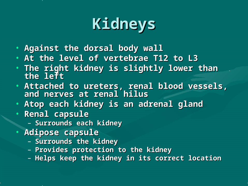
KidneysKidneys• Against the dorsal body wallAgainst the dorsal body wall• At the level of vertebrae T12 to L3At the level of vertebrae T12 to L3• The right kidney is slightly lower than the The right kidney is slightly lower than the
leftleft• Attached to ureters, renal blood vessels, and Attached to ureters, renal blood vessels, and
nerves at renal hilusnerves at renal hilus• Atop each kidney is an adrenal glandAtop each kidney is an adrenal gland• Renal capsuleRenal capsule
– Surrounds each kidneySurrounds each kidney• Adipose capsuleAdipose capsule
– Surrounds the kidneySurrounds the kidney– Provides protection to the kidneyProvides protection to the kidney– Helps keep the kidney in its correct locationHelps keep the kidney in its correct location

Kidney Internal Kidney Internal StructureStructure
• Renal cortex – outer Renal cortex – outer regionregion
• Renal medulla – Renal medulla – inside the cortexinside the cortex
• Renal pelvis – inner Renal pelvis – inner collecting tubecollecting tube
• Medullary pyramids – Medullary pyramids – triangular regions of triangular regions of tissue in the medullatissue in the medulla
• Renal columns – Renal columns – extensions of cortex-extensions of cortex-like material inwardlike material inward
• Calyces – cup-shaped Calyces – cup-shaped structures that structures that funnel urine towards funnel urine towards the renal pelvisthe renal pelvis
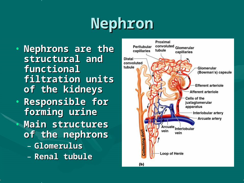
NephronNephron
• Nephrons are the Nephrons are the structural and structural and functional functional filtration units of filtration units of the kidneysthe kidneys
• Responsible for Responsible for forming urineforming urine
• Main structures Main structures of the nephronsof the nephrons– GlomerulusGlomerulus– Renal tubuleRenal tubule

Nephron: GlomerulusNephron: Glomerulus• A specialized capillary A specialized capillary
bedbed• Attached to arterioles Attached to arterioles
on both sides on both sides (maintains high blood (maintains high blood pressure)pressure)– Large afferent Large afferent
arteriolearteriole– Narrow efferent Narrow efferent
arteriolearteriole• Capillaries are Capillaries are
covered with covered with podocytes from the podocytes from the renal tubulerenal tubule
• The glomerulus sits The glomerulus sits within a glomerular within a glomerular capsule (the first part capsule (the first part of the renal tubule)of the renal tubule)

Nephron: Renal TubuleNephron: Renal Tubule
• Glomerular Glomerular (Bowman’s) (Bowman’s) capsulecapsule
• Proximal Proximal convoluted convoluted tubuletubule
• Loop of HenleLoop of Henle• Distal convoluted Distal convoluted
tubuletubule

Peritubular CapillariesPeritubular Capillaries
• Arise from efferent Arise from efferent arteriole of the arteriole of the glomerulusglomerulus
• Normal, low Normal, low pressure capillariespressure capillaries
• Attached to a venuleAttached to a venule• Cling close to the Cling close to the
renal tubulerenal tubule• Reabsorb (reclaim) Reabsorb (reclaim)
some substances some substances from collecting from collecting tubestubes

Urine FormationUrine Formation
• a. Filtrationa. Filtration• b. Reabsorptionb. Reabsorption• c. Secretionc. Secretion

FiltrationFiltration
• Nonselective passive Nonselective passive processprocess
• Water and solutes Water and solutes smaller than proteins smaller than proteins are forced through are forced through capillary wallscapillary walls
• Blood cells cannot Blood cells cannot pass out to the pass out to the capillariescapillaries
• Filtrate is collected in Filtrate is collected in the glomerular the glomerular capsule and leaves via capsule and leaves via the renal tubulethe renal tubule

ReabsorptionReabsorption• The peritubular The peritubular
capillaries reabsorb capillaries reabsorb several materialsseveral materials– Some waterSome water– GlucoseGlucose– Amino acidsAmino acids– Ions ( sodium)Ions ( sodium)
• Some reabsorption is Some reabsorption is passive, most is activepassive, most is active
• Most reabsorption occurs Most reabsorption occurs in the proximal in the proximal convoluted tubuleconvoluted tubule
• Materials not reabsorbed:Materials not reabsorbed:Nitrogenous waste Nitrogenous waste
productsproducts• UreaUrea• Uric acidUric acid• CreatinineCreatinine
Excess waterExcess water
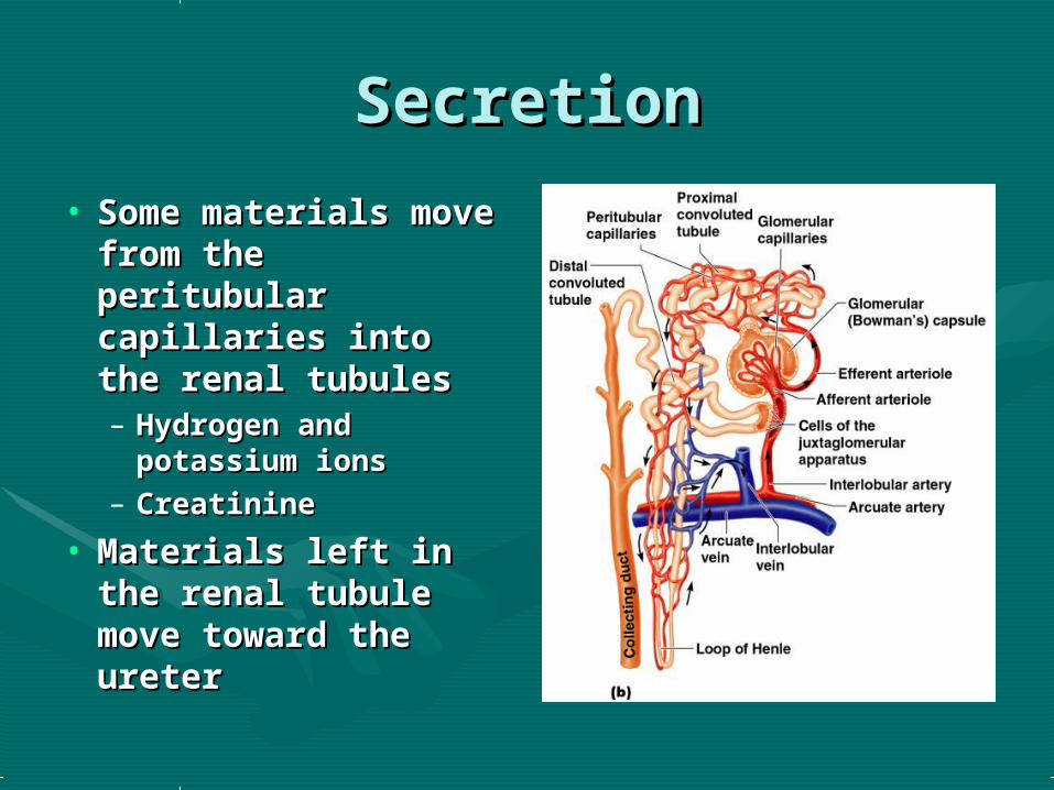
SecretionSecretion
• Some materials Some materials move from the move from the peritubular peritubular capillaries into the capillaries into the renal tubulesrenal tubules– Hydrogen and Hydrogen and
potassium ionspotassium ions– CreatinineCreatinine
• Materials left in the Materials left in the renal tubule move renal tubule move toward the uretertoward the ureter

Urine FormationUrine Formation
Characteristics of Characteristics of Normal UrineNormal Urine
• Colored somewhat yellow due to Colored somewhat yellow due to the pigment urochrome (from the pigment urochrome (from the destruction of hemoglobin) the destruction of hemoglobin) and solutesand solutes
• SterileSterile• Slightly aromatic (has an odor)Slightly aromatic (has an odor)• Normal pH of around 6Normal pH of around 6• Specific gravity of 1.001 to 1.035Specific gravity of 1.001 to 1.035

UretersUreters
• Slender tubes Slender tubes attaching the kidney attaching the kidney to the bladderto the bladder– Continuous with the Continuous with the
renal pelvisrenal pelvis– Enter the posterior Enter the posterior
aspect of the bladderaspect of the bladder• Runs behind the Runs behind the
peritoneumperitoneum• Peristalsis aids Peristalsis aids
gravity in urine gravity in urine transporttransport
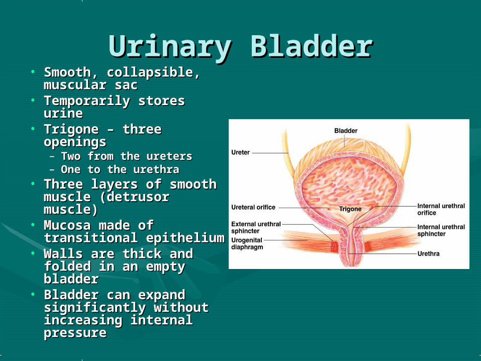
Urinary BladderUrinary Bladder• Smooth, collapsible, Smooth, collapsible,
muscular sacmuscular sac• Temporarily stores Temporarily stores
urineurine• Trigone – three Trigone – three
openingsopenings– Two from the uretersTwo from the ureters– One to the urethraOne to the urethra
• Three layers of smooth Three layers of smooth muscle (detrusor muscle (detrusor muscle)muscle)
• Mucosa made of Mucosa made of transitional epitheliumtransitional epithelium
• Walls are thick and Walls are thick and folded in an empty folded in an empty bladderbladder
• Bladder can expand Bladder can expand significantly without significantly without increasing internal increasing internal pressurepressure

UrethraUrethra• Thin-walled tube that Thin-walled tube that
carries urine from the carries urine from the bladder to the outside of bladder to the outside of the body by peristalsisthe body by peristalsis
• Release of urine is Release of urine is controlled by two controlled by two sphincter muscles:sphincter muscles:– Internal urethral Internal urethral
sphincter (involuntary)sphincter (involuntary)– External urethral External urethral
sphincter (voluntary)sphincter (voluntary)• LengthLength
– Females – 3–4 cm (1 Females – 3–4 cm (1 inch)inch)
– Males – 20 cm (8 inches)Males – 20 cm (8 inches)• LocationLocation
– Females – along wall of Females – along wall of the vaginathe vagina
– Males – through the Males – through the prostate and penisprostate and penis
• FunctionFunction– Females – only carries Females – only carries
urineurine– Males – carries urine and Males – carries urine and
is ais a passageway for passageway for sperm cellssperm cells
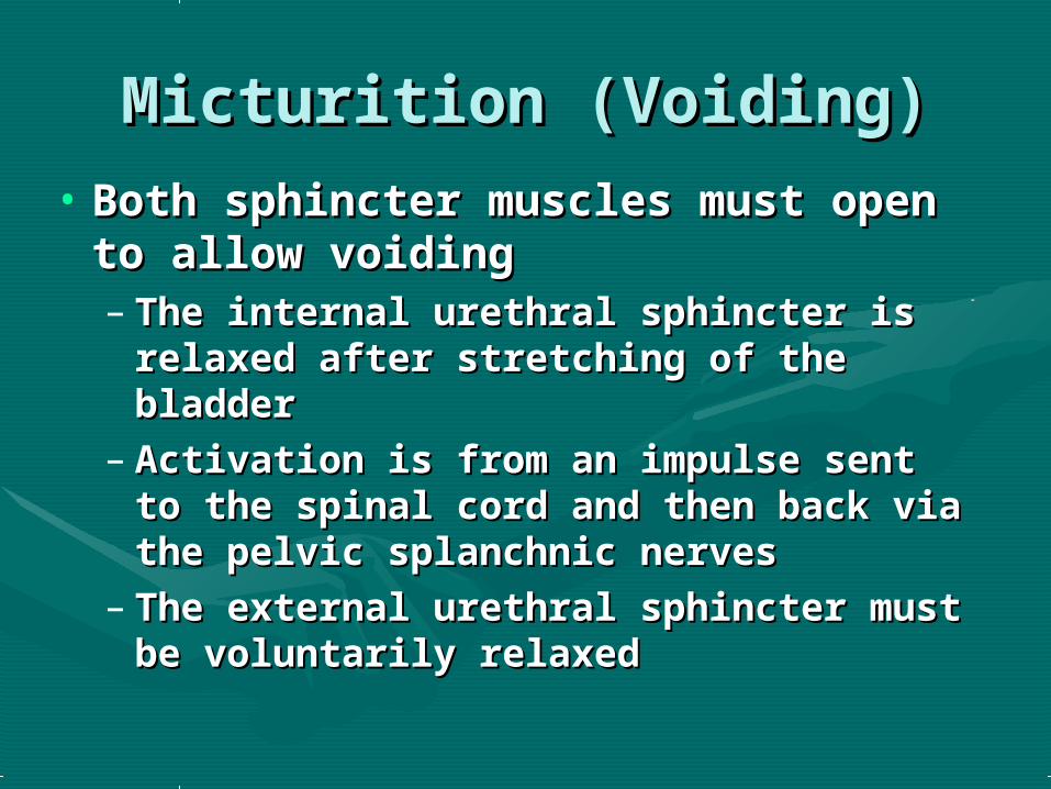
Micturition (Voiding)Micturition (Voiding)• Both sphincter muscles must Both sphincter muscles must
open to allow voidingopen to allow voiding– The internal urethral sphincter is The internal urethral sphincter is
relaxed after stretching of the relaxed after stretching of the bladderbladder
– Activation is from an impulse sent to Activation is from an impulse sent to the spinal cord and then back via the the spinal cord and then back via the pelvic splanchnic nervespelvic splanchnic nerves
– The external urethral sphincter must The external urethral sphincter must be voluntarily relaxedbe voluntarily relaxed

Water BalanceWater Balance• Water intake must equal water outputWater intake must equal water output• Sources for water intakeSources for water intake
– Ingested foods and fluidsIngested foods and fluids– Water produced from metabolic processesWater produced from metabolic processes
• Sources for water outputSources for water output– Vaporization out of the lungsVaporization out of the lungs– Lost in perspirationLost in perspiration– Leaves the body in the fecesLeaves the body in the feces– Urine productionUrine production
• Dilute urine is produced if water intake is excessiveDilute urine is produced if water intake is excessive• Less urine (concentrated) is produced if large amounts of Less urine (concentrated) is produced if large amounts of
water are lostwater are lost• Proper concentrations of various electrolytes must be Proper concentrations of various electrolytes must be
presentpresent• Regulation is primarily by hormonesRegulation is primarily by hormones
– Antidiuretic hormone (ADH) prevents excessive water loss in Antidiuretic hormone (ADH) prevents excessive water loss in urineurine
– Diuretics are substances which block the production (alcohol) Diuretics are substances which block the production (alcohol) or inhibit the action of ADH on the collecting tubules (caffeine) or inhibit the action of ADH on the collecting tubules (caffeine) in either event they increase urine output.in either event they increase urine output.
Diseases and Disorders of Diseases and Disorders of the Urinary Systemthe Urinary System
• CystitisCystitis: Bladder infection due to yeast or : Bladder infection due to yeast or bacteria which irritate the lining of the bladder bacteria which irritate the lining of the bladder resulting in inflammation, often producing a resulting in inflammation, often producing a feeling of a need to void constantly, more feeling of a need to void constantly, more common in females than males due to common in females than males due to anatomical differences.anatomical differences.
• Glomerular nephritisGlomerular nephritis: Destruction of the : Destruction of the nephron of the kidney, genetic, hypertension, or nephron of the kidney, genetic, hypertension, or renal infections can result in this disorder. Can renal infections can result in this disorder. Can lead to renal failure and the necessity for lead to renal failure and the necessity for dialysis by a kidney machine to function as the dialysis by a kidney machine to function as the kidney.kidney.
Diseases and Disorders of Diseases and Disorders of the Urinary Systemthe Urinary System
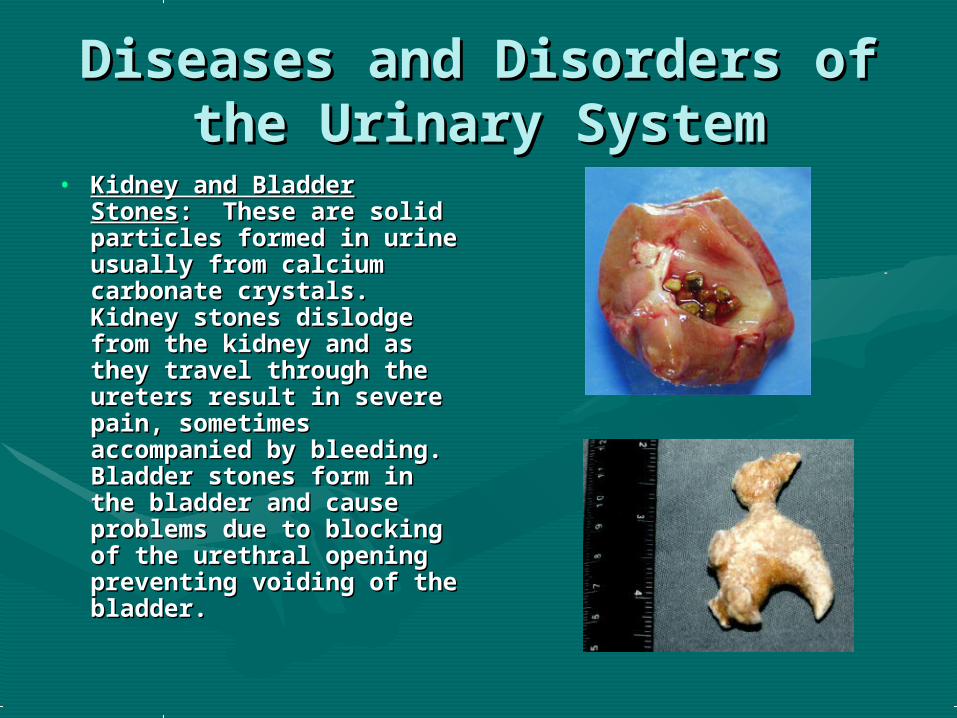
• Kidney and Bladder Kidney and Bladder StonesStones: These are solid : These are solid particles formed in urine particles formed in urine usually from calcium usually from calcium carbonate crystals. carbonate crystals. Kidney stones dislodge Kidney stones dislodge from the kidney and as from the kidney and as they travel through the they travel through the ureters result in severe ureters result in severe pain, sometimes pain, sometimes accompanied by accompanied by bleeding. Bladder stones bleeding. Bladder stones form in the bladder and form in the bladder and cause problems due to cause problems due to blocking of the urethral blocking of the urethral opening preventing opening preventing voiding of the bladder.voiding of the bladder.