the use of advanced imaging in the diagnosis of tb
TRANSCRIPT

The use of advanced imaging in the diagnosis of TB
Pierre Goussard , Robert Gie
Tygerberg Children`s Hospital and University of Stellenbosch

Imaging
• Bronchoscopy
• Tracheo-bronchograms
• Chest CT-scan
• Ultrasound
• TBNA
• FDG scan
• MRI
• Virtual bronchoscopy

TB airway obstruction model

TB airway obstruction : The present • We we were able to demonstrate that bronchoscopy has an important role to
play in accurately assessing the site and degree of airway narrowing due to TB lymph node compression.
• The correct assessment is essential in determining whether the affected child requires surgical or medical treatment.
• There was a statistically significant correlation between bronchoscopic and Chest CT scan findings with both of these investigations identifying bronchus intermedius and the left main bronchus as the most common sites as well as the most severely compressed regions of the airways.
• One third of children with clinical and radiologically significant airway narrowing will need surgical intervention.
• Outcome of surgery was excellent resulting in significant improvement in airway size with low morbidity and no mortality.
• The diagnostic yield during bronchoscopy was been improved by the application of TBNA and Xpert MTB/RIF, although there is still significant room for improvement.

TB airway obstruction : The past
• Well known that TB cause airway obstruction • Locations and cause of airway obstruction was not
well defined• Imaging of airway obstruction was lacking• Bronchoscopy was mostly used to collect sample for
culture• Rigid bronchoscopy was the only instrument
available • Natural history of TB airway obstruction was not
well documented• Which children and the timing of surgical
intervention was not sorted out

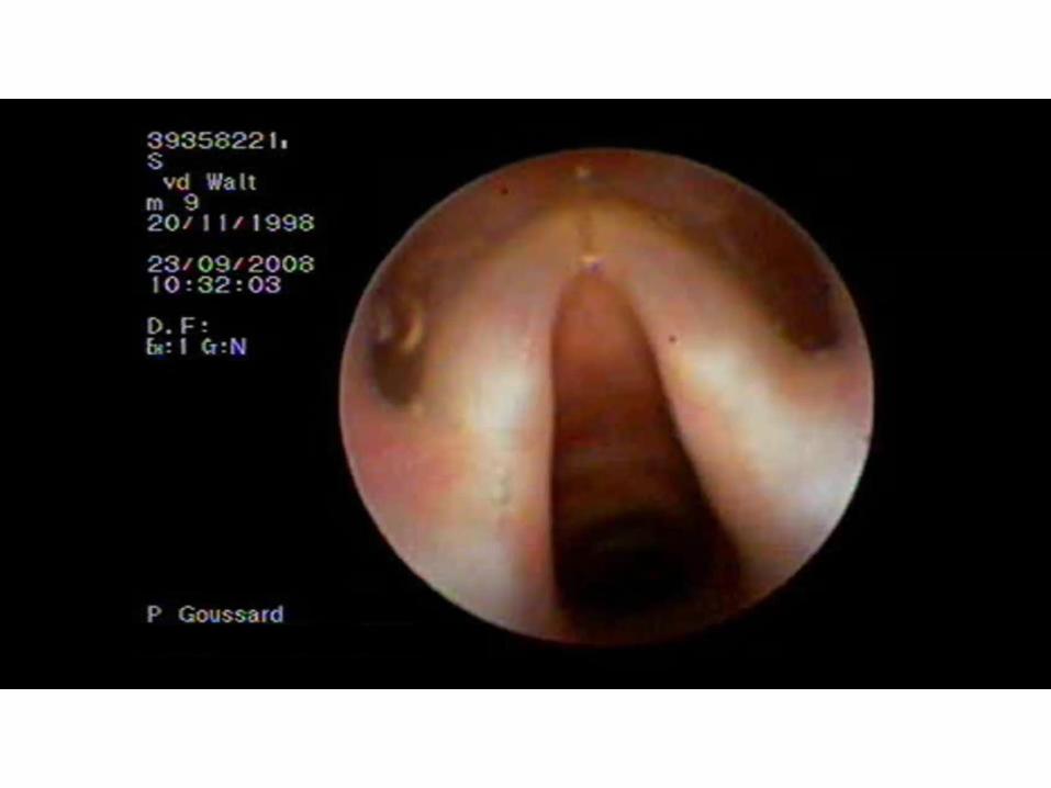
Tracheo-bronchogram

Bronchoscopy
• Diagnostic
• Samples for cultures

Indications for bronchoscopy• Not indicated for routine specimen collection as the
yield from BAL is less than gastric lavage• To confirm the diagnosis of PTB. Bronchial obstruction
may be present in the absence of visibly enlarged hilarand mediastinal lymph nodes on a chest radiograph.
• To determine the degree of airway obstruction in complicated disease.
• For the endoscopic enucleation of glands that have ulcerated into the airway, and for the removal of granulation tissue and caseous material causing airway obstruction.
• To evaluate the response to treatment in children with complicated airway disease

Gland herniating into airway
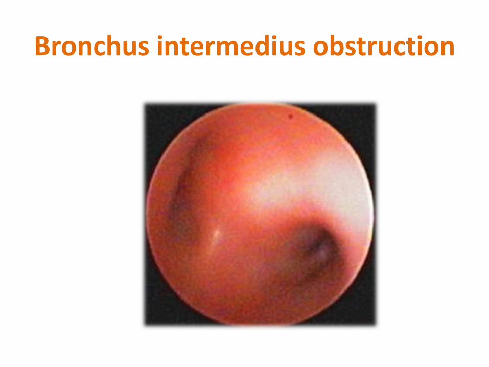
Bronchus intermedius obstruction

tracheal compression

Bronchus intermedius obstruction

Samples for culture
• BAL
• TBNA
• Endobronchial tissue
• Transbronchial biopsy tissue

Endobronchial lumen biopsy

TBNA


TBNA

Excamples of diagnosis made by TBNA
Ziehl–Neelsen stain showing numerous acid fast bacilli. ZN 1,000.

TBB

Bronchus intermedius obstruction


Chest CT-scan


Lymphadenopathy was common, but only 46 patients had lymph nodes greater than 1 cm.Enhancement characteristics of tuberculous adenopathy differ from that described previously. Typical enhancement was ghost-like rather than discreet ring enhancing with alow-density centre. The subcarinal region is most frequently involved and is also the site of the largest lymph-node masses. The presence of lymph-node groups at other recognisedsites adds confidence when there is doubt, as multifocal involvement is common. 25% of patients with hilar adenopathy may have bronchial compression in childhood

• Chest CT--scan should be done in children with clinically and radiological significant airway compression to determine the location of glandular involvement and the relationship of these glands to the airways.
• CT –scan is useful to determine the nature of mediastinal glands and to demonstrate the typical ghost like enhancement of the rims of the lymph glands after contrast administration .
• The most common location for tuberculosis lymph gland enlargement in children with tuberculosis:– subcarinal (90%)– right hilar (74%)– left hilar (72%),– bilateral hilar (61%)– anterior mediastinum (79%)
• The largest group of nodes is located in the subcarinal area (87%). The most common site of airway compression was the left main bronchus (21%), right main bronchus (14%) and bronchus intermedius (8%)
Andronikou S et al. CT scanning for the decection of tuberculous
mediastinal and hilar lymphadenopathy in hildren. Ped Radiology
2004;34:232-236

Ghostlike enhancement Ring-enhancing with low-density centres.

Lymphobronchial TB
1 2
3 4
Obstruction Consolidation
Necrosis Cavitation

Age
2
8
19
19
50
0 10 20 30 40 50 60
10-12 years
6-10 years
2-6 years
1-2 years
0-12 months
Number of patients

Why so young?
• It is well documented that children under 5 are more likely to have nodal disease
• Younger airways are softer and more compressible
• Younger airways are narrower; BI in a 1 year old is only 3 mm in diameter!
• However, not all children with PTB will develop LBTB; incidence in the literature varies from 28%(1) to 38%(2)
1.) Weber AL, Bird KT, Janower ML. Primary tuberculosis in childhood with particular emphasis on changes affecting the tracheobronchial tree. Am J Roentgenol Radium Ther Nucl Med 1968; 103(1):123-132.2.) Theart AC, Marais BJ, Gie RP, Hesseling AC, Beyers N. Criteria used for the diagnosis of childhood tuberculosis at primary health care level in a high-burden, urban setting. Int J Tuberc Lung Dis 2005; 9(11):1210-1214

Sites of nodes
• Subcarinal
• Right paratracheal
• Right hilar
• Right azygo-oesophageal
• Left hilar
• Right paracardiac

Sites of Obstructions
Trachea
LMB
LUL
LLL
RMB
BI
RLL

Number of compressions by site and severity
Site Mild Moderate Severe Complete Total
BI 15 24 18 16 73
LMB 22 25 7 9 63
Trachea 44 16 2 0 62
RLL 16 6 1 3 26
RMB 10 9 1 0 20
LLL 7 2 0 1 10
LUL 3 0 1 1 5
All sites 117 82 30 30 259

Tracheal compressions mostly “mild”
Airway most likely to be compressed is BI
Airway most likely to show “severe” or “complete” compression is BI
“Severe” and “complete” compressions made up almost half of the BI compressions

Number of compressions by age and severity
Age N Mild Moderate Severe Complete Total Comp/pt
0-1 yrs 50 58 41 22 19 140 2.8
1-2 yrs 19 26 15 4 8 53 2.8
3-6 yrs 19 20 20 2 1 43 2.3
6-10 yrs 8 10 5 2 1 18 2.3
10-13 yrs 2 3 1 0 1 5 2.5
All Ages 98 117 82 30 30 259 2.6


Coronal minimum intensity projection (MiniP) image showing compression of the bronchus intermedius in a “nutcracker” fashion. (arrows) by lymphadenopathy (black stars)

Axial CT scan post contrast demonstrates right hilar and subcarinal lymphadenopathy resulting in severe circumferential compression of the bronchus intermedius (collaring) (white arrow) and moderate compression of the left main bronchus against the left pulmonary artery

Axial CT scan on lung windows demonstrates air-trapping of the right lung as a result of mild to moderate compression of the bronchus intermedius

Milliary TB


BEF

Contrast study done with water-soluble contrast medium : demonstrating BOF too the left main bronchus . BOF demonstrated in
88% of cases (n =15)

Figure 2 a and b: CT scan chest: A direct communication is demonstrated between the oesophagus and the left main bronchus.

Figure 1 a and b: Axial CT scans in a child with BOF involving the left main bronchusContrast enhanced axial CT immediately caudal to the carina demonstrates a calcified subcarinal lymph node that abuts the left main bronchus and has eroded into the oesophagus and projects into the lumen. In addition there is compression of the bronchus intermedius between the subcarinal node and a large right hilar node. Lung window more caudally demonstrates a large amount of air in the distal third of the oesophagus. This axial slcie also demonstrates a solitary parenchymal nodule representing a Ghon focus at the right cardiophrenic angle.

Bronchoscopy demonstrates left sided fistula just below the carina
Bronchoscopy demonstrates methylene blue in the airway after instillation in oesophagusdemonstrating the BOF

Which is better CT-scan or scope?

Conclusions: Flexible bronchoscopy remains a very relevant tool in the diagnostic and therapeutic management of childhood pulmonary tuberculosis but resulted in treatment modifi cation or microbiological proof in a minority of our patients. We propose thatfl exible bronchoscopy in children with pulmonary tuberculosis be limited to those who show tracheobronchial luminal narrowing on an initial CT scan.
Arch Dis Child 2010;95:125–129.

Conclusion 3-D VR demonstrates a very good correlation with FTB in
determining airway compression caused by TB lymphadenopathy in children.
In combination with FTB, 3- D VR adds confidence to the bronchoscopy
findings and complements FTB by adding additional information on the
status of the airway distal to severe obstructions unreachable by FTB.

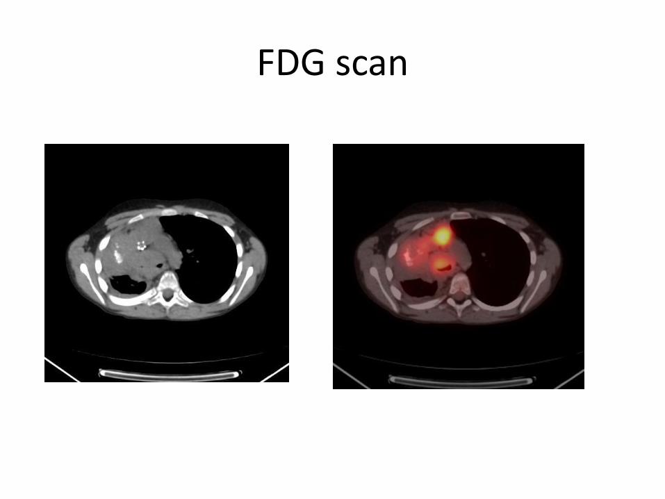
FDG scan

MR
Conclusion: Lung parenchymal necrosis in primary pulmonary TB in children may be of low signal intensity on T2 and STIR magnetic resonance imaging. This may be distal to lymphobronchial obstruction and is probably due to the caseating necrosis.
J Thorac Imaging 2011;00:000–000

Postcontrast CT scan of the lungs demonstrating central areas of lowdensity (arrows) within an area of airspace consolidation. Notethe lack of vessels, bronchograms, and enhancement in the areasof low density, representing lung necrosis. In contrast, theairspace consolidation demonstrates enhancement, vessels, andair bronchograms.

STIR MRI demonstrates that one area of CT low density demonstrates high signal (white arrow) intensity, whereas the other demonstrates low signal intensity (black arrow) presumably due to different stages of necrosis.

Postgadolinium T1-weighted MRI demonstrates that the areasof CT low density lack enhancement (arrows), whereas theairspace consolidation enhances.


Why should we treat TB correctly…... Most get better anyway
• Airway related complications– Broncheal stenosis – Balloon Dilatations
• Parenchymal complications– Cystic lung disease and volume loss – lobectomy /
pneumonectomy
• Bronchiectasis– HIV/ BOF related – asymptomatic / symptomatic
• Fibrosing mediastinitis: – Lethal disease

Complications of PTB
1. Bronchial stenosis2. Bronchiectasis3. Hemoptysis4. Lung cysts5. Fibrosing
mediastinitis


Fibrosing mediastinatis

TB empyema
• Pleural involvement due to TB is believed to result from direct spread of caseous material from a sub-pleural parenchymal or lymph node focus, or from haematogenous spread.
• The degree of pleural pathology depends on the dose and virulence of bacilli that enter the pleural space and the immune status of the patient.
• The presence of caseous material in the pleural space triggers a hypersensitivity inflammatory response which leads to the accumulation of serous straw-colored fluid containing few tuberculosis bacilli.
• If there is active caseation in the pleural space, thick loculated pus, containing many tuberculosis bacilli will develop.
• The chest CT scan picture includes pleural thickening with enhancement, fluid collections with mediastinal lymph nodes and parenchymal lesions.
• The presence of caseating empyema is normally indicated by a persistent high swinging fever and aspiration of pus can be very difficult due to the fact that it is very thick.

AB


Tuberculosis of the spine


Why should we treat TB correctly…... Most get better anyway
• Airway related complications– Broncheal stenosis – Balloon Dilatations
• Parenchymal complications– Cystic lung disease and volume loss – lobectomy /
pneumonectomy
• Bronchiectasis– HIV/ BOF related – asymptomatic / symptomatic
• Fibrosing mediastinitis: – Lethal disease

Lung biopsy for TB diagnosis


5/51 cases = 9.8%

• The present study is one of the first to describe TB as a cause of DLD in infants and children.
• Our study emphasises that, in a high TB burden country, TB pneumonia cannot be excluded using conventional investigations.
• In the one child treated for pulmonary TB before the OLB, MDR-TB was isolated, necessitating a change of treatment.
• Few studies have described the clinical picture of acute pneumonia caused by M. tuberculosis.
• In a study from Uganda, risk factors associated with severe pneumonia caused by M. tuberculosis were children aged ,5 years (odds ratio [OR] 2.4, 95%CI 1.05–5.9) and contact with a smear-positive TB case (OR 3.0, 95%CI 1.3–6.5).14


The Future
• PC models for predicting PTB
• Virtual bronchoscopy
• EBUS
• Electromagnetc navigation system to biopsy peripheral lung nodules

Presents two methods to analyse airway shape and detect airway pathology from CTimages. Features were extracted using (1) the principal components ofthe airway surface mesh and (2) branch radius and orientation features.These methods were applied to a dataset of 61 TB and non-TB paediatricpatients. Nested cross-validation of the support vector classifier found the sensitivity of detecting TB to be 86% and a specificity of 91% for the first 10 PCA modes while radius based features had a sensitivity of 86% and a specificity of 94%. These methods show the potential of computer assisted detection of TB and other airway pathology fromairway shape deformation.


Virtual bronchoscopy

Not PTB
• Lymphoma
• Foreign body aspiration
• Vascular and cardiac abnormalities
• Bronchogenic cyst
• Cryptococcus neoformans


Chest CT-scan: Fig 1 anterior mediastinal lymphnodes and para tracheal lymphnodes causing tracheal compression from the right side. Fig 2 Subcarinal lymphnodes and anterior mediastinal glands causing compression of carina and right main bronchus
Fig 1 Fig 2

Bronchoscopy
Bronchoscopy: Fig a demonstrates tracheal compression and fig b external compression of both Right upper lobe bronchus and bronchus intermedius
Fig a Fig b

Special investigations
•Enucleation of lymph glands
– Paratracheal and Subcarinal
– Histology• Granulomatous
inflammation
• Stain positive for yeast cells
– Cytology• Cryptococcus neoformans
• Tuberculous Zn and culture negative