the use of chlorobenzene as a probe molecule in molecular
TRANSCRIPT
The Use of Chlorobenzene as a Probe Molecule in MolecularDynamics SimulationsYaw Sing Tan,†,‡ David R. Spring,† Chris Abell,† and Chandra Verma*,‡,§,⊥
†Department of Chemistry, University of Cambridge, Lensfield Road, Cambridge CB2 1EW, United Kingdom‡Bioinformatics Institute (A*STAR), 30 Biopolis Street, #07-01 Matrix, Singapore 138671§Department of Biological Sciences, National University of Singapore, 14 Science Drive 4, Singapore 117543⊥School of Biological Sciences, Nanyang Technological University, 60 Nanyang Drive, Singapore 637551
*S Supporting Information
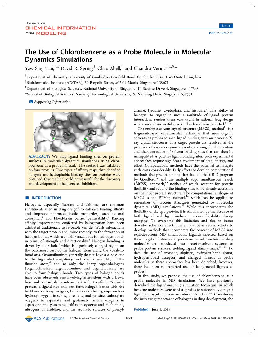
ABSTRACT: We map ligand binding sites on proteinsurfaces in molecular dynamics simulations using chlor-obenzene as a probe molecule. The method was validatedon four proteins. Two types of affinity maps that identifiedhalogen and hydrophobic binding sites on proteins wereobtained. Our method could prove useful for the discoveryand development of halogenated inhibitors.
■ INTRODUCTION
Halogens, especially fluorine and chlorine, are commonsubstituents used in drug design1 to enhance binding affinityand improve pharmacokinetic properties, such as oralabsorption2 and blood-brain barrier permeability.3 Bindingaffinity improvements conferred by halogenation have beenattributed traditionally to favorable van der Waals interactionswith the target protein and, more recently, to the formation ofhalogen bonds, which are highly analogous to hydrogen bondsin terms of strength and directionality.4 Halogen bonding isdriven by the σ-hole,5 which is a positively charged region onthe outermost part of the halogen atom along the covalent-bond axis. Organofluorines generally do not have a σ-hole dueto the high electronegativity and low polarizability of thefluorine atom,6 and so only the heavy organohalogens(organochlorines, organobromines and organoiodines) areable to form halogen bonds. Two types of halogen bondshave been observed: one involving interactions with a Lewisbase and one involving interactions with π-surfaces. Within aprotein, a ligand not only can form halogen bonds with thebackbone carbonyl oxygens, but also side chain groups such ashydroxyl oxygens in serine, threonine, and tyrosine, carboxylateoxygens in aspartate and glutamate, amide oxygens inasparagine and glutamine, sulfurs in cysteine and methionine,nitrogens in histidine, and the aromatic surfaces of phenyl-
alanine, tyrosine, tryptophan, and histidine.7 The ability ofhalogens to engage in such a multitude of ligand−proteininteractions renders them very useful in rational drug designwhere several successful case studies have been reported.8−10
The multiple solvent crystal structure (MSCS) method11 is afragment-based experimental technique that uses organicsolvents as probes to map ligand binding sites on proteins. X-ray crystal structures of a target protein are resolved in thepresence of various organic solvents, allowing for the locationand characterization of solvent binding sites that can then bemanipulated as putative ligand binding sites. Such experimentalapproaches require significant investment of time, energy, andeffort. Computational methods have the potential to mitigatesuch costs considerably. Early efforts to develop computationalmethods that predict binding sites include the GRID programby Goodford12 and the multiple copy simultaneous search(MCSS) approach,13 neither of which account for proteinflexibility and require the binding sites to be already accessibleon the input protein structure. The computational analogue ofMSCS is the FTMap method,14 which can be applied toensembles of protein structures generated by moleculardynamics (MD) simulations.15 While this incorporates theflexibility of the apo protein, it is still limited by the absence ofboth ligand and ligand-induced protein flexibility duringmapping. To overcome this limitation and also to betterdescribe solvation effects, there have been recent efforts todevelop methods that incorporate the concept of MSCS intoexplicit-solvent MD simulations. Ligands selected based ontheir drug-like features and prevalence as substructures in drugmolecules are introduced into protein−solvent systems toprobe protein surfaces, yielding ligand affinity maps.16−21 Todate, the use of aromatic, aliphatic, hydrogen-bond donor,hydrogen-bond acceptor, and charged ligands as probemolecules in these approaches has been described; however,there has been no reported use of halogenated ligands asprobes.In this study, we propose the use of chlorobenzene as a
probe molecule in MD simulations. We have previouslydescribed the ligand-mapping simulation technique, in whichbenzene molecules were used as probes to successfully design aligand to target a protein−protein interaction.20 Consideringthe increasing importance of halogens in drug development, the
Published: June 8, 2014
Letter
pubs.acs.org/jcim
© 2014 American Chemical Society 1821 dx.doi.org/10.1021/ci500215x | J. Chem. Inf. Model. 2014, 54, 1821−1827
ligand-mapping method was refined to probe for both aromaticand halogen interaction sites. Four proteins, for whichstructural data on halogenated ligands is available and whichexhibit significant conformational changes upon ligand binding,were chosen to validate our enhanced ligand-mapping method-ology.
■ RESULTS AND DISCUSSIONHalogenation of compounds is commonly used during the drugdevelopment process. In particular, chlorine is a moderatehalogen-bond acceptor, and the C−Cl bond is relatively stable,which may explain why it is the most prevalent halogen amongsmall-molecule modulators of protein−protein interactions22
and launched drugs listed in Thomson Reuters Pharma over thelast century.23 In order to explore the sites for halogeninteractions, we chose chlorobenzene as the candidate probe sothat in addition to chlorine interactions, the phenyl group ofchlorobenzene would concurrently also be able to probe forhydrophobic binding sites.We chose four proteins involved in protein−protein
interactions to test the ability of our method to reproduceexperimental data: MDM2, MCL-1, interleukin-2, and Bcl-xL.These proteins have available structural data of halogenatedligands in the Protein Data Bank (PDB) and display significantconformational changes on ligand binding. In particular, some(MDM2, MCL-1, Bcl-xL) or all (interleukin-2) of the halogenbinding sites are absent in both the unbound states of theseproteins as well as when they are bound to their natural proteinor peptide partners. The protein structures were specificallyextracted from these complexes and used to initiate the ligand-mapping simulations (see Supporting Information for details),thus ensuring a rigorous test of the utility of our method.Several changes to the protocol were introduced compared
to our previous study in which benzene was used as the probemolecule.20 Simulation conditions were initially refined usingMDM2, which has the most extensive structural data ofhalogenated ligands among the four test proteins, beforeextending them to the other three proteins. Chlorobenzene wasused at a lower concentration of 0.15 M, as compared to 0.2 M
for benzene, since significant aggregation of chlorobenzene wasobserved at the higher concentration. Such aggregation isundesirable because it lowers the effective ligand concentrationand prevents thorough sampling of the protein surface.17 Thelower ligand concentration necessitated an increase in thesimulation length to 10 ns from 5 ns used previously, for bettersampling. Ligand-induced denaturation of the target protein24
due to the increased simulation length was not an issue asstable trajectories were observed for all four proteins (FigureS1). Lastly, due to the increased lipophilicity of chlorobenzenecompared to benzene, a lower proportion of chlorobenzeneswas observed to remain in solution during the simulations. Thisnecessitated the utilization of a higher cutoff isocontour valuefor visualization of the occupancy grids. This was set at fivetimes the threshold bulk value, which is defined as the highestisovalue at which chlorobenzene carbon or chlorine atoms arerespectively detected in the bulk solvent. A comparison of thearomatic carbon occupancy maps of MDM2 generated frombenzene-mapping and chlorobenzene-mapping simulationsshowed that they concurred on the prediction of mosthydrophobic binding sites (Figure S2). The nonconsensussites are not known to bind any ligands. This suggests thatchlorobenzene is a suitable replacement for benzene as a probefor hydrophobic binding sites.
MDM2. The MDM2−p53 interaction is one of the moststudied protein−protein interactions and there is extensivestructural data available in the PDB of MDM2 complexes. Theprotein structure used to initiate the ligand-mapping simu-lations was obtained from the complex of MDM2 with the p53transactivation domain peptide (PDB 1YCR).25
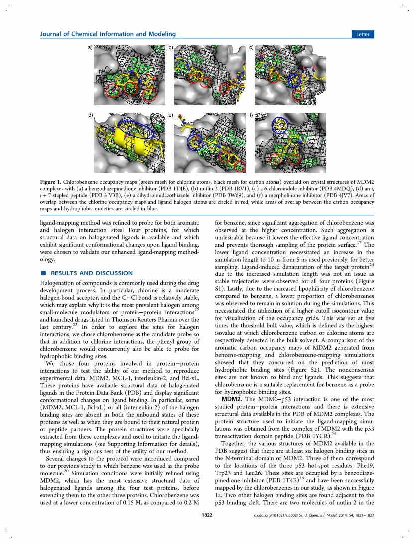
Together, the various structures of MDM2 available in thePDB suggest that there are at least six halogen binding sites inthe N-terminal domain of MDM2. Three of them correspondto the locations of the three p53 hot-spot residues, Phe19,Trp23 and Leu26. These sites are occupied by a benzodiaze-pinedione inhibitor (PDB 1T4E)26 and have been successfullymapped by the chlorobenzenes in our study, as shown in Figure1a. Two other halogen binding sites are found adjacent to thep53 binding cleft. There are two molecules of nutlin-2 in the
Figure 1. Chlorobenzene occupancy maps (green mesh for chlorine atoms, black mesh for carbon atoms) overlaid on crystal structures of MDM2complexes with (a) a benzodiazepinedione inhibitor (PDB 1T4E), (b) nutlin-2 (PDB 1RV1), (c) a 6-chloroindole inhibitor (PDB 4MDQ), (d) an i,i + 7 stapled peptide (PDB 3 V3B), (e) a dihydroimidazothiazole inhibitor (PDB 3W69), and (f) a morpholinone inhibitor (PDB 4JV7). Areas ofoverlap between the chlorine occupancy maps and ligand halogen atoms are circled in red, while areas of overlap between the carbon occupancymaps and hydrophobic moieties are circled in blue.
Journal of Chemical Information and Modeling Letter
dx.doi.org/10.1021/ci500215x | J. Chem. Inf. Model. 2014, 54, 1821−18271822
structure of an MDM2/nutlin-2 complex (PDB 1RV1),27 asshown in Figure 1b. One molecule is bound to the main p53binding site, and the other is bound, through one of itsbromophenyl moieties, to a second nutlin interaction site; thissite is occluded in the initial MDM2 structure. The chlorinedensities coincide with the location of the bromine atom of thelatter nutlin molecule, indicating a halogen binding site. This isstabilized by a halogen bond with the carbonyl oxygen ofTyr100. This second nutlin interaction site was also mapped bythe carbon atoms of the chlorobenzene probes, indicating thatit might be a potential druggable site that could be explored forligand binding. The fifth detected halogen binding site isadjacent to the second nutlin interaction site. Similar to theobservation of two nutlin binding sites in the MDM2/nutlin-2complex structure, two chloroindole-based inhibitor moleculesare complexed to MDM2 in the crystal structure of the complex(PDB 4MDQ).28 One is bound at the main binding cleft, whilethe second molecule is bound at a site between the two nutlininteraction sites (Figure 1c). The locations of the phenyl andchloroindole groups of the second molecule were recapitulatedby the chlorobenzene occupancy maps. The sixth halogenbinding site is found in the vicinity of this ‘intermediate site’and is formed partially by residues 17−24 of MDM2 (PDB4MDN).28 These N-terminal residues were absent in our inputMDM2 structure and could explain why this last halogenbinding site was not detected by the chlorobenzenes. Anadditional set of chlorobenzene-mapping simulations wasinitiated using this MDM2 structure to see if the last halogenbinding site could be detected in the presence of a longer N-terminal tail. The chlorobenzene map generated showeddistinct chlorine density close to the chloro substituent of theligand in the 4MDN structure (Figure S3), suggesting that amore complete MDM2 structure was indeed necessary forsuccessful (i.e., more accurate) detection of this halogenbinding site.
Besides mapping halogen binding sites, the chlorobenzenesalso found hydrophobic sites, even those that require significantprotein conformational changes. A total of four such cryptichydrophobic binding sites, which are not present in the initialMDM2 structure, were identified in the ligand-mappingsimulations. The ‘intermediate site’ mentioned earlier is anextension of the Leu26 binding site (Figure 1c) and is one ofthe cryptic hydrophobic sites revealed by the chlorobenzeneprobes. Similarly, although the binding site for the hydrocarbonstaple of an i, i + 7 stapled peptide (PDB 3V3B)29 was notvisible in the initial protein structure, the chlorobenzenes wereable to successfully map it. (Figure 1d). In Figure 1e, thepyrrolidine moiety of the inhibitor (PBD 3W69)30 pushesagainst the walls of the Phe19 binding site, creating anotherhydrophobic interaction site by induced-fitting. Finally, inFigure 1f, the benzyl group of a morpholinone inhibitor (PDB4JV7)31 engages in an edge-face π−π interaction with Phe55 ona shallow hydrophobic binding region that is obscured by sidechains in the input structure.
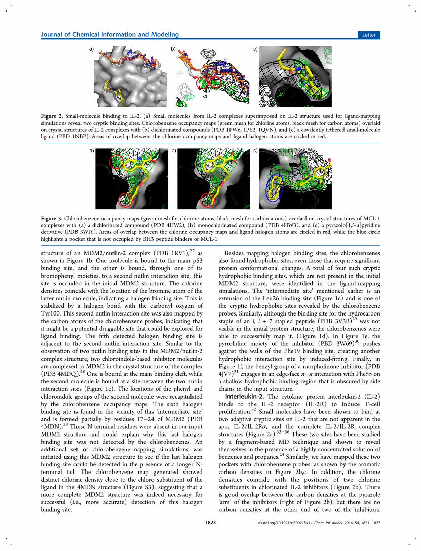
Interleukin-2. The cytokine protein interleukin-2 (IL-2)binds to the IL-2 receptor (IL-2R) to induce T-cellproliferation.32 Small molecules have been shown to bind attwo adaptive cryptic sites on IL-2 that are not apparent in theapo, IL-2/IL-2Rα, and the complete IL-2/IL-2R complexstructures (Figure 2a).33−36 These two sites have been studiedby a fragment-based MD technique and shown to revealthemselves in the presence of a highly concentrated solution ofbenzenes and propanes.24 Similarly, we have mapped these twopockets with chlorobenzene probes, as shown by the aromaticcarbon densities in Figure 2b,c. In addition, the chlorinedensities coincide with the positions of two chlorinesubstituents in chlorinated IL-2 inhibitors (Figure 2b). Thereis good overlap between the carbon densities at the pyrazole‘arm’ of the inhibitors (right of Figure 2b), but there are nocarbon densities at the other end of two of the inhibitors.
Figure 2. Small-molecule binding to IL-2. (a) Small molecules from IL-2 complexes superimposed on IL-2 structure used for ligand-mappingsimulations reveal two cryptic binding sites. Chlorobenzene occupancy maps (green mesh for chlorine atoms, black mesh for carbon atoms) overlaidon crystal structures of IL-2 complexes with (b) dichlorinated compounds (PDB 1PW6, 1PY2, 1QVN), and (c) a covalently tethered small-moleculeligand (PBD 1NBP). Areas of overlap between the chlorine occupancy maps and ligand halogen atoms are circled in red.
Figure 3. Chlorobenzene occupancy maps (green mesh for chlorine atoms, black mesh for carbon atoms) overlaid on crystal structures of MCL-1complexes with (a) a dichlorinated compound (PDB 4HW2), (b) monochlorinated compound (PDB 4HW3), and (c) a pyrazolo[1,5-a]pyridinederivative (PDB 3WIY). Areas of overlap between the chlorine occupancy maps and ligand halogen atoms are circled in red, while the blue circlehighlights a pocket that is not occupied by BH3 peptide binders of MCL-1.
Journal of Chemical Information and Modeling Letter
dx.doi.org/10.1021/ci500215x | J. Chem. Inf. Model. 2014, 54, 1821−18271823

Experimental data have shown that binding affinity wasenhanced when a furoic acid fragment was added, but notwhen it was replaced with a neutral phenylacetamide group.36
This suggests that the extension provides only favorableelectrostatic interactions and does not actually occupy aparticular hydrophobic binding site, which is in agreementwith the lack of carbon density in the region.MCL-1. MCL-1 is an antiapoptotic protein that suppresses
apoptosis by sequestering the pro-apoptotic Bcl-2 familyproteins37 and is found to be overexpressed in a variety ofhuman cancers.38−40 It contains a hydrophobic binding groovethat binds the BH3 α-helices of these proteins. BH3 helixmimicry has been the basis for the development of MCL-1inhibitors to sensitize cancer cell apoptosis for anticancertherapy.41 Such efforts have led to the development of smallmolecules that bind at the BH3 binding pocket and three ofthem are shown in Figure 3.The crystal structure of MCL-1 bound to a dichlorinated
compound (Figure 3a) shows that there are at least twohalogen interaction sites within the binding cavity,42 the deeperof which is occluded in the starting MCL-1 conformationderived from an MCL-1/Bim BH3 peptide complex structure(PDB 3KJ0). Both sites were successfully detected in theligand-mapping simulations. One of the chlorines forms ahalogen bond with the carbonyl oxygen of Ala227, whereas theother chlorine found at the lower part of the pocket engages inpurely van der Waals interactions with the protein (Figure 3b).The occupancy maps also indicated chlorine density at one ofthe meta-positions of the chlorinated phenyl group (Figure3a,b). This points to a possible halogen binding site, where ahalogen bond could be formed with the carbonyl oxygen of thenearby Leu246.The BH3 binding pocket of MCL-1 is very deep, but its full
capacity is not utilized by the BH3 peptides. The full depth ofthe pocket has been explored by small molecules, however, asshown by the 4-chloro-3,5-dimethylphenyl groups of theligands in Figure 3a,b and the naphthyl group of the ligandin Figure 3c. Significant protein backbone movements arerequired for the accommodation of these hydrophobicmoieties,42,43 but this does not prevent the chlorobenzene
probes from inducing these changes and reproducing the ligandinteractions at the bottom of the pocket.
Bcl-xL. Like MCL-1, Bcl-xL is an antiapoptotic protein thatneutralizes proapoptotic proteins such as Bax and Bak bybinding to their α-helical BH3 domains. It is frequentlyoverexpressed in cancer cells, leading to chemotherapyresistance.44,45 The use of BH3 mimetics to inhibit itsinteractions with proapoptotic proteins is therefore of greatinterest in cancer therapy.41
There are two large binding pockets in Bcl-xL, as shown inFigure 4a. The deeper and more well-defined pocket isoccupied by the 4-chlorophenyl group of the ligand, whilethe shallower pocket is occupied by a thiophenol group. Thechlorine interaction site in the deep pocket is occluded in theapo structures46−48 as well as in the structure of the complexwith the Bak peptide (PDB 1BXL) that was used as the initialstructure for the ligand-mapping simulations. Nevertheless, thisinteraction was detected by the chlorobenzene probes, as thechlorine densities derived from the simulations coincide withthe position of the chlorine atom in the 4-chlorophenyl group(Figure 4a). Another chlorine interaction site was identified inthe shallow pocket, near the meta-position of the phenyl group.Currently, there is no structural data showing ligands engagingthe shallow pocket with halogenated groups. Known Bcl-xLinhibitors having a similar binding mode as the ligand in Figure4a could be modified to present a 3-chlorophenyl group at theshallow pocket to enhance their binding affinities.The compound in Figure 4b is bound to a different Bcl-xL
conformation from that in Figure 4a but engages the samebinding pockets. The benzothiazole group of the compound isshown fully utilizing the capacity of the deeper Bcl-xL pocket,just like the 4-chlorophenyl group in Figure 4a. Although thelocation of these two hydrophobic moieties in Bcl-xL wasoccluded in the starting structure used for the ligand-mappingsimulations, chlorobenzene probes were able to expose them byinducing side chain movements to expand the pocket, asevidenced by the presence of carbon densities deep in thepocket.Recent crystal structures of Bcl-xL complexes reported by
Brady et al.49 show the difluorophenyl groups of twobenzoylurea inhibitors projecting into a previously unreported
Figure 4. Chlorobenzene occupancy maps (green mesh for chlorine atoms, black mesh for carbon atoms) overlaid on crystal structures of Bcl-xLcomplexes with (a) a chlorinated inhibitor (PDB 2YXJ), (b) a benzothiazole inhibitor (PDB 3ZLO), and (c) a benzoylurea inhibitor (PDB 4C5D).Areas of overlap between the chlorine occupancy maps and ligand halogen atoms are circled in red, while the blue circle highlights a pocket that isnot occupied by BH3 peptide partners of Bcl-xL. (d) Comparison of the position of helix α1 in monomeric (green) and dimeric (orange) Bcl-xL,with the benzoylurea inhibitor (yellow sticks) superimposed (PDB 4C5D).
Journal of Chemical Information and Modeling Letter
dx.doi.org/10.1021/ci500215x | J. Chem. Inf. Model. 2014, 54, 1821−18271824

pocket near the BH3 binding groove (Figure 4c). Thishydrophobic pocket was not detected in the ligand-mappingsimulations; however, another halogen interaction site in Bcl-xL, as indicated by the bromophenyl group of the benzoylureainhibitor, was detected by the chlorobenzene probes. Thestructure of Bcl-xL in one of the crystal structures obtained byBrady et al. was compared to the Bcl-xL solution structure usedfor the simulations (Figure 4d). The most striking differencewas in the position of helix α1, which was seen to undergo asignificant conformational change between the two structures.Bcl-xL exists as a dimer in the newly reported crystal structures.On dimerization, a domain swap involving helix α1 occursbetween the monomers. The original position of α1 is occupiedby the helix α1 from another protein chain. This could possiblyhave altered the dynamics of the Bcl-xL subunit significantlyenough to allow the opening of this cryptic pocket, whichwould otherwise remain closed in the monomeric state, thusexplaining why it was not detected in the ligand-mappingsimulations.
■ CONCLUSION
We have described a reliable method of mapping halogen andhydrophobic binding sites on protein surfaces by incorporationof chlorobenzene molecules into MD simulations. This is anevolution of our previous implementation of ligand-mappingsimulations that used benzene probes,20 since two types ofaffinity maps can now be obtained in a single set of simulations.We have also demonstrated that the chlorobenzene probes areable to expose binding sites that are occluded in the inputstructures of four proteins involved in protein−proteininteractions. Such sites are of utmost importance in the designof small molecule inhibitors of protein−protein interactions, asthe target protein may appear flat and undruggable even whenbound to its protein or peptide partner.50 The ensemble ofsimulated protein conformations with these cryptic sitesexposed may then be used for small molecule docking instructure-based virtual screening. Additionally, affinity mapsobtained from ligand-mapping simulations could be used toguide the elaboration of fragments and hit compounds, byrevealing favorable sites for the addition of halogens andhydrophobic groups.The ability of the method to reproduce known halogen
binding sites in our test set of proteins is rather remarkable,taking into consideration the inaccuracies of the force field usedto describe halogen bonding between chlorobenzene probesand the protein. Like all other classical force fields used inbiomolecular modeling, the general AMBER force field(GAFF)51 assigns isotropic partial charges to atoms and doesnot account for the anisotropic distribution of the electrostaticpotential on halogen atoms. Halogen atoms are usually assignednegative partial charges in classical force fields (Table S1),making electrostatic interactions with Lewis bases and π-surfaces incorrectly unfavorable. Efforts are underway to modifyconventional force fields to account for halogen bonding.52−55
We believe that by demonstrating the reproduction ofexperimental data, our results could provide a platform to aidin the discovery of novel halogenated inhibitors and at the sametime, offer optimism for the development of more accuratehalogen-mapping strategies that incorporate modified forcefields.
■ ASSOCIATED CONTENT*S Supporting InformationSimulation setups and analysis procedures. Figures and tablereferred to in the paper. This material is available free of chargevia the Internet at http://pubs.acs.org.
■ AUTHOR INFORMATIONCorresponding Author*E-mail: [email protected] authors declare no competing financial interest.
■ ACKNOWLEDGMENTSWe are grateful for funding from BMSI, Agency for Science,Technology and Research (Singapore). Y.S.T. was supportedby an A*STAR Graduate Scholarship.
■ REFERENCES(1) Hernandes, M. Z.; Cavalcanti, S. M. T.; Moreira, D. R. M.; deAzevedo, W. F., Jr.; Lima Leite, A. C. Halogen atoms in the modernmedicinal chemistry: hints for the drug design. Curr. Drug Targets2010, 11, 303−314.(2) Gerebtzoff, G.; Li-Blatter, X.; Fischer, H.; Frentzel, A.; Seelig, A.Halogenation of drugs enhances membrane binding and permeation.ChemBioChem 2004, 5, 676−684.(3) Gentry, C. L.; Egleton, R. D.; Gillespie, T.; Abbruscato, T. J.;Bechowski, H. B.; Hruby, V. J.; Davis, T. P. The effect of halogenationon blood-brain barrier permeability of a novel peptide drug. Peptides1999, 20, 1229−1238.(4) Metrangolo, P.; Resnati, G. Halogen bonding: a paradigm insupramolecular chemistry. Chem.Eur. J. 2001, 7, 2511−2519.(5) Clark, T.; Hennemann, M.; Murray, J. S.; Politzer, P. Halogenbonding: the sigma-hole. J. Mol. Model. 2007, 13, 291−296.(6) Metrangolo, P.; Neukirch, H.; Pilati, T.; Resnati, G. Halogenbonding based recognition processes: a world parallel to hydrogenbonding. Acc. Chem. Res. 2005, 38, 386−395.(7) Wilcken, R.; Zimmermann, M. O.; Lange, A.; Joerger, A. C.;Boeckler, F. M. Principles and applications of halogen bonding inmedicinal chemistry and chemical biology. J. Med. Chem. 2013, 56,1363−1388.(8) Xu, Z.; Liu, Z.; Chen, T.; Chen, T.; Wang, Z.; Tian, G.; Shi, J.;Wang, X.; Lu, Y.; Yan, X.; Wang, G.; Jiang, H.; Chen, K.; Wang, S.; Xu,Y.; Shen, J.; Zhu, W. Utilization of halogen bond in lead optimization:a case study of rational design of potent phosphodiesterase type 5(PDE5) inhibitors. J. Med. Chem. 2011, 54, 5607−5611.(9) Hardegger, L. A.; Kuhn, B.; Spinnler, B.; Anselm, L.; Ecabert, R.;Stihle, M.; Gsell, B.; Thoma, R.; Diez, J.; Benz, J.; Plancher, J.-M.;Hartmann, G.; Banner, D. W.; Haap, W.; Diederich, F. Systematicinvestigation of halogen bonding in protein-ligand interactions. Angew.Chem., Int. Ed. 2011, 50, 314−318.(10) Rohde, L. A. H.; Ahring, P. K.; Jensen, M. L.; Nielsen, E. O.;Peters, D.; Helgstrand, C.; Krintel, C.; Harpsoe, K.; Gajhede, M.;Kastrup, J. S.; Balle, T. Intersubunit bridge formation governs agonistefficacy at nicotinic acetylcholine α4β2 receptors: unique role ofhalogen bonding revealed. J. Biol. Chem. 2012, 287, 4248−4259.(11) Mattos, C.; Ringe, D. Locating and characterizing binding siteson proteins. Nat. Biotechnol. 1996, 14, 595−599.(12) Goodford, P. J. A computational procedure for determiningenergetically favorable binding sites on biologically importantmacromolecules. J. Med. Chem. 1985, 28, 849−857.(13) Miranker, A.; Karplus, M. Functionality maps of binding sites: amultiple copy simultaneous search method. Proteins 1991, 11, 29−34.(14) Brenke, R.; Kozakov, D.; Chuang, G.-Y.; Beglov, D.; Hall, D.;Landon, M. R.; Mattos, C.; Vajda, S. Fragment-based identification ofdruggable ’hot spots’ of proteins using Fourier domain correlationtechniques. Bioinformatics 2009, 25, 621−627.
Journal of Chemical Information and Modeling Letter
dx.doi.org/10.1021/ci500215x | J. Chem. Inf. Model. 2014, 54, 1821−18271825
(15) Ivetac, A.; McCammon, J. A. Mapping the druggable allostericspace of G-protein coupled receptors: a fragment-based moleculardynamics approach. Chem. Biol. Drug Des. 2010, 76, 201−217.(16) Seco, J.; Luque, F. J.; Barril, X. Binding site detection anddruggability index from first principles. J. Med. Chem. 2009, 52, 2363−2371.(17) Guvench, O.; MacKerell, A. D., Jr. Computational fragment-based binding site identification by ligand competitive saturation. PLoSComp. Biol. 2009, 5, e1000435.(18) Lexa, K. W.; Carlson, H. A. Full protein flexibility is essential forproper hot-spot mapping. J. Am. Chem. Soc. 2011, 133, 200−202.(19) Bakan, A.; Nevins, N.; Lakdawala, A. S.; Bahar, I. Druggabilityassessment of allosteric proteins by dynamics simulations in thepresence of probe molecules. J. Chem. Theory Comput. 2012, 8, 2435−2447.(20) Tan, Y. S.; Sledz, P.; Lang, S.; Stubbs, C. J.; Spring, D. R.; Abell,C.; Best, R. B. Using ligand-mapping simulations to design a ligandselectively targeting a cryptic surface pocket of polo-like kinase 1.Angew. Chem., Int. Ed. 2012, 51, 10078−10081.(21) Zhu, M.; De Simone, A.; Schenk, D.; Toth, G.; Dobson, C. M.;Vendruscolo, M. Identification of small-molecule binding pockets inthe soluble monomeric form of the Aβ42 peptide. J. Chem. Phys. 2013,139, 035101.(22) Basse, M. J.; Betzi, S.; Bourgeas, R.; Bouzidi, S.; Chetrit, B.;Hamon, V.; Morelli, X.; Roche, P. 2P2Idb: a structural databasededicated to orthosteric modulation of protein-protein interactions.Nucleic Acids Res. 2013, 41, D824−D827.(23) Xu, Z.; Yang, Z.; Liu, Y.; Lu, Y.; Chen, K.; Zhu, W. Halogenbond: its role beyond drug−target binding affinity for drug discoveryand development. J. Chem. Inf. Model. 2014, 54, 69−78.(24) Foster, T. J.; Mackerell, A. D., Jr.; Guvench, O. Balancing targetflexibility and target denaturation in computational fragment-basedinhibitor discovery. J. Comput. Chem. 2012, 33, 1880−91.(25) Kussie, P. H.; Gorina, S.; Marechal, V.; Elenbaas, B.; Moreau, J.;Levine, A. J.; Pavletich, N. P. Structure of the MDM2 oncoproteinbound to the p53 tumor suppressor transactivation domain. Science1996, 274, 948−953.(26) Grasberger, B. L.; Lu, T. B.; Schubert, C.; Parks, D. J.; Carver, T.E.; Koblish, H. K.; Cummings, M. D.; LaFrance, L. V.; Milkiewicz, K.L.; Calvo, R. R.; Maguire, D.; Lattanze, J.; Franks, C. F.; Zhao, S. Y.;Ramachandren, K.; Bylebyl, G. R.; Zhang, M.; Manthey, C. L.; Petrella,E. C.; Pantoliano, M. W.; Deckman, I. C.; Spurlino, J. C.; Maroney, A.C.; Tomczuk, B. E.; Molloy, C. J.; Bone, R. F. Discovery and cocrystalstructure of benzodiazepinedione HDM2 antagonists that activate p53in cells. J. Med. Chem. 2005, 48, 909−912.(27) Vassilev, L. T.; Vu, B. T.; Graves, B.; Carvajal, D.; Podlaski, F.;Filipovic, Z.; Kong, N.; Kammlott, U.; Lukacs, C.; Klein, C.; Fotouhi,N.; Liu, E. A. In vivo activation of the p53 pathway by small-moleculeantagonists of MDM2. Science 2004, 303, 844−848.(28) Bista, M.; Wolf, S.; Khoury, K.; Kowalska, K.; Huang, Y.; Wrona,E.; Arciniega, M.; Popowicz, G. M.; Holak, T. A.; Domling, A.Transient protein states in designing inhibitors of the MDM2-p53interaction. Structure 2013, 21, 2143−2151.(29) Baek, S.; Kutchukian, P. S.; Verdine, G. L.; Huber, R.; Holak, T.A.; Lee, K. W.; Popowicz, G. M. Structure of the stapled p53 peptidebound to Mdm2. J. Am. Chem. Soc. 2012, 134, 103−106.(30) Miyazaki, M.; Naito, H.; Sugimoto, Y.; Yoshida, K.; Kawato, H.;Okayama, T.; Shimizu, H.; Miyazaki, M.; Kitagawa, M.; Seki, T.;Fukutake, S.; Shiose, Y.; Aonuma, M.; Soga, T. Synthesis andevaluation of novel orally active p53-MDM2 interaction inhibitors.Biorg. Med. Chem. 2013, 21, 4319−4331.(31) de Turiso, F. G.-L.; Sun, D.; Rew, Y.; Bartberger, M. D.; Beck,H. P.; Canon, J.; Chen, A.; Chow, D.; Correll, T. L.; Huang, X.; Julian,L. D.; Kayser, F.; Lo, M.-C.; Long, A. M.; McMinn, D.; Oliner, J. D.;Osgood, T.; Powers, J. P.; Saiki, A. Y.; Schneider, S.; Shaffer, P.; Xiao,S.-H.; Yakowec, P.; Yan, X.; Ye, Q.; Yu, D.; Zhao, X.; Zhou, J.; Medina,J. C.; Olson, S. H. Rational design and binding mode duality ofMDM2-p53 inhibitors. J. Med. Chem. 2013, 56, 4053−4070.
(32) Nelson, B. H.; Willerford, D. M., Biology of the interleukin-2receptor. In Advances in Immunology, Dixon, F. J., Ed. 1998; Vol. 70, pp1−81.(33) Arkin, M. R.; Randal, M.; DeLano, W. L.; Hyde, J.; Luong, T.N.; Oslob, J. D.; Raphael, D. R.; Taylor, L.; Wang, J.; McDowell, R. S.;Wells, J. A.; Braisted, A. C. Binding of small molecules to an adaptiveprotein-protein interface. Proc. Natl. Acad. Sci. U. S. A. 2003, 100,1603−1608.(34) Braisted, A. C.; Oslob, J. D.; Delano, W. L.; Hyde, J.; McDowell,R. S.; Waal, N.; Yu, C.; Arkin, M. R.; Raimundo, B. C. Discovery of apotent small molecule IL-2 inhibitor through fragment assembly. J.Am. Chem. Soc. 2003, 125, 3714−3715.(35) Thanos, C. D.; Randal, M.; Wells, J. A. Potent small-moleculebinding to a dynamic hot spot on IL-2. J. Am. Chem. Soc. 2003, 125,15280−15281.(36) Thanos, C. D.; DeLano, W. L.; Wells, J. A. Hot-spot mimicry ofa cytokine receptor by a small molecule. Proc. Natl. Acad. Sci. U. S. A.2006, 103, 15422−15427.(37) Adams, J. M. Ways of dying: multiple pathways to apoptosis.Genes Dev. 2003, 17, 2481−2495.(38) Wuilleme-Toumi, S.; Robillard, N.; Gomez, P.; Moreau, P.; LeGouill, S.; Avet-Loiseau, H.; Harousseau, J. L.; Amiot, M.; Bataille, R.Mcl-1 is overexpressed in multiple myeloma and associated withrelapse and shorter survival. Leukemia 2005, 19, 1248−1252.(39) Boisvert-Adamo, K.; Longmate, W.; Abel, E. V.; Aplin, A. E.Mcl-1 Is Required for Melanoma Cell Resistance to Anoikis. Mol.Cancer Res. 2009, 7, 549−556.(40) Ding, Q.; He, X.; Xia, W.; Hsu, J.-M.; Chen, C.-T.; Li, L.-Y.; Lee,D.-F.; Yang, J.-Y.; Xie, X.; Liu, J.-C.; Hung, M.-C. Myeloid cellleukemia-1 inversely correlates with glycogen synthase kinase-3βactivity and associates with poor prognosis in human breast cancer.Cancer Res. 2007, 67, 4564−4571.(41) Lessene, G.; Czabotar, P. E.; Colman, P. M. BCL-2 familyantagonists for cancer therapy. Nat. Rev. Drug Discovery 2008, 7, 989−1000.(42) Friberg, A.; Vigil, D.; Zhao, B.; Daniels, R. N.; Burke, J. P.;Garcia-Barrantes, P. M.; Camper, D.; Chauder, B. A.; Lee, T.;Olejniczak, E. T.; Fesik, S. W. Discovery of potent myeloid cellleukemia 1 (Mcl-1) inhibitors using fragment-based methods andstructure-based design. J. Med. Chem. 2013, 56, 15−30.(43) Tanaka, Y.; Aikawa, K.; Nishida, G.; Homma, M.; Sogabe, S.;Igaki, S.; Hayano, Y.; Sameshima, T.; Miyahisa, I.; Kawamoto, T.;Tawada, M.; Imai, Y.; Inazuka, M.; Cho, N.; Imaeda, Y.; Ishikawa, T.Discovery of potent Mcl-1/Bcl-xL dual inhibitors by using ahybridization strategy based on structural analysis of target proteins.J. Med. Chem. 2013, 56, 9635−9645.(44) Labi, V.; Erlacher, M.; Kiessling, S.; Villunger, A. BH3-onlyproteins in cell death initiation, malignant disease and anticancertherapy. Cell Death Differ. 2006, 13, 1325−1338.(45) Amundson, S. A.; Myers, T. G.; Scudiero, D.; Kitada, S.; Reed, J.C.; Fornace, A. J. An informatics approach identifying markers ofchemosensitivity in human cancer cell lines. Cancer Res. 2000, 60,6101−6110.(46) Muchmore, S. W.; Sattler, M.; Liang, H.; Meadows, R. P.;Harlan, J. E.; Yoon, H. S.; Nettesheim, D.; Chang, B. S.; Thompson, C.B.; Wong, S. L.; Ng, S. C.; Fesik, S. W. X-ray and NMR structure ofhuman Bcl-x(L), an inhibitor of programmed cell death. Nature 1996,381, 335−341.(47) Manion, M. K.; O’Neill, J. W.; Giedt, C. D.; Kim, K. M.; Zhang,K. Y. Z.; Hockenbery, D. M. Bcl-X-L mutations suppress cellularsensitivity to antimycin A. J. Biol. Chem. 2004, 279, 2159−2165.(48) Wysoczanski, P.; Mart, R. J.; Loveridge, E. J.; Williams, C.;Whittaker, S. B. M.; Crump, M. P.; Allemann, R. K. NMR solutionstructure of a photoswitchable apoptosis activating Bak peptide boundto Bcl-x(L). J. Am. Chem. Soc. 2012, 134, 7644−7647.(49) Brady, R. M.; Vom, A.; Roy, M. J.; Toovey, N.; Smith, B. J.;Moss, R. M.; Hatzis, E.; Huang, D. C. S.; Parisot, J. P.; Yang, H.;Street, I. P.; Colman, P. M.; Czabotar, P. E.; Baell, J. B.; Lessene, G.De-novo designed library of benzoylureas as inhibitors of BCL-XL:
Journal of Chemical Information and Modeling Letter
dx.doi.org/10.1021/ci500215x | J. Chem. Inf. Model. 2014, 54, 1821−18271826
synthesis, structural and biochemical characterization. J. Med. Chem.2014, 57, 1323−1343.(50) Stauber, D. J.; Debler, E. W.; Horton, P. A.; Smith, K. A.;Wilson, I. A. Crystal structure of the IL-2 signaling complex: paradigmfor a heterotrimeric cytokine receptor. Proc. Natl. Acad. Sci. U. S. A.2006, 103, 2788−2793.(51) Wang, J. M.; Wolf, R. M.; Caldwell, J. W.; Kollman, P. A.; Case,D. A. Development and testing of a general amber force field. J.Comput. Chem. 2004, 25, 1157−1174.(52) Ibrahim, M. A. A. Molecular mechanical study of halogenbonding in drug discovery. J. Comput. Chem. 2011, 32, 2564−2574.(53) Rendine, S.; Pieraccini, S.; Forni, A.; Sironi, M. Halogenbonding in ligand-receptor systems in the framework of classical forcefields. Phys. Chem. Chem. Phys. 2011, 13, 19508−19516.(54) Jorgensen, W. L.; Schyman, P. Treatment of halogen bonding inthe OPLS-AA force field: application to potent anti-HIV agents. J.Chem. Theory Comput. 2012, 8, 3895−3901.(55) Du, L.; Gao, J.; Bi, F.; Wang, L.; Liu, C. A polarizable ellipsoidalforce field for halogen bonds. J. Comput. Chem. 2013, 34, 2032−2040.
Journal of Chemical Information and Modeling Letter
dx.doi.org/10.1021/ci500215x | J. Chem. Inf. Model. 2014, 54, 1821−18271827