the use of chromatin templates to recreate transcriptional regulatory phenomena in vitro
TRANSCRIPT

Mini-review
The use of chromatin templates to recreate transcriptional regulatoryphenomena in vitro
Karen M. Robinson, James T. Kadonaga *Department of Biology, 0347, and Center for Molecular Genetics, University of California, San Diego, 9500 Gilman Drive, La Jolla,
CA 92093-0347, USA
Received 23 March 1998; accepted 14 April 1998 z 1998 Elsevier Science B.V.
Keywords: Transcriptional regulation; RNA polymerase II; Chromatin; Nucleosome mobilization factor
1. Biochemical analysis of transcription withchromatin templates ^ an overview
In the eukaryotic nucleus, gene transcription oc-curs with a chromatin template, and hence, in theanalysis of transcriptional regulation in vitro, it isuseful to investigate the function of transcription fac-tors and coactivators in the context of chromatin. Inthis brief essay, we will provide a limited perspectiveon some recent experiments in which sequence-spe-ci¢c transcription factors have been studied withchromatin templates that consist of periodic arraysof nucleosomes that can be mobilized by ATP-utiliz-ing chromatin remodeling factors. We will primarilyfocus on the properties of such mobile nucleosomearrays, because they are likely to recreate the state ofchromatin in vivo more closely than naked DNA ortemplates containing static nucleosomes.
Current data suggest that the packaging of DNAinto chromatin speci¢cally represses basal transcrip-tion, and that promoter- and enhancer-binding fac-tors function, at least in part, to counteract the chro-matin-mediated repression ^ a process that issometimes referred to as `antirepression' [1^11].
Transcriptional activation with chromatin templatesis more sophisticated, however, than a matter of re-pression and derepression. Some properties of tran-scription factors, such as the response of a nuclearreceptor to agonists or antagonists or the require-ment for a transcriptional activation motif, can beseen in vitro with chromatin templates but not withnaked DNA templates. These results indicate thatchromatin has an integral role in the function ofmany transcription factors. We, therefore, suggestthe use of chromatin templates in biochemical experi-ments be directed toward the elucidation of the mo-lecular mechanisms by which transcription factorsand coactivators control gene activity.
2. Experimental approach to the analysis oftranscription with chromatin templates
To study transcription with chromatin templates,we have used the following experimental approach.First, periodic nucleosome arrays are assembled withplasmid DNA, puri¢ed core histones, and a crudechromatin assembly extract, termed the S-190, whichis derived from Drosophila embryos [12,13]. Then,the resulting chromatin, in the continued presenceof the nucleosome remodeling and assembly factorsin the S-190 extract (which cannot, by itself, mediate
0304-419X / 98 / $19.00 ß 1998 Elsevier Science B.V. All rights reserved.PII S 0 3 0 4 - 4 1 9 X ( 9 8 ) 0 0 0 0 8 - 0
* Corresponding author. Fax: +1 (619) 5340555;E-mail : [email protected]
BBACAN 87408 19-7-98
Biochimica et Biophysica Acta 1378 (1998) M1^M6

transcription in vitro), is subjected to standard invitro transcription and primer extension analysiswith a nuclear transcription extract derived from ei-ther Drosophila embryos [14,15] or HeLa cells [16].Where appropriate, sequence-speci¢c DNA-bindingfactors are added either to the preassembled chroma-tin or to naked DNA prior to chromatin assembly.The detailed methodologies for these procedureshave been described elsewhere [13,15,17,18].
3. The analysis of Gal4 derivatives leads to a modelfor transcriptional activation
We carried out the initial studies of S-190-as-sembled chromatin templates using derivatives ofthe Gal4 protein, which is a sequence-speci¢cDNA-binding transcription factor in Saccharomycescerevisiae. These widely used Gal4 derivatives con-tained the N-terminal DNA-binding domain ofGal4 either by itself (typically, as Gal4(1^147), whichcontains the N-terminal 147 amino acid residues ofGal4 protein but lacks its transcriptional activationmotifs) or as a fusion protein with a transcriptionalactivation motif from the herpesvirus VP16 protein(Gal4(1^147)-VP16, which is more commonly termedGal4-VP16). Gal4-VP16 is a potent transcriptionalactivator both in vivo and in vitro, whereasGal4(1^147) is a weak activator in vivo but can bea strong activator in vitro with naked DNA tem-plates [19^21].
The transcriptional properties of the chromatintemplates were investigated as follows. First, in theabsence of sequence-speci¢c activators, the packag-ing of DNA into chromatin was found to result instrong repression (v100-fold) of basal transcription,as in state I of Fig. 1. In contrast, we also observedthat Gal4-VP16 binds to DNA and activates tran-scription to a comparable extent when it is addedeither to preassembled chromatin or to nakedDNA prior to chromatin assembly [22]. Thus, chro-matin strongly represses basal transcription, but doesnot inhibit the function of transcriptional activatorssuch as Gal4-VP16.
Because Gal4(1^147), which lacks a strong tran-scriptional activation motif, has been found to be aweak activator in vivo and a strong activator in vitrowith naked DNA templates [21], the activity of
Gal4(1^147) was tested with chromatin versus nakedDNA templates under otherwise identical conditions.These experiments revealed that Gal4(1^147) is aweak activator with chromatin templates, as seen invivo, and a strong activator (comparable to Gal4-VP16) with naked DNA templates [22]. Hence, thereis a more speci¢c requirement for the VP16 transcrip-tional activation motif with chromatin templatesthan with naked DNA templates.
We then examined the changes in chromatin struc-ture that occurred upon binding of the Gal4 deriva-tives in the presence of the S-190 extract. These stud-ies indicated that the Gal4 DNA-binding domain (inthe absence of the VP16 motif) is su¤cient to inducean ATP-dependent recon¢guration of chromatinstructure that results in the positioning of nucleo-somes to locations that £ank the Gal4-binding sites,as depicted in state II of Fig. 1 ([22]; also see [23]).These ¢ndings suggested that the ATP-dependent nu-cleosome mobilization activity might participate intranscriptional activation. We, therefore, tested thishypothesis by carrying out transcription reactions inwhich Gal4-VP16 was added to preassembled chro-matin in either the presence or the absence of ATP,and it was found that ATP stimulated (about 10-fold) the amount of transcriptional activation byGal4-VP16 when the factor was added to preas-sembled chromatin templates ([22]; also see [24],for a related study with Gal4-HSF and puri¢edNURF). Hence, these data suggest that ATP-utiliz-ing nucleosome mobilization factors can facilitatetranscription from chromatin templates.
3.1. A model for transcriptional activation in thecontext of a chromatin template
These experiments with the Gal4 derivatives, alongwith related results from other laboratories, suggest atentative working hypothesis for a mechanism bywhich transcriptional activation can occur with achromatin template (Fig. 1). A few features of thismodel are as follows. First, the packaging of DNAinto chromatin represses basal transcription; yetmany (but not all) promoter- and enhancer-bindingfactors function readily in chromatin to counteractchromatin-mediated repression of basal transcrip-tion. Second, there is an ATP-dependent recon¢gu-ration of chromatin structure that is induced by the
BBACAN 87408 19-7-98
K.M. Robinson, J.T. Kadonaga / Biochimica et Biophysica Acta 1378 (1998) M1^M6M2
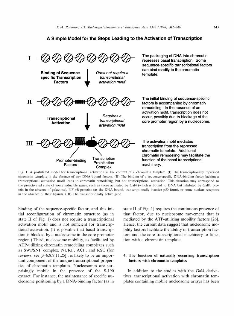
binding of the sequence-speci¢c factor, and this ini-tial recon¢guration of chromatin structure (as instate II of Fig. 1) does not require a transcriptionalactivation motif and is not su¤cient for transcrip-tional activation. (It is possible that basal transcrip-tion is blocked by a nucleosome in the core promoterregion.) Third, nucleosome mobility, as facilitated byATP-utilizing chromatin remodeling complexes suchas SWI/SNF complex, NURF, ACF, and RSC (forreviews, see [3^6,8,9,11,25]), is likely to be an impor-tant component of the unique transcriptional proper-ties of chromatin templates. Nucleosomes are sur-prisingly mobile in the presence of the S-190extract. For instance, the maintenance of speci¢c nu-cleosome positioning by a DNA-binding factor (as in
state II of Fig. 1) requires the continuous presence ofthat factor, due to nucleosome movement that ismediated by the ATP-utilizing mobility factors [26].Hence, the current data suggest that nucleosome mo-bility factors facilitate the ability of transcription fac-tors and the core transcriptional machinery to func-tion with a chromatin template.
4. The function of naturally occurring transcriptionfactors with chromatin templates
In addition to the studies with the Gal4 deriva-tives, transcriptional activation with chromatin tem-plates containing mobile nucleosome arrays has been
Fig. 1. A postulated model for transcriptional activation in the context of a chromatin template. (I) The transcriptionally repressedchromatin template in the absence of any DNA-bound factors. (II) The binding of a sequence-speci¢c DNA-binding factor lacking atranscriptional activation motif leads to chromatin remodeling, but not transcriptional activation. This situation may correspond tothe preactivated state of some inducible genes, such as those activated by Gal4 (which is bound to DNA but inhibited by Gal80 pro-tein in the absence of galactose), NF-UB proteins (as the DNA-bound, transcriptionally inactive p50 form), or some nuclear receptorsin the absence of their ligands. (III) The transcriptionally active gene.
BBACAN 87408 19-7-98
K.M. Robinson, J.T. Kadonaga / Biochimica et Biophysica Acta 1378 (1998) M1^M6 M3
observed with naturally occurring transcription fac-tors, which include Sp1, NF-UB p65, LEF-1, Ets-1,TFE-3, CREB, and estrogen receptor [27^30]. Inthese experiments, many biological phenomenawere observed to be recreated in vitro with chroma-tin templates but not with naked DNA templates. Afew examples are as follows.
4.1. Transcriptional synergy between Sp1 and otheractivators
In studies of the HIV-1 LTR promoter, Sp1 andNF-UB p65 [28] as well as Sp1 and the combinationof LEF-1, Ets-1, and TFE-3 [27,28] were observed toactivate transcription synergistically with chromatintemplates but not with naked DNA templates. In theanalysis of the function of Sp1 and NF-UB with theHIV-1 LTR promoter [28], it was found that NF-UBp65 (which functions as an activator in vivo), but notNF-UB p50 (which does not function as an activatorin vivo), activates transcription synergistically withSp1. In addition, Sp1 and NF-UB p65 (but notNF-UB p50) were observed to bind cooperatively tochromatin templates but not to naked DNA tem-plates. Moreover, the combination of Sp1 and NF-UB (either p50 or p65) was found to induce DNase Ihypersensitivity and nucleosome positioning that re-semble those seen in the promoter region of the in-tegrated form of the HIV-1 LTR in vivo. Notably,with Sp1 and NF-UB p50, the chromatin structure isrecon¢gured but the promoter is transcriptionally in-active, as seen in the HIV-1 provirus in the absenceof NF-UB induction (depicted schematically in stateII of Fig. 1). These results collectively indicate thatkey features of both the chromatin structure andtranscriptional regulation of the HIV-1 LTR pro-moter can be reconstituted in vitro.
4.2. Regulation of the transcriptional activity ofCREB by phosphorylation
In this analysis of the distal enhancer of the T-cellreceptor K chain gene, it was found that the phos-phorylation (at serine 133) of the cyclic AMP-re-sponsive binding protein (CREB) by protein kinaseA results in a 10-fold induction of the transcriptionalactivity of CREB with chromatin templates but notwith naked DNA templates ([29]; T. Mayall and K.
Jones, personal communication). Upon mutation ofserine 133 of CREB to alanine, transcriptional en-hancement by protein kinase A was not seen. DNaseI footprinting experiments revealed that both thephosphorylated and the non-phosphorylated formsof CREB were bound to the chromatin template,and hence, the enhancement of CREB transcription-al activity by phosphorylation by protein kinase Aoccurs by a mechanism that is distinct from thebinding of CREB to its site in chromatin. Thesestudies provide additional evidence that the bindingof a factor to chromatin does not necessarily corre-late with transcriptional activation, but instead, thatkey events in transcriptional activation can occursubsequent to the initial DNA binding and chroma-tin recon¢guration (as outlined in states II and III ofFig. 1).
4.3. Ligand-regulated transcriptional activation by theestrogen receptor and the coactivator, p300
In these studies, estrogen- and antiestrogen-regu-lated transcriptional activation by the puri¢ed full-length human estrogen receptor were achieved in vi-tro with chromatin templates but not with nakedDNA templates under otherwise identical conditions[30]. Moreover, it was observed that a single aminoacid substitution mutation in the helix 12/AF-2 re-gion of the estrogen receptor (leucine 540 to gluta-mine) does not a¡ect ability of estrogen receptor tobind to ligand or to DNA, but does render the estro-gen receptor unable to activate transcription in vitrowith chromatin templates (but not with naked DNAtemplates) [30], as seen in vivo [31]. DNase I foot-print analyses of the estrogen receptor with the chro-matin templates indicated that DNA binding by theestrogen receptor is not su¤cient for transcriptionalactivation. Hence, as seen with other regulatory fac-tors, the limiting step(s) in transcriptional activationby the estrogen receptor occur after the binding ofthe protein to the chromatin template.
With this experimental system, puri¢ed humanp300 was found to function as a coactivator of es-trogen receptor with chromatin but not with nakedDNA templates [30]. In the absence of ligand ac-tivated estrogen receptor, p300 was seen to have lit-tle e¡ect (less than 2-fold activation) upon basaltranscription. In the presence of estrogen receptor,
BBACAN 87408 19-7-98
K.M. Robinson, J.T. Kadonaga / Biochimica et Biophysica Acta 1378 (1998) M1^M6M4
however, p300 was observed to enhance transcrip-tion cooperatively by a mechanism that is stimulatedby estrogens and inhibited by antiestrogens. Thus,using chromatin templates in vitro, it has been pos-sible to recreate the regulation of transcription bythe estrogen receptor and the p300 coactivator. Itseems likely that the function of many transcriptionfactors and coactivators, including other nuclear re-ceptors, could be analyzed in vitro with chromatintemplates.
5. Summary and perspectives
Gene expression is a multidimensional phenomen-on that is a¡ected by many di¡erent parameters,including the structure and modi¢cation of thechromatin template. In many respects, it is usefulto view chromatin as an active participant in generegulation, rather than as an unchanging, staticstructure that serves as unwieldy obstruction to thetranscription process. In this review, we have pre-sented a few examples of the use of chromatin tem-plates to recreate in vitro an apparently close resem-blance of the function of transcription factors invivo. Moreover, other data suggest that chromatintemplates can be employed to study the participationof HMG proteins in the transcriptional process (see,for example, [32]), the role of histone acetylation intranscriptional regulation (see, for example, [33]),long-distance activation of transcription (see, for in-stance, [12,34]), and transcriptional elongation inchromatin (see, for example, [35]). Hence, it seemsreasonable to have an optimistic outlook towardour current and future ability to incorporate chro-matin structure into the biochemical analysis of tran-scriptional regulation.
Acknowledgements
We are grateful to Mike Pazin for critical readingof the manuscript, and to Drs. T. Mayall and K.Jones for communication of unpublished data. Weapologize for any errors or omissions in this review.Studies of chromatin structure and transcriptionalregulation in the laboratory of J.T.K. are supportedby the National Institutes of Health (GM46995).
Studies of chromatin assembly with ACF are sup-ported by the National Science Foundation (MCB9631121).
References
[1] J.E. Brownell, C.D. Allis, Curr. Opin. Genet. Dev. 6 (1996)176^184.
[2] M. Grunstein, Nature 389 (1997) 349^352.[3] G.A. Hartzog, F. Winston, Curr. Opin. Genet. Dev. 7 (1997)
192^198.[4] J.T. Kadonaga, Cell 92 (1998) 307^313.[5] R.E. Kingston, C.A. Bunker, A.N. Imbalzano, Genes Dev.
10 (1996) 905^920.[6] T. Krude, S.C.R. Elgin, Curr. Biol. 6 (1996) 511^515.[7] M.J. Pazin, J.T. Kadonaga, Cell 89 (1997) 325^328.[8] C.L. Peterson, Curr. Opin. Genet. Dev. 6 (1996) 171^175.[9] T. Tsukiyama, C. Wu, Curr. Opin. Genet. Dev. 7 (1997)
182^191.[10] A.P. Wol¡e, D. Pruss, Cell 84 (1996) 817^819.[11] C. Wu, J. Biol. Chem. 272 (1997) 28171^28174.[12] R.T. Kamakaka, M. Bulger, J.T. Kadonaga, Genes Dev. 7
(1993) 1779^1795.[13] M. Bulger, J.T. Kadonaga, Methods Mol. Genet. 5 (1994)
241^262.[14] R.T. Kamakaka, C.M. Tyree, J.T. Kadonaga, Proc. Natl.
Acad. Sci. USA 88 (1991) 1024^1028.[15] R.T. Kamakaka, J.T. Kadonaga, Methods Cell Biol. 44
(1994) 225^235.[16] J.D. Dignam, R.M. Lebovitz, R.G. Roeder, Nucleic Acids
Res. 11 (1983) 1475^1489.[17] C.P. George, J.T. Kadonaga, in: P.A. Krieg (Ed.), A Labo-
ratory Guide to RNA: Isolation, Analysis, and Synthesis,J. Wiley and Sons, New York, 1996, pp. 133^139.
[18] M.J. Pazin, J.T. Kadonaga, in: H. Gould (Ed.), Chromatin:a Practical Approach, IRL Press, Oxford University Press,Oxford, 1998, in press.
[19] I. Sadowski, J. Ma, S. Triezenberg, M. Ptashne, Nature 335(1988) 563^564.
[20] D.I. Chasman, J. Leatherwood, M. Carey, M. Ptashne, R.D.Kornberg, Mol. Cell. Biol. 9 (1989) 4746^4749.
[21] Y.-S. Lin, M.F. Carey, M. Ptashne, M.R. Green, Cell 54(1988) 659^664.
[22] M.J. Pazin, R.T. Kamakaka, J.T. Kadonaga, Science 266(1994) 2007^2011.
[23] T. Tsukiyama, P.B. Becker, C. Wu, Nature 367 (1994) 525^532.
[24] G. Mizuguchi, T. Tsukiyama, J. Wisniewski, C. Wu, Mol.Cell 1 (1997) 141^150.
[25] M.J. Pazin, J.T. Kadonaga, Cell 88 (1997) 737^740.[26] M.J. Pazin, P. Bhargava, E.P. Geiduschek, J.T. Kadonaga,
Science 276 (1997) 809^812.[27] P.L. Sheridan, C.T. Sheline, K. Cannon, M.L. Voz, M.J.
Pazin, J.T. Kadonaga, K.A. Jones, Genes Dev. 9 (1995)2090^2104.
BBACAN 87408 19-7-98
K.M. Robinson, J.T. Kadonaga / Biochimica et Biophysica Acta 1378 (1998) M1^M6 M5
[28] M.J. Pazin, P.L. Sheridan, K. Cannon, Z. Cao, J.G. Keck,J.T. Kadonaga, K.A. Jones, Genes Dev. 10 (1996) 37^49.
[29] T.P. Mayall, P.L. Sheridan, M.R. Montminy, K.A. Jones,Genes Dev. 11 (1997) 887^899.
[30] W.L. Kraus, J.T. Kadonaga, Genes Dev. 12 (1998) 331^342.[31] C.K. Wrenn, B.S. Katzenellenbogen, J. Biol. Chem. 268
(1993) 24089^24098.[32] S.M. Paranjape, A. Krumm, J.T. Kadonaga, Genes Dev. 9
(1995) 1978^1991.
[33] P.L. Sheridan, T.P. Mayall, E. Verdin, K.A. Jones, GenesDev. 11 (1997) 3327^3340.
[34] M.C. Barton, B.M. Emerson, Genes Dev. 8 (1994) 2453^2465.
[35] G. Orphanides, G. LeRoy, C.-H. Chang, D.S. Luse, D.Reinberg, Cell 92 (1998) 105^116.
BBACAN 87408 19-7-98
K.M. Robinson, J.T. Kadonaga / Biochimica et Biophysica Acta 1378 (1998) M1^M6M6