the use of dental morphology to identify an ontario iroquois
TRANSCRIPT

THE USE OF DENTAL MORPHOLOGY TO IDENTIFY AN
ONTARIO IROQUOIS OSSUARY POPULATION
by .
Kathryn E. Wright, B.A.
A Thesis
Submitted to the School of Graduate Studies
in Partial Fulfilment of the Requirements
fO:r:" the Degree
Master of Arts
McMaster University
October 1977
HASTER OF ARTS (1977) (Anthropology)
McMASTER UNIVERSITY Hamilton, Ontario
TITLE:
AUTHOR:
SUPERVISOR:
The use of Dental Morphology to Identify an Ontario Iroquois Ossuary Population
Kathryn E. Wright, B.A. (Toronto)
Professor Emake J.E. Szathmary
NUMBER OF PAGES: vii, 79

ABSTRACT
The Kleinburg ossuary population is known to be a
protohistoric Iroquois group, but little else has been
discovered. The non-metric dental morphology was observed
and compared to that of three contemporary Iroquois groups
known in an archaeological context in an attempt to more
precisely identify the Kleinburg population. Twenty-eight
characters were used for comparisons. Two statistical
methods were chosen, both giving estimates of overall
divergence between samples. A modern white sample was
included to test the validity of the method.
iii
ACKNOWLEDGEMENTS
l would like to thank Dr. F.J. Melbye, University of
Toronto, for permission to use the Kleinburg material, and the
Burlington Research Centre, Faculty of Dentistry, University
of Toronto for access to their records. Ms. S. Saunders
kindly provided the computer programme for the transformation
of the data. Lastly l am grateful for the criticism and
encouragement given by Dr. J.T. Mayhall, University of Toronto
and Dr. E.J~E. Szathmary.
iv
ABSTRACT
LIST OF TABLES
CHAPTER 1.
CHAPTER II.
C'HAPTER III.
CHAPTER IV.
TABLE OF CONTENTS
INTRODUCTION
OBJECTIVES AND APPROACH
Historical Perspective
Objectives
Approach
The dental traits
Genetical control of traits
General methodology
Recording
Terminology
DEFINITION OF TRAITS
Maxillary Incisors
Maxillary Canines
Maxillary premolars
Maxillary Molars
Mandibular Incisors
Mandibular Canines
Mandibular Premolars
Mandibular Molars
OBSERVATIONS OF DENTAL MORPHOLOGY
Maxillary Incisors
Maxillary Canines
Maxillary premolars
Maxillary Molars, First and Second
Maxillary Molars, Third
Mandibular Incisors
Mandibular Canines
v
iii
vii
1
4
4
5
7
7
7
9
Il
Il
12
12
15
16
18
23
24
24
26
33
34
35
35
36
38
40
40
CHAPTER V.
CHAPTER VI.
CHAPTER VII.
REFERENCES
Mandibular premolars 41
Mandibular Molars, First and Second 42
Mandibular Molars, Third 44
Anomalous Conditions 46
STATISTICAL ANALYSIS
Statistical Method
Computations
RESULTS AND INTERPRETATION
Results
Indian-Burlington
Indian-Indian
Sanghvi' s X2
Correlation measurements
Interpretation
CONCLUSIONS AND SU~rnARY
Conclusions
Sununary
vi
48
53
60
60
60
63
63
65
69
70
72
LIST OF TABLES
TABLE 4.1 KLEINBURG DENTAL SAMPLE 33
TABLES 4.2-4.11 OBSERVATIONS OF DENTAL MORPHOLOGY
4.2
4.3
4.4
4.5
4.6
4.7
4.8
4.9
4.10
4.11
TABLE 5.1
TABLE 5.2
TABLE 6.1
TABLE 6.2
TABLE 6.3
TABLE 6.4
TABLE 6.5
Maxi11ary Incisors 34
Maxi11ary Canines 35
Maxi11ary premo1ars 35
Maxi11ary Ho1ars, First and Second 36
Maxi11ary Mo1ars, Third 37
Handibu1ar Incisors 40
Mandibu1ar Canines 40
Mandibu1ar Premo1ars 41
Mandibu1ar Mo1ars, First and Second 42
Mandibu1ar Molars, Third 43
INCIDENCE OF DENTAL VARIANTS 55
TRAITS FOR ADDITIONAL SANGHVI'S X2 59
THETA VALUES, Left and Right 61+ 62
MEASURE OF DIVERGENCE AND STANDARD DEVIATIONS 67
MEASURE OF DIVERGENCE CALCULATED WITH POOLED DATA
SANGHVI'S X2 VALUES FOR SAMPLES TAKEN IN PAIRS
SANGHVI'S X2 VALUES FOR 7 AND 2 TRAITS
vii
67.
68
68
CHAPTER l
INTRODUCTION
The dentition has been a traditional tool used by
both primate and human palaeontologists to study evolutionary
changes. It has only been recently that the dentition has
come into focus in the study of modern human groups. As
with the cranium, discrete morphological variations rather
th an metrical seem to be more suitable for differentiating
or relating populations.
The teeth have many advantages for this type of study.
Once erupted, the form of the tooth is not affected by
environmental factors other than attrition and caries. Of the
commonly studied traits, only tooth size has been shown to be
affected by internal environmental influences during develop
ment (Kirveskari 1974). The dentition is generally resilient
to factors affecting the development of other tissues (Wright
1975). The teeth are the hardest and most durable of body
tissues. They are available from archaeological collections
even when skeletal material has been badly damaged. Because
teeth are easily studied in living populations, it is possible
to observe changes in a particular dentition over time. There
are also a large number of traits readily recorded on the
dentition as a whole, and on each individual tooth.
l
2
The number of studies using the dentition to relate
populations is gradually increasing. Early works generally
dealt with large populations. We find papers on the dentition
of the North American Indian (Dahlberg 1949; Wissler 1931) or
comparisons of Mongoloid and Indo-European populations
(Tratman 1950). This led to the definition of a basic
Mongoloid dental complex (Hanihara 1967; Moorrees 1962). With
the accumulation of data on dental variation, the emphasis
has shifted from general patterns to more specific differences
between smaller groups of people. Moorrees (1957) demonstrated
differences between the East and West Aleuts and this
variation was later shown to be due to local evolution rather
than European admixture (Turner 1967). Papago and Pecos
Indians can be differentiated using dental morphology
(Morris 1965). Kirveskari (1974) studied dental characters
in an attempt to determine the origins of Skolt Lapps. He
concluded that their dentition is distinct from that of both
Mongoloids and Caucasoids, but the frequency of traits
suggested closer affiliations with the latter. Using
deciduous molar characters, Japanese-White and Japanese-
Negro hybrids could be distinguished from Japanese, American
White and American Negro groups (Hanihara 1963) . Recently
dental morphology has been used to compute distance
statistics representative of the genetic distance between
populations (Brewer-Carias et al. 1976; Sofaer et al. 1972).
The latter concluded "that tooth morphology has the
3
potential of providing moderately good discrimination"
(Sofaer et al. 1972:364). The former also had positive
results, and state that "the pattern of micro-differentiation
suggested by dental traits is roughly comparable to that
projected by other types of biological traits whose genetic
basis is more firmly established" (Brewer-Carias et al.
1976:13).
CHAPTER II
OBJECTIVES AND APPROACH
Historical Perspective
The Kleinburg ossuary was excavated in the summer of
1970 by F.J. Melbye, University of Toronto. The site is
located in Kleinburg, Ontario, north of Toronto. An associated
village site has yet to be discovered. A date of 1600 ±15
years has been assigned the site based on the presence of
certain limited trade goods (Melbye pers. comm.).
At that time two distinct branches of the Late Ontario
Iroquois Stage existed; the Neutral-Erie branch in southwestern
Ontario and New York, and the Huron-Petun branch in south
eastern Ontario (Wright 1972). Both branches had evolved from
the Middleport substage of the Middle Ontario Iroquois Stage
by 1400 A.D. (Wright 1966). Pn~historically the Huron-Petun
branch was divided into a Northern division in Huronia proper
and a Southern division along the north shore of Lake Ontario.
The Southern division gradually shifted northward. Fusion
with the Northern division by 1550 A.D. resulted in the
historic Huron and Petun groups. The Neutral division of the
Neutral-Erie bran ch was centred in the Hamilton-Brantford area
(Wright 1974). It was at this time, 1615-1625 A.D., that
the Recollets and Jesuits began their missionary work in
Huronia. By 1654 A.D. all the Ontario Iroquois had been
absorbed into the Iroquois League of Nations (Wright 1972) .
4

Both branches of the Ontario Iroquois practised
ossuary burials. The Neutral lined their ossuary pits with
clay, a practice apparently not followed by the Huron-Petun
(Wright 1966). In other aspects the Huron and Neutral were
similar. Corn was the staple food, supplemented by beans,
squash and sorne meat and fish. Hunting and fishing were of
greater importance in the Neutral-Erie brancha The material
culture of both branches was generally the same although
specifie differences allow archaeological separation of the
two.
Objectives
5
It will be my primary purpose to place the Kleinburg
ossuary in this general scheme of Ontario archaeology. The
date and location of the site suggest it was a Huron ossuary.
Data on the dentition of three contemporary ossuary popula
tions are available for study (Wright 1974) and it is through
comparison with these groups that l hope to shed sorne light
on the Kleinburg people. Although analysis of the entire
Kleinburg ossuary is nearing completion, no works have been
published, and no comparative analyses have been done. This
thesis will be the first attempt to relate the populations to
other contemporary groups.
Having placed the ossuary in the archaeological
context, this thesis will provide a thorough and extensive
description of an Iroquois dentition, be it Neutral or Huron.
Such a complete consideration of Ontario dental morphology
has been ~acking. Osteological studies have largely ignored
6
the teeth, mentioning only the more striking traits of shovel
shaping, protostylid, Carabelli's cusp and cusp and groove
pattern.
The most complete work on Iroquoian odontology has
been Wright's The Dental Morphology of Three Ontario Iroquois
Ossuary Populations (1974). However, this work is wanting in
several aspects. Wright has used several traits unsuitable
because a subjective judgement of size or orientation is
necessary. Other traits which are easily studied in samples
of loose teeth, and which may be important are missing; root
morphology has been ignored. Perhaps the main weakness in
Wright's work is the small size of his samples. The Kleinburg
sample is many times larger than Wright's three samples
pooled. Wright has provided excellent references for
standardization of the traits and categories. Where appro
priate, these same references will be included here.
A completé description of such a large ossuary as
Kleinburg will be a baseline tofurther comparative studies.
It will allow comparisons not only with other protohistoric
groups, but also with living Iroquois groups and will
contribute to our growing knowledge of dental variability.
It is impossible to form hypotheses of the adaptiveness of
tooth form, a question which concerns palaeontologists and
anthropologists, unless a total range of possible variation
is known.
As Turner states:
"It is no longer sufficient to talk of the
American Indian dentition as if the teeth
of every Indian had been cast in the same
mold." (1969:25)
Approach
7
1. The Dental Traits. The traits selected for study are those
with known discriminatory value which were readily recorded
and which are thought to be independently expressed (Hanihara
1963; Kirveskari 1974j Mayhall 1976j Sofaer et al. 1972;
Wright 1974). Some traits which have yet to be studied in
detail but which promise to be of value were also included.
The criteria for classification were largely those used by
previous researchers, although some simplification was
necessary. All traits are defined in Chapter III. Those
used for distance analyses are marked with an asterisk.
2. Genetical Control of Traits. The genetic component of
dental traits has long beèn known (Hrdli~ka 1911; Krogman
1960; Lasker 1950). This was orginally concluded from the
observed population differences in the frequencies of traits.
Since these early conclusions, studies on monozygotic (MZ) and
dizygotic (DZ) twins have further demonstrated that tooth
from is largely under genetic control. Lundstrorn (1963) was
able to correctly diagnose zygosity in 117 of 124 pairs of
twins on the busis of permanent tooth morphology. Korkhaus
(1930) observed shovel shaping, fissure pattern and cusp
formation in twins and concluded that these traits were
8
inherited. Considering only traits on the mandibular first
premolar, Kraus (1957) found that MZ twins showed significantly
more concordances than DZ twins and unrelated pairs. He and
his associates (1959) found that premolar morphology equalled
dermatoglyphics as a method of diagnosing monozygosity.
Wasser (1953) observed five traits on upper premolars and
found the number of concordances in MZ twins to be more than
expected by chance. More recently Biggerstaff (1973) found
he was able to distinguish MZ t~ins on the basis of
Carabelli's trait alone.
Little is known about actual mechanisms of inheritance
but polygenic inheritance appears to be most likely
(Dahlberg 1962; Bailit et al. 1974). This, plus the quasi
continuous nature of the traits has made genetic analysis
difficult. A quasicontinuous trait behaves somewhat as a
discrete trait in that it is present or absent. However there
is a continuous gradation of variation for eaoh trait.. When
a trait is present it is so in varying intensities. Below a
certain point on this scale of variation the trait does not
appear at all (Grüneberg 1952) .
Some attempts have been made to determine modes of·
inheritance. Turner (1967) assurned equilibrium and applied
the Hardy-Weinberg theorem to arrive at expected values of
incidences for various traits. This approach has since been
shown to be invalid (Sofaer 1970). Kraus (1951), Goose and
Lee (1971) and Fortin and Alvesalo (1974) have also attempted

to determine modes of inheritance. These studies will be
considered in more detail when traits are discussed
individually.
9
3. General Methodology. Two major problems are encountered
ln this type of study. The first is that of bilaterality of
traits, the second is sex differences.
Most traits are expressed on both sides of the mouth.
The degree of expression may differ, however, although this
is rarely by more than one category (Biggerstaff 1970). Kraus
(1957) found 20-30% occurrences of asymmetrical traits on the
mandibular premolars. Garn ~ ~ (1966a) found asymmetry
was not a problem in mandibular first molars but it was more
pronounced in second molars. Biggerstaff (1973) found an
unexpectedly high incidence of asymmetry in the expression
of Carabelli's trait in twins, but the frequency of asymmetry
ln the general population was less. Goose and Lee (1971)
found left and right sides to be the same. Only the occurrence
of three-rooted mandibular molars has been shown to be signi
ficantly asymmetrical (Tratman 1938; Turner 1967). This
problem has been handled in various ways. Sorne workers have
used only one antimere for analysis on the assumption that
symmetry existed (Moorrees 1957). Kirveskari (1974) compared
the total frequencies for each side, and finding no signifi
cant differences followed the same procedure as Moorrees.
Turner (1967) considered one side only if there was less than
5% asymmetry for a trait. If there was more, he used each

10
tooth separately. The frequencies from these methods are not
directly comparable. In dealing with an ossuary one must use
the latter method, what Turner terms 'tooth' count. Each
tooth is considered, and any comparisons are made by side
(Wright 1974). There are then two sets of observations for
the sample.
Sex differences ln the expression of traits are
evident, but rarely are they significant (Kirveskari 1974;
Pedersen 1949; Turner 1967). Turner (1967) found only two
traits with significant differences; first maxillary molar
cusp number and the occurrence of three-rooted mandibular
molars. This latter trait shows a sex difference in symmetry
as well (Tratman 1938;Turner 1967). There is a male preponder
ance for shovelling in Whites, Negroes, Texas Indians and
Japanese (Goldstein 1948; Hrdlitka 1920;Suzuki and Sakai 1966).
The opposite seems to be true in Chinese and Teso (Barnes
1969; Hrdli~ka 1920). Males have more lingual tubercles on
the incisors in Whites and coloured populations (Hrdli~ka 1921).
Carabelli's trait is more common in males, as is larger cusp
number in mandibular molars (Garn et al 1966bj Goose and Lee
1971j Meredith and Hixon 1954jTurner 1967). However, none of
these differences proved to be significant. Most researchers
have pooled their samples for analysis (Bang and Hasund 1972;
Kirveskari 1974; Moorrees 1957). It then seems reasonable to
assume that in the Kleinburg sample, which cannot be sorted
by sex j sex differences will not significantly affect the

11
observed frequencies regardless of the true ratio of sexes.
4. Recording. Where possible standard models or photographs
were used to maintain scoring consistency. Detailed references
are given below. Categories for each trait were numbered for
recording purposes and are presented as such. To test for
repeatabilitya sample of loose teeth was observed by J.T.
Mayhall. In no instance did our results differ by more th an
one category, nor was there discordance in present/absent
classification. Repeatability was not determined for individual
traits, but overall repeatability was in the order of 85%.
5. Terminology. For the purposes of this work the major
molar cusps are named by location. These terms with the older
terms suggested by Osborn (1888) are given below:
Maxillary Molars
Paracone
Protocone
Metacone
Hypocone
Mandibular Molars
protoconid
Metaconid
Hypoconid
Entoconid
Hypoconulid
Mesiobuccal cusp
Mesiolingual cusp
Distobuccal cusp
Distolingual cusp
Mesiobuccal cusp
Mesiolingual cusp
Distobuccal cusp
Distolingual cusp
Distal cusp
CHAPTER III
DEFINITION OF TRAITS
Maxillary Incisors*
1. Lingual Cervical Area
The lingual cervical area of the incisors and canines
is often referred to as the cingulum. A true cingulum, how
ever is rarely seen in hominids (Kirveskari 1974). Tubercles
and ridges in this area are considered to be the remnants of
a cingulum (Carbonell 1963; Kirveskari 1974). Tubercles
occur most frequently on maxillary canines, then lateral
incisors and rarely on central and mandibular incisors.
There do occur population differences in the
appearance of tubercles, but the frequency of occurence lS
generally low except in Arctic populations (Carbonell 1963)
The expression of lingual tubercles and the degree of
expression of shovel-shaping are related in Japanese (Suzuki
& Sakai 1966). Other data suggest the traits are not gener
ally related (Carbonell 1963; Hrdli~ka 1921; Lasker 1950).
The categories for recording are taken largely from Wright
(1974:19) and Barnes (1969:184).
1.0 Absence of pits, tubercles or ridges.
1.1 Presence of one or more small tubercles.
A tubercle must have a groove between it
and the rest of the tooth (Barnes 1969).
If the groove was slight and the tubercles
had a partly-free apex, it was recorded as
small.
12
1.2 Presence of one or more large tubercles.
A large tubercle is clearly elevated with
a free apex (Kirveskari 1974).
1.3 Presence of finger-like projections.
13
Parallel ridges of elevated enamel separated
by grooves were seen. Shallow grooves with no
clear enamel elevations were not included
(Barnes 1969).
1.4 Presence of one or more pits.
1.5 Presence of both one or more pits and one or
more tubercles.
1.6 Presence of a single groove where marginal
ridges meet.
2. Lingual Marginal Grooves
Single or multiple grooves may dissect either the
mesial distal or both marginal ridges, extending from the
lingual cervical area to the cementum (Kraus et al. 1969:21)
Any groove which could be felt by the fingernail and extended
onto the cementum was recorded.
Kraus, Jordan and Abrams (1969) mention this trait
as a variation on lateral incisors only. The incidence on
both incisors was recorded here using the following categories.
2.0 Absence of marginal grooves.
2.1 Presence of one or more mesial grooves (see
photograph: Kraus et al. 1969:22, Fig.1-30)

14
2.2 Presence of one or more distal grooves.
2.3 Presence of both mesial and distal grooves.
3. Lingual Shovel Shape*
The mesial and distal lingual ridges of the incisors
may be elevated producing a 'shovel-shaped' incisor. The
lateral incisors are more often affected by shovelling
(Carbonell 1963; Lasker & Lee 1957). The degree of expression
is concordant for monozygotic twins (Korkhaus 1930; Lasker
1950). Tests for modes of inheritance have been made by
Turner (1967) and Portin and Alvesalo (1974). In both
instances a model of two cod0minant alleles fit the data,
although the distribution of expressions differed. Portin
and Alvesalo did not rule out the possibility of polygenic
inheritance.
The categories used were based originally on Hrdli6ka's
classification with some alterations (Carbonell 1963: Fig. li
Hrdliëka 1920: 449). Plaque 1 of Dahlberg's reference series
was used to record categories 3.1 to 3.4.
3.0 Absence of shovel trait; no trace, faint or
imperfect rim.
3.1 Trace of shovel shape; slight but distinct
ridges.
3.2 Semi-shovel shape; enamel rim is distinct
but fossa is still shallow.
3.3 Shovel shape; enamel rim and fossa are well
developed.

3.4 Marked shovel shape; enamel rim is
extremely developed.
3.5 Peg-shaped; reduced incisor with
cylindrical_ crown.
3.6 Barrel-shaped; shovelling is so extreme
as to form a continuous rim of enamel
which projects incisally for at least
2/3 the length of the crown.
4. Labial Ridging*
15
A build-up of enamel may occur on the labial surface
of incisors both on the margins and in the central lobe
(Carbonell 1963:218; Dahlberg 1949:141).
Central incisors are affected more than laterals
(Kirveskari 1974; Snyder 1960). The relative intensity of
the expression of labial ridging compared to that of shovel
shaping is not the same for all populations (Kirveskari 1974)
When either of the marginal ridges or both were
elevated more than the central lobe, labial ridging was said
to be present (Kirveskari 1974) .
4.0 Absence of labial ridging.
4.1 Presence of labial ridging.
Maxillary Canines*
1. Lingual Cervical Area
As in the incisors, tubercles sometimes occur on the
canines. These are also of cingular origin (Carbonell 1963).

16
The method for recording was taken from Wright (1974:23).
1.0 Absence of pits or tubercles.
1.1 Presence of one tubercle with no pits.
1.2 Presence of one tubercle with one or multiple
pits.
1.3 Presence of multiple tubercles.
1.4 Presence of multiple tubercles and oné or
multiple pits.
1.5 Presence of one or multiple pits.
Maxillary premolars*
1. Distal Transverse Ridge
An accessory ridge may lie between the buccal
triangular ridge and the distal marginal ridge on the
occlusal surface (Kraus et al 1969:60; Wright 1974:24). The
ridge is more frequently seen on second premolars (Kraus et
al. 1969).
1.0 Absence of distal transverse ridge.
1.1 Presence of distal transverse ridge
(see photograph: Kraus et al. 1969:60,
Fig. 1-97).
2. Occlusal Marginal Ridge Continuity
Both the mesial and distal marginal ridges may be
continuous or may be cut by one or more grooves. Tubercles
may also be present both mesially and distally (Kraus et al.
1969:60; Morris 1965:33; Wright 1974:23). The system for
scoring is an expansion of that used by Wright (1974:23).

2.0 Absence of mesial or distal marginal
grooves or tubercles.
2.1 Presence of mesial marginal groove.
2.2 Presence of distal marginal groove.
17
2.3 Presence of a mesial marginal accessory tubercle
with or without a distal groove.
2.4 Presence of a distal marginal accessory tubercle
with or without a mesial groove.
2.5 Presence of both a mesial and distal marginal
groove.
2.6 Presence of both a mesial and distal accessory
tubercle.
3. Root Number
First premolars usually have two roots, second
premolars one (Kraus et al. 1969). The double-rooted form lS
rarer in Mongoloid populations (Pedersen 1949; Turner 1967) .
The definitions for single and double roots follow Pedersen's
system for root classification (1949:74).
3.1 Presence of a single root.
3.2 Presence of two rootsi roots must be free for
at least ~ their length.
3.3 Presence of two roots fused; roots are
free for less than ~ their length.

Maxillary Molars
1. Cusp Number*
18
Variation in cusp number is due to presence or absence
of the distolingual cusp, although other cusps vary in size
(Dahlberg 1949:168i Turner 1967:58). Reduction is rare in
the first molar, more common in the second and third
(Dahlberg 1949) .
Reduction of the distolingual cusp in the maxillary
molars is associated with third molar agenesis (Keene 1965) .
Cusp reduction seems to be of more value in distinguisbing
between two closely related groups than as a diagnostic
racial trait (Kirveskari 1974) .
Dahlberg's diagrams of degrees of cusp reduction
(1949:168) were used for recording. Two new categories were
introduced to accomodate the observationsi 1.6 and 1.7.
1.1 Presence of four well-developed cusps.
1.2 Presence of four CUSpSi distolingual cusp
reduced.
1.3
1.4
1.5
1.6
1.7
Presence of three cusps with faint
of the distolingual cusp.
Presence of three cusps only.
Presence of two cusps with a distal
Presence of two cusps only.
Presence of more than four cusps.
expression
tubercle.
19
2. Carabelli's Trait*
Carabelli's trait is a series of expressions ranging
from pits to large cusps. The trait occurs on the mesio
lingual cusp. The frequency of occurence decreases from first
to third molars (Dahlberg 1949) .
The trait is a relatively recent derivative from the
cingulum (Dahlberg 1949: Kirveskari 1974). It has been
suggested that the fullest expression of the trait compen
sates for reduction in tooth size (Dahlberg 1965). Keene
(1965) found a negative correlation between Carabell's cusp
expression and third molar agenesis. Others have found no
correlation between trait expression and molar size or
suppression of the third molar (Bang & Hasund 1972; Garn et
al. 1966b).
Several possible genetic mechanisms have been
suggested. A model of simple 1'1endelian dominant was proposed
(Lasker 1950) and was supported by application of the Hardy
Weinberg theory to population data (Hanihara 1963). The same
method also supported a model of two autosomal codominant
alleles (Turner 1967) which had earlier beensuggested from
pedigree analysis (Kraus 1951). As this model has been
shown to be inadequate and invalid (Goose & Lee 1971; Kraus
1959; Sofaer 1970) it seems probable that inheritance is
multifactorial.
The system used for classification and recording was
Dahlberg's (1963:159).

2.0 Absence of Carabelli's trait.
2.1 Presence of a single furrow; any groove was
recorded however shallow.
2.2 Presence of a pit.
2.3 Presence of a Y-shaped groove.
2.4 Presence of a double furrow.
20
2.5 Presence of bulging on the tooth surface with
or without a Y-shaped groove or double furrows.
2.6 Presence of a small cusp.
2.7 Presence of a large cusp.
3. Lingual Enamel Extension
An extension of enamel onto the root may occur at the
cemento-enamel junction. These occur on lingual or buccal
aspects, or both (Pedersen 1949:74; Turner 1967:101). Enamel
pearls are often found in the same populations which have
extensions (Lasker 1950) .
Mandibular molars show a greater incidence of the
trait (Andrews 1975iTurner 1967). The second molar in each
arch is more frequently affected (Andrews 1975; Wright 1974) .
Three-quarters of all extensions occur on buccal surfaces
and are slight (Andrews 1975).
Pedersen (1949:74) proposed a system of classification
which has been reduced for the purposes of this analysis.
3.0 Absence of lingual extension.
3.1 Presence of lingual extension which does
not ex tend between roots (see diagram:
Andrews 1975:51, Fig.8).
3.0 Absence of lingual extension.
3.1 Presence of lingual extension which does
not extend between roots (see diagram:
Andrews 1975:51, Fig. 8).
3.2 Presence of a lingual extension which
extends between the roots.
4. Buccal Enamel Extension
The categories for buccal enamel extensions are the
same as for lingual extensions.
5. Anterior Transverse Ridge *
21
An extra ridge may be present on the occlusal surface
between the mesial marginal ridge and the triangular ridge
of the mesiobuccal cusp (Kraus et al. 1969:90). The ridge
can be present in a range of sizes, but because of attrition
presence or absence only was considered.
5.0 Absence of an anterior transverse ridge.
5.1 Presence of an anterior transverse ridge
(see photograph: Kraus et al. 1969:91,
Fig. l-145A,B).
6. Lingual Groove Termination
Both the lingual and buccal grooves vary in forme
They may be shallow and blend into the tooth surface; they may
end abruptly, or in a pit (Kraus et al. 1969:90; Morris 1965:
45; Wright 1975:31). Observations were made on four-cusped
molars only and were recorded as suggested by Kraus et al.
(1969: 90) .
6.0 Absence of lingual groove.
6.1 Lingual groove blends smoothly into the
tooth surface.
6.2 Lingual groove ends abruptly.
6.3 Lingual groove ends in a pit.
7. Buccal Groove Termination
22
Buccal groove termination was observed on aIl molars.
Again method of recording is taken from Kraus et al. (1969:90).
7.0 Absence of buccal groove.
7.1 Buccal groove blends smoothly into the tooth
surface.
7.2 Buccal groove ends abruptly.
7.3 Buccal groove ends in a pit.
8. Root Number
Maxillary molars usually have three roots although
this can vary. Both Pedersen (1949) and Turner (1967)
considered root number. A derivative of pedersen's class
ification (1949:75) has been used here. Any roots partially
fused but free for at least half their length were considered
as separate.
8.1 Presence of single root.
8.2 Presence of two roots fusedi i.e. single
root with double apex.
8.3 Presence of two roots with double or triple
apex; the condition of two roots results from
fusion of any two of three roots.
8.4 Presence of three roots.
8.5 Presence of three fused rootsi i.e. single
root with three apices.
8.6 There is an increase in the number of roots
regardless of fusion; this includes any
extra rootlets even if the normal roots are
fused.
Mandibular Incisors
1. Lingual Cervical Area
Tubercles can appear on the. mandibular incisors
although the variation seen in the maxillary anterior teeth
is missing (Morris 1965:46; Wright. 1974:33). Categories
used are similar to those used for Maxillary Incisors.
1.0 Absence of tubercles or grooves.
1.1 Presence of a slight tubercle.
1.2 Presence of a prominent tubercle having
a free apex.
1.3 Presence of one or more pits.
1.4 Presence of a single groove in the centre
where marginal ridges meet.
2. Lingual Shovel Shape*
23
Shovel shaping on mandibu1ar incisors is less pro
nounced than on the maxillary (Dahlberg 1949~ Hrd1i~ka 1920).
A condensed system of scoring was used, taken from Mayhall
(l976: 62).
2.0 Absence of shovel trait.
2.1 Trace of shovel shape.
2.2 Semi-shovel shape.
2.3 Marked shovel shape.
2.4 Peg-shaped incisor.
2.5 Barrel-shaped incisor.
3. Labial Ridging*
3.0 Absence of labial ridging.
3.1 Presence of labial ridging.
Mandibular Canines
1. Lingual Cervical Area
24
Tubercles are seen less frequently on mandibular
canines than on maxillary (Turner 1967). Scoring is the same
as for maxillary canines.
1.0 Absence of pits or tubercles.
1.1 Presence of one tubercle with no pits.
1.2 Presence of one tubercle with one or more pits.
1.3 Presence of two or more tubercles.
1.4 Presence of two or more tubercles with pits.
1.5 Presence of one or more pits.
Mandibular Premolars
1. Lingual Cusp Number*
Mandibular molars generally have one buccal cusp and
one or two lingual cusps (Kraus & Furr 1953:559; Turner 1967:
51). The condition of two lingual cusps is less conmlon in
25
the first premolar (Kraus et al. 1969).
Any cusp with a partially or wholly indepcndent apex
was recorded.
1.0 Absence of lingual cusp (see photograph:
Kraus et al. 1969:71, Fig. l-114A).
1.1 Presence of one lingual cusp.
1.2 Presence of two lingual cusps (see photo
graph: Kraus et al. 1969:71, Fig. l-114C)
1.3 Presence of three or more lingual cusps.
2. Occlusal Marginal Ridge Continuity
The occlusal marginal ridges connect the buccal and
lingual cusps mesially and distally. The ridges can be
continuous, divided by a groove or can possess a tubercle
(Kraus & Furr 1953:559-560; Kraus et al. 1969:61; Morris 1965:
52; vvright 1974:37).
2.0 Both margins are continuous.
2.1 Presence of a mesial marginal groove.
2.2 Presence of a distal marginal groove.
2.3 Both margins grooved.
2.4 Presence of a mesial marginal tubercle.
3. Accessory Transverse Ridges
The main transverse or occlussal ridge may be
bifurcated. As well, extra transverse ridges may be present
(Kraus & Furr 1953:560; Kraus et al. 1969:72; Ludwig 1957:
267-268). Only accessory ridges and not the total nurnber of
ridges were counted (Ludwig 1957) .
3.0 Absence of accessory ridges.
3.1 Presence of one accessory ridge.
3.2 Presence of two or more accessory ridges.
3.3 Presence of bifurcated main ridge.
3.4 Presence of bifurcated ridge with one
accessory ridge.
3.5 Presence of bifurcated ridge with two or
more accessory ridges.
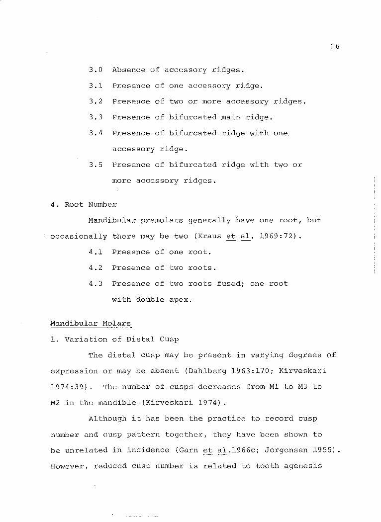
4. Root Number
Mandibu1ar premo1ars genera11y have one root, but
occasiona11y there may be two (Kraus et al. 1969:72).
4.1 Presence of one root.
4.2 Presence of two roots.
4.3 Presence of two roots fuSedi one root
with double apex.
Mandibu1ar Mo1ars
1. Variation of Distal Cusp
26
The distal cusp may be present in varying degrees of
expression or may be absent (Dah1berg 1963:170; Kirveskari
1974:39). The number of cusps decreases from Ml to M3 to
M2 in the mandib1e (Kirveskari 1974).
A1though it has been the practice to record cusp
number and cusp pattern togethcr, they have been shown to
be unre1ated in incidence (Garn et a1.1966ci Jorgensen 1955)
However, reduced cusp number is re1ated to tooth agenesis

(Davies 1968; Keene 1965) rand there is a tendency for
smaller teeth ta have fewer cusps (Dahlberg 1962) .
The mode of inheritance for the distal cusp is
unknown. Simple models are inadequate ta explain the
collected data (Biggerstaff 1970) .
Some researchers have recorded size (Morris 1965;
Wright 1974) while others have recorded presence or absence
only (Kirveskari 1974; Turner 1969). The former was chosen
here and Wright's categories (1974:42l were used.
1.0 - Absence of distal cusp.
1.1 Presence of a distal cusp smaller than ~ the
size of any other cusp.
1.2 Presence of a distal cusp at least ~ the size
of any other cusp.
1.3 Presence of a distal cusp equal in size ta
any other cusp.
27
1.4 Presence of a distal cusPr size undeterminable.
2. Tuberculum Sextum*
An extra cusp may occur on the distal marginal ridge
between the distal and distobuccal cusps (Hellman 1928:164;
Kirveskari 1974:46; Kraus et al. 1969:110). The frequency
of this cusp tends ta increase from Ml ta M3 (Kirveskari
1974). This cusp occurs independently of the distal cusp
(Mayhall:pers. comm.).

28
The method of recording is taken from Kirveskari
(1974:47).
2.0 Absence of tubercu1um sextum.
2.1 Presence of grooves or sma1l tubercle.
2.2 Presence of a distinct cusp of small or
medium size.
2.3 Presence of a distinct cusp of large size;
comparable to distal cusp in size (see
photograph: Kirveskari 1974:46, Fig. 15).
2.4 Presence of a cusp, size undeterminable.
3. Tuberculum Intermedium*
A small cusp, sometimes term C7, may be found on the
distal ridge of the mesiolingual cusp (Hellman 1928:164;
Kirveskari 1974:46, Kraus et al. 1969:110). This cusp
decreases in frequency from Ml to M3 (Kirveskari 1974). An
association between the occurrence of this cusp and Carabelli's
cusp was found in the Teso (Barnes 1969). Tuberculi inter-
rnedii are seen less often in individuals missing third molars
(Keene 1965).
The method of recording is taken from Kirveskari
(1974:48) 0
3.0 Absence of tuberculurn intermedium.
3.1 Presence of double grooves or weak CUSPi
without a free apex.
3.2 Presence of a distinct cusp of small or
medium size.

3.3 Presence of a distinct cusp of large
size (see photograph: Kirveskari 1974:
46, Fig. 15).
3.4 Presence of a cusp, size undeterminable.
4. Protostylid*
29
The protostylid is a continuous variable occurring on
the buccal or mesiobuccal portion of the mesiobuccal cusp
(Dahlberg 1950~15). Expressions of the trait range from pits
and furrows to large cusps. The incidence and degree of
expression decrease from Ml to M2 (Dahlberg 1950, 1963),
although Turner (1967) found the second molar of Northern
Indians and Eskimos to be most affected.
Carabelli's trait and the protostylid were found to
be associated in the Japanese (Suzuki & Sakai 1954i Sakai
1955 cited in Kirveskari 1974). This association was not
evident in Peruvians where Protostylid cusps appear more often
with 'advanced' cusp patterns (Goaz & Meller 1966). The
genetic mechanism controlling the trait is most likely
multifactorial (Dahlberg 1950) .
Dahlberg's classifications were used (1963:163) as
suggested by Mayhall (1976:80). Pits occurring at the end
of the buccal groove were not included here.
4.0 Absence of protostylid.
4.1 Presence of a pit or furrow.
4.2 Presence of distal deviation of the buccal
groove.

30
4.3 Presence of irregularities of the cusp
surface.
4.4 Presence of a prorninence.
4.5 Presence of a prorninence with a furrow,
including diagonal grooves.
4.6 Presence of a srnall cusp.
4.7 Presence of a large cusp.
5. Buccal Groove Terrnination
The buccal groove, separating the rnesiobuccal and
distobuccal cusps can be very prorninent or barely
discernible (Dahlberg 1949:160; Kraus et al. 1969:110;
Morris 1965:61). It often ends .in a pit. Dahlberg (1963)
includes this pit as an expression of the protostylid. The
distribution of the pit appears to be independent of other
protostylid expressions (Kirveskari 1974; Mayhall 1976).
Devoto et al. (1972) found an association between the occur-
rence of buccal pits and protostylid, but did not consider
them to be the same trait.
The classification suggested by Kraus et al. (1969:
Ill, Fig. 1-181) was used.
5.0 Absence of buccal groove.
3.1 Buccal groove blends smoothly into the tooth
surface.
5.2 Buccal groove ends abruptly.
5.3 Buccal groove ends in a pit.

31
6. Lingual Enamel Extension
Lingual enamel extensions were recorded in the same
manner as for maxillary molar extensions. Both lingual and
buccal extensions are more frequent in mandibular teeth
(Andrews 1975).
6.0 Absence of lingual enamel extension.
6.1 Presence of lingual extension which does not
extend between the roots.
6.2 Presence of lingual extension which extends
between the roots.
7. Buccal Enamel Extension
7.0 Absence of buccal enamel extension.
7.1 Presence of buccal extension which does not
extend between the roots.
7.2 Presence of buccal extension which extends
between the roots.
8. Cusp and Groove Pattern*
Cusp and groove pattern until recently has been
recorded with cusp number. Three patterns are commonly
recorded; Y, +, and X (J~rgensen 1955:195). The incidence of
the Y pattern decreases from Ml to M3i the opposite is true
for the X pattern (Kirveskari 1974) .
8.1 Presence of Y pattern; ML and DB cusps
touch, MB and DL cusps do note
8.2 Presence of + patterni all four cusps are
adjacent.
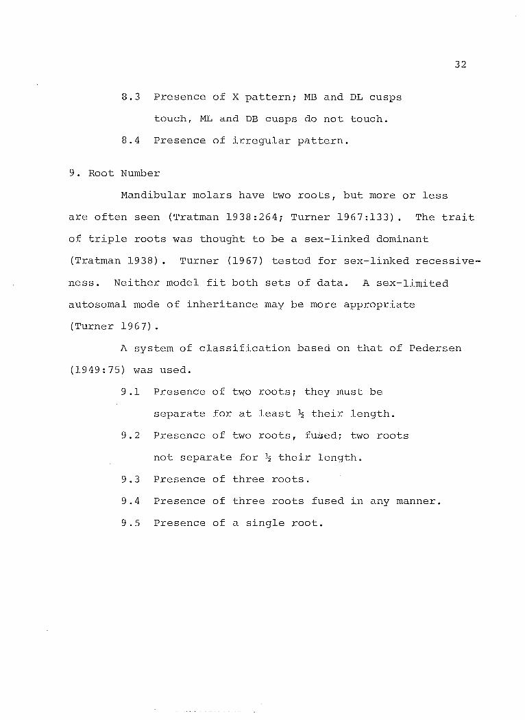
8.3 Presence of X pattern; MB and DL cusps
touch, ML and DB cusps do not toucha
8.4 Presence of irregular pattern.
9. Root Number
Mandibular molars have two roots, but more or less
32
are often seen (Tratman 1938:264; Turner 1967:133). The trait
of triple roots was thought to be a sex-linked dominant
(Tratman 1938). Turner (1967) tested for sex-linked recessive
ness. Neither model fit both sets of data. A sex-limited
autosomal mode of inheritance may be more appropriate
(Turner 1967) .
A system of classification based on that of Pedersen
(1949:75) was used.
9.1 Presence of two rootsi they must be
separate for at least ~ their length.
9.2 Presence of two roots, fusedi two roots
not separate for ~ their length.
9.3 Presence of three roots.
9.4 Presence of three roots fused in any manner.
9.5 Presence of a single root.

CHAPTER IV
OBSERVATIONS OF DENTAL MORPHOLOGY OF THE KLEINBURG SAMPLE
The analysable dental sample includes only those teeth
which could be accurately indentified and which were suffi
ciently intact to allow scoring of at least one trait. The
total sample numbers 8,459 teeth. Of these 2,653 were in
454 mandibular and 533 maxillary fragments. The remainder
were loose. The composition by tooth group is given in
Table 4.1.
Tooth Type
Maxillary
Maxillary
Maxillary
Maxillary
Mandibular
Mandibular
Mandibular
Mandibular
Total
TABLE 4.1
KLEINBURG DENTAL SAMPLE
Left
incisors 483
canines 359
premolars 471
molars 935
incisors 420
canines 336
premolars 523
molars 735
4262
All observations for all tooth groups
although not all were used in the comparative
33
Right
474
334
467
932
405
333
509
743
4197
are presented
analyses.
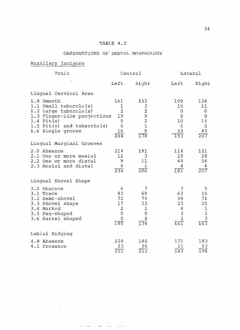
34
TABLE 4.2
OBSERVATIONS OF DENTAL MORPHOLOGY
Maxillary Incisors
Trait 2entral Lateral
Left Right Left Right
Lingual Cervical Area
1.0 Smooth 161 152 109 134 1.1 Small tubercle(s) 1 3 15 11 1.2 Large tubercle(s) 2 2 0 0 1.3 Finger-like projections 19 9 0 0 1.4 Pites) 5 2 10 15 1.5 Pit (s) and tubercleCs) 0 1 6 2 1.6 Single groove 16 9 53 45
204 178 193 207
Lingual Marginal Grooves
2.0 Absence 214 191 114 121 2.1 One or more mesial 11 3 19 28 2.2 One or more distal 9 11 44 54 2.3 Mesial and distal 0 1 4 4
234 206 181 207
Lingual Shovel Shape
3.0 Absence 6 7 7 5 3.1 Trace 83 69 63 55 3.2 Semi-shovel 72 74 59 71 3.3 Shovel shape 17 23 23 25 3.4 Marked 2 1 4 1 3.5 Peg-shaped 0 0 3 3 3.6 Barrel shaped 0 0 2 3
180 174 161 163
Labial Ridging
4.0 Absence 228 186 171 183 4.1 Presence 23 26 11 13
251 212 182 196
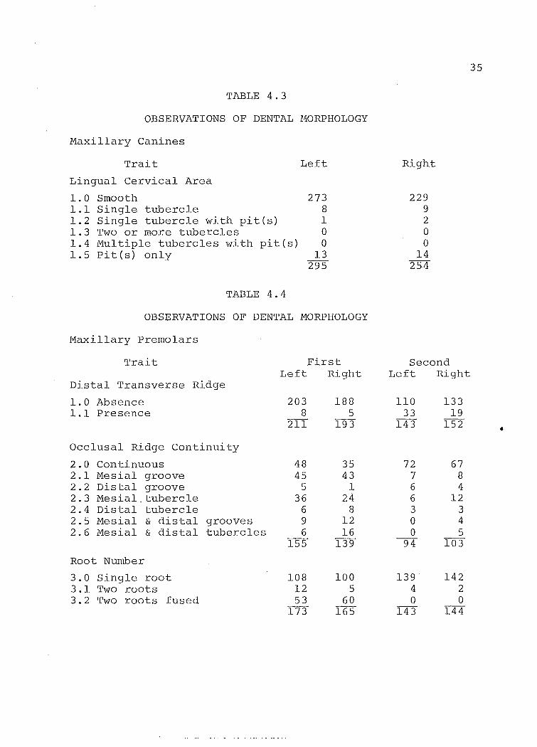
TABLE 4.3
OBSERVATIONS OF DENTAL 110RPHOLOGY
Maxillary Canines
Trait
Lingual Cervical Area
1.0 Smooth 1.1 Single tubercle 1.2 Single tubercle with pit(s) 1.3 Two or more tubercles 1.4 Multiple tubercles with pit(s) 1.5 Pit(s) only
TABLE 4.4
Left
273 8 1 0 0
13 295
OBSERVATIONS OF DENTAL MORPHOLOGY
Maxillary Premolars
Trait
Distal Transverse Ridge
1.0 Absence 1.1 Presence
Occlusal Ridge Continuity
2.0 Continuous 2.1 Mesial groove 2.2 Distal groove 2.3 Mesial. tubercle 2.4 Distal tubercle 2.5 Mesial & distal grooves 2.6 Mesial & distal tubercles
Root Number
3.0 Single root 3.1 Two roots 3.2 Two roots fused
First Left Right
203 8
211
48 45
5 36
6 9 6
155
108 12 53
173
188 5
193
35 43
1 24
8 12 16
139
100 5
60 165
Right
229 9 2 0 0
14 254
Second Left Rig-ht
110 33
143
72 7 6 6 3 0 0
94
139 4 0
143
133 19
152
67 8 4
12 3 4 5
103
142 2 0
144
35
•
36
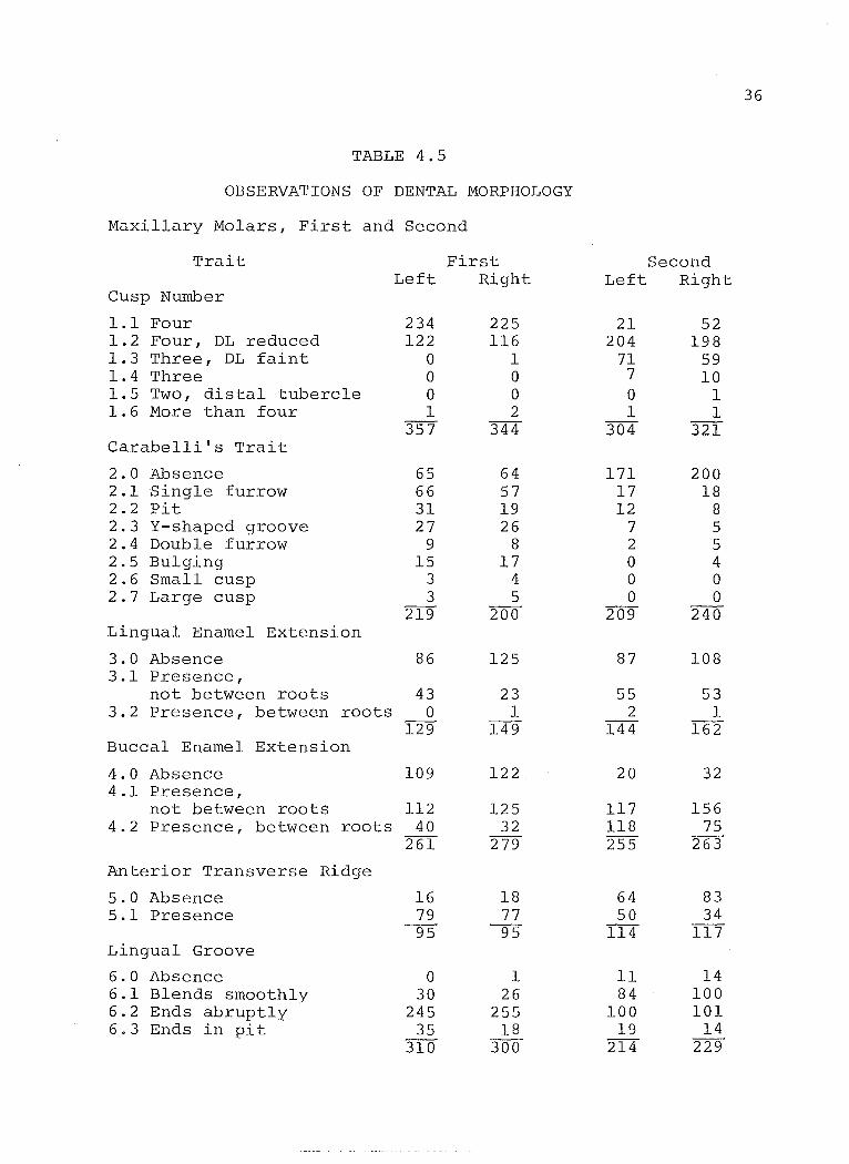
TABLE 4.5
OBSERVATIONS OF DENTAL MORPHOLOGY
Maxillary Molars, First and Second
Trait First Second Left Right Left Right
Cusp Number
1.1 Four 234 225 21 52 1.2 Four, DL reduced 122 116 204 198 1.3 Three, DL faint 0 1 71 59 1.4 Three 0 0 7 10 1.5 Two, distal tubercle 0 0 0 1 1.6 More than four 1 2 1 1
357 344 304 321 Carabelli's Trait
2.0 Absence 65 64 171 200 2.1 Single furrow 66 57 17 18 2.2 Pit 31 19 12 8 2.3 Y-shaped groove 27 26 7 5 2.4 Double furrow 9 8 2 5 2.5 Bulging 15 17 0 4 2.6 Small cusp 3 4 0 0 2.7 Large cusp 3 5 0 0
219 200 209 240 Lingual Enamel Extension
3.0 Absence 86 125 87 108 3.1 Presence,
not between roots 43 23 55 53 3.2 Presence, between roots 0 1 2 1
129 149 144 162 Buccal Enamel Extension
4.0 Absence 109 122 20 32 4.1 Presence,
not between roots 112 125 117 156 4.2 Presence, between roots 40 32 118 75
261 279 255 263
Anterior Transverse Ridge
5.0 Absence 16 18 64 83 5.1 Presence 79 77 50 34
95 95 114 117 Ling-ual Groove
6.0 Absence 0 1 Il 14 6.1 Blends smoothly 30 26 84 100 6.2 Ends abruptly 245 255 100 101 6.3 Ends in pit 35 18 19 14
310 300 214 229
37
TABLE 4.5 (cont'd)
Trait First Second Left Right Left Right
Buccal Groove
7.0 Absence a a 1 a 7.1 Blends smoothly 279 284 227 273 7.2 Ends abruptly 20 20 25 20 7.3 Ends in pit 15 13 la 12
314 317 263 305 Root Number
8.1 Single root a a 12 15 8.2 Two roots, fused a 1 1 2 8.3 Two roots, three fused 3 a 26 37 8.4 Three roots 108 90 90 93 8.5 One root, three fused a a 2 2 8.6 Increase a 0 a 1
111 9T 131 150

TABLE 4.6
OBSERVATIONS OF DENTAL MORPHOLOGY
Maxillary Molars, Third
Trait
Cusp Number
1.1 Four cusps 1.2 Four cusps, DL reduced 1.3 Three cusps, faint DL 1.4 Three 1.5 Two cusps, distal 1.6 Two cusps 1.7 More than four
Carabelli's Trait
2.0 Absence 2.1 Single furrow 2.2 Pit 2.3 Y-shaped groove 2.4 Double furrow 2.5 Bulging 2.6 Small cusp 2.7 Large cusp
tubercle
Lingual Enamel Extension
3.0 Absence 3.1 Presence, not between roots 3.2 Presence between roots
Buccal Enamel Extension
4.0 Absence 4.1 Presence, not between roots 4.2 Presence, between roots
Anterior Transverse Ridge
5.0 Absence 5.1 Presence
Left
1 20 56
120 22
2 1
222
158 8
10 1 1 4 o 1
183
116 60
3 179
23 84 98
205
85 38
123
Right
0 38 45 75 25 10
0 193
144 5
15 2 2 1 o o
169
111 43
5 159
20 91 81
192
95 27
122
38

T~ble 4.6 (cont'd)
Trait
Lingual Groove
6.0 Absence 6.1 Blends smoothly 6.2 Ends ~bruptly 6.3 Ends in pit
Buccal Groove
7.0 Absence 7.1 Blends smoothly 7.2 Ends abruptly 7.3 Ends in pit
Root Number
8.1 Single root 8.2 Two roots, fused 8.3 Two roots, three fused 8.4 Three roots 8.5 One root, three fused 8.6 Increase
Left.
17 39 17
9 82
14 175
22 10
221
85 18 21 19
7 2
152
Right
34 39 14
2 89
28 133
20 Il
192
83 Il 14 13
9 2
132
39
40
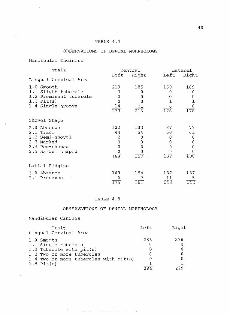
TABLE 4.7
OBSERVATIONS OF DENTAL NORPHOLOGY
Nandibular Incisors
Trait Central Lateral Left
1 Right Left Right Lingual Cervical Area
1.0 Smooth 219 185 169 169 1.1 Slight tubercle 0 0 0 0 1.2 Prominent tubercle 0 0 0 0 1.3 Pit (s) 0 0 1 1 1.4 Single g"roove 14 31 6 8
233 216 176 178
Shovel Shape
2.0 Absence 122 103 87 77 2.1 Trace 44 54 50 61 2.2 Semi-shovel 2 0 0 0 2.3 Marked a a 0 0 2.4 Peg-shaped a a a a 2.5 Barrel shaped a a a a
168 157 137 138
Labial Ridging
3.0 Absence 169 154 137 137 3.1 Presence 6 7 11 5
175 161 148 142
TABLE 4.8
OBSERVATIONS OF DENTAL NORPHOLOGY
Mandibular Canines
Trait Left Right Lingual Cervical Area
1.0 Smooth 283 278 1.1 Single tubercle a a 1.2 Tubercle with pit(s) 0 a 1.3 Two or more tubercles a a 1.4 Two or more tubercles w i th. pit ( s ) 0 a 1.5 Pit (s) 1 1
284 279
41
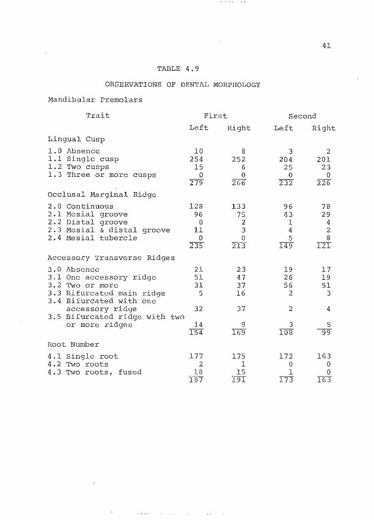
TABLE 4.9
OBSERVATIONS OF DENTAL MORPHOLOGY
Mandibular premolars
Trait First Second
Left Right Left Right
Lingual Cusp
1.0 Absence 10 8 3 2 1.1 Single cusp 254 252 204 201 1.2 Two cusps 15 6 25 23 1.3 Three or more cusps 0 0 0 0
279 266 232 226
Occlus a: Marginal Ridge
2.0 Continuous 128 133 96 78 2.1 Mesial groove 96 75 43 29 2.2 Distal groove 0 2 1 4 2.3 Mesial & distal groove 11 3 4 2 2.4 Mesia1 tubercle 0 0 5 8
235 213 149 121
Accessory Transverse Ridges
3.0 Absence 21 23 19 17 3.1 One accessory ridge 51 47 26 19 3.2 Two or more 31 37 56 51 3.3 Bifurcated main ridge 5 16 2 3 3.4 Bifurcated with one
accessory ridge 32 37 2 4 3.5 Bifurcated ridge with two
or more ridges 14 9 3 5 154 169 108 99
Root Number
4.1 Single root 177 175 172 163 4.2 Two roots 2 1 0 0 4.3 Two roots, fused 18 15 1 0
197 191 173 163
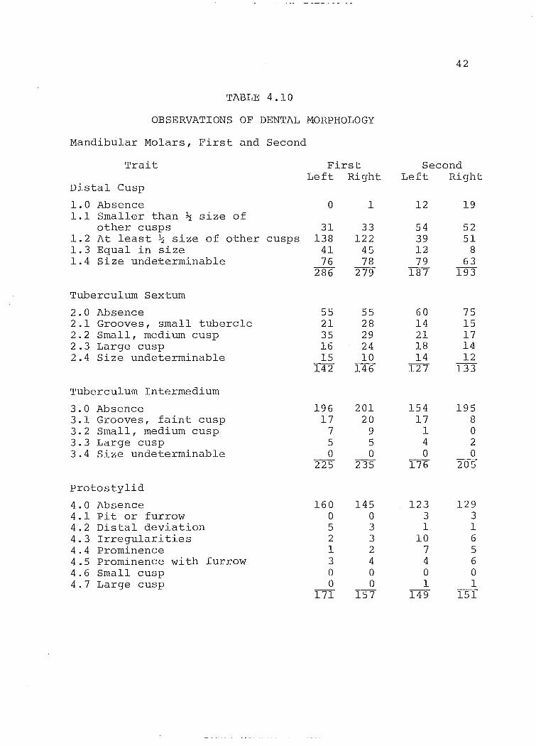
TABLE 4.10
OBSERVATIONS OF DENTAL MORPHOLOGY
Mandibu1ar Mo1ars, First and Second
Trait
Distal Cusp
1.0 Absence 1.1 Sma11er than ~ size of
other cusps 1.2 At 1east ~ size of other cusps 1.3 Equa1 in size 1.4 Size undeterminab1e
Tubercu1um Sextum
2.0 Absence 2.1 Grooves, sma11 tuberc1e 2.2 Sma11, medium cusp 2.3 Large cusp 2.4 Size undeterminab1e
Tubercu1urn Intermedium
3.0 Absence 3.1 Grooves, faint cusp 3.2 Sma11 , medium cusp 3.3 Large cusp 3.4 Size undeterminab1e
protosty1id
4.0 Absence 4.1 pit or furrow 4.2 Distal deviation 4.3 Irregu1arities 4.4 prominence 4.5 prominence with furrow 4.6 Sma11 cusp 4.7 Large cusp
First Left Right
o
31 138
41 76
286
55 21 35 16 15
142
196 17
7 5 o
225
160 o 5 2 1 3 o o
171
1
33 122
45 78
279
55 28 29 24 10
146
201 20
9 5 o
235
145 o 3 3 2 4 o o
157
42
Second Left Right
12
54 39 12 79
187
60 14 21 18 14
127
154 17
1 4 o
176
123 3 1
10 7 4 o 1
149
19
52 51
8 63
193
75 15 17 14 12
133
195 8 o 2 o
205
129 3 1 6 5 6 o 1
151

TABLE 4.10 (cont1d)
OBSERVATIONS OF DENTAL MORPHOLOGY
Trait
Buccal Groove
5.0 Absence 5.1 Blends smoothly 5.2 Ends abruptly 5.3 Ends in pit
Lingual Enamel Extension
6.0 Absence 6.1 Presence, not between roots 6.2 Presence, between roots
Buccal Enamel Extension
7.0 Absence 7.1 Presence, not between roots 7.2 Presence, between roots
Cusp & Groove Pattern
8.1 y Pattern 8.2 + pattern 8.3 X pattern 8.4 Irregular
Root Number
9.1 Two roots 9.2 Two roots, fused 9.3 Three roots 9.4 Three roots, fused 9.5 Single root
First Le-ft Right
1 8
201 63
273
103 138
2 243
60 118
42 220
196 16
9 o
221
95 1 3 o o
99
1 5
184 74
264
97 132
2 231
58 125
41 224
186 18 10
1 215
81 o 3 o o
84
43
Second Left Right
6 33 98 85
222
116 98 o
214
18 90
102 210
28 107
46 12
193
86 25
3 o o
114
5 36
119 75
235
126 94
2 222
22 100 104 226
13 III
62 6
192
68 30 o o o
98
/
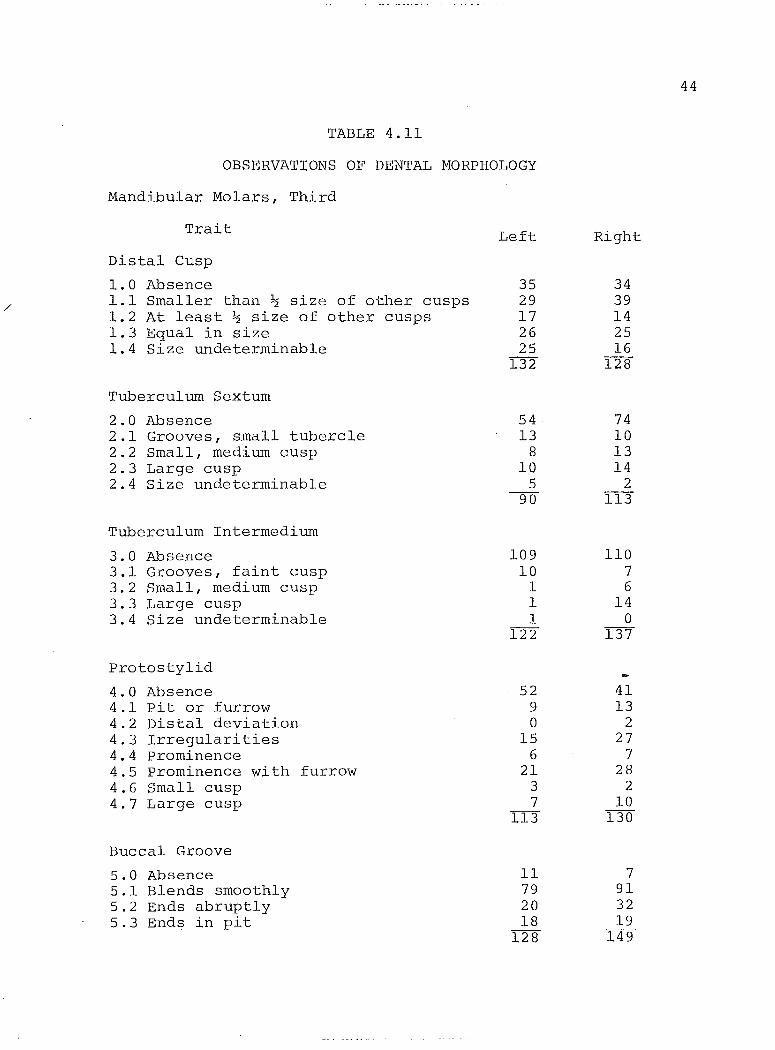
TABLE 4.11
OBSERVATIONS OF DENTAL MORPHOLOGY
Mandibular Molars, Third
Trait
Distal Cusp
1.0 Absence 1.1 Smaller than ~ size of other cusps 1.2 At least ~ size of other cusps 1.3 Equal in size 1.4 Size undeterminable
Tuberculum Sextum
2.0 Absence 2.1 Grooves, small tubercle 2.2 Small, medium cusp 2.3 Large cusp 2.4 Size undeterminable
Tuberculum Intermedium
3.0 Absence 3.1 Grooves, faint cusp 3.2 Small, medium cusp 3.3 Large cusp 3.4 Size undeterminable
Protostylid
4.0 Absence 4.1 pit or furrow 4.2 Distal deviation 4.3 Irregularities 4.4 prominence 4.5 prominence with furrow 4.6 Smal1 cusp 4.7 Large cusp
Buccal Groove
5.0 Absence 5.1 Blends smoothly 5.2 Ends abruptly 5.3 Ends in pit
Left
35 29 17 26 25
132
54 13
8 10
5 90
109 10
1 1 1
122
52 9 o
15 6
21 3 7
113
Il 79 20 18
128
Right
34 39 14 25 16
128
74 10 13 14
2 113
110 7 6
14 o
137
41 13
2 27
7 28
2 10
130
7 91 32 19
149
44
45
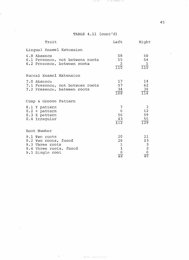
TABLE 4.11 (cont'd)
Trait Left Right
Lingual Ename 1 Extension
6.0 Absence 58 60 6.1 Presence, not between roots 55 54 6.2 Presence, between roots 2 1
115 115
Buccal Ename1 Extension
7.0 Absence 17 14 7.1 Presence, not between roots 57 62 7.2 Presence, between roots 34 38
108 114
Cusp & Groove Pattern
8.1 y pattern 7 3 8.2 + pattern 6 12 8.3 X pattern 56 59 8.4 Irregu1ar 43 55
112 129
Root Number
9.1 Two roots 20 21 9.2 Two roots, fused 26 23 9.3 Three roots 1 3 9.4 Three roots, fused 1 2 9.5 Single root 0 0
48 49
46
Anomalous Conditions
Although anomalies were not recorded for purposes of
comparisons, sorne unusual conditions were noted. Three
supernumerary maxillary incisors were found, two on the left
and three on the right out of 403 maxillary pieces with the
front part intact. There was evidence of one congenitally
missing incisor in 487 mandibles. One instance of fused
mandibular central incisors was discovered.
The canines in both maxillae and mandibles seemed to
be free from striking anomalies, as did the maxillary
premolars. Four interesting cases of what appear to be
supernumerary premolars were found in the mandibles. One
mandible was affected on both sides, and two others were
affected unilaterally. There were a total of 567 mandibular
pieces which could be examined in the premolar region. There
was one instance of an occlusal tubercle.
Several anomalies were noticed on the molars. On
the maxillary teeth, 46 enamel pearls were scored. Ali but
two of these were on the second and third molars. The break
down is as follows: left Ml-l, right Ml-l, left M2-12,
right M2-11, left M3-10, right M3-11. Mesiobuccal para
styles or pits seemed to be common although l am not aware
of other researchers noting this type of condition. One
parastyle was seen on a first molar, and twenty were seen on
second molars. The incidences were the same for each side;
twelve on the left and eight on the right. These numbers
were increased slightly on the third molars with thirteen
on the left and eighteen on the right.
47
Enamel pearls were also noted on the mandibular
molars. Only one left and two right second molars had enamel
pearls. Only three pearls were noted on third molars also.
No other conditions occurred on the mandibular molars.
CHAPTER V
STATISTICAL ANALYSIS
The primary purpose of this thesis as stated above, is
to find biological affinities of the Kleinburg sample with
hopes of contributing to the knowledge of the origins of this
population. The dentition of three contemporary ossuary
groups has been analysed by Wright (1974) and these data were
available for comparison. Shaver Hill,dated 1600-1620 A.D.
and Carton dated 1590-1610 A.D. are considered to be Neutral
ossuaries on the basis of arachaeological evidence. Sopher,
dated 1580-1610 A.D. is Huron (Wright 1974). Wright
concluded that the two Neutra1 populations were closer
biologically than either were to the Huron population. If
his conclusions are valid, it should be possible to relate
the K1einburg people to one or the other group. In addition,
an unrelated sample of North American whites was included to
test the validity of the methods chosen, and as a reference
point with which to compare the results of the Indian-Indian
comparisons. Casts from the Burlington Growth Centre were
kindly made available. A total of 52 casts were examined,
26 of each sex.
Statistica1 Method
Rather than compare each trait separately it was
decided to use a statistic that allowed an estimation of
48
49
overall genetic relationship. Two techniques were chosen
with the intent of comparing results; Sanghvi's X2 (1953)
and C.A.B. Smith's Measure of Divergence as first used by
Grewal (1962). Both methods have successfully been applied
in studies of dental morphology (Berry 1976; Sofaer et al.
1972) .
Sanghvi's X2 allows direct comparison of several
characters even when the number of categories differs for
each trait. It is essentially a cumulative X2 possible
only when the traits considered are independent of one
another.
It is not known that the dental characters are
unrelated. Some traits appear to be related in some
populations but not in others. For example, Carabelli's
cusp was found to be associated with the protostylid cusp
in the Japanese (Suzuki & Sakai 1954) but not in the
Peruvians (Goaz & Meller 1966). Data concerning the other
traits are equally inconclusive. Specific instances are
included in Chapter III where individual traits are discussed.
Generally, there may be a correlation between extra cusps
and tooth size (Dahlberg 1961, Garn et al. 1966b, Keene 1965).
There were no correlations between the 17 traits on mandibular
premolars studied by Kraus and Furr (1953). Out of 31 traits
considered throughout the mouth by Berry (1976) seven showed
associations with others, and of these five were in the
anterior teeth. The only significant correlation in the

50
molar region was between Carabelli's cusp and the condition
of more than four cusps on first maxillary molars r and this
was only found in one of her two groups. Sofaer et al (1972)
felt correlations were not important when only considering
relative distances between groupsI so assumed independence.
Brewer-Carias et al (1976) chose traits which from the
available literature appeared to be most independent, but
did not consider the problem further. Others not using
cumulative statistical techniques have not dealt with the
possibility of associations between traits (Kirveskari 1974,
wright 1974). The inconclusive data, plus the fact that
the traits considered here are spread throughout the mouth,
support the treatment of the characters as independent.
The X2 value obtained is a mean of the individual X2
values and is therefore an estimate of mean divergence
between a pair of populations. Sanghvi's X2 is equal to:
n r I I (Pl-Q)2- (P2-Q)2 l l Q Q
df
where Pl and P2 are the percentage incidences of each of r
classes in which a character is recorded in two populations,
Q = (Pl + P2)/2, n is the number of characters and
d.f. = n(r-l). Sanghvi (1953) used this value to represent
the distance between populations. It is the square root of
this value which is a true distance function (Sofaer et al.
1972). This method allows comparison of several X2 values

51
based on relative size of the values. A larger figure
indicates a larger genetic distance between the populations.
The significance of any one value cannot be tested.
Smith's Measure of Divergence (Berry & Berry 1967;
Grewal 1962) likewise gives a mean measure of divergence for
the total number of traits, and is "a quantitative expression
of the separation of the populations" (Berry & Berry 1967:373).
In other words the larger the value, the greater the
difference between the groups. The procedure first involves
the angular transformation of the frequencies of the traits
into a Theta value measured in radians by the following
formula
8 = arcsin (1-2k/n)
where k/n is the observed frequency of the trait. The
variance of Theta is l/n where n is the sample size. For
small sample sizes this relationship no longer holds true
and variance of Theta is not l/n (Green & Suchey 1976). An
alternate transformation, that of Freeman and Tukey (1950)
has been shown to be the best transformation for stabilizing
variance with small samples (Green & Suchey 1976). The
formula for this method is:
8 k . l - 2k + k - = 2 arcsln (n-l) 2 arcsin 1-2(k+l)
(n+l)
where k/n is the observed frequency for a trait. Because
four out of the five samples were of small size, this trans-
formation was chosen. Transformed Theta values are presented
in Table 6.1.
52
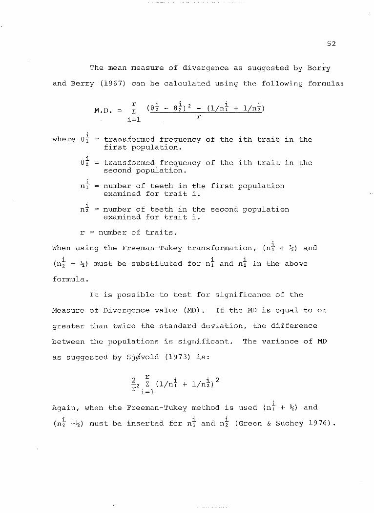
The mean measure of divergence as suggested by Berry
and Berry (1967) can be calculated using the following formula:
r M.D. = L: (et - e~) 2 - (lin} + l/nt)
i=l r
where 8~ = transformed frequency of the ith trait in the first population.
8~ transformed frequency of the ith trait in the second population.
n} = number of teeth in the first population examined for trait i.
i number of teeth in the second population n2 = examined for trait i.
r = number of traits.
When using the Freeman-Tukey transformation, i (nl + ~) and
(n~ + ~) must be substituted for n} and nt in the above
formula.
It is possible to test for significance of the
t'leasure of Divergence value (IvID). If the MD is equal to or
greater than twice the standard deviation, the difference
between the populations is significant. The variance of ~ID
as suggested by Sj~vold (1973) is:
2 ri' 2 -2 L: (l/nl + lin}) r i=l
i Again, when the Freeman-Tukey method is used (nl + ~) and
(n~ +~) must be inserted for n~ and n~ (Green & Suchey 1976).
53
Computations
A total of 90 sets of observations were made on the
Kleinburg material. This figure was reduced to 28 for the
following reasons. The 28 included those traits which were
observed on aIl five groups, which could be reduced to
present/absent categories and which were scored using the
same objective aids as Wright (1974) to maximize scoring
consistency. The traits and frequencies for each population
are presented in Table 5.1.
In aIl computations present/absent categories only
were considered. Calculations for left and right sides were
done separately. That is, left sides were compared to left
and right sides to right. A correlation coefficient was
calculated to compare results for each side using Spearmanls
rank method (Arkin & Cotton 1970). Each sample was compared
ta every other sample using bath statistical methods.
Spearmanls rank coefficient of correlation was calculated to
compare the results from each method. Additional Sanghvils
2 X were computed for the Indian groups using only seven and
two traits (see Table 5.2). These groupings correspond to
traits found by Sofaer et al. (1972) to have greater than 90%
and 95% repeatability. Those traits with greatest
repeatability were most discriminating between closely
related samples (Sofaer et al. 1972) . Individual traits
were not tested for repeatability here, but it seemed a
fair assumption that the same traits having the most
54
discriminating power in Southwest Indians would also show
differences in Ontario Indians (Mayhall pers. comm.).
TABLE 5.1 (Part 1) lI) mCIDENCE OF DHITAL VARIANTS - Left Side lI)
Charactcr Present l<leinburg Shaver Hill Ca.rton Sopher Bur1in"ton
lIo. % No. 9é No. % No. g6 No. % :'laxilla
Lingual Shovel on Il 114/180 96.66 15/15 100 3/3 100 12/12 100 18/50 36. 00
Labi3l Rid~ing on Il 23/251 9.16 6/16 37.50 1/3 33.33 4/10 t+O.OO 5/51 9. 80
Lingual Tubcrcles on Il 36/204 1.47 3/17 17.65 1/3 33.33 0/11 oê 1/46 2. 17
Lingual Shove1 on 12 154/161 95.65 9/10 90.00 12/12 100 9/9 100 20/47 42. 55
Labial Ridf,ing on 12 11/182 6. Ott 4/10 40.00 1/10 10.00 2/8 25.00 0/51 00
LinGual Tubercles on 12 15/193 7.77 3/11 27.27 1/12 8.23 2/10 20.00 3/45 6.67
Lingual Tuberc1es on C 9/2'95 3.05 0/15 0 10/32 31.25 2/10 20,00 3/58 5.17
Anterior transverse ridgc on PmI 8/211 3.79 3/26 11.54 0/35 00 3/14 21.43 3/45 6.67
" " 11 Il " ft " ft on Prn2 33/143 23.08 4/9 44.44 16/28 57.14 10/19 52.63 25}48 52.08
Carabc1li's trait on Ml 154/2]9 70.32 13/32 40.62 26/54 48.15 14/32 43 .75 44/52 86.21
Antcrior transverse rid3;e on Hl 79/95 83.16 12/14 85.72 17/19 89.47 13/21 61.91 25/29 84.62
four cusps on M2 225/304 74.01 7/25 28.00 15/27 55.56 7/21 33.33 32/48 66.67
Carabclli's Trait on M2 38/209 18.18 3/24 12.50 1/32 3,12 1/20 5.00 16/49 78.38
Antcrior transverse ridge on H2 50/114 43.86 5/11 45.45 5/12 41.67 1/16 6.25 29/37 32. 51

~ Lf)
TABLE 5.1 (Part 1) ctd. Caaracter Present Kleinburg Shaver Hill Carton Sopher Burlington
_________________ ~_ poo __ -'-:_~ %~ ___ ~.~No_. ___ !6 _____ I1~. _ % ____ J'T6_.~ __ _ % ~(). % Mandible
Lingual shovelling on Il
Labial ridging on Il
Lingual shovelling on I2
Labial ridging on I2
Two or more lingual cusps on Pml
" Il " " " " Il " Pm 2
Protostylid on Ml
+ groove pattern on Ml
Tuberculum sextum on Ml
46/168 23.38
6/175 3.43
50/137 36.50
11/148 7.43
15/279 5.38
25/232 10.76
11/171 6.42
16/221 7.24
87/142 61.27
Tuberculum intermedium on Ml 29/225 12.89
Protostylid on M2
+ groove pattern on M2
Tuberculum sextum on M2
26/149 17.44
107/193 55.44
67/127 52.75
Tuberculum intermedium on M2
22/176 12.50
7/9 77.77 5/9 55.56 5/6 83.33 1/50 2.00
2/8 25.00 0/8 00 0/7 00 3/52 13.46
8/9 88.89 7/11 63.64 6/6 100 3/52 5.82
2/7 28.57 1/9 11.11 1/6 16;67 2/51 3.93
5/19 26.32 4/22 16.67 6/19 31.58 12/47 25.53
1/13 7.69 6/25 24.00 1/18 5.56 35/50 60.00
0/26 00 2/51 3.92 5/34 14.71 3/49 6/J..2
1/30 3.33 5/45 11.11 1/33 3.02 2/17 11. 76
8/22 36.36 9/20 45.00 12/33 36.36 10134 29.41
6/27 22.22 8/42 19.05 7/34 20.59 11/48 22.92
2/26 7.69 2/46 4.35 1/21 4.76 1/49 2.04
3/14 21.44 25/46 50.00 8/21 47.62 15/25 60,00
3/15 20.00 7/23 30.43 4/18 22.22 1/14 7.14
2/24 8.33 1/42 2.38 5/21 23.81 4/38 10.53
TABLE 5.1 (Part 2) r-- INCIDENCE OF DENTAL VARIANTS - Right Side lI)
Character Present Kleinburg Shaver Hill Carton ScEher Burlin~ton
. No. % No. % No. % No. % No. % Maxilla
Lingual Shovel on Il 166/174 95.98 12/12 100 7/7 100 15/15 100 15/45 33.33
Labial Ridging on Il 26/212 12.26 9/13 69.23 4/6 66.67 7/15 46.67 5/52 9.62
Lingual Tubercles on Il 6/193 3,37 0/13 00 1/7 14.29 2/15 13 .33 1/47 2.13
Lingual Shovel on 12 158/163 96.93 10/11 90.91 14/14 100 7/7 100 19/50 38.00
Labial Ridging on 12 13/196 6.63 5/8 62.50 3/12 25.00 7/15 46.67 1/52 1. 92
Lingual Tubercles on 12 13/207 6.28 2/11 18.18 2/13 15.38 1/8 12.50 5/48 10.42
Lingual Tubercles on C 11/254 4.33 5/17 29.41 6/19 31.58 5/18 27.78 6/49 12.24
Anterior transverse ridge on Pm
1 5/193 2.59 1/11 9.09 3/33 9.09 5/22 22.73 8/46 17.39
" " " " " " 11 " on Pm 2 19/152 12.50 5/19 26.32 11/25 44.00 5/9 55.56 23/48 47.92
Carabel1i's trait on Ml 136/200 68.00 22/42 52.38 33/64- 51.56 27/36 75.00 28/32 87.50
Anterior transverse ridge on Ml 77/96 81. 05 20/20 100 20/23 86.96 17/25 68.00 47/54 87.04
Four cusps on M2 250/321 77 .88 2/22 9.09 16/38 42.11 10/17 58.82 31/46 67.39
Carabel1i's Trait on M2 40/240 16.66 0/21 00 0/39 00 3/17 17.65 31/44 70.45
Anterior transverse ridge on M2 34/117 29. 06 3/9 33.33 10/22 45.45 1/14 7.14- 17/39 43.59

co TABLE 5. 1 (Part 2) ctd. 11) INCIDENCE OF DENTAL VARIANTS - Right Side
Character Present Kleinburg Shaver Hill Carton Sopher Burlington Mandible
.
Lingual Shovel on Il 54/157 34.39 6/9 66.67 1/3 33.33 5/7 71.43 3/52 5.,;77
Labial ridging on Il 7/161 4.35 0/7 00 0/2 00 0/8 00 5/52 9.62
Lingual shovelling on 12 61/138 44.20 8/8 100 4/8 50.00 7/10 70.00 2/52 3.84
Labial ridging on 12 5/142 3.52 3/8 37.50 0/6 00 0/10 00 1/52 1.92
Two or more.lingual cusps on PmI 6/266 2.25 2/12 16.67 9/22 40.91 5/20 25.00 12/47 25.53
Il " " " " " " " on Pm2 23/226 10.18 7/19 36.84 4/27 14.81 4/14 28.57 22/50 44.00
Protostylid on Hl 12/157 7.64 2/33 6.45 3/57 5.26 1/18 5.56 3/51 5.88
+ groove pattern on Ml 18/215 8.37 3/32 9.37 4/52 7.69 1/18 5.56 3/18 16.67
Tuberculum sextum on Ml 91/146 62.33 8/26 30.77 11/22 50.00 6/16 37.50 7/25 28.00
Tuberculum intermedium on Ml 34/235 14.47 8/32 25.00 15/48 31. 25 4/18 22.22 7/50 14.00
Protostylid on M2
22/151 14.56 1/24 4.17 4/39 10.26 0/26 00 1/48 2.08
+ groove pattern on M2 111/192 57.81 6/21 28.57 14/40 35.00 9/25 36.00 10/28 35.71
Tuberculum sextum on ~12 58/133 43.61 4/11 36.36 7/22 31.82 7/20 35.00 2/16 12.50
Tuberculum intermedium on M2
10/2Q5 4.88 2/22 9.09 2/35 5.71 5/25 20.00 7/41 17.07
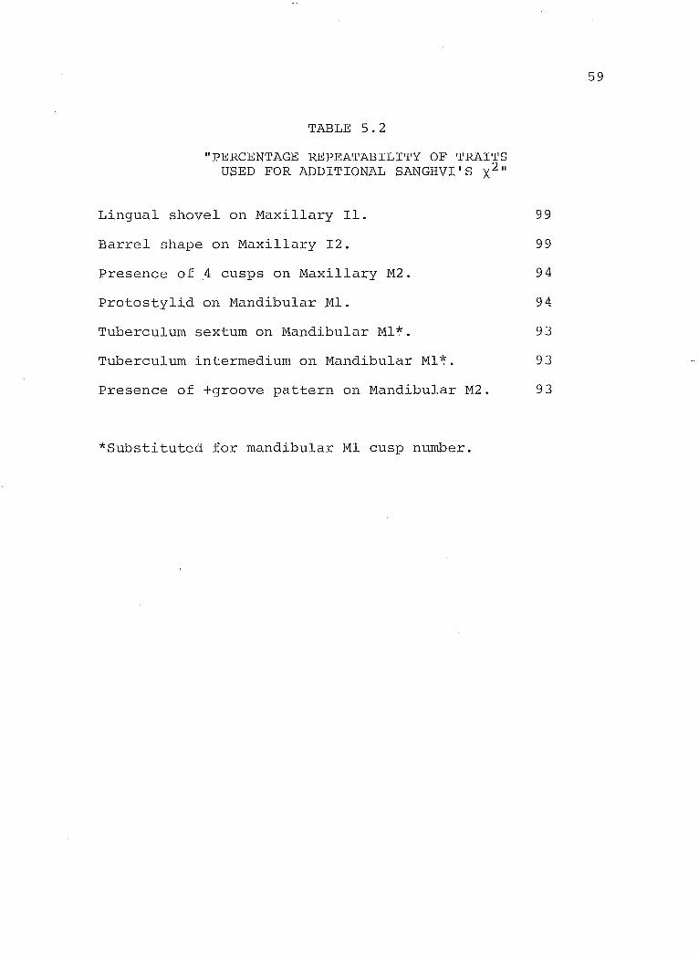
TABLE 5.2
"PERCENTAGE REPEATABILITY OF TRAITS USED FOR ADDITIONAL SANGHVI'S X211
Lingual shovel on Maxillary Il. 99
Barrel shape on Maxillary 12. 99
Presence of 4 cusps on Maxillary M2. 94
Protostylid on Mandibular Ml. 94
Tuberculum sextum on Mandibular Ml~. 93
Tuberculum intermedium on Mandibular Ml~. 93
Presence of +groove pattern on Mandibular M2. 93
*Substituted for mandibular Ml cusp number.
59
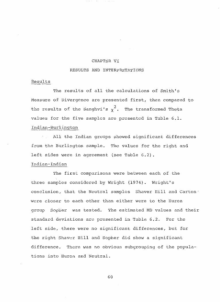
CHAPTER VI
RESULTS AND INTERPRETATIONS
Results
The results of all the calculations of Smith's
Measure of Divergence are presented first, then compared to
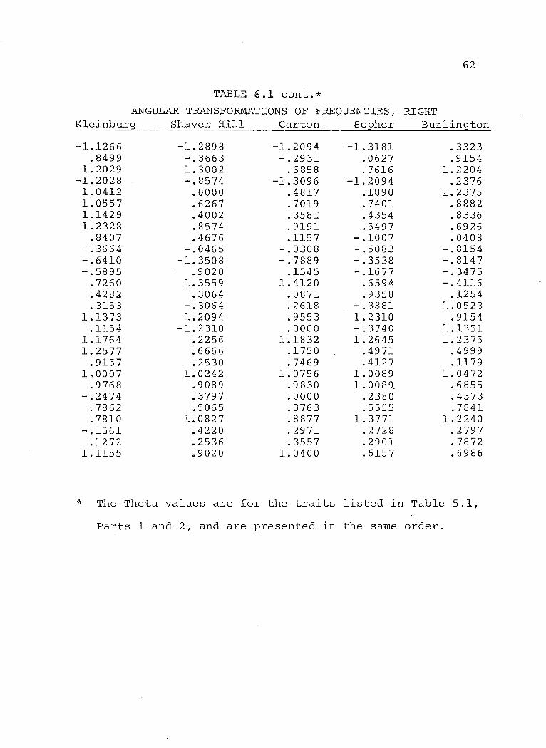
the results of the Sanghvi's X2 . The transformed Theta
values for the five samples are presented in Table 6.1.
Indian-Burlington
All the Indian groups showed significant differences
from the Burlington sample. The values for the right and
left sides were in agreement (see Table 6.2) .
Indian-Indian
The first comparisons were between each of the
three samples considered by Wright (1974). Wright's
conclusion, that the Neutral samples Shaver Hill and Carton'
were closer to each other than either were to the Huron
group Sopher was tested. The estimated MD values and their
standard deviations are presented in Table 6.2. For the
left side f there were no significant differences, but for
the right Shaver Hill and Sopher did show a significant
difference. There was no obvious subgrouping of the popula
tions into Huron and Neutral.
60

61
The Kleinburg sample differed significantly from the
three Indian samples for both right and left sides. The data
from Shaver Hill, Carton and Sopher were pooled and another
MD was calculated. Again the Kleinburg sample differed
significantly for both sides (Table 6.3).
TABLE 6.1*
ANGULAR TRANSFORMATIONS OF FREQUENCIES,LEFT
Kleinburg
-.2684 .9502
1.3094 -1.1373 1.0641
.9977 1.2107 1.1678
.5642 -.4165 -.7159 -.4992
.6859
.1220
.4664 1.1843
.2714 1. 0079 1.0958
.8965 1. 0478 1.0186 -.2257
.8315
.7032 -.1085 -.0547
.8417
Shaver Hill
-1.3181 .2380 .6594
-.8240 .1836 .4317
1.3181 .8359 .1007 .1829
-.7334 .4373 .8055 .0837
-.5275 .4644
-.7854 .3881 .4676 .9126
1.3771 1.1334
.2642
.5659
.9553
.5645
.5994
.9303
Carton
-1.0472 .2618 .2618
-1.2898 .8240 .8867 .3724
1. 4033 -.1385
.0364 -.8513 -.1074 1.1471
.1549 -.1007 1. 2310 -.2536
.7854
.6556
.5242 1.1308
.8653
.0955
.6497 1.1076 ·-.0852
.3848 1.2003
Sopher
-1. 2898 .1836
1.2780 -1. 2490
.4644
.5808
.5808
.5645 -.0501 .1216
-~2295
.3242 1. 0371
.9757 -.6193 1.2094
-1.1832 .6194 .3581
1. 0089 .7563
1.1535 .2680 .6087
1.0497 .2295 .5555 .5244
Burlington
.2781
.9089 1. 2166
.1464 1.4317 1.0132 1. 0799 1. 0132 -.0408 -.7470 -.7765 -.3327
.3470 -.5858 1.2310 1.0523 1.0523 1.1308
.4999 -.4031 1. 0366
.8104
.4117
.5595 1.2275 -.1937
.9358
.8786
* The Theta values are for the traits listed in Table 5.1,
Parts 1 and 2, and are presented in the same order.
62
TABLE 6.1 cont.*
AN GULAR TRANSFORMATIONS OF FREQUENCIES r RIGHT K1einburs;r Shaver Hill Carton Sopher Bur1ington
-1.1266 -1.2898 -1.2094 -1.3181 .3323 .8499 -.3663 -.2931 .0627 .9154
1.2029 1.3002. .6858 .7616 1.2204 -1.2028 -.8574 -1.3096 -1.2094 .2376 1. 0412 .0000 .4817 .1890 1.2375 1.0557 .6267 .7019 .7401 .8882 1.1429 .4002 .3581 .4354 .8336 1. 2328 .8574 .9191 .5497 .6926
.8407 .4676 .1157 -.1007 .0408 -.3664 -.0465 -.0308 -.5083 -.8154 -.6410 -1.3508 -.7889 -.3538 -.8147 -.5895 .9020 .1545 -.1677 -.3475
.7260 1. 3559 1.4120 .6594 -.4116
.4282 .3064 .0871 .9358 .1254
.3153 -.3064 .2618 -.3881 1.0523 1.1373 1.2094 .9553 1.2310 .9154
.1154 -1.2310 .0000 -.3740 1.1351 1.1764 .2256 1.1832 1.2645 1. 2375 1.2577 .6666 .1750 .4971 .4999
.9157 .2530 .7469 .4127 .1179 1.0007 1.0242 1.0756 1.0089 1.0472
.9768 .9089 . 9830 1.0089 . .6855 -.2474 .3797 .0000 .2380 .4373
.7862 .5065 .3763 .5555 .7841
.7810 1.0827 .8877 1.3771 1.2240 -.1561 .4220 .2971 .2728 .2797
.1272 .2536 .3557 .2901 .7872 1.1155 .9020 1.0400 .6157 .6986
* The Theta values are for the traits 1isted in Table 5.1,
Parts 1 and 2, and are presented in the same order.
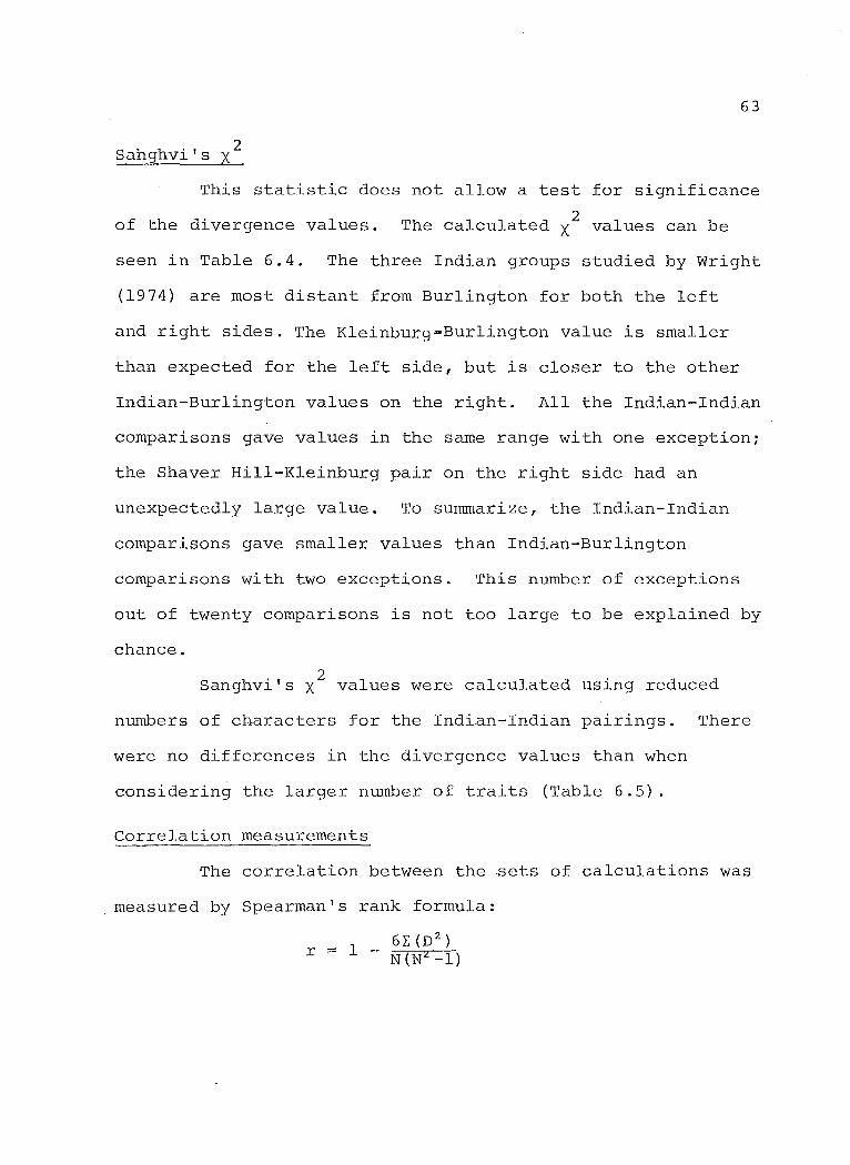
2 Sahghvi's X
63
This statistic does not allow a test for significance
of the divergence values. 2 The calculated X values can be
seen in Table 6.4. The three Indian groups studied by Wright
(1974) are most distant from Burlington for both the left
and right sides. The Kleinburg=Burlington value is smaller
than expected for the left side, but is closer to the other
Indian-Burlington values on the right. AIl the Indian-Indian
comparisons gave values in the same range with one exception;
the Shaver Hill-Kleinburg pair on the right side had an
unexpectedly large value. To summarize, the Indian-Indian
comparisons gave smaller values than Indian-Burlington
comparisons with two exceptions. This number of exceptions
out of twenty comparisons is not too large to be explained by
chance.
2 Sanghvi's X values were calculated using reduced
numbers of characters for the Indian-Indian pairings. There
were no differences in the divergence values than when
considering the larger number of traits (Table 6.5).
Correlation measurements
The correlation between the sets of calculations was
measured by Spearman's rank formula:

where r ~ measure of correlation
D difference between the ranks of each pair of populations for each set of calculations.
N number of pairs, in this case the number of comparisons.
64
The pairs are ranked from the smallest MD value to the largest
for each series. The difference between the ranking for the
two sets being measured is squared then added and inserted
into the formula. The closer the value to 1.0, the greater
the correlation between series.
The coefficient of correlation between right and
left sides for Smith's Measure of Divergence is +.867. The
side to side coefficient for Sanghvi's is +.752. The critical
value for r at a = .05 and N = 10 is +.564; at a = .01 the
value is +.764 (Thomas 1976, Table A.13). The observed
correlation in both cases is more than the critical values,
therefore is statistically significant.
The measure of correlation between the Smith and
Sanghvi values is +.964 for the left side and +.879 for the
right. The same critical values used above apply here, and
the correlation between the two methods is again significant.
65
Interpretation
In general it is expected that Indian and White
groups differ significantly, and this in fact is what was
found. The MD value for the Kleinburg-Burlington comparisons
are smaller than expected, even though significant. Ossenberg
(1976) found that within race MD values were significantly
smaller than between race MD values. The results for the
three known Indian samples paired with themselves, then
Burlington support this. However the Kleinburg-Burlington
MD value for the left side appears to be no larger than the
MD value for the Kleinburg-Indian comparisons. The number
of MD values was not sufficiently large to allow testing for
differences bétween within-race "and b'etween-race values.
The rather large and significant MD values between
Kleinburg, a postulated Huron population and the other
Ontario Iroquois are surprising and must be explained. Again
the values for the Sopher, Shaver Hill and Carton cbmparisons
agree with Indian-Indian values found using cranial traits
(Ossenberg 1976), and it was expected that the Kleinburg
population would be no less close. The Kleinburg ossuary
was very large, containing over 600 individuals. In this
respect it is atypical. It is also atypical in that no
associated village site has been found. The Kleinburg
sample may not be representative of any one population.
Or the data assigned the site, 1600 A.D. may be incorrect.
A larger temporal gup bctween the populations may account
66
Alternately the traits chosen may not be those traits
best reflecting the genetic differences between the popula
tions. Those traits demonstrating between-race differences
may not be suitable for within-race comparisons. Lastly,
the reduction of the categories to present/absent scoring
causes a loss of information which may affect MD values.

TABLE 6.2
MEASURES OF DIVERGENCE AND THEIR STANDARD DEVIATIONS (brackets) OF SAMPLES TAKEN IN PAIRS
Left Right
Shaver Hi11-Sopher .0028 .0911* (.0405) (.0400)
Shaver Hill-Carton .0314 .0473 (.0452) (.0479)
Carton-Sopher .0556 -.1416 (.0485) (.0476)
Kleinburg-Carton .1197* .1416* (.0300) (.0320)
K1einburg-Bur1ington .2663* .4045* (.0100) (.0100)
K1einburg-Shaver Hill .2701* .3478* (.0200) ( .0223)
K1einpurg-Sopher .2950* .1675* (.0236) (.0200)
Carton-Bür1ington .3727* .4291* (.0339) (.0254)
Shaver Hi11-Bur1ingon .4796* .6695* (.0258) (.0264)
Sopher-Bur1ington .6995* .4160* (.0282) ( .0083)
TABLE 6.3
MEASURES OF DIVERGENCE CALCULATED WITH POOLED DATA
K1einburg-Poo1ed data
* indicates a significant value.
Left
.2644* (.0100)
Right
.2142* (.0083)
67

TABLE 6.4
SANGHVI'S X2 V~LUES FOR SAMPLES TAKEN IN PAIRS (/)
Left Right
Shaver Hi11-Sopher 2.7112 3.3205
Shaver Hill-Carton 3.1344 3.1835
Carton-Sopher 3.0871 2.5883
K1einburg-Carton 3.0403 3.2016
K1einburg-Bur1ington 3.9074 5.1039
K1einburg-Shaver Hill 3.9441 4.8468
K1einburg-Sopher 3.9312 3.4425
Carton-Bur1ington 5.3468 4.4807
Shaver Hi11-Bur1ington 5.8780 4.7340
Sopher-Bur1ington 5.4390 5.7297
TABLE 6.5
SANGHVI' S X2 VALUES FOR 7 AND 2 TRAITS ( 1)
7 Traits 2 Traits Left Right Left Right
K1einburg-Carton 2.6972 2.7932 2.2009 1. 7263
K1einburg-Sopher 2.3805 2.5024 2.4294 2.3660
K1einburg-Shaver Hill 3.6704 4.5237 1.5241 1. 7263
Shaver Hill-Carton 2.3778 2.3825 .4365 .2739
Shaver Hi11-Sopher 2.0330 3.1200 2.4252 2.2851
Carton-Sopher 1.6541 1. 6480 .2125 2.0180
68
Conclusions
CHAPTER VII
CONCLUSIONS AND Sm·1MARY
Having demonstrated that there is no difference
biologically between Huron and Neutral groups it is not
possible ta identify Kleinburg as one or the other. The
population is distinct from the other Indian groups and from
the White population. When considering"the magnitude of the
MD values, Kleinburg seems ta fall somewhere in the middle of
the continuum from the three known Indian groups ta the White
sample. The location of the site suggests it is Huron. The
evidence from the dental morphology indicates that Kleinburg
is significantly different from a known Huron sample (Sopher).
There are several factors which might provide an
explanation for this intermediate relationship of the Klein
burg sample. Microevolutionary trends become apparent in
morphological traits over time (Anderson 1968). If aIl the
Indian groups are contemporary, microevolutionary changes
will be of little significance. If, however, the assigned
date of 1600 A~D. for Kleinburg is tao early, sorne micro
evolutionary change might be expected, increasing the
divergence between Kleinburg and the other Indian groups.
Wright (1974) points out the apparent parallel between bio
logical divergence and archaeological divergence. If this
69

70
parallel is real, biological divergence between Ontario
Iroquois would increase over time from prehistoric to
historic. Again, if the Kleinburg population dates later
than 1600 A.D. it would support this hypothesis. Another
consideration is the amount of contact between the Kleinburg
population and other Indian groups. The large size of the
ossuary hints at a mixed population. Little is known about
the extent of trading during protohistoric times nor about
the practice of adopting individuals from outside the popula
tion. In historic times the Huron acted as middlemen between
the French and other Indian groups in trading (Wright 1966) .
It is also known that in times of famine Neutral groups at
least, visited Huronia (Wright 1974). Continuous contact
would have produced a population quite different from Shaver
Hill, Carton and Sopher.
The results of the investigation do not allow any
definite conclusions. Hopefully further research on the
Kleinburg ossuary sample will clarify the identity of the
group. The accumulation of information on microevolutionary
change in Ontario Iroquois will permit a more conclusive
interpretation of the data presented here.
Summary
1. The non-metric morphology of the permanent dentition
of the Kleinburg population was observed and compared
to that of three contemporary Indian populations;
Shaver Hill, Carton and Sopher. All four Indian groups
71
were compared to a modern white population, Burlington.
2. For comparative purposes twenty-eight traits were
considered. These included those traits observed
on all five populations, which could be reduced to
present/absent categories and which were scored using
the same objective aids.
3. The Kleinburg population is distinct from other
contemporary Iroquois groups and from a white
population. It cannot be conclusively identified
as either Huron or Neutral.
4. Sanghvi's X2 and Smith's Measure of Divergence give
comparable estimates of biological distance when
considering relative distances.

Anderson, J.C. 1968
Andrews, N.H. 1975
LIST OF REFERENCES
Skeletal 'Anomalies' as Genetic Indicators. In The Skeletal Biology of Human Populations. D.R. Brothwell, ed. London, pergamon Press.
periodontal significance of cervical enamel projections. J. Canad. Dent. Assn. 41:50-52.
Arkin, H. & R.R. Cotton 1970 Statistical Methods, 5th ed. New York,
Barnes & Noble Inc.
Bang, G. & A. 1972
Bailit, H.L., 1970
Barnes, D.S. 1969
Berry, A.C. 1976
Biggerstaff, 1970
Hasund Morphological characteristics of the Alaskan Eskimo dentition. II. Carabelli's cusp. Am. J. Phys. Anthrop. 37:35-40 . .
P.L. Workman, J.D. Niswander, C.J. MacLean Dental asymmetry as an indicator of genetic and environmental conditions in human populations. Human Biol. 42:43-47.
R.H.
Tooth morphology and other aspects of Teso dentition. Am. J. Phys. Anthrop. 30:183-193.
The anthropological value of minor dental variants of the dental crown. Am. J. Phys. Anthrop. 45:257-268.
Morphological variations for the permanent mandibular first molars in human momozygotic and dizygotic twins. Archs. Oral. Biol. 15: 721-730.
Biggerstaff, R.H. 1973 Heritability of the Carabelli's cusp in twins.
J. Dent. Res. 52:42-44.
72
73
Brewer-Carias, C.A. & S. LeBlanc & J.V. Neel 1976 Genetic structure of a tribal
the Yanomamo Indians. XIII. differentiation. Am. J. Phys. 44:5-14.
population, Dental microAnthrop.
Carbonell, V.M. 1963
Dahlberg, A.A. 1945
Dahlberg, A.A. 1949
Dahlberg, A.A. 1950
Dahlberg, A.A. 1961
Dahlberg, A.A. 1962
Dahlberg, A.A. 1963
Dahlberg, A.A. 1965
Davies, P.L. 1968
Variations in the frequency of shovel-shaped incisors in different populations. In Dental Anthropology. D.R. Brothwell, ed. Oxford, pergamon Press.
The changing dentition in man. J. Am. Dent. Assn. 32:676-690.
The dentition of the American Indian. Papers on the Physical Anthropology of American Indian. W. Laughlin, ed. New Viking Fund.
In the York,
The evolutionary significance of the protostylid. Am. J. Phys. Anthrop. 8:15-25.
Relationship of tooth size to cusp number and groove conformation of occlusal surface patterns of lower molar teeth. J. Dent. Res. 40:34-38.
Genetic aspects of evolution of the human dentition. In Genetics and Dental Health. C.J. Witkop, ed. New York, McGraw Hill Book Co.
Analysis of the American Indian dentition. In Dental Anthropology. D.R. Brothwell, ed. Oxford, pergamon Press.
Geographic distribution and origin of dentitions. Internat. Dent. J. 15:311-422.
Relationship of cusp reduction in the permanent mandibular molar to agenesis of the teeth. J. Dent. Res. 47:499.
Devoto, F.C.H., 1972
Garn, S.M., A.B. 1966a
Garn, S.M., A.B. 1966b
74
B.M. Perrotto, N.H. Arias Related inheritance of the foremen caecma to the protostylid cusp-inhibiting allele. J. Dent. Res. 51:873.
Lewis & R.S. Kerewsky Bilateral asymmetry in cusp number and crown morphology of the mandibular first molar. J. Dent. Res. 45:1820.
Lewis, R.S. Kerewsky & A.A. Dahlberg Genetic independence of Carabelli's trait from tooth size of crown morphology. Archs. Oral Biol. 11:745-747.
Garn, S.M. A.A. Dahlberg, A.B. Lewis & R.S. Kerewsky 1966c Groove pattern cusp number and tooth size.
J. Dent. Res. 45:970.
Goaz, P.W. & 1966
M.C. Miller
Goldstein, M.S. 1948
A preliminary description of the dental morphology of the Peruvian Indian. J. Dent. Res. 45:106-119.
Dentition of Indian crania from Texas. Am. J. Phys. Anthrop. 6:63-69.
Goose, D.H. & G.T.R. Lee 1948 The mode of inheritance of Carabelli's trait.
HUffian Biol. 43:64-69.
Green, R.F. 1976
& J.M. Suchey
Greene, D.L. 1967
Grewal, M.S. 1962
Grüneberg, H. 1952
The use of inverse sine transformations in the analysis of non-metric cranial data. Am. J. Phys. Anthrop. 45:61-68.
Genetics, Dentition and Taxonomy. University of Wyoming Publ. Vol. XXXIII, Nos. 1,2, and 3.
The rate of genetic divergence of sublines ln the C57BL strain of mice. Genet. Res. 3:226-237.
Genetical studies on the skeleton of the mouse. IV. Quasi-continuous variations. J. Genet. 51-95-114.
Hanihara, K. 1963
Hanihara, K. 1967
Hellman, M. 1928
Hrdliéka, A. 1911
Hrdliè5"ka, A. 1920
Hrdlicka, A. 1921
J;6rgensen, K.D. 1955
Keene, H.J. 1965
Kirveskari, P. 1974
Korkhaus, G. 1930
Kraus, B.S. 1951
75
Crown characters of the deciduous dentition of Japanese-American hybrids. In Dental Anthropology. D.R. Brothwell, ed. Oxford, pregamon Press.
Racial characteristics in the dentition. J. Dent. Res. 46:923-926.
Racial characters in human dentition. Proc. Amer. Philos. Soc. 67:157-174.
Human dentition and teeth from the evolutionary and racial standpoint. Dominion Dental J. 23:403-417.
Shovel-shaped teeth. Am. J. Phys. Anthrop. 3:429.
Further studies of tooth morphology, Am. J. Phys. Anthrop. 4:141-176
The Dryopithecus pattern in recent Danes and Dutchmen. J. Dent. Res. 34:195-208.
The relationship between third molar agenesis and the morphological variability of the molar teeth. Angle Orthodont. 35:289-298.
Morphological Traits of the Permanent Dentition of Living Skolt Lapps. Academic Dissertation, Instit~te of Dentistry, University of Turku, Turku, Finland.
Anthropologic and odontologic studies of twins. Int. J. Orthodont. 16:640-647.
Carabelli's anomaly of the maxillary molar teeth. Am. J. Hum. Genet. 3:348-355.

Kraus, B.S. 1957
Kraus, B.S. 1959
The genetics of the human dentition. J. Forensic Sei. 2:419-427.
76
Occurrence of the Carabelli trait in southwest ethnie groups. Am. J. Phys. Anthrop. 17:117-123.
Kraus, B.S. & M. Furr 1953 Lower first premolars. Part 1. A definition
and classification of discrete morphologie traits. J. Dent. Res. 32:554-564.
Kraus, B.S., R.E. Jordan & L. Abrams 1969 Dental Anatomy and Occlusion. Baltimore,
Williams & Wilkins.
Kraus, B.S., W.J. Wise & R.H. Frei 1959 Heredity and the craniofacial complex.
Krogman, W.M. 1960
Lasker, G.W. 1950
Am. J. Orthodont. 45:172-217.
Oral structures genetically and anthropologically considered. N.Y. Acad. Sei. Annals 85:17-41.
Genetic analysis of racial traits of the teeth. Cold Spring Harbor Symposia on Quantitative Biology 15:191-203.
Lasker, G.W. & M.M.C. Lee 1957 Racial traits in the human teeth. J. Forensic
Sei. 2:401-419.
Ludwig, F.J. 1957
Lundstrom, A. 1963
Mayhall, J.T. 1976
The mandibular second premolars: morphologie variation and inheritance. J. Dent. Res. 36:263-273.
Tooth morphology as a basis for distinguishing monozygotic and dizygotic twins. Am J. Hum. Gen. 15:34-43.
The Morphology of the Permanent Dentition of prehistoric and Modern Central Arctic Eskimoid Peoples: A Study of Their Biological Relationships. Ph.D. Dissertation, U. of Chicago.
77
Meredith, H.V. & E.H. Hixon 1954 Frequency, size and bi1atera1ism of
Carabelli's tubercle. J. Dent. Res. 33:435-440.
Moorrees, C.F.A. 1957
Moorrees, C.F.A. 1961
Morris, D.H. 1965
Osborn, H.F. 1888
Ossenberg, N. 1976
Pedersen, P.O. 1949
Portin, P. & L. 1974
Sanghvi, L.D. 1953
Snyder, R. 1960
Sofaer, J.A. 1970
The Aleut Dentition: A Correlative Study of Dental characteristics in an Eskimoid People. Cambridge, Harvard U. Press.
Genetic considerations in dental anthropology. In Genetics and Dental Health, C.J. Witkop, ed. New York, McGrall Hill Book Co.
The Anthropological Utility of Dental Morphology. Ph.D. Dissertation, u. of Arizona.
The nomenclature of the mammalian molar cusps. Amer. Naturalist 22:926-928.
Within and between race distances in population studies based on discrete traits of the human skull. Am. J. Phys. Anthrop. 45:701-716.
The East Greenland dentition. Meddeleser Om Gronland 142, 244 pages.
Alvesalo The inheritance of shovel shape in maxillary central incisors. Am. J. Phys. Anthrop. 41~59-62.
Comparison of genetical and morphological methods for a study of biological differences. Am. J. Phys. Anthrop. 11:385-404.
Mesial marginal ridging of incisor labial surfaces. J. Dent. Res. 39:361-364.
Dental morphological variation and the HardyWeinberg Law. J. Dent. Res. 49:1505-1508.
78
Sofaer, 1972
J.A.,J.D. Niswander, C.J. MacLean & P.L. Workman Population studies on Southwestern Indian tribes. V. Tooth morpho1ogy as an indicator of bio1ogica1 distance. Am. J. Phys. Anthrop. 37:357-365.
Suzuki, M. & T. Sakai 1954 On the 'protosty1id' of the Japanese. Zin.
Zassi 63:81-84.
Suzuki, M. & T. Sakai 1965 Labial surface pattern on permanent upper
incisors of the Japanese. Zin. Zassi 73:1-8.
Suzuki, M. & T. Sakai 1966 Morpho1ogica1 ana1ysis of shove1 shaped
teeth. Zin. Zassb 74:202-218.
Sj;tvo1d, T. 1973
Thomas, D.H. 1976
Tratman, E.K. 1938
Tratman, E.K. 1950
Turner, C.G. 1967
Turner, C.G. 1969
Wasser, R. 1953
The occurrence of minor non-metric variants in the ske1eton and their quantitative treatment for population comparisons. Homo 24:204-233.
Figuring Anthropo1ogy. New York, Ho1t, Rinehart & Winston.
Three-rooted lower mo1ars in man and their racial distribution. Brit. Dent. J. 64:264-274.
A comparison of the teeth of people, IndoEuropean racial stock with Mongoloid racial stock. Am. J. Phys. Anthrop. 6:272-314.
The Dentition of Arctic Peop1es. Ph.D. Dissertation, U. of Wisconsin.
Microevo1utionary interpretations from the dentition. Am. J. Phys. Anthrop. 30:421-426.
Morpho1ogica1 Traits of Maxi11ary First Premolars. Master's Thesis, U. of Arizona.
Wissler, C. 1931
Wright, J.V. 1966
Wright, J.V. 1972
Wright, K.E. 1975
Wright, P.J. 1974
79
Observations on the face and teeth of the North American Indians. Anth. Papers of the American Museum of Natural History 33:1-33.
The Ontario Iroquois Tradition. Ottawa, National Museum of Canada, Bulletin 210.
Ontario Prehistory. Ottawa, Archaeological Survey, National Museum of Man.
1 The effects of endocrine disease on the developing dentition. Unpublished paper.
The Dental Morphology of Three Ontario Iroquois Ossuary Populations. M.A. Thesis, McMaster University, Hamilton.