the use of polyhydroxylated carboxylic acids and lactones
TRANSCRIPT
RSC Advances
PAPER
Ope
n A
cces
s A
rtic
le. P
ublis
hed
on 0
9 A
pril
2019
. Dow
nloa
ded
on 1
/27/
2022
10:
19:5
1 A
M.
Thi
s ar
ticle
is li
cens
ed u
nder
a C
reat
ive
Com
mon
s A
ttrib
utio
n 3.
0 U
npor
ted
Lic
ence
.
View Article OnlineView Journal | View Issue
The use of polyh
aDepartment of Biology, Lund University, SobCentre for Analysis and Synthesis, Centre f
Lund University, P. O. Box 124, SE-221 00
chem.lu.secDepartment of Obstetrics and Gynecology
Lund, Sweden
† Electronic supplementary information (time-lapse microscopy. See DOI: 10.1039/
Cite this: RSC Adv., 2019, 9, 10983
Received 16th February 2019Accepted 22nd March 2019
DOI: 10.1039/c9ra01204d
rsc.li/rsc-advances
This journal is © The Royal Society of C
ydroxylated carboxylic acids andlactones to diminish biofilm formation of thepathogenic yeast Candida albicans†
Olena P. Ishchuk, ab Olov Sterner,b Helena Strevens,c Ulf Ellervik b
and Sophie Manner *b
The vaginal microbiome of healthy women is a diverse and dynamic system of various microorganisms. Any
sudden change in microbe composition can increase the vaginal pH and thus lead to vaginal infections,
conditions that affect a large percentage of women each year. The most common fungal strains
involved in infections belong to the yeast species Candida albicans. The main virulence factor of C.
albicans is the ability to transform from planktonic yeast-form cells into a filamentous form (hyphae or
pseudohyphae), with the subsequent formation of biofilm. The hyphal form, constituted by filamentous
cells, has the ability to invade tissue and induce inflammation. Our hypothesis is that certain
polyhydroxylated carboxylic acids, that may serve as an alternative carbohydrate source and at the same
time lower the pH, function as an indicator of a nutrient-rich environment for C. albicans, which favors
planktonic cells over hyphae, and thus diminish the formation of biofilm. We have shown that the biofilm
formation in C. albicans and other Candida species can be significantly reduced by the addition of
glucono-d-lactone (GDL).
Introduction
The vaginal microbiome is a diverse and dynamic system ofvarious microorganisms. This microbial community dependslargely upon Lactobacillus species that produce lactic acid andmaintain the weakly acidic environment (typically pH 3.5 to 4.5)of healthy individuals. The exact composition of Lactobacillusspecies may be important to explain the propensity for somewomen to contract health problems such as viral, bacterial, orfungal pathogen infections, as well as premature deliveries.1,2 Asudden change in the vaginal microora can increase thevaginal pH and consequently create a more favorable environ-ment for the establishment of vaginal pathogens, which growoptimally at a pH over 5. The vaginal infections caused by thesepathogens affect a large percentage of women of reproductiveage each year. The most common strains, causing 85–95% of allfungal infections, belong to the species Candida albicans.3,4
Vaginal Candida infection is a common problem, affectingmost women at times. Between 70–75% of all women are
lvegatan 35, SE-223 62 Lund, Sweden
or Chemistry and Chemical Engineering,
Lund, Sweden. E-mail: sophie.manner@
, Skanes Universitetssjukhus, SE-221 85
ESI) available: Calcouor experiments,c9ra01204d
hemistry 2019
believed to be affected at least once in their lifetime, whileapproximately 5–8% experience extremely bothersome andrecurrent infections.3 The symptoms include itching, sorenessor irritation, reddened and swollen vaginal tissues, pain withurination and intercourse, typically adhesive white and clumpydischarge or normal to thin and watery discharge. C. albicans isnormally present in smaller amounts in the vagina, mouth,digestive tract, and on the skin of healthy individuals withoutcausing infection, but with changes in the normal microora,caused for example by antibiotic treatments, C. albicans canbecome more abundant and cause infections. Data concerningthe precise occurrence of vulvovaginal infections is likely to beincomplete due to the psychosocial stigma associated withgenital infections, and the numbers may well be higher thanpreviously described.3,4
C. albicans thrives on the glycogen present in vaginalmucosa, and infections are facilitated by the effect of increasedestrogen levels on the mucosa during pregnancy as well as bythe weakened immune system during gestation. Contraceptivepills, menstruation, diabetes, and other stress factors can alsoenhance the occurrence of the infections.3
Although vulvovaginal candidiasis is not a life-threateningcondition, it can become chronic and thus reduce the qualityof life, sex life, work, and the ability to concentrate on imme-diate tasks; a chronic condition can eventually lead to depres-sions. The chronic condition can also cause debilitatingvestibulitis, which can be exceedingly difficult to treat.5
RSC Adv., 2019, 9, 10983–10989 | 10983
RSC Advances Paper
Ope
n A
cces
s A
rtic
le. P
ublis
hed
on 0
9 A
pril
2019
. Dow
nloa
ded
on 1
/27/
2022
10:
19:5
1 A
M.
Thi
s ar
ticle
is li
cens
ed u
nder
a C
reat
ive
Com
mon
s A
ttrib
utio
n 3.
0 U
npor
ted
Lic
ence
.View Article Online
It is estimated that approximately 80% of infections inhumans, including vulvovaginal Candida infections, are relatedto the formation of biolm, i.e. the formation of complex three-dimensional structures of the pathogens bound to host cellwalls as well as to other pathogen cells.6 The formation of bio-lm also reduces the efficiency of anti-fungal drugs by 10–100times. It has been shown that biolm formation is required forvulvovaginal Candida infections.6 The prerequisite for biolmformation by C. albicans is the morphological transition ofplanktonic yeast-form cells into lamentous hyphae or pseu-dohyphae, which have increased adhesion properties.7 Thehyphal form also has the ability to invade tissue and induceinammation, which is mediated by candidalysin, a cytotoxicpeptide toxin that facilitates the penetration of the hyphae intothe epithelial cells.8
Current treatment of Candida infections include topicalapplication or oral administration of azole antifungals, such asuconazole.3 However, although side effects of uconazole aremild and infrequent (stomach upset, headache and rash), u-conazole may interact with a number of medications and is notrecommended during pregnancy due to the potential risk ofharm to the fetus. Alternative treatments of vulvovaginal can-didosis involve the use of lactic acid and lactic acid bacteria.
Our hypothesis is that certain polyhydroxylated carboxylicacids, that may serve as alternative carbohydrate sources and atthe same time lower the pH, may function as a trigger ofa favorable environment for C. albicans, which favors plank-tonic yeast-form cells over the hyphal form, and thus diminishthe formation of biolm.
Experimental
DL-Lactic acid, DL-glyceric acid, D-xylonic acid, D-gluconic acid,citric acid, and D-glucono-d-lactone were obtained fromcommercial suppliers. The following Candida strains were used:C. albicans SC5314,9 C. glabrata CBS138,10 C. tropicalis siliconeisolate U3-3 (Atos Medical AB), C. tropicalis silicone isolate A6-1(Atos Medical AB), C. krusei silicone isolate U3-5 (Atos MedicalAB), C. krusei silicone isolate U2-12 (Atos Medical AB), C. kruseisilicone isolate A5-2 (Atos Medical AB), and C. krusei siliconeisolate A4-1 (Atos Medical AB).
Biolm formation assay
Yeast strains were grown at 37 �C in complete medium YPD(0.5% (weight/volume) yeast extract, 1% (weight/volume)peptone, 2% (weight/volume) glucose) or minimal mediumconsisting of YNB (yeast nitrogen base without amino acids andammonium sulphate, FORMEDIUM™, CYN0505) supple-mented with 0.45% (weight/volume) ammonium sulphate,0.2% (weight/volume) glucose and 100 mM L-proline. If needed,2% (weight/volume) agar was used to solidify media. The liquidminimal medium (YNB (yeast nitrogen base without aminoacids and ammonium sulphate, FORMEDIUM™, CYN0505)supplemented with 0.45% ammonium sulphate, 0.2% (weight/volume) glucose and 100 mM L-proline) was used for biolmassay (biolm medium). In the experiments on the impact of
10984 | RSC Adv., 2019, 9, 10983–10989
different acids, lactic acid, glyceric acid, xylonic acid, gluconicacid, or citric acid were added to a nal concentration of 0.06%(weight/volume). In the experiments on the impact of pH onbiolm the pH values (from 2.6 to 6.6) were obtained usingeither different potassium phosphate buffers at the nalconcentration 0.25 M, or by the addition of lactic acid, citricacid, gluconic acid, or GDL to the biolm medium.
Biolm was measured in liquid culture as described11,12 withsome modications. Prior the biolm assay, yeast cultures weregrown in liquid YPD medium for 24 h until stationary phase.Cells were then pelleted by centrifugation (1699 � g), washedwith sterile MQ water and the cells were further inoculated intotest biolm medium (YNB (yeast nitrogen base without aminoacids and ammonium sulphate) supplemented with 0.45%ammonium sulphate, 0.2% glucose and 100 mM L-proline pH7.0) at nal concentration of 0.2 OD600 mL�1 and incubated in96-well at-bottom polystyrene microtiter plates (Sigma Aldrich,Corning® Costar® culture plates, CLS3596-50EA) for 72 h at37 �C. At dened time points crystal violet (HT901-8FOZ; SigmaAldrich) was added to the media at the nal concentration0.05%. Aer 24 h of cells staining, plate wells were washed fourtimes with 200 mL of water to remove planktonic (non-adherent)cells. Biolms were then dried and dissolved in 200 mL of 96%ethanol. In addition, total biomass (biolm and planktoniccells) was measured spectrophotometrically in unstained wells;for this the cells in the well were re-suspended by pipetting (toobtain both biolm and planktonic cells). Optical density (OD)measurements of both total biomass and crystal violet biolmaer staining were performed at 560 nm with FLUOstarOPTIMA plate reader, BMG LABTECH spectrophotometer.Crystal violet biolm measurements were normalized to thetotal biomass (OD560biolm/OD560total biomass). For investi-gations of mature biolm, the biolm was allowed to grow for48 h. Then, medium and planktonic cells were removed andnew media added.
Biolm viability assay
Viability of biolms aer treatment with GDL at differentconcentrations and different time periods was evaluated by animproved XTT method.13 The mature biolm was exposed toGDL for 24 h. Then the cells were washed 2 times with PBS toremove planktonic cells, aer which 200 mL of reaction mixturewas added to each well of microtiter plate to adherent cells: PBSbuffer containing 200 mM glucose, 0.2 mM XTT (2,3-bis(2-methoxy-4-nitro-5-sulfophenyl)-5-[(phenylamino)carbonyl]-2H-tetrazolium hydroxide, X4626, Sigma-Aldrich), and 4 mMmenadione.13 Aer 0.5 h of incubation in the dark, the opticaldensity was measured at 485 nm. The viable cells reduce theXTT to colored formazan.
Sensitivity to calcouor white as indicator of cell wall damage
To deduce cell wall damage,14 cells from biolm experimentswere plated onto YPD solid media with calcouor white at 10and 70 mg mL�1 with or without addition of 0.5 M sucrose(osmotic stabilizer) and incubated at 37 �C.
This journal is © The Royal Society of Chemistry 2019
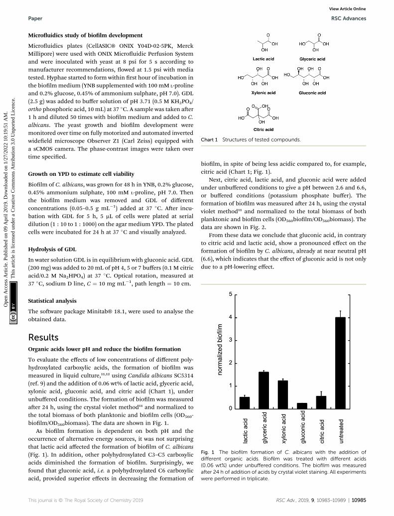
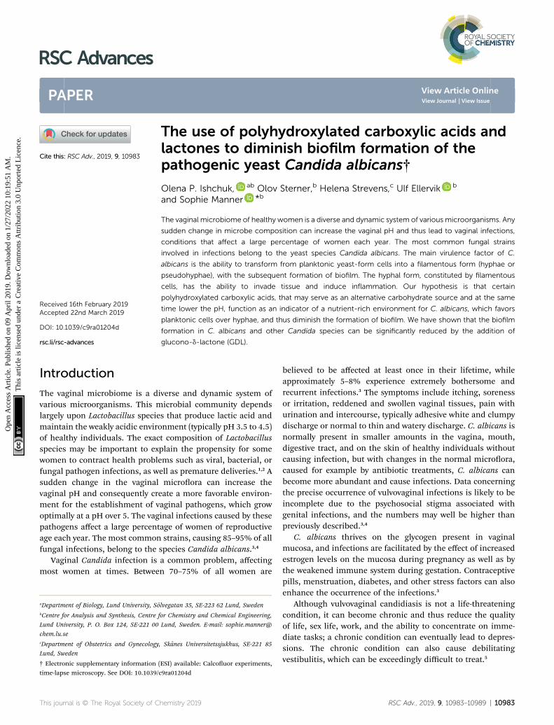
Chart 1 Structures of tested compounds.
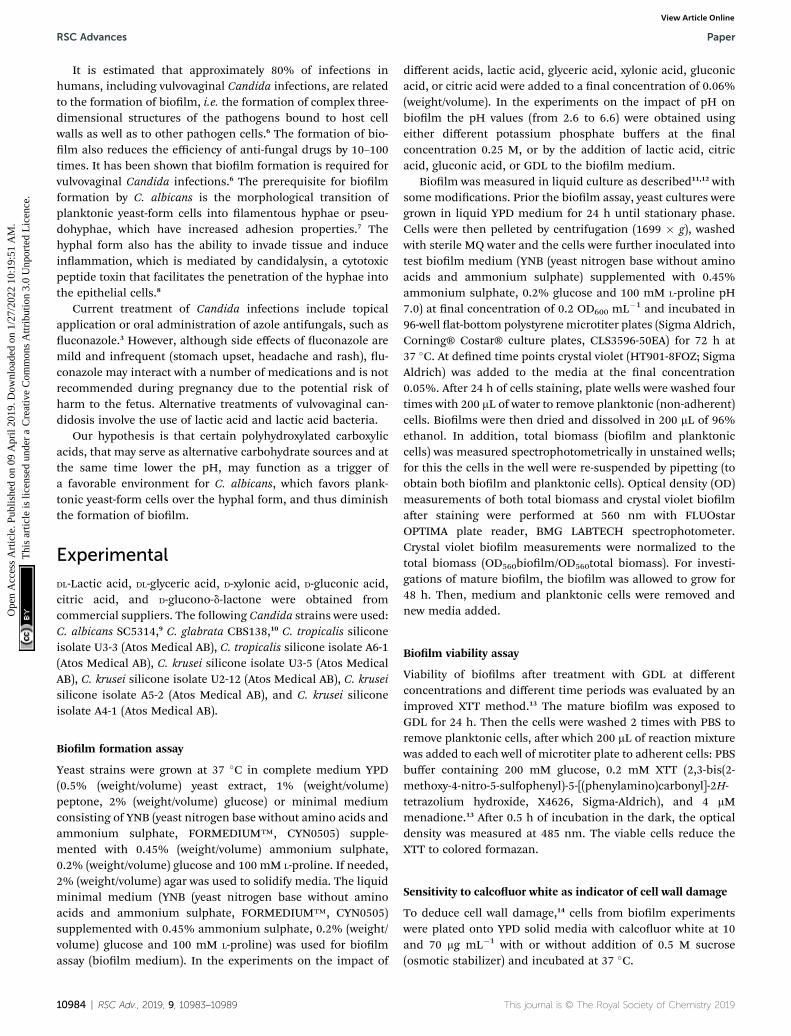
Fig. 1 The biofilm formation of C. albicans with the addition ofdifferent organic acids. Biofilm was treated with different acids(0.06 wt%) under unbuffered conditions. The biofilm was measuredafter 24 h of addition of acids by crystal violet staining. All experimentswere performed in triplicate.
Paper RSC Advances
Ope
n A
cces
s A
rtic
le. P
ublis
hed
on 0
9 A
pril
2019
. Dow
nloa
ded
on 1
/27/
2022
10:
19:5
1 A
M.
Thi
s ar
ticle
is li
cens
ed u
nder
a C
reat
ive
Com
mon
s A
ttrib
utio
n 3.
0 U
npor
ted
Lic
ence
.View Article Online
Microuidics study of biolm development
Microuidics plates (CellASIC® ONIX Y04D-02-5PK, MerckMillipore) were used with ONIX Microuidic Perfusion Systemand were inoculated with yeast at 8 psi for 5 s according tomanufacturer recommendations, owed at 1.5 psi with mediatested. Hyphae started to form within rst hour of incubation inthe biolm medium (YNB supplemented with 100 mM L-prolineand 0.2% glucose, 0.45% of ammonium sulphate, pH 7.0). GDL(2.5 g) was added to buffer solution of pH 3.71 (0.5 M KH2PO4/ortho phosphoric acid, 10 mL) at 37 �C. A sample was taken aer1 h and diluted 50 times with biolm medium and added to C.albicans. The yeast growth and biolm development weremonitored over time on fully motorized and automated invertedwideeld microscope Observer Z1 (Carl Zeiss) equipped witha sCMOS camera. The phase-contrast images were taken overtime specied.
Growth on YPD to estimate cell viability
Biolm of C. albicans, was grown for 48 h in YNB, 0.2% glucose,0.45% ammonium sulphate, 100 mM L-proline, pH 7.0. Thenthe biolm medium was removed and GDL of differentconcentrations (0.05–0.5 g mL�1) added at 37 �C. Aer incu-bation with GDL for 5 h, 5 mL of cells were plated at serialdilution (1 : 10 to 1 : 1000) on the agar medium YPD. The platedcells were incubated for 24 h at 37 �C and visually analyzed.
Hydrolysis of GDL
In water solution GDL is in equilibrium with gluconic acid. GDL(200 mg) was added to 20 mL of pH 4, 5 or 7 buffers (0.1 M citricacid/0.2 M Na2HPO4) at 37 �C. Optical rotation, measured at37 �C, sodium D line, C ¼ 10 mg mL�1, path length ¼ 10 cm.
Statistical analysis
The soware package Minitab® 18.1, were used to analyse theobtained data.
ResultsOrganic acids lower pH and reduce the biolm formation
To evaluate the effects of low concentrations of different poly-hydroxylated carboxylic acids, the formation of biolm wasmeasured in liquid culture,11,12 using Candida albicans SC5314(ref. 9) and the addition of 0.06 wt% of lactic acid, glyceric acid,xylonic acid, gluconic acid, and citric acid (Chart 1), underunbuffered conditions. The formation of biolm was measuredaer 24 h, using the crystal violet method11 and normalized tothe total biomass of both planktonic and biolm cells (OD560-biolm/OD560biomass). The data are shown in Fig. 1.
As biolm formation is dependent on both pH and theoccurrence of alternative energy sources, it was not surprisingthat lactic acid affected the formation of biolm of C. albicans(Fig. 1). In addition, other polyhydroxylated C3–C5 carboxylicacids diminished the formation of biolm. Surprisingly, wefound that gluconic acid, i.e. a polyhydroxylated C6 carboxylicacid, provided superior effects in decreasing the formation of
This journal is © The Royal Society of Chemistry 2019
biolm, in spite of being less acidic compared to, for example,citric acid (Chart 1; Fig. 1).
Next, citric acid, lactic acid, and gluconic acid were addedunder unbuffered conditions to give a pH between 2.6 and 6.6,or buffered conditions (potassium phosphate buffer). Theformation of biolm was measured aer 24 h, using the crystalviolet method11 and normalized to the total biomass of bothplanktonic and biolm cells (OD560biolm/OD560biomass). Thedata are shown in Fig. 2.
From these data we conclude that gluconic acid, in contraryto citric acid and lactic acid, show a pronounced effect on theformation of biolm by C. albicans, already at near neutral pH(6.6), which indicates that the effect of gluconic acid is not onlydue to a pH-lowering effect.
RSC Adv., 2019, 9, 10983–10989 | 10985
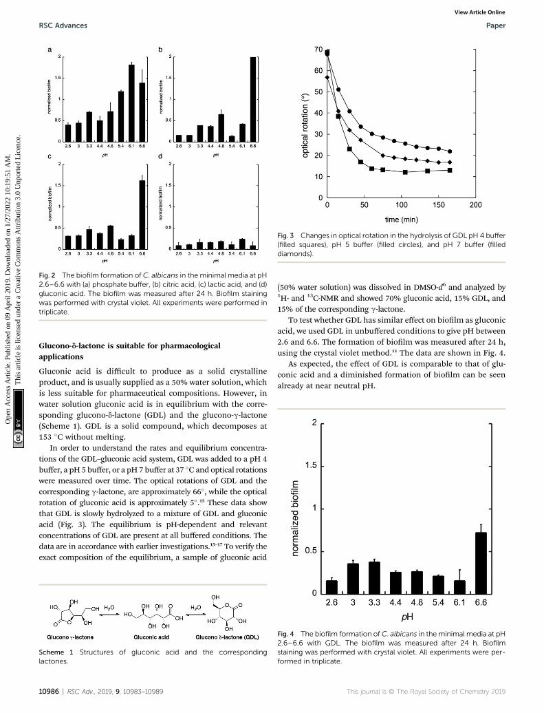
Fig. 2 The biofilm formation of C. albicans in the minimal media at pH2.6–6.6 with (a) phosphate buffer, (b) citric acid, (c) lactic acid, and (d)gluconic acid. The biofilm was measured after 24 h. Biofilm stainingwas performed with crystal violet. All experiments were performed intriplicate.
Fig. 3 Changes in optical rotation in the hydrolysis of GDL pH 4 buffer(filled squares), pH 5 buffer (filled circles), and pH 7 buffer (filleddiamonds).
RSC Advances Paper
Ope
n A
cces
s A
rtic
le. P
ublis
hed
on 0
9 A
pril
2019
. Dow
nloa
ded
on 1
/27/
2022
10:
19:5
1 A
M.
Thi
s ar
ticle
is li
cens
ed u
nder
a C
reat
ive
Com
mon
s A
ttrib
utio
n 3.
0 U
npor
ted
Lic
ence
.View Article Online
Glucono-d-lactone is suitable for pharmacologicalapplications
Gluconic acid is difficult to produce as a solid crystallineproduct, and is usually supplied as a 50% water solution, whichis less suitable for pharmaceutical compositions. However, inwater solution gluconic acid is in equilibrium with the corre-sponding glucono-d-lactone (GDL) and the glucono-g-lactone(Scheme 1). GDL is a solid compound, which decomposes at153 �C without melting.
In order to understand the rates and equilibrium concentra-tions of the GDL–gluconic acid system, GDL was added to a pH 4buffer, a pH 5 buffer, or a pH 7 buffer at 37 �C and optical rotationswere measured over time. The optical rotations of GDL and thecorresponding g-lactone, are approximately 66�, while the opticalrotation of gluconic acid is approximately 5�.15 These data showthat GDL is slowly hydrolyzed to a mixture of GDL and gluconicacid (Fig. 3). The equilibrium is pH-dependent and relevantconcentrations of GDL are present at all buffered conditions. Thedata are in accordance with earlier investigations.15–17 To verify theexact composition of the equilibrium, a sample of gluconic acid
Scheme 1 Structures of gluconic acid and the correspondinglactones.
10986 | RSC Adv., 2019, 9, 10983–10989
(50% water solution) was dissolved in DMSO-d6 and analyzed by1H- and 13C-NMR and showed 70% gluconic acid, 15% GDL, and15% of the corresponding g-lactone.
To test whether GDL has similar effect on biolm as gluconicacid, we used GDL in unbuffered conditions to give pH between2.6 and 6.6. The formation of biolm was measured aer 24 h,using the crystal violet method.11 The data are shown in Fig. 4.
As expected, the effect of GDL is comparable to that of glu-conic acid and a diminished formation of biolm can be seenalready at near neutral pH.
Fig. 4 The biofilm formation of C. albicans in the minimal media at pH2.6–6.6 with GDL. The biofilm was measured after 24 h. Biofilmstaining was performed with crystal violet. All experiments were per-formed in triplicate.
This journal is © The Royal Society of Chemistry 2019
Paper RSC Advances
Ope
n A
cces
s A
rtic
le. P
ublis
hed
on 0
9 A
pril
2019
. Dow
nloa
ded
on 1
/27/
2022
10:
19:5
1 A
M.
Thi
s ar
ticle
is li
cens
ed u
nder
a C
reat
ive
Com
mon
s A
ttrib
utio
n 3.
0 U
npor
ted
Lic
ence
.View Article Online
GDL affects the morphological transition to hyphae form
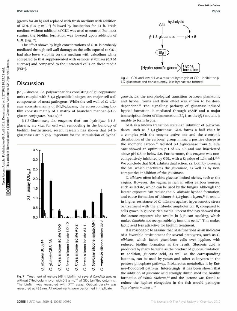
To monitor the cell morphology of C. albicans, yeast cells wereinoculated in the biolm medium (YNB supplemented with100 mM L-proline and 0.2% glucose, pH 7.0) with or without theaddition of GDL (5 mg mL�1). In the untreated sample, hyphaestarted to form within the rst hour of incubation, whiletreatment with GDL resulted in mainly yeast-form cells (Fig. 5).The process was also followed by time-lapse microscopy (ESI†)showing that the addition of GDL prevents the formation ofhyphae.
High concentrations of GDL inhibits C. albicans viability
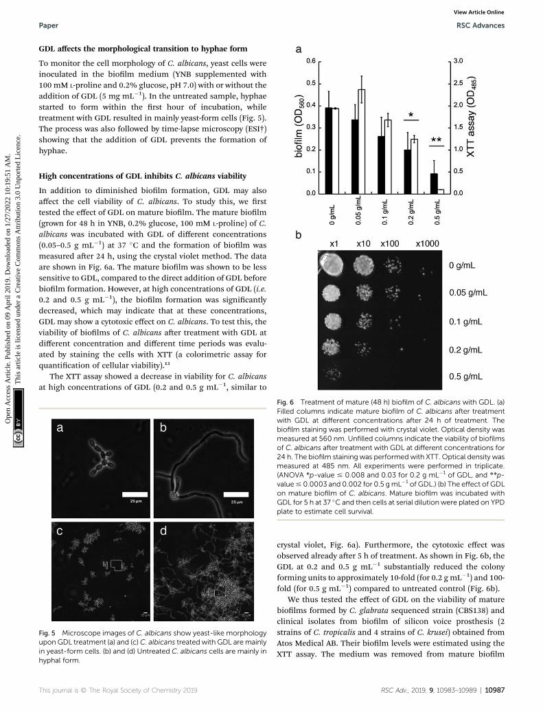
In addition to diminished biolm formation, GDL may alsoaffect the cell viability of C. albicans. To study this, we rsttested the effect of GDL on mature biolm. The mature biolm(grown for 48 h in YNB, 0.2% glucose, 100 mM L-proline) of C.albicans was incubated with GDL of different concentrations(0.05–0.5 g mL�1) at 37 �C and the formation of biolm wasmeasured aer 24 h, using the crystal violet method. The dataare shown in Fig. 6a. The mature biolm was shown to be lesssensitive to GDL, compared to the direct addition of GDL beforebiolm formation. However, at high concentrations of GDL (i.e.0.2 and 0.5 g mL�1), the biolm formation was signicantlydecreased, which may indicate that at these concentrations,GDL may show a cytotoxic effect on C. albicans. To test this, theviability of biolms of C. albicans aer treatment with GDL atdifferent concentration and different time periods was evalu-ated by staining the cells with XTT (a colorimetric assay forquantication of cellular viability).13
The XTT assay showed a decrease in viability for C. albicansat high concentrations of GDL (0.2 and 0.5 g mL�1, similar to
Fig. 5 Microscope images of C. albicans show yeast-like morphologyupon GDL treatment (a) and (c)C. albicans treated with GDL aremainlyin yeast-form cells. (b) and (d) Untreated C. albicans cells are mainly inhyphal form.
Fig. 6 Treatment of mature (48 h) biofilm of C. albicans with GDL. (a)Filled columns indicate mature biofilm of C. albicans after treatmentwith GDL at different concentrations after 24 h of treatment. Thebiofilm staining was performed with crystal violet. Optical density wasmeasured at 560 nm. Unfilled columns indicate the viability of biofilmsof C. albicans after treatment with GDL at different concentrations for24 h. The biofilm staining was performedwith XTT. Optical density wasmeasured at 485 nm. All experiments were performed in triplicate.(ANOVA *p-value # 0.008 and 0.03 for 0.2 g mL�1 of GDL, and **p-value# 0.0003 and 0.002 for 0.5 gmL�1 of GDL.) (b) The effect of GDLon mature biofilm of C. albicans. Mature biofilm was incubated withGDL for 5 h at 37 �C and then cells at serial dilution were plated on YPDplate to estimate cell survival.
This journal is © The Royal Society of Chemistry 2019
crystal violet, Fig. 6a). Furthermore, the cytotoxic effect wasobserved already aer 5 h of treatment. As shown in Fig. 6b, theGDL at 0.2 and 0.5 g mL�1 substantially reduced the colonyforming units to approximately 10-fold (for 0.2 g mL�1) and 100-fold (for 0.5 g mL�1) compared to untreated control (Fig. 6b).
We thus tested the effect of GDL on the viability of maturebiolms formed by C. glabrata sequenced strain (CBS138) andclinical isolates from biolm of silicon voice prosthesis (2strains of C. tropicalis and 4 strains of C. krusei) obtained fromAtos Medical AB. Their biolm levels were estimated using theXTT assay. The medium was removed from mature biolm
RSC Adv., 2019, 9, 10983–10989 | 10987
RSC Advances Paper
Ope
n A
cces
s A
rtic
le. P
ublis
hed
on 0
9 A
pril
2019
. Dow
nloa
ded
on 1
/27/
2022
10:
19:5
1 A
M.
Thi
s ar
ticle
is li
cens
ed u
nder
a C
reat
ive
Com
mon
s A
ttrib
utio
n 3.
0 U
npor
ted
Lic
ence
.View Article Online
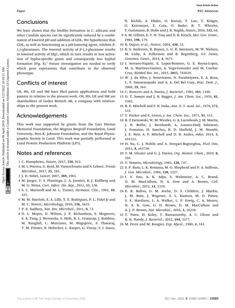
(grown for 48 h) and replaced with fresh medium with additionof GDL (0.5 g mL�1) followed by incubation for 24 h. Freshmedium without addition of GDL was used as control. For moststrains, the biolm formation was lowered upon addition ofGDL (Fig. 7).
The effect shown by high concentrations of GDL is probablymediated through cell wall damage as the cells exposed to GDLhad also lower viability on the medium with calcouor whitecompared to that supplemented with osmotic stabilizer (0.5 Msucrose) and compared to the untreated cells on these media(ESI†).
Fig. 8 GDL and low pH, as a result of hydrolysis of GDL, inhibit the b-1,3-glucanase and consequently, less hyphae are formed.
Discussionb-1,3-Glucans, i.e. polysaccharides consisting of glucopyranoseunits coupled with b-1,3-glycosidic linkages, are major cell wallcomponents of most pathogens. While the cell wall of C. albi-cans consists mainly of b-1,3-glucans, the corresponding bio-lm consists mainly of a matrix of branched mannan-b-1,6-glucan conjugates (MGCx).18
b-1,3-Glucanases, i.e. enzymes that can hydrolyze b-1,3-glucans, are vital for cell wall remodeling in the build-up ofbiolm. Furthermore, recent research has shown that b-1,3-glucanases are highly important for the stimulation of hyphal
Fig. 7 Treatment of mature (48 h) biofilm of several Candida specieswithout (filled columns) or with 0.5 g mL�1 of GDL (unfilled columns).The biofilm was measured with XTT assay. Optical density wasmeasured at 485 nm. All experiments were performed in triplicate.
10988 | RSC Adv., 2019, 9, 10983–10989
growth, i.e. the morphological transition between planktonicand hyphal forms and their effect was shown to be dose-dependent.19 The signalling pathway of glucanase-inducedhyphal formation is mediated through cAMP and a majortranscription factor of lamentation, Efg1, as the efg1mutant isunable to form hypha.
GDL is a known transition state-like inhibitor of b-glycosi-dases, such as b-1,3-glucanase. GDL forms a half chair ina complex with the enzyme active site and the electronicdistribution of the carbonyl group mimic a positive charge atthe anomeric carbon.20 Isolated b-1,3-glucanase from C. albi-cans showed an optimum pH of 5.5–5.6 and was inactivatedabove pH 6.5 or below 5.0. Furthermore, this enzyme was non-competitively inhibited by GDL, with a Ki value of 1.34 mM.21,22
We conclude that GDL exhibits dual action, i.e. both by loweringthe pH, which inactivates the glucanase, as well as by non-competitive inhibition of the glucanase.
C. albicans oen inhabits glucose limited niches, such as thevagina. However, the vagina is rich in other carbon sources,such as lactate, which can be used by the fungus. Although thelactate exposure can reduce the C. albicans hyphae formation,and cause formation of thinner b-1,3-glucan layers,23 it resultsin higher resistance of C. albicans against hyperosmotic stressor treatment with the antibiotic amphotericin B, compared tocells grown in glucose rich media. Recent ndings showed thatthe lactate exposure also results in b-glucan masking, whichmakes Candida not recognizable by immune cells.24 This makeslactic acid less attractive for biolm treatment.
It is reasonable to assume that GDL functions as an indicatorof a favorable environment for several pathogens, such as C.albicans, which favors yeast-form cells over hyphae, withreduced biolm formation as the result. Gluconic acid isproduced by many bacteria as the product of glucose oxidation.In addition, gluconic acid, as well as the correspondinglactones, can be used by yeasts and other eukaryotes in thepentose phosphate pathway. Prokaryotes metabolize it by Ent-ner–Doudoroff pathway. Interestingly, it has been shown thatthe addition of gluconic acid strongly diminished the biolmformation of Vibrio cholerae,25 and the lactone was found toreduce the hyphae elongation in the sh mould pathogenSaprolegnia monoica.26
This journal is © The Royal Society of Chemistry 2019
Paper RSC Advances
Ope
n A
cces
s A
rtic
le. P
ublis
hed
on 0
9 A
pril
2019
. Dow
nloa
ded
on 1
/27/
2022
10:
19:5
1 A
M.
Thi
s ar
ticle
is li
cens
ed u
nder
a C
reat
ive
Com
mon
s A
ttrib
utio
n 3.
0 U
npor
ted
Lic
ence
.View Article Online
Conclusions
We have shown that the biolm formation in C. albicans andother Candida species can be signicantly reduced by a combi-nation of lowered pH and addition of GDL. We hypothesize thatGDL, as well as functioning as a pH-lowering agent, inhibits b-1,3-glucanases. The lowered activity of b-1,3-glucanase resultsin lowered activity of Efg1, which in turn results in less activa-tion of hypha-specic genes and consequently less hyphalformation (Fig. 8).7 Future investigation are needed to verifywhich gene expressions that contribute to the observedphenotype.
Conflicts of interest
OS, HS, UE and SM have led patent applications and holdpatents in relation to the present work. OS, HS, UE and SM areshareholders of Gedea Biotech AB, a company with relation-ships to the present work.
Acknowledgements
This work was supported by grants from the Lars HiertasMemorial Foundation, the Magnus Bergvall Foundation, LundUniversity, Sten K. Johnson Foundation, and the Royal Physio-graphic Society in Lund. This work was partially performed atLund Protein Production Platform (LP3).
Notes and references
1 C. Humphries, Nature, 2017, 550, S12.2 M. I. Petrova, G. Reid, M. Vaneechoutte and S. Lebeer, TrendsMicrobiol., 2017, 25, 182.
3 J. D. Sobel, Lancet, 2007, 369, 1961.4 M. Jaeger, T. S. Plantinga, L. A. Joosten, B. J. Kullberg andM. G. Netea, Curr. Infect. Dis. Rep., 2013, 15, 136.
5 S. C. Marinoff and M. L. Turner, Dermatol. Clin., 1992, 10,435.
6 M. M. Harriott, E. A. Lilly, T. E. Rodriguez, P. L. Fidel Jr andM. C. Noverr, Microbiology, 2010, 156, 3635.
7 P. E. Sudbery, Nat. Rev. Microbiol., 2011, 9, 73.8 D. L. Moyes, D. Wilson, J. P. Richardson, S. Mogavero,S. X. Tang, J. Wernecke, S. Hofs, R. L. Gratacap, J. Robbins,M. Runglall, C. Murciano, M. Blagojevic, S. Thavaraj,T. M. Forster, B. Hebecker, L. Kasper, G. Vizcay, S. I. Iancu,
This journal is © The Royal Society of Chemistry 2019
N. Kichik, A. Hader, O. Kurzai, T. Luo, T. Kruger,O. Kniemeyer, E. Cota, O. Bader, R. T. Wheeler,T. Gutsmann, B. Hube and J. R. Naglik, Nature, 2016, 532, 64.
9 A. M. Gillum, E. Y. H. Tsay and D. R. Kirsch,Mol. Gen. Genet.,1984, 198, 179.
10 B. Dujon, et al., Nature, 2004, 430, 35.11 K. S. Andersen, R. Bojsen, L. G. R. Sørensen, M. W. Nielsen,
M. Lisby, A. Folkesson and B. Regenberg, G3: Genes,Genomes, Genet., 2014, 4, 1671.
12 I. Serrano-Fujarte, E. Lopez-Romero, G. E. Reyna-Lopez,M. A. Martinez-Gamez, A. Vega-Gonzalez and M. Cuellar-Cruz, BioMed Res. Int., 2015, 2015, 783639.
13 W. J. da Silva, J. Seneviratne, N. Parahitiyawa, E. A. Rosa,L. P. Samaranayake and A. A. Del Bel Cury, Braz. Dent. J.,2008, 19, 364.
14 C. Roncero and A. Duran, J. Bacteriol., 1985, 163, 1180.15 D. T. Sawyer and J. B. Bagger, J. Am. Chem. Soc., 1959, 81,
5302.16 R. E. Mitchell and F. R. Duke, Ann. N. Y. Acad. Sci., 1970, 172,
131.17 Y. Pocker and E. Green, J. Am. Chem. Soc., 1973, 95, 113.18 R. Z Zarnowski, W. M.Westler, G. A. Lacmbouh, J. M. Marita,
J. R. Bothe, J. Bernhardt, A. Lounes-Hadj Sahraoui,J. Fontaine, H. Sanchez, R. D. Hateld, J. M. Ntambi,J. E. Nett, A. P. Mitchell and D. R. Andes, mBio, 2014, 5,e01333.
19 H. Xu, C. J. Nobile and A. Dongari-Bagtzoglou, PLoS One,2013, 8, e63736.
20 T. M. Gloster and G. J. Davies, Org. Biomol. Chem., 2010, 8,305.
21 V. Notario, Microbiology, 1982, 128, 747.22 S. P. Ram, L. K. Romana, M. G. Shepherd and P. A. Sullivan,
J. Gen. Microbiol., 1984, 130, 1227.23 I. V. Ene, A. K. Adya, S. Wehmeier, A. C. Brand,
D. M. MacCallum, N. A. Gow and A. Brown, Cell.Microbiol., 2012, 14, 1319.
24 E. R. Ballou, G. M. Avelar, D. S. Childers, J. Mackie,J. M. Bain, J. Wagener, S. L. Kastora, M. D. Panea,S. E. Hardison, L. A. Walker, L. P. Erwig, C. A. Munro,N. A. R. Gow, G. D. Brown, D. M. MacCallum andA. J. P. Brown, Nat. Microbiol., 2016, 2, 16238.
25 T. Patra, H. Koley, T. Ramamurthy, A. C. Ghose andR. K. Nandy, J. Bacteriol., 2012, 194, 3377.
26 M. Fevre and M. Rougier, Exp. Mycol., 1980, 4, 343.
RSC Adv., 2019, 9, 10983–10989 | 10989