the utility of a high-intensity exercise protocol to
TRANSCRIPT

The utility of a high-intensity exercise protocol to prospectively assess
ACL injury risk
Fransiska M. Bossuyt1, Felipe García-Pinillos2, Raja M.F. Raja Azidin1, Jos Vanrenterghem1,
Mark A. Robinson1
1Research Institute for Sport and Exercise Sciences, Liverpool John Moores University, Liverpool, United Kingdom 2Department of Corporal Expression, University of Jaen, Jaen, Spain
International Journal of Sports Medicine
http://dx.doi.org/10.1055/s-0035-1555930
Abstract
This study investigated the utility of a 5-min high-intensity exercise protocol
(SAFT5) to include in prospective cohort studies investigating ACL injury risk. 15
active females were tested on 2 occasions during which their non-dominant leg was
analysed before SAFT5 (PRE), immediately after (POST0), 15 min after (POST15),
and 30 min after (POST30). On the first occasion, testing included 5 maximum
isokinetic contractions for eccentric and concentric hamstring and concentric
quadriceps and on the second occasion, 3 trials of 2 landing tasks (i.e. single-leg
hop and drop vertical jump) were conducted. Results showed a reduced eccentric
hamstring peak torque at POST0, POST15 and POST30 (p <.05) and a reduced
functional HQ ratio (Hecc/Qcon) at POST15 and POST30 (p < .05). Additionally,
a more extended knee angle at POST30 (p < .05) and increased knee internal
rotation angle at POST0 and POST15 (p < .05) were found in a single-leg hop.
SAFT5 altered landing strategies associated with increased ACL injury risk and
similar to observations from match simulations. Our findings therefore support the
utility of a high-intensity exercise protocol such as SAFT5 to strengthen injury
screening tests and to include in prospective cohort studies where time constraints
apply.
Corresponding Author:
Fransiska Marie Bossuyt : [email protected]
brought to you by COREView metadata, citation and similar papers at core.ac.uk
provided by LJMU Research Online

INTRODUCTION
Risk factors of ACL injuries can only be defined with the highest level of evidence when
prospectively assessed.[39] Muscular and biomechanical ACL injury risk factors have been
studied extensively as they are modifiable through training.[41] Muscular risk factors include
reduced eccentric hamstring peak torque (Hecc) and reduced hamstring/quadriceps ratio
(H/Q).[45] Reduced hamstring strength is believed to permit increased anterior tibial translation
and in turn increase ACL strain.[2] Biomechanical risk factors include increased peak knee
abduction moment and peak vertical ground reaction force (VGRFpeak).[21] Reduced knee
flexion at initial contact (IC) during landing has been associated with increased anterior tibial
translation[6, 7] which causes increased ACL loading.[6, 8, 9] Additionally, increased knee
internal rotation has been associated with increased ACL loading.[9, 10] It is important to note
that fatigue (i.e., loss of maximum or potential performance) alters these factors which may
further increase injury risk.[11, 12] Additionally, evidence shows that most injuries occur at the
end of a soccer match.[12, 13] These findings suggest that fatigue plays a crucial role on
muscular and biomechanical risk factors in the mechanism of ACL injury.[19] To date,
however, no prospective studies have included exercise-induced fatigue in their screening
protocols, most likely due to the conflict between screening a large cohort and the time-
consuming nature of inducing fatigue.
Fatigue has been induced in a number of ways, some of which are more related to
dynamic activities than others. Previous research has suggested, however, that including
functional movements as part of the protocol is key to revealing specific match-play-induced
deficits which can increase ACL injury risk.[14, 15] As such the effects of a shuttle run revealed
changes in transverse plane kinematics in sidestep cutting as increased external rotation of hip,
knee and ankle angle at IC and increased knee internal rotation angle during stance.[40] In
another study, a treadmill-based soccer match simulation reduced H/Q.[16] Finally, a soccer
specific match simulation (SAFT90) which included multidirectional movements, high
accelerations and decelerations, and which was shown to be a valid simulation of match
play[26], caused significant reductions in Hecc and in H/Q.[36,43,44] Fatigue induced by soccer
match play (90 min) has been suggested to be a combination of both central (altered motor
commands from the motor cortex) and peripheral fatigue (metabolite accumulation, limitations
in energy supply, reduced blood flow and neuromuscular mechanisms)[37,50]. Additionally it
has been suggested that during soccer, players experience fatigue in several different ways: (1)
disturbed muscle ion homeostasis during temporary fatigue after short bursts of high-intensity

exercises, (2) lowered muscle temperature (e.g., at the beginning of the second half) and (3)
through muscle glycogen and dehydration as experienced towards the end of a game.[32]
Whilst full-length match simulations would be considered most ideal for simulating the
effects of match play, the development and evaluation of short-term protocols is needed to
include match-play-induced fatigue assessment within prospective studies. It is important to
acknowledge the influence of intensity, duration and type of contraction on the mechanisms of
fatigue.[3] As far as we are aware however, no previous study has directly compared
neuromuscular responses between short-term protocols and full-length match simulations.
Nevertheless, previous findings [27] found similar biomechanical alterations in response to a
short-duration high-intensity protocol and a longer duration protocol (30min). 2 studies
involving short-term protocols, the first including vertical jumps followed by 30-m sprint, and
the second including series of athletic exercises (countermovement jump (CMJ), step up/down,
squat and shuttle run), resulted in decreased knee flexion angles at IC and increased knee
abduction moments in sidestep cutting[10] and stop-jump tasks.[8] Finally, a short-term
protocol based on continuous drills (step up/down and plyometric bounding) caused increased
knee abduction angles/moments and increased knee internal rotation angles in a drop vertical
jump (DVJ).[31] It is important to note that all these protocols induced fatigue until maximum
exhaustion which is not representative of match play, and inappropriate for the inclusion in
prospective studies as this implies differences in duration or amount of repetitions.
Few if any studies have focused on short-term protocols that simulate match play of
dynamic sports (i.e., sport which involves high accelerations and decelerations and typically
involves interactions with an object (ball, racket, etc.)). Therefore, the aim of this study was to
investigate how a short-term high-intensity exercise protocol (SAFT5), based on a long-duration
match-play simulation (SAFT90) [26], affects muscular and biomechanical markers of ACL
injury in recreationally active females. It was hypothesized that SAFT5 would result in changes
in markers of ACL injury risk.
METHODS
Participants
15 females (age: 22±3 years, height: 1.68±0.07 m, mass: 70.8±8.9 kg) were tested
following a sample-size estimation from a functional fatigue protocol to observe a 5° difference
in knee flexion angle at 80 % statistical power and alpha=0.05.[11,31] All participants met the
inclusion criteria: (1) female, (2) recreationally active (i.e., 3 sessions per week) in dynamic
sports (hockey, netball, etc.), (3) did not suffer from an ACL injury and (4) did not suffer from
any other lower limb injury within the last 6 months before data collection. Ethical approval
was granted by the university ethics committee and all participants provided informed consent
according to IJSM ethical standards.[18]
Design
This was a cross-sectional study with a repeated measures design. Initially participants
were familiarized with the protocol and assessment methods, height and weight were recorded,
and maximum jump height (JumpHeightmax) was defined. This was followed by 2 testing
sessions which were separated by at least 3 days. Both sessions included a dynamic warm-up,
the SAFT5 protocol, a pre-test (PRE), a test immediately following SAFT5 (POST0), after 15
min of passive rest (POST15) and after 30 min of passive rest (POST30). These moments in
time were selected in order to define the prolonged effect of SAFT5. This information is
required for the inclusion of SAFT5 in a prospective study protocol which could consist of
measurements before and after SAFT5. In addition, this information would be useful for the
design of activity-rest cycles to minimize the potential for fatigue-related injury. Different
parameters were assessed during each session and between participants the order of sessions
was randomly assigned. Participants were instructed to avoid strenuous exercises 48 hours prior
to testing.
Exercise protocol
SAFT5 is based on the first five min of SAFT90.[26,36] The distance of the SAFT90
protocol was modified to 15m in order to make the SAFT5 course feasible in our laboratory
(Figure 1 and Table 1). The intensity of SAFT5 was increased by adding high-intensity
exercises, based on previous studies [11,31,51] and pilot work [9,54]. Pilot work aimed at
defining the activity profile of the protocol by investigating the implementation of functional

high-intensity movements. Several variations of the protocol based on different high-intensity
exercises, amount of repetitions and order of exercises were explored by monitoring HR and
RPE during the protocol which represented the intensity. The final protocol was selected in
accordance to the following criteria: (1) HR and RPE presented a similar overall pattern as
during SAFT90 and actual game play [24,36], and (2) practical and personal observations of the
researchers. As such it was decided to include the following 3 exercises: a CMJ at 80% of their
JumpHeightmax, an agility ladder drill (one foot per square) and a ‘jump scissors’ task (jumping
from unilateral lunge with left leg forward and hands placed on the hips, to unilateral lunge
with right leg forward)(Table 1). During the protocol, white tape was placed onto a wall which
represented 80% JumpHeightmax, participants had to touch the white tape during every jump
and received verbal feedback if they didn’t reach the tape.
Data collection
At one test session maximum voluntary contractions (MVCs) of concentric hamstring strength
(Hcon), concentric quadriceps strength (Qcon) and Hecc were assessed by an isokinetic
dynamometer (IKD, Biodex System 3, Shirley, NY). The non-dominant leg (i.e., non-preferred
leg to kick a ball with) was tested as it has been identified as the most vulnerable to ACL injury
in females.[6,38] Concentric MVCs consisted of repeated knee flexion and extension
contractions within 90° RoM at 120°/s. During eccentric MVCs, participants resisted against
the passive external knee extension moment of the IKD over 90° RoM and at 120°/s.[35,
43]Participants were verbally encouraged and 5 repetitions were measured for each task with
one min rest between different contractions. The order of assessment was randomly assigned.
At the other test session, participants wore tight-fitting clothes and measurements
consisted of 3D motion and force analysis of DVJ and single-leg hop (SLH). For the DVJ,
participants were instructed to drop off a 30-cm high box (feet 20 cm apart), and land with each
foot on separate force platforms, immediately rebounding for a maximum vertical jump. For
the SLH, participants were instructed to stand on the non-dominant leg and hop forward to
cover a distance of 75% of body height [33] in order to use a standardised distance adjusted to
personal dimensions. 3 successful trials of each task were recorded with trials excluded if the
participant lost balance less than 2s after landing.
10 optoelectronic cameras sampling at 250 Hz (OQUS 3, Qualysis AB, Gothenburg,
Sweden) were used to collect 3D motion data. Spherical reflective markers were attached to
lower limb and trunk according to the LJMU kinematic model [49], which has established

reliability.[28] One static and 4 functional motion trials were recorded to define functional hip
and knee joint axes, after which anatomy-defining markers were removed. GRF were collected
simultaneously from 2 force platforms at 1500 Hz (Kistler, Winterthur, Switzerland).
Additional measurements for both sessions, recorded every 5 min throughout the
sessions, and during SAFT5, included JumpHeightmax with a jump mat (Probotics, Inc.,
Huntsville, AL) in order to assess fatigue as a reduction of performance, heart rate (HR) (Polar
heart rate system, Electro, Finland) and rate of perceived exertion (RPE) (20-point Borg scale).
Timing and order of each measurement is represented in Table 2.
Data analysis
Kinematic and kinetic data were calculated within Visual 3D (C-Motion, Germantown,
MD). Marker trajectories and forces were filtered through a Butterworth and a critically damped
low-pass filter with 20-Hz cut-off frequencies and normalized to 101 time nodes. IC and take-
off were defined as the instant when GRF exceeded or reached below 10N.[10] The stance
phase of an SLH was defined as one second after IC. Only the first landing of the DVJ was used
for analysis.[1] Euler rotations (X-Y-Z) were used for joint angle calculations and knee
moments were obtained by inverse dynamics [31] and are reported as external moments. IKD
data were gravity-corrected and analysed with a custom Matlab (MathWorks, Inc., Natick, MA,
USA) program. Peak torques were calculated from a polynomial fit of data points that met the
criteria within a 10% tolerance (velocity: 120°/s, RoM at least 70° for Hcon and Qcon, and RoM
at least 50° for Hecc). Finally, the functional HQ ratio (Hecc/Qcon) which was previously
presented as a valid representation of muscle-specific exertion induced by football match-play
[12] and conventional HQ ratio (Hcon/Qcon) were calculated. Dependent variables of interest
were JumpHeightmax, HR, RPE, Hecc/Qcon, Hcon/Qcon, Hcon, Hecc and Qcon, knee flexion angle and
transverse plane knee angle at IC, peak knee abduction moment and VGRFpeak.
Statistical treatment
Data are presented as means ± standard deviation. One-way repeated measures
ANOVAs were used to assess meaningful variations across time for each variable. Percentage
differences of jump height, Hecc, Hcon, Qcon, Hecc/Qcon and Hcon/Qcon were calculated and HR was
presented as a percentage of the estimated maximum HR (HRmax=220-age). If statistical
significance was found, pairwise comparisons were applied with Bonferroni corrections to
reduce the risk of type-1 errors. Reliability of the 2 testing sessions was confirmed by (1) low
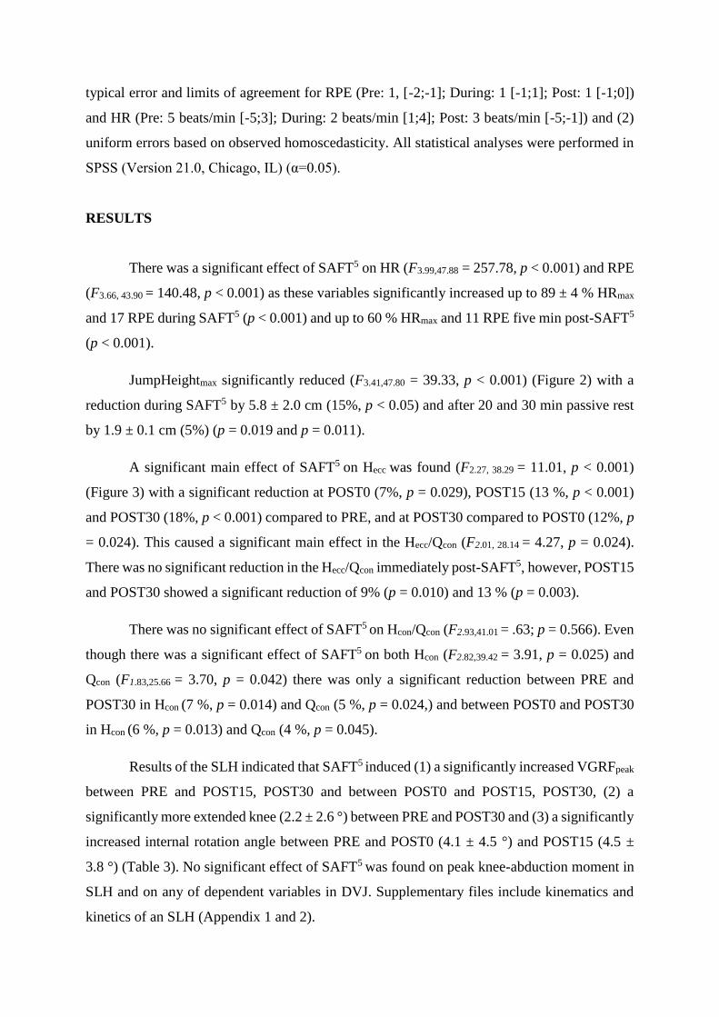
typical error and limits of agreement for RPE (Pre: 1, [-2;-1]; During: 1 [-1;1]; Post: 1 [-1;0])
and HR (Pre: 5 beats/min [-5;3]; During: 2 beats/min [1;4]; Post: 3 beats/min [-5;-1]) and (2)
uniform errors based on observed homoscedasticity. All statistical analyses were performed in
SPSS (Version 21.0, Chicago, IL) (α=0.05).
RESULTS
There was a significant effect of SAFT5 on HR (F3.99,47.88 = 257.78, p < 0.001) and RPE
(F3.66, 43.90 = 140.48, p < 0.001) as these variables significantly increased up to 89 ± 4 % HRmax
and 17 RPE during SAFT5 (p < 0.001) and up to 60 % HRmax and 11 RPE five min post-SAFT5
(p < 0.001).
JumpHeightmax significantly reduced (F3.41,47.80 = 39.33, p < 0.001) (Figure 2) with a
reduction during SAFT5 by 5.8 ± 2.0 cm (15%, p < 0.05) and after 20 and 30 min passive rest
by 1.9 ± 0.1 cm (5%) (p = 0.019 and p = 0.011).
A significant main effect of SAFT5 on Hecc was found (F2.27, 38.29 = 11.01, p < 0.001)
(Figure 3) with a significant reduction at POST0 (7%, p = 0.029), POST15 (13 %, p < 0.001)
and POST30 (18%, p < 0.001) compared to PRE, and at POST30 compared to POST0 (12%, p
= 0.024). This caused a significant main effect in the Hecc/Qcon (F2.01, 28.14 = 4.27, p = 0.024).
There was no significant reduction in the Hecc/Qcon immediately post-SAFT5, however, POST15
and POST30 showed a significant reduction of 9% (p = 0.010) and 13 % (p = 0.003).
There was no significant effect of SAFT5 on Hcon/Qcon (F2.93,41.01 = .63; p = 0.566). Even
though there was a significant effect of SAFT5 on both Hcon (F2.82,39.42 = 3.91, p = 0.025) and
Qcon (F1.83,25.66 = 3.70, p = 0.042) there was only a significant reduction between PRE and
POST30 in Hcon (7 %, p = 0.014) and Qcon (5 %, p = 0.024,) and between POST0 and POST30
in Hcon (6 %, p = 0.013) and Qcon (4 %, p = 0.045).
Results of the SLH indicated that SAFT5 induced (1) a significantly increased VGRFpeak
between PRE and POST15, POST30 and between POST0 and POST15, POST30, (2) a
significantly more extended knee (2.2 ± 2.6 °) between PRE and POST30 and (3) a significantly
increased internal rotation angle between PRE and POST0 (4.1 ± 4.5 °) and POST15 (4.5 ±
3.8 °) (Table 3). No significant effect of SAFT5 was found on peak knee-abduction moment in
SLH and on any of dependent variables in DVJ. Supplementary files include kinematics and
kinetics of an SLH (Appendix 1 and 2).

DISCUSSION
This study determined whether the SAFT5, a high-intensity exercise protocol, could be
affectively used to induce changes in the muscular and biomechanical characteristics that are
typically associated to ACL injury risk. The results partly confirmed the hypothesis. Most
notably, there was a significant reduction in Hecc and Hecc/Qcon, and altered landing strategies in
SLH. The protocol caused a 15% drop in jump performance and RPE was rated between ‘hard’
and ‘very hard’ with the average HR (89±4% HRmax) similar to that of female football match
play (87-97% HRmax[24]). The intensity of the current protocol therefore appears to induce
physiological responses similar to those in longer duration match-play situations. In general,
current findings imply the importance of screening athletes after a high-intensity functional
exercise protocol such as SAFT5.
The significant reduction in Hecc immediately after SAFT5 and POST15 is in agreement
with previous studies investigating the effect of a soccer-specific field test over 90 min
(SAFT90).[17, 43] The selective occurrence of fatigue in Hecc has been explained previously by
the presence of more fatigable type-2 muscle fibres [14], and the high eccentric requirements
of the hamstrings in repetitive sprinting and kicking [53] to counteract the anterior shear forces
created by the quadriceps. Despite the significant reduction in Hecc between PRE and every
post-test, Hecc/Qcon was only significantly reduced POST15 and POST30.[17, 43] It should be
especially noted that at POST30, Hecc/Qcon fell below the at-risk threshold of 0.71.[52] The
delayed reduction of Hecc/Qcon can be explained by the short-term high-intensity characteristics
of SAFT5. Firstly, the occurrence of post activation potentiation (PAP) (i.e., improved muscular
performance in response to conditioning stimulus [22]) could have interfered with the effects
immediately following SAFT5, as previous work has found an improvement in peak force
within the first 5 min following exercise.[46] Hecc/Qcon is, however, not increased post-SAFT5
which assumes that PAP is not the main contributor. Secondly, it could be suggested that there
is a delay in peripheral fatigue (i.e., fatigue occurred within the muscle itself) immediately post-
SAFT5 as a recent study concluded that central fatigue (i.e., alterations in the nervous system)
manifests prior to reductions in knee-flexor maximal torque in response to SAFT90.[30] This
finding was explained by the earlier and more pronounced effect of central fatigue on explosive
force-producing exercises. The delayed onset of peripheral fatigue is represented in the
significant reduction in Hecc/Qcon after 15 min passive rest and is in agreement with previous
research.[17, 26, 43] There was no significant reduction in Hcon/Qcon post-SAFT5 which further
supports the use of Hecc/Qcon for the evaluation of hamstrings function during dynamic

activities. Based on Hecc and HQ ratios, it could be assumed that SAFT5 induces detrimental
effects on muscle performance similar to changes after match-play exertion as SAFT90.
Detrimental effects are, however, best observed with a 15 min delay.
In accordance with the hypothesis, SAFT5 altered landing strategies in the non-
dominant leg of an SLH. The reduced knee-flexion angle and increased knee internal rotation
are in accordance with other studies exploring the effect of a short-term fatigue protocol on
sidestep and stop-jump tasks.[8,10,11] The significant increase in VGRFpeak post-SAFT5 is in
contrast with a previous study investigating the effect of a short-term fatigue protocol.[11] It
should be noted that SAFT5 aimed to replicate match-play exertion and deviated from the
mentioned short-term protocols by the lack of maximum exhaustion which increases the stress
applied on the body and may cause greater detrimental effects on landing strategies. Several
possible speculations could relate to the significantly reduced knee-flexion angle only after 30
minutes passive rest: (1) the suggested relationship between exercise-related changes in fatigue
(e.g. decreased landing forces) and knee laxity (e.g., greater knee-extensor loads and knee shear
forces), which is dependent on the baseline knee laxity [42], (2) altered muscular activation
patterns in response to fatigue as reduced pre-activation of the hamstrings and gastrocnemius
could affect the sagittal and transverse plane differently [15] and (3) post-activation
potentiation, as described previously, could play a role [46]. Further studies, which take these
variables into account, will need to be undertaken. Maximum exhaustion may alter landing
strategies which are more related to reduced power generation than to altered stabilization
mechanisms. This may explain the different findings of the VGRFpeak. Previous studies found
increased peak knee internal rotation post-fatigue [5,31,40,47] but to date, no study reported
increased knee internal rotation at IC during an SLH. Nevertheless, an extended knee position
at IC during landing has been associated with increased ACL loading especially during single-
leg landing.[25] Additionally, increased internal rotation of the tibia [13,29] and increased
VGRFpeak [7] have been associated with increased ACL loading. In summary, SAFT5 induced
kinematic changes in landing strategies of an SLH which are thought to be associated with
increased ACL injury risk. This evidence is in support of the notion that biomechanical
screening after a functional exercise protocol such as SAFT5 may be better suited to identify at-
risk individuals than observations without prior high-intensity functional exercises. This was
evident in an SLH and not in a bilateral DVJ.
Several limitations to the present study need to be acknowledged. Firstly, due to short-
term characteristics of SAFT5, different physiological processes will be triggered compared to

long-duration match play. More specifically, high-intensity exercises (5 min) induce
accumulation of metabolites and ions resulting in metabolic acidosis [48], whereas long-
duration exercises impose a greater aerobic demand and may deplete energy supply and cause
dehydration.[24] This results in different loading on the body. Secondly, as training status was
reported to influence fatigue-induced mechanisms [4], findings of the present study
investigating recreational active athletes cannot be generalised to elite athletes. Athletes with a
higher percentage of type-1 fibres (i.e., slow/fatigue resistant fibres) and a higher aerobic fitness
are suggested to have, for instance, a greater capability of resisting fatigue.[4] Finally, whilst
our study involved a closer simulation of match play than maximal exhaustion protocols may
do, its findings do not directly pertain to the effects of actual match play.
The present study was designed to determine the effect of SAFT5 on muscular and
biomechanical markers of ACL injury risk, primarily to inform the development of future
prospective studies that aim at including screening after functional exercises. SAFT5 reduced
Hecc and Hecc/Qcon, and caused altered landing patterns in SLH. All of the above have previously
been associated with increased ACL injury risk and suggest that testing after a high-intensity
functional exercise protocol such as SAFT5 may strengthen the identification of individuals
with increased ACL injury risk within a sporting population.

REFERENCES
1 Bates N a, Ford KR, Myer GD, Hewett TE. Timing differences in the generation of ground
reaction forces between the initial and secondary landing phases of the drop vertical jump. Clin
Biomech (Bristol, Avon) 2013; 28: 796–799
2 Behrens M, Mau-Moeller A, Wassermann F, Bruhn S. Effect of fatigue on hamstring reflex
responses and posterior-anterior tibial translation in men and women. PLoS One 2013; 8: e56988
3 Bishop DJ. Fatigue during intermittent-sprint exercise. Clin Exp Pharmacol Physiol 2012; 39:
836–841
4 Bogdanis GC. Effects of physical activity and inactivity on muscle fatigue. Front Physiol 2012;
3: 1–15
5 Borotikar BS, Newcomer R, Koppes R, McLean SG. Combined effects of fatigue and decision
making on female lower limb landing postures: central and peripheral contributions to ACL
injury risk. Clin Biomech 2008; 23: 81–92
6 Brophy R, Silvers HJ, Gonzales T, Mandelbaum BR. Gender influences: the role of leg
dominance in ACL injury among soccer players. Br J Sports Med 2010; 44: 694–897
7 Cerulli G, Benoit DL, Lamontagne M, Caraffa A, Liti A. In vivo anterior cruciate ligament strain
behaviour during a rapid deceleration movement: case report. Knee Surgery, Sport Traumatol
Arthrosc 2003; 11: 307–311
8 Chappell JD, Herman DC, Knight BS, Kirkendall DT, Garrett WE, Yu B. Effect of fatigue on
knee kinetics and kinematics in stop-jump tasks. Am J Sports Med 2005; 33: 1022–1029
9 Chavez A, Knudson D, Harter ROD. Activity-Specific Effects of Fatigue Protocols May
Influence Landing Kinematics : A Pilot Study. Int J Exerc Sci 2013; 6: 242–249
10 Cortes N, Greska E, Kollock R, Ambegaonkar J, Onate J a. Changes in lower extremity
biomechanics due to a short-term fatigue protocol. J Athl Train 2013; 48: 306–313
11 Cortes N, Quammen D, Lucci S, Greska E, Onate J. A functional agility short-term fatigue
protocol changes lower extremity mechanics. J Sports Sci 2012; 30: 797–805
12 Delextrat a, Gregory J, Cohen D. The use of the functional H:Q ratio to assess fatigue in soccer.
Int J Sports Med 2010; 31: 192–197
13 Fleming BC, Renstrom PA, Beynnon BD, Engstrom B, Peura GD, Badger GJ, Johnson RJ. The
effect of weightbearing and external loading on anterior cruciate ligament strain. J Biomech
2001; 34: 163–170
14 Garrett WE, Califf JC, Bassett FH. Histochemical correlates of hamstring injuries. Am J Sports
Med 1984; 12: 98–103
15 Gehring D, Melnyk M, Gollhofer A. Gender and fatigue have influence on knee joint control
strategies during landing. Clin Biomech (Bristol, Avon) 2009; 24: 82–87

16 Greco CC, Da Silva WL, Camarda SRA, Denadai BS. Fatigue and rapid hamstring/quadriceps
force capacity in professional soccer players. Clin Physiol Funct Imaging 2013; 33: 18–23
17 Greig M. The influence of soccer-specific fatigue on peak isokinetic torque production of the
knee flexors and extensors. Am J Sports Med 2008; 36: 1403–1409
18 Harriss DJ, Atkinson G. Ethical standards in sports and exercise science research: 2014 update.
Int J Sports Med 2013; 34: 1025–1028
19 Hashemi J, Breighner R, Chandrashekar N, Hardy DM, Chaudhari AM, Shultz SJ, Slauterbeck
JR, Beynnon BD. Hip extension, knee flexion paradox: a new mechanism for non-contact ACL
injury. J Biomech 2011; 44: 577–585
20 Hawkins RD, Hulse MA, Wilkinson C, Hodson A, Gibson M. The association football medical
research programme: an audit of injuries in professional football. Br J Sports Med 2001; 35: 43–
47
21 Hewett TE, Myer GD, Ford KR, Heidt RS, Colosimo AJ, McLean SG, van den Bogert AJ, Paterno
M V, Succop P. Biomechanical measures of neuromuscular control and valgus loading of the
knee predict anterior cruciate ligament injury risk in female athletes: a prospective study. Am J
Sports Med 2005; 33: 492–501
22 Hodgson M, Docherty D, Robbins D. Post-Activation Potentiation. Sports Med 2005; 35: 585–
596
23 Hughes G, Watkins J. A risk-factor model for anterior cruciate ligament injury. Sports Med 2006;
36: 411–428
24 Krustrup P, Mohr M, Ellingsgaard H, Bangsbo J. Physical Demands during an Elite Female
Soccer Game: Importance of Training Status. Med Sci Sport Exerc 2005; 37: 1242–1248
25 Laughlin WA, Weinhandl JT, Kernozek TW, Cobb SC, Keenan KG, O’Connor KM. The effects
of single-leg landing technique on ACL loading. J Biomech 2011; 44: 1845–1851
26 Lovell R, Knapper B, Small K. Physiological responses to SAFT90: a new soccer-specific match
simulation. Coach Sport Sci 2008; 3: 46–76
27 Lucci S, Cortes N, Van Lunen B, Ringleb S, Onate J. Knee and hip sagittal and transverse plane
changes after two fatigue protocols. J Sci Med Sport 2011; 14: 453–459
28 Malfait B, Sankey S, Firhad Raja Azidin RM, Deschamps K, Vanrenterghem J, Robinson M a,
Staes F, Verschueren S. How reliable are lower-limb kinematics and kinetics during a drop
vertical jump? Med Sci Sports Exerc 2014; 46: 678–685
29 Markolf KL, Burchfield DM, Shapiro MM, Shepard MF, Finerman G a, Slauterbeck JL.
Combined knee loading states that generate high anterior cruciate ligament forces. J Orthop Res
1995; 13: 930–935
30 Marshall PWM, Lovell R, Jeppesen GK, Andersen K, Siegler JC. Hamstring Muscle Fatigue and
Central Motor Output during a Simulated Soccer Match. PLoS One 2014; 9: e102753
31 McLean SG, Fellin RE, Suedekum N, Calabrese G, Passerallo A, Joy S. Impact of fatigue on
gender-based high-risk landing strategies. Med Sci Sports Exerc 2007; 39: 502–514
32 Mohr M, Krustrup P, Bangsbo J. Fatigue in soccer : A brief review. J Sports Sci 2007; 23: 37–
41
33 Oberländer KD, Brüggemann G-P, Höher J, Karamanidis K. Reduced knee joint moment in
ACL deficient patients at a cost of dynamic stability during landing. J Biomech 2012; 45: 1387–
1392
34 Oh YK, Lipps DB, Ashton-Miller JA, Wojtys EM. What strains the anterior cruciate ligament
during a pivot landing? Am J Sports Med 2012; 40: 574–583
35 Rahnama N, Reilly T, Lees A, Graham-Smith P. Muscle fatigue induced by exercise simulating
the work rate of competitive soccer. J Sports Sci 2003; 21: 933–942
36 Raja Azidin RMF, Sankey S, Drust B, Robinson M a, Vanrenterghem J. Effects of treadmill
versus overground soccer match simulations on biomechanical markers of anterior cruciate
ligament injury risk in side cutting. J Sports Sci 2015; 1–10
37 Rampinini E, Bosio A, Ferraresi I, Petruolo A, Morelli A, Sassi A. Match-related fatigue in soccer
players. Med Sci Sports Exerc 2011; 43: 2161–2170
38 Ruedl G, Webhofer M, Helle K, Strobl M, Schranz A, Fink C, Gatterer H, Burtscher M. Leg
dominance is a risk factor for noncontact anterior cruciate ligament injuries in female recreational
skiers. Am J Sports Med 2012; 40: 1269–1273
39 Samuelsson K, Desai N, McNair E, van Eck CF, Petzold M, Fu FH, Bhandari M, Karlsson J.
Level of evidence in anterior cruciate ligament reconstruction research: a systematic review. Am
J Sports Med 2013; 41: 924–934
40 Sanna G, O’Connor KM. Fatigue-related changes in stance leg mechanics during sidestep cutting
maneuvers. Clin Biomech 2008; 23: 946–954
41 Shultz SJ, Schmitz RJ, Benjaminse A, Chaudhari AM, Collins M, Padua D a. ACL Research
Retreat VI: an update on ACL injury risk and prevention. J Athl Train 2012; 47: 591–603
Available from:
42 Shultz SJ, Schmitz RJ, Cone JR, Henson R a., Montgomery MM, Pye ML, Tritsch AJ. Changes in
Fatigue, Multiplanar Knee Laxity, and Landing Biomechanics During Intermittent Exercise. J
Athl Train 2015; 49: Epub ahead of print
43 Small K, McNaughton L, Greig M, Lovell R. The effects of multidirectional soccer-specific
fatigue on markers of hamstring injury risk. J Sci Med Sport 2010; 13: 120–125
44 Small K, McNaughton LR, Greig M, Lohkamp M, Lovell R. Soccer fatigue, sprinting and
hamstring injury risk. Int J Sports Med 2009; 30: 573–578
45 Söderman K, Alfredson H, Pietilä T, Werner S. Risk factors for leg injuries in female soccer
players: a prospective investigation during one out-door season. Knee Surg Sports Traumatol
Arthrosc 2001; 9: 313–321
46 Tobin DP, Delahunt E. The acute effect of a plyometric stimulus on jump performance in
professional rugby players. J Strength Cond Res 2014; 28: 367–372

47 Tsai L-C, Sigward SM, Pollard CD, Fletcher MJ, Powers CM. Effects of fatigue and recovery
on knee mechanics during side-step cutting. Med Sci Sports Exerc 2009; 41: 1952–1957
48 Tschakert G, Kroepfl J, Hofmann P, Mueller A, Moser O, Groeschl W. How to Regulate the
Acute Physiological Response to “ Aerobic ” High-Intensity Interval Exercise. J Sport Sci Med
2015; 14: 29–36
49 Vanrenterghem J, Gormley D, Robinson MA, Lees A. Solutions for representing the whole-body
centre of mass in side cutting manoeuvres based on data that is typically available for lower limb
kinematics. Gait Posture 2010; 31: 517–521
50 Vøllestad NK. Measurement of human muscle fatigue. J Neurosci Methods 1997; 74: 219–227
51 Wikstrom E a, Powers ME, Tillman MD. Dynamic Stabilization Time After Isokinetic and
Functional Fatigue. J Athl Train 2004; 39: 247–253
52 Yeung SS, Suen AMY, Yeung EW. A prospective cohort study of hamstring injuries in competitive
sprinters: preseason muscle imbalance as a possible risk factor. Br J Sports Med 2009; 43: 589–
594
53 Yu B, Queen RM, Abbey AN, Liu Y, Moorman CT, Garrett WE. Hamstring muscle kinematics
and activation during overground sprinting. J Biomech 2008; 41: 3121–3126
54 Zebis MK, Bencke J, Andersen LL, Alkjaer T, Suetta C, Mortensen P, Kjaer M, Aagaard P. Acute
fatigue impairs neuromuscular activity of anterior cruciate ligament-agonist muscles in female
team handball players. Scand J Med Sci Sports 2011; 21: 833–840

TABLES
Table 1: Activity profile of SAFT5 compared with the activity profile of 5 min of SAFT90 with the order, speed and
duration of each task represented.
No Activity SAFT5 Activity SAFT90 Speed (m/s) Time (s)
0 10x Scissors
1 Stand Stand 0 4
2 Jog Jog 2.86 10
3 CMJ (1x CMJmax + 9x 80% JumpHeightmax) Walk 1.39 17
4 Stride Stride 4.17 7
5 Agility ladder drill Walk 1.39 17
6 Jog Jog 2.86 10
7 Sprint Sprint 5.58 6
8 Stand Stand 0 4
9 Jog Jog 2.86 10
10 10x Scissors Walk 1.39 17
11 Jog Jog 2.86 10
12 CMJ (1x CMJmax + 9x 80% JumpHeightmax) Walk 1.39 17
13 Stride Stride 4.17 7
14 Agility ladder drill Walk 1.39 17
15 Stand Stand 0 4
16 Jog Jog 2.86 10
17 10x Scissors Walk 1.39 17
18 Jog Jog 2.86 10
19 Jog Jog 2.86 10
20 Jog Jog 2.86 10
21 CMJ (1x CMJmax + 9x 80% JumpHeightmax) Walk 1.39 17
22 Jog Jog 2.86 10
23 Agility ladder drill Walk 1.39 17
24 Stride Stride 4.17 7
25 Stand Stand 0 4
26 10x Scissors Walk 1.39 17
27 Jog Jog 2.86 10
28 Sprint Walk 1.39 17

Table 2: Timing and order of measurements (one maximum counter movement jump (CMJ), heart rate (HR), rate of perceived exertion (RPE), kinematics and kinetics of a single leg
hop (SLH) and drop vertical jump (DVJ) of the non-dominant leg and maximum voluntary contractions of concentric and eccentric hamstring strength and concentric quadriceps
(IKD)) during the 2 testing sessions.
PRE POST 0 POST 15 POST 30
Time (min) -15 -10 -5 0 5 10 15 20 25 30
1. CMJ
2. HR
3. RPE
4. SLH&DVJ
4. IKD
Test 1&2
Test 1
Test 2
SAFT 5

Table 3: Mean ± SD peak vertical ground reaction force (VGRFpeak), knee joint rotations at initial contact (IC), and peak knee abduction moments during single-leg hop (SLH) and
drop vertical jump (DVJ) prior to SAFT5 (PRE), immediately after (POST0), after 15 min passive rest (POST15) and after 30 min passive rest (POST30).
SLH DVJ
PRE POST0 POST15 POST30 F PRE POST0 POST15 POST30 F
GRF (N)
VGRFpeak 1857 ± 325 1864 ± 375 2099 ± 363** 2027 ± 395** F2.18, 30.51 =
10.26 §
860 ± 246 898 ± 286 914 ± 257 930 ± 266 F2.03, 26.39 =
1.38
Knee Angle (°)
Flexion (-) -13.7 ± 4.7 -11.5 ± 5.9 -11.3 ± 4.4 -11.4 ± 4.7* F3, 42 =
5.31 §
-32.4 ± 8.6 -29.3 ± 8.7 -28.6 ± 8.2 -28.7 ±
10.2
F2.14, 29.89 =
2.44
Internal rotation (+) -0.5 ± 6.5 3.6 ± 7.7* 3.6 ± 7.6* 2.6 ± 8.0 F1.89, 26.49 =
8.68 §
-2.6 ± 5.4 -0.8 ± 6.9 -1.6 ± 6.9 -1.1 ± 6.2 F1.42, 19.82 =
0.85
Knee Moment (Nm.kg-1)
Abduction (+) 0.30 ± 0.26 0.29 ± 0.25 0.36 ± 0.26 0.34 ± 0.26 F1.77, 24.80 =
2.19
0.36 ± 0.14 0.44 ± 0.16 0.40 ± 0.15 0.39 ± 0.15 F1.98, 27.58 =
0.80
Note. * denotes statistically significant differences between marked POST-test and PRE (p<.05). ** denotes statistically significant differences between marked POST-test and both
PRE- and POST0. § denotes statistically significant main effect over time (p<.05).

FIGURES
Figure 1: The SAFT5 course. The participant received anticipatory instructions about the task and the
speed for 5 min. The dotted line at the start of the course represents either upwards and backwards (“up
jog”) or sideways running (“side”) around the second cone. The solid line represents forward running
and sidestep cutting around the middle pole. The speed of the task was either “jog” or “stride”. Stride
represented a speed between a jog and sprint. Once the participant arrived back at the first pole a second
instruction was given. This instruction was either: (1) “sprint” or “jog”, which meant that the participant
jogged or sprinted; (2) “agility ladder”, which meant that the participant performed the agility drill of
running forwards with one foot per square and performing a final sprint once finished with the agility
ladder drill; (3) “CMJ” or “Scissors” which meant that the participant performed 10 CMJ’s (1 maximum
jump on a jump mat and 9 jumps at 80% of JumpHeightmax) or 10 scissors at the black square that is
represented on the figure.
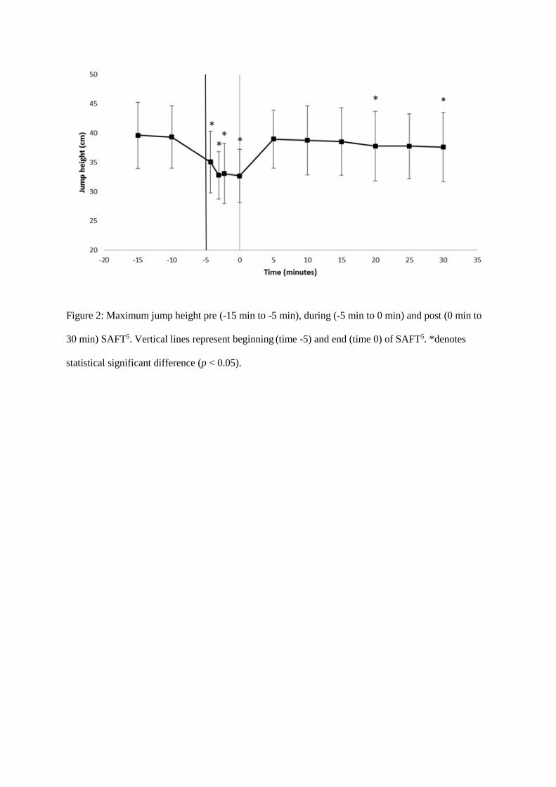
Figure 2: Maximum jump height pre (-15 min to -5 min), during (-5 min to 0 min) and post (0 min to
30 min) SAFT5. Vertical lines represent beginning (time -5) and end (time 0) of SAFT5. *denotes
statistical significant difference (p < 0.05).

Figure 3: Functional HQ ratio (Heccentric/Qconcentric) and hamstringeccentric peak torque pre, during and post
(0 min, 15 min and 30 min) SAFT5. *denotes statistical significant difference (p < .05).

SUPPLEMENTARY DATA
Appendix 1: Ankle, knee and hip angles (°) (mean ± SD) of sagittal, frontal and transversal plane during
stance phase (initial contact + 1 s) of a single-leg hop pre and immediately post SAFT5. Black solid line
represents PRE and red solid line represents POST.

Appendix 2: Ankle, knee and hip moments (Nm.kg-1) (Mean ± SD) of the sagittal, frontal and transversal
plane during the stance phase (initial contact + 1 s) of a single-leg hop pre and immediately post SAFT5.
Black solid line represents PRE and red solid line represents POST.