the utility of presentation and 4-hour high sensitivity ... · pdf fileaccepted manuscript...
TRANSCRIPT
�������� ����� ��
The utility of presentation and 4-hour high sensitivity troponin I to rule-outacute myocardial infarction in the emergency department
John W. Pickering, Joanna M. Young, Peter George, Sally Aldous, LouiseCullen, Jaimi H. Greenslade, A. Mark Richards, Richard Troughton, MichaelArdagh, Christopher M. Frampton, Martin P. Than
PII: S0009-9120(15)00313-6DOI: doi: 10.1016/j.clinbiochem.2015.07.033Reference: CLB 9100
To appear in: Clinical Biochemistry
Please cite this article as: Pickering John W., Young Joanna M., George Peter, AldousSally, Cullen Louise, Greenslade Jaimi H., Richards A. Mark, Troughton Richard, ArdaghMichael, Frampton Christopher M., Than Martin P., The utility of presentation and 4-hour high sensitivity troponin I to rule-out acute myocardial infarction in the emergencydepartment, Clinical Biochemistry (2015), doi: 10.1016/j.clinbiochem.2015.07.033
This is a PDF file of an unedited manuscript that has been accepted for publication.As a service to our customers we are providing this early version of the manuscript.The manuscript will undergo copyediting, typesetting, and review of the resulting proofbefore it is published in its final form. Please note that during the production processerrors may be discovered which could affect the content, and all legal disclaimers thatapply to the journal pertain.
ACC
EPTE
D M
ANU
SCR
IPT
ACCEPTED MANUSCRIPT
1
The utility of presentation and 4-hour high sensitivity troponin I to rule-out
acute myocardial infarction in the emergency department.
Authors:
John W Pickering1,4
, Joanna M Young2, Peter George
3,4, Sally Aldous
5, Louise Cullen
6,7,8, Jaimi
H Greenslade6, 7,8
, A Mark Richards4, Richard Troughton
4,5, Michael Ardagh
1,4, Christopher M
Frampton4, Martin P Than
1
1. Emergency Department, Christchurch Hospital, Christchurch, New Zealand
2. Lipid and Diabetes Research Group, Christchurch, New Zealand
3. Canterbury Health Laboratories, Christchurch, New Zealand
4. University of Otago, Christchurch, New Zealand
5. Cardiology Department, Christchurch Hospital, Christchurch, New Zealand
6. Department of Emergency Medicine, Royal Brisbane and Women’s Hospital, Brisbane,
Australia
7. School of Medicine, The University of Queensland, Brisbane, Australia
8. School of Public Health, The Queensland University of Technology, Brisbane, Australia
Correspondence to:
Dr Martin Than
Email address: [email protected]
Running Title: 0 and 4h hs-cTnI to rule-out AMI
Word Counts:
Abstract: 235
Body: 2150
Abbreviations
ADP: Accelerated Diagnostic Pathway
AMI: Acute Myocardial Infarction
NSTEMI: Non-ST Elevated Myocardial Infarction
ECG: Electrocardiogram
ESC: European Society of Cardiology
hs-cTnI: high sensitivity cardiac Troponin I
hs-cTnT: high sensitivity cardiac Troponin T
LoD: Limit of Detection
NICE: National Institute for Health Care and Excellence

ACC
EPTE
D M
ANU
SCR
IPT
ACCEPTED MANUSCRIPT
2
Abstract
a) Objectives: International guidance recommends that early serial sampling of high sensitivity
troponin be used to accurately identify acute myocardial infarction (AMI) in chest pain patients.
The background evidence for this approach is limited. We evaluated whether on presentation and
4-hour high-sensitivity troponin I (hs-cTnI) could be used to accurately rule-out AMI.
b) Design and Methods: hs-cTnI was measured on presentation and at 4-hours in adult patients
attending an emergency department with possible acute coronary syndrome. We determined the
sensitivity for AMI for at least one hs-cTnI above the 99th percentile for a healthy population or
alone or in combination with new ischemic ECG changes. Both overall and sex-specific 99th
percentiles were assessed.. Patients with negative tests were designated low-risk.
c) Results: 63 (17.1%) of 368 patients had AMI. The median (interquartile range) time from
symptom onset to first blood sampling was 4.8 hours (2.8-8.6). The sensitivity of the
presentation and 4h hs-cTnI using the overall 99th percentile was 92.1% (95% CI 82.4% to
97.4%) and Negative Predictive Value 95.4% (92.3% to 97.4%) with 78.3% low-risk. Applying
the sex-specific 99th percentile did not change the sensitivity. The addition of ECG did not
change the sensitivity.
d) Conclusion: Hs-cTnI >99th percentile thresholds measured on presentation and at 4-hours was
not a safe strategy to rule-out AMI in this clinical setting irrespective of whether sex-specific
99th percentiles were used, or whether hs-cTnI was combined with ECG results.
Key Words: high sensitivity troponin; acute myocardial infarction; emergency department;
emergency room; accelerated diagnostic pathway; acute coronary syndrome; STEMI; NSTEMI

ACC
EPTE
D M
ANU
SCR
IPT
ACCEPTED MANUSCRIPT
3
1. Introduction
Twin imperatives drive the assessment of patients presenting with chest pain to Emergency
Departments (ED), namely early identification of patients with an acute myocardial infarction
(AMI) and early identification of those at very low short-term risk of harm from AMI or
ischemic heart disease. The former facilitates earlier planning of treatment and the latter helps
avoids unnecessary inpatient admissions. Historically, most patients with symptoms suggestive
of AMI undergo prolonged assessment, either in the ED or as hospital in-patients even though
three quarters of these patients ultimately do not have a final diagnosis of AMI[1-3].
High sensitivity cardiac troponin assays (hs-cTn) produce analytically reliable results at the 99th
percentile of a healthy population which may facilitate identification of patients suitable (i.e.
safe) for rapid discharge to outpatient care with potentially major benefits for health services
costs and ED and hospital overcrowding[4-7]. A second measurement of hs-cTn from a blood
sample drawn two to four-hours after hospital attendance time-point may be useful. Whereas
accelerated chest pain pathways incorporating presentation and 2h sampling can identify an
increased proportion of low-risk patients[8,9] a presentation and 3h or 4h timeframe is still short
enough so that patients could remain in the ED under the care of the original clinicians without
transfer to another hospital area or handover to other staff.
The 2011 European Society of Cardiology (ESC) guidelines for management of acute coronary
syndrome without persistent ST-segment elevation recommended a rapid rule-out of AMI
protocol with only serial sampling of high sensitivity cardiac troponin[10]. A presentation and 3h
sampling time was proposed based on two studies, one utilised a small sample size and the other
utilised only one sample per patient[11,12]. An earlier generation troponin assay had yielded no
ACC
EPTE
D M
ANU
SCR
IPT
ACCEPTED MANUSCRIPT
4
statistical difference in the positivity for AMI using greater than 3h or greater than 6h between
samples[1,13,14]. The 2011 guidelines additionally recommended that where both troponin
values are less than the 99th
percentile of a healthy population that the Global Registry of
Coronary Events (GRACE) be applied to confirm low risk. Since publication of the ESC
guidelines a study by Keller and colleagues in a cohort of patients presenting with chest pain
found that a prototype high sensitivity cardiac Troponin I (hs-cTnI) at a 99th
percentile threshold
of 30 ng/L had a 98.2% (95.9% to 99.4%) sensitivity for AMI with serial sampling at
presentation and 3h[5].
The more recent National Institute for Health Care and Excellence (NICE) diagnostics guidance
for hs-cTn assays recommending that “the assays are recommended for use with early rule-out
protocols, which typically include a blood sample for cardiac troponin I or T taken at initial
assessment in an emergency department and a second blood sample taken after 3 hours”. Also
recommended is that the high sensitivity troponin be used in conjunction with electrocardiogram
(ECG) for diagnosis of NSTEMI[15].
We aimed to prospectively validate that plasma troponin levels analysed with a hs-cTn assay at
presentation (0h) and 4h from hospital presentation can rule-out AMI in patients presenting
acutely to Emergency Departments with chest discomfort that might be due to an AMI. We
assessed both the troponin alone (ESC) and troponin plus ECG (NICE) strategies. For each
strategy we also compared the performance of overall 99th
percentile to the sex-specific
percentiles. Additionally, we assessed the sensitivity for AMI and the proportion of patients who
could be designated as low risk for AMI when the threshold used was the limit of detection of
the high sensitive troponin.

ACC
EPTE
D M
ANU
SCR
IPT
ACCEPTED MANUSCRIPT
5
2. Methods
An observational cohort study design was used. Patients were recruited in conjunction with a
randomised controlled trial comparing an ‘accelerated’ (2 hour) chest pain diagnostic pathway
against the standard investigative process at Christchurch Hospital. This trial has been described
in detail elsewhere[8] and was carried out in accordance with The Code of Ethics of the World
Medical Association (Declaration of Helsinki) for experiments involving humans, received
regional ethics approval and was registered on anzctr.org.au as ACTRN12610000766011.
Briefly, eligible patients were aged ≥18 years, presenting acutely from the community to the ED
with symptoms suggestive of AMI for whom, following initial clinical assessment the attending
physician(s) planned to investigate for AMI with serial biomarker tests. In accordance with
American Heart Association case definitions, possible cardiac symptoms included: the presence
of acute chest, epigastric, neck, jaw or arm pain or discomfort or pressure without apparent non-
cardiac source[2]. Patients were excluded if any of the following conditions were satisfied: ST
Segment Elevation Myocardial Infarction (STEMI) as demonstrated by an ST elevation on any
electrocardiograph (ECG) at presentation (Note: patients with all other ECG changes, including
ST depression, were included); chest pain episode began >12 hours prior to assessment, proven
or suspected non-coronary pathology as the cause of chest pain; need for admission regardless of
a cTn <99th
percentile, due to other medical conditions, or need for other investigations;
previously enrolled in this study; anticipated problem with follow-up (e.g. resident outside New
Zealand or terminal illness); or unable or unwilling to provide informed consent. Enrolment was
consecutive during the hours of available research nurse (normally 0800 to 2300, 7 days a week).
This analysis was limited to patients with sufficient stored plasma sample available for hs-cTnI
assay at both presentation and 4 hours.
ACC
EPTE
D M
ANU
SCR
IPT
ACCEPTED MANUSCRIPT
6
2.1 Reference standard
Classification of AMI was based upon global taskforce recommendations requiring evidence of
myocardial necrosis together with evidence of myocardial ischaemia (ischaemic symptoms, ECG
changes or imaging evidence)[4] Necrosis was diagnosed on the basis of a rising or falling
pattern of the laboratory cardiac troponin (ARCHITECT troponin I [TnI] assay; Abbott) level,
with at least one value above the 99th percentile (28 ng/L). The manufacturer-specified LOD for
the assay was 10 ng/mL, and 10% coefficient of variation was at 32 ng/L. Outcomes and
investigations were reported using predefined standardised reporting guidelines[16]. The
presence of AMI was adjudicated independently by local cardiologists using these reporting
guidelines and blinded to the results of the hs-cTnI (index test). If the reference troponin was
above the reference range, but there was no rise or fall, other causes of a raised troponin were
considered. If no clear alternative cause of the troponin rise was apparent then, if the clinical
presentation was suggestive of ACS, an adjudicated diagnosis of AMI was made. A panel of two
cardiologists performed the adjudication independent of each other with a third cardiologist
making an independent adjudication in cases of disagreement.
2.2 Index Tests
Hs-cTnI sampling was on presentation to the ED and 4 hours later. Blood was drawn into 1x4mL
Lithium heparin tubes, spun at 3220 RCF at 4°C for 10 minutes, 1ml of plasma was then
transferred to one or two 1.5mL tubes and immediately stored at -80°C for later thawing and
assay. Samples were assayed 12 to 36 months following collection. An ischaemic ECG was
defined as ST-segment depression of at least 0.05 mV in 2 or more contiguous leads (including
reciprocal changes), T-wave inversion of at least 0.1 mV, or Q-waves >30 ms in width and
greater than or equal to 0.1 mV in depth in at least 2 contiguous leads. Patients with other
ACC
EPTE
D M
ANU
SCR
IPT
ACCEPTED MANUSCRIPT
7
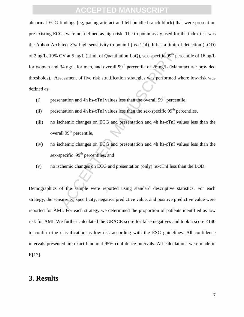
abnormal ECG findings (eg, pacing artefact and left bundle-branch block) that were present on
pre-existing ECGs were not defined as high risk. The troponin assay used for the index test was
the Abbott Architect Stat high sensitivity troponin I (hs-cTnI). It has a limit of detection (LOD)
of 2 ng/L, 10% CV at 5 ng/L (Limit of Quantitation LoQ), sex-specific 99th
percentile of 16 ng/L
for women and 34 ng/L for men, and overall 99th
percentile of 26 ng/L (Manufacturer provided
thresholds). Assessment of five risk stratification strategies was performed where low-risk was
defined as:
(i) presentation and 4h hs-cTnI values less than the overall 99th
percentile,
(ii) presentation and 4h hs-cTnI values less than the sex-specific 99th
percentiles,
(iii) no ischemic changes on ECG and presentation and 4h hs-cTnI values less than the
overall 99th
percentile,
(iv) no ischemic changes on ECG and presentation and 4h hs-cTnI values less than the
sex-specific 99th
percentiles, and
(v) no ischemic changes on ECG and presentation (only) hs-cTnI less than the LOD.
Demographics of the sample were reported using standard descriptive statistics. For each
strategy, the sensitivity, specificity, negative predictive value, and positive predictive value were
reported for AMI. For each strategy we determined the proportion of patients identified as low
risk for AMI. We further calculated the GRACE score for false negatives and took a score <140
to confirm the classification as low-risk according with the ESC guidelines. All confidence
intervals presented are exact binomial 95% confidence intervals. All calculations were made in
R[17].
3. Results

ACC
EPTE
D M
ANU
SCR
IPT
ACCEPTED MANUSCRIPT
8
There were 368 patients. The first blood sample was taken a median (interquartile range) 4.8
(2.8-8.6) hours after onset of symptoms. The median time difference between the presentation
and the 4h sample was 4.4 h (4.25 to 4.63). Sixty-three (17.1%) were diagnosed with AMI.
Demographics are given in table 1.
Table 1: Cohort characteristics
Variable Value
Age (years) 61±13
Female 35% (129)
Weight (kg) 85±19
Ethnicity (self identified)
Maori 9
New Zealander/ New Zealand European 329
Other 30
Risk Factors and History (patient
reported)
Hypertension 36.8% (174)
Dyslipidaemia 53.8% (198)
Diabetes 16.0% (57)
Current smoker 18.5% (68)
Family history of Ischemic Heart Disease 60.3% (222)
Prior:
Myocardial Infarction 26.6% (98)
Angina 37.0% (136)
Ventricular Tachycardia 4.6% (17)
CAD 44.0% (162)
Atrial Arrhythmia 9.5% (35)
Congestive Heart Failure 4.3% (16)
Stroke or Transient Ischemic Attack 11.1% (41)
Peripheral Arterial Disease 4.6% (17)
Coronary Artery Bybass Graft 7.6% (28)
Coronary Angioplasty 27.7% (102)
Rheumatoid Arthritis 2.2% (8)
Outcomes
ECG positive 5.4% (20)
STEMI 1 (0.3%)
NSTEMI 62 (16.8%)
Hospital length of stay (Days) 1.2 (0.9-3.1)
Data presents as n (%) or mean±SD or median (lower quartile - upper quartile)
ACC
EPTE
D M
ANU
SCR
IPT
ACCEPTED MANUSCRIPT
9
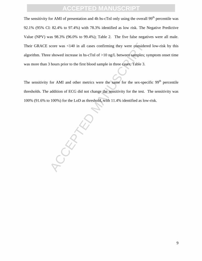
The sensitivity for AMI of presentation and 4h hs-cTnI only using the overall 99th
percentile was
92.1% (95% CI: 82.4% to 97.4%) with 78.3% identified as low risk. The Negative Predictive
Value (NPV) was 98.3% (96.0% to 99.4%); Table 2. The five false negatives were all male.
Their GRACE score was <140 in all cases confirming they were considered low-risk by this
algorithm. Three showed increase in hs-cTnI of >10 ng/L between samples; symptom onset time
was more than 3 hours prior to the first blood sample in three cases, Table 3.
The sensitivity for AMI and other metrics were the same for the sex-specific 99th
percentile
thresholds. The addition of ECG did not change the sensitivity for the test. The sensitivity was
100% (91.6% to 100%) for the LoD as threshold, with 11.4% identified as low-risk.
ACC
EPTE
D M
ANU
SCR
IPT
ACCEPTED MANUSCRIPT
10
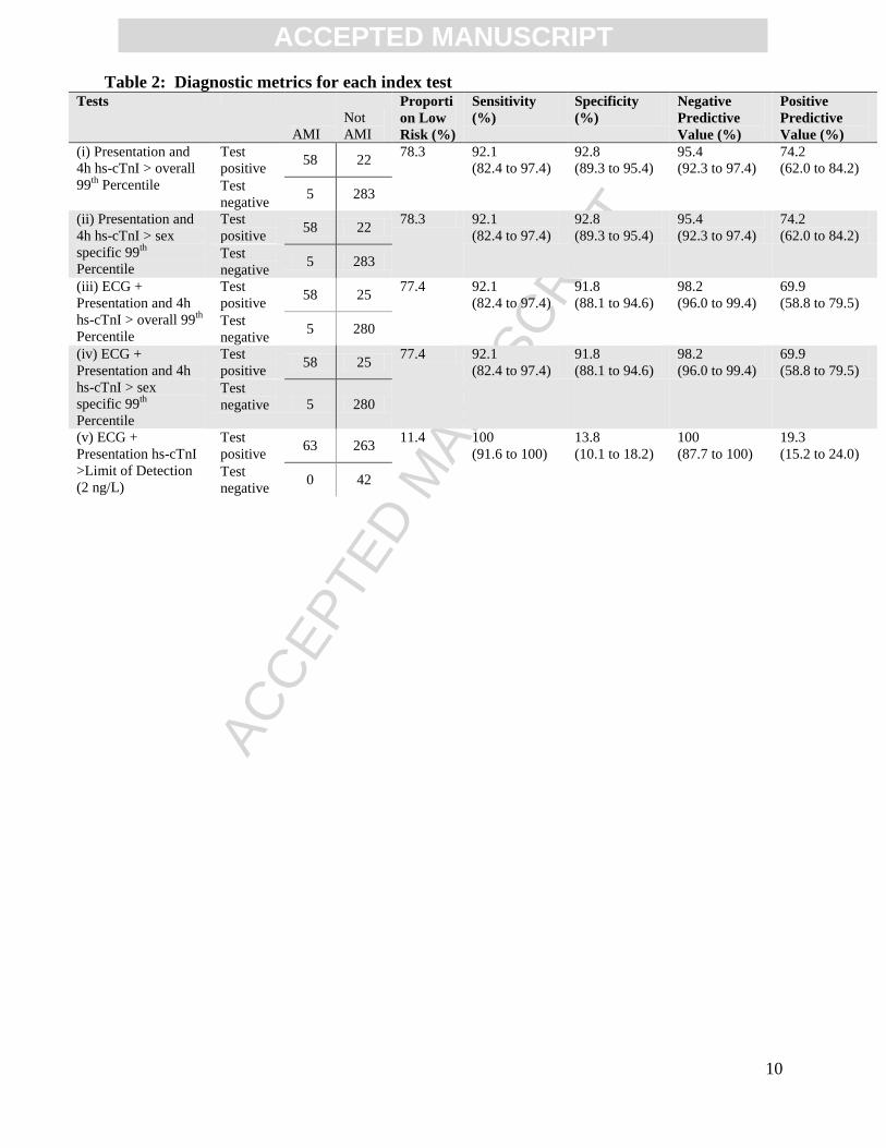
Table 2: Diagnostic metrics for each index test Tests
AMI
Not
AMI
Proporti
on Low
Risk (%)
Sensitivity
(%)
Specificity
(%)
Negative
Predictive
Value (%)
Positive
Predictive
Value (%)
(i) Presentation and
4h hs-cTnI > overall
99th
Percentile
Test
positive 58 22
78.3 92.1
(82.4 to 97.4)
92.8
(89.3 to 95.4)
95.4
(92.3 to 97.4)
74.2
(62.0 to 84.2)
Test
negative 5 283
(ii) Presentation and
4h hs-cTnI > sex
specific 99th
Percentile
Test
positive 58 22
78.3 92.1
(82.4 to 97.4)
92.8
(89.3 to 95.4)
95.4
(92.3 to 97.4)
74.2
(62.0 to 84.2)
Test
negative 5 283
(iii) ECG +
Presentation and 4h
hs-cTnI > overall 99th
Percentile
Test
positive 58 25
77.4 92.1
(82.4 to 97.4)
91.8
(88.1 to 94.6)
98.2
(96.0 to 99.4)
69.9
(58.8 to 79.5)
Test
negative 5 280
(iv) ECG +
Presentation and 4h
hs-cTnI > sex
specific 99th
Percentile
Test
positive 58 25
77.4 92.1
(82.4 to 97.4)
91.8
(88.1 to 94.6)
98.2
(96.0 to 99.4)
69.9
(58.8 to 79.5)
Test
negative 5 280
(v) ECG +
Presentation hs-cTnI
>Limit of Detection
(2 ng/L)
Test
positive 63 263
11.4 100
(91.6 to 100)
13.8
(10.1 to 18.2)
100
(87.7 to 100)
19.3
(15.2 to 24.0)
Test
negative 0 42

ACC
EPTE
D M
ANU
SCR
IPT
ACCEPTED MANUSCRIPT
11
Table 3: Details of the False Negatives for index test (i) Presentation and 4h hs-cTnI >
overall 99th Percentile Study ID CP150 CP157 CP170 CP464 CP530
sex M M M M M
age 47 73 84 72 69
Hs-cTnI at presentation (ng/L) 7.2 13.8 6.5 7.1 7
Hs-cTnI at 4h (ng/L) 17.9 24.1 23 8.3 8.3
cTnI (ng/L) at presentation <10 <10 <10 10 20
First cTnI (ng/L) at >6h 60 120 30 20‡ 40
ECG Negative Negative Negative Negative Negative
Relative difference (%) 148.6 74.6 253.8 16.9 18.6
Absolute difference (ng/L) 10.7 10.3 16.5 1.2 1.3
Time from symptom onset to
presentation sample (h) 2.58 4.83 2.83 3.75 5.92
Heart rate (bpm) 82 58 60 70 70
Systolic blood pressure 121 126 160 124 190
Creatinine (μmol/L) 130* 81 91 148** 65
Killip Class NA 1 2 1 NA
GRACE score (Death or MI in hospital) <133† 101 124 113 <132†
Pain
Pleuritic No No No No Yes
On palpitation No No No No No
Radiates to arm No No Yes Yes No
Diaphoresis Yes Yes Yes Yes No
Risk Factors and History (patient
reported)
Hypertension Yes Yes Yes No No
Dyslipidaemia Yes Yes Yes Yes No
Diabetes No Yes No No No
Current smoker No No No No No
Family history of Ischemic Heart
Disease Yes Yes Yes Yes No
Use of Aspirin in last 7 days No No No Yes No
Prior:
Myocardial Infarction No No Yes Yes No
Angina No Yes Yes Yes No
Ventricular Tachycardia No No No No No
CAD No Yes Yes Yes No
Atrial Arrhythmia No No No Yes No
Congestive Heart Failure No No No No No
Stroke or Transient Ischemic Attack No No Yes Yes No
Peripheral Arterial Disease No Yes No No No
Coronary Artery Bybass Graft No No No No No
Coronary Angioplasty No No Yes Yes No
Rheumatoid Arthritis No No No No No
Modified TIMI risk score†† 0 3 3 3 1
Investigations and Treatments
Revascularisation
Urgent PCI
at 3 days
Urgent
CABG at 12
days
No No Urgent
PCI at 6
days
Angiostenosis Yes None None Yes Yes
Echocardiogram ejection fraction NA 62 36 62 65
EMRA NA No Yes No Yes
* Possibly Acute Kidney Injury; **Probably a chronic elevation given the patient creatinine history.
† Assumes a maximum Killip class of 3 because class 4 is cardiogenic shock and this was not recorded in any case.
†† modified because the score for the troponin and ECG are not included.
‡ a subsequent troponin on the next morning (13 hours later) was 40 ng/L (ie positive).

ACC
EPTE
D M
ANU
SCR
IPT
ACCEPTED MANUSCRIPT
12
NA: Not Available; PCI: Percutaneous Coronary Intervention; MI: Myocardial Infarction; EMRA:
Echocardiograph regional wall motion abnormality
ACC
EPTE
D M
ANU
SCR
IPT
ACCEPTED MANUSCRIPT
13
4. Discussion
The sensitivity for AMI of either presentation and 4h hs-cTnI at the 99th
percentile on its own or
in combination with ECG on presentation to rule-out AMI was considerably less than 99%, a
threshold considered optimal by ED physicians[18]. Even at the best sensitivity for AMI the
upper confidence interval of 97.4% was below this level. Notably this is less than the 98.2%
(95.9% to 99.4%) found by Keller and colleagues for presentation and 3h hs-cTnI above the 99th
percentile for a prototype hs-cTnI assay and without the inclusion of ECG in the test (the
inclusion of which should only increase sensitivity). This and the false negatives we observed
highlights the dangers of a biomarker only approach, especially where a relatively high threshold
(99th
percentile of a healthy population) is applied and that findings during clinical assessment
must be incorporated into the assessment in a Baysean manner together with biochemical
markers. Previously, we have demonstrated in the primary trial that a minimum level of 99%
sensitivity for AMI may be reached with risk stratification using the Thrombolysis in Myocardial
Infarction (TIMI) risk score in addition to a presentation ECG and presentation and 2 hour
contemporary cardiac troponin[8]. The TIMI score incorporates age, risk factors including
family history of coronary artery disease, and history. Other risk scoring strategies that may also
improve identification of a low-risk cohort, for example the EDACS, HEART, Modified
Goldman and Vancouver Chest Pain rule, include the same class of factors with different
weightings and variation in the components that make up the history and risk factor scores in
particular[9,19-21]. EDACS also includes sex. An EDACS-ADP may classify with high
sensitivity over 40% of patients[9].
We also noted that in three of the false negatives there was a small, but measureable change in
hs-cTnI of greater than 10 ng/L between samples. Although both samples remained below the
ACC
EPTE
D M
ANU
SCR
IPT
ACCEPTED MANUSCRIPT
14
99th
percentile, a cautious clinician may not wish to classify these patients as low risk. Further
work is needed to decide on the role of deltas below the 99th
percentile.
The ESC guideline recommendation of a presentation and typically after 3h high sensitivity
troponin samples to rule out AMI are based on only two studies. The first assessed the
performance of hs-cTnT with a threshold of 14 ng/L in a population of 57 patients without
impaired renal function and with retrospectively confirmed unstable angina and evolving
NSTEMI[11]. A sensitivity of 100% for diagnosis of NSTEMI with a wide confidence interval
(95% confidence interval: 75.1% to 100%) was achieved for serial sampling on admission and a
second sample within 3 hours. The second study was comprised of two cohorts where the
performance of a single hs-cTnT measure within a median 3 hours (range 0 to 7 hours) was
assessed. Only in one cohort, the Bad Nauheim ACS registry comprising 1023 patients who had
been referred for coronary angiography or percutaneous coronary intervention (because of ACS
within the previous 48 hours), was the performance of hs-cTnT assessed for diagnosis of
AMI[12]. The sensitivity for AMI was 96% and negative predictive value 80%. Both these
studies were published at a time when they may have been affected by the calibration issues
leading to inaccurate reporting of numerical results for hs-cTnT[22,23].
The NICE guidelines were based on a systematic review of the literature which was dominated
by hs-cTnT rather than hs-cTnI studies[24] and only 6 studies which reported multiple testing.
The recommendation of a second hs-cTn test ‘typically’ at 3-hour time point was based on
consideration of the possibility of ruling in AMI with a suitable delta.
ACC
EPTE
D M
ANU
SCR
IPT
ACCEPTED MANUSCRIPT
15
This present study aimed to employ a 4-hour time period between presentation and the second
sample. This was deliberate because the primary study already had presentation and 2 hour time
points, 4 hours was still short enough to enable discharge from ED to outpatient care within the
national 6 hour ED target stay, and was only likely to enhance sensitivity and specificity for AMI
over a 3 hour sampling. The study was limited by its size meaning the confidence intervals are
broad.
Eggers and colleagues have previously considered the use of the 99th percentile for consecutive
presentation and 2-hour hs-cTnI samples in combination with ECG to rule-out NSTEMI[25].
They found the sensitivity for NSTEMI to be 96.9%; as with the current study, too low to be
clinically useful. When the authors considered the lower 97.5% threshold (15.5 ng/L) the
sensitivity for NSTEMI improved to 98.2% with 54.4% ruled-out by this strategy. Similarly the
TRAPID AMI protocol employing an hs-cTnT threshold less than the 99th
threshold (ie 12 ng/L)
along with a delta of <3 ng/L in 1 hour ruled out 60% of patients with 100% sensitivity in a
derivation cohort[26] and 59.5% of patients with 99.6% sensitivity in a validation cohort[27].
Cullen and colleagues assessed the sensitivity for 30-d AMI or cardiac death of a Siemens
contemporary troponin I at the 99th
percentile (56 ng/L) at 0 and 2h and found a sensitivity of
92.2% [28]. Druey and colleagues also recently assessed the use of contemporary troponin I to
rule-out AMI using a 0 and 2h algorithm with a lower threshold of 10ng/L[29]. This ruled-out
44% of patients with a sensitivity of 98.4% in a derivation cohort and 62% of patients with a
sensitivity of 94.5% in a validation cohort. The present study was not powered to discover an
optimal threshold for rule-out. Nevertheless, we could rule-out 58% of patients with 100%
sensitivity (95%CI: 91.6% to 100%) for AMI with a threshold of 8 ng/L. The differences are
probably due to differences in cohort characteristics and because the current study had only 63
ACC
EPTE
D M
ANU
SCR
IPT
ACCEPTED MANUSCRIPT
16
AMI resulting in broad confidence intervals. Nevertheless, the Eggers analysis and the present
study suggest that a threshold between the limit of detection and 99th
percentile may be used with
serial samples and in combination with ECG to rule-out a significant proportion of patients. We
recommend that such strategies be compared in the same cohort with those which also utilise a
risk stratification score.
The use of an undetectable hs-cTnT [30,31] or hs-cTnI [5,32] in conjunction with a negative
ECG has recently been demonstrated to rule-out AMI on presentation with excellent sensitivity.
This study supports those findings with a sensitivity for AMI of 100% allowing 11.4% of
patients to be ruled out of having an AMI shortly after presentation to ED. In a cohort of patients
with identical exclusion and inclusion criteria Greenslade and colleagues also had 100%
sensitivity for AMI for undetectable hs-cTnI and found 17.8% of patients were low risk[15,32].
Similarly Keller and colleagues had a 100% sensitivity with 27.4% low risk[5,15]. On the other
hand Body and colleagues found AMI could not be excluded by this method (sensitivity: 97.1%)
and recommended further work on serial sampling to improve sensitivity[33]. Although the
sensitivity for AMI in our study was 100%, the study size was limited and the lower limit 95%
confidence intervals was only 91.6%. Therefore, we too recommend further work.
A limitation of our study potentially affecting our conclusion that performance did not differ
with sex-specific thresholds was that the diagnosis of AMI was based on overall (not sex-
specific) values. This may have biased against females with low, but abnormal troponin
elevations using sex-specific cut points.
5. Conclusion

ACC
EPTE
D M
ANU
SCR
IPT
ACCEPTED MANUSCRIPT
17
The sensitivity for AMI of hs-cTnI <99th
percentile at presentation and 4 hours alone or in
combination of a non-ischaemic ECG was too low to be reliably clinically useful for rule out
when used without reference to clinical indicators. The proposed use of classifying patients with
hs-cTnI less than the limit of detection on presentation also had good sensitivity and would
enable approximately 10% of patients to be classified as low-risk as soon as the first blood
results became available.
6. Acknowledgements
This study was funded by the Heart Foundation of New Zealand (Grant 1457). JP is supported by
a Senior Research Fellowship from the Canterbury Medical Research Foundation, Emergency
Care Foundation, and Canterbury District Health Board. JY is funded by a Lottery Health
Postdoctoral Fellowship. The Queensland Emergency Research Foundation supports LC with a
Research Fellowship. LC has received research funding and honorarium from Abbott
Diagnostics, Alere, Roche, Siemens and Radiometer Pacific. We thank the research nurses and
staff of the Christchurch Heart Institute for data collection and John Wallace of Canterbury
Health Laboratories for assaying hs-cTnI.
ACC
EPTE
D M
ANU
SCR
IPT
ACCEPTED MANUSCRIPT
18
7. References
[1] Hollander JE. The continuing search to identify the very-low-risk chest pain patient. Acad Emerg Med
1999;6:979–81.
[2] Luepker RV, Apple FS, Christenson RH, Crow RS, Fortmann SP, Goff D, et al. Case definitions for acute
coronary heart disease in epidemiology and clinical research studies: a statement from the AHA Council
on Epidemiology and Prevention; AHA Statistics Committee; World Heart Federation Council on
Epidemiology and Prevention; the European Society of Cardiology Working Group on Epidemiology and
Prevention; Centers for Disease Control and Prevention; and the National Heart, Lung, and Blood
Institute. Circulation 2003;108:2543–9.
[3] Than MP, Cullen L, Reid CM, Lim SH, Aldous S, Ardagh MW, et al. A 2-h diagnostic protocol to assess
patients with chest pain symptoms in the Asia-Pacific region (ASPECT): a prospective observational
validation study. Lancet 2011;377:1077–84.
[4] Thygesen K, Alpert JS, White HD, on behalf of the Joint ESC/ACCF/AHA/WHF Task Force for the
Redefinition of Myocardial Infarction. Universal definition of myocardial infarction. Circulation
2007;116:2634–53.
[5] Keller T, Zeller T, Ojeda F, Tzikas S, Lillpopp L, Sinning C, et al. Serial changes in highly sensitive
troponin I assay and early diagnosis of myocardial infarction. Jama 2011;306:2684–93.
[6] Reichlin T, Hochholzer W, Bassetti S, Steuer S, Stelzig C, Hartwiger S, et al. Early diagnosis of
myocardial infarction with sensitive cardiac troponin assays. N Engl J Med 2009;361:858–67.
[7] Cullen L, French JK, Briffa TG, Redfern J, Hammett CJK, Brieger DB, et al. Availability of highly
sensitive troponin assays and acute coronary syndrome care: insights from the SNAPSHOT registry. Med
J Aust 2015;202:36–9.
[8] Than MP, Aldous S, Lord SJ, Goodacre S, Frampton CMA, Troughton R, et al. A 2-hour diagnostic
protocol for possible cardiac chest pain in the emergency department: a randomized clinical trial. JAMA
Intern Med 2014;174:51–8.
[9] Than MP, Flaws D, Sanders S, Doust J, Glasziou P, Kline J, et al. Development and validation of the
Emergency Department Assessment of Chest pain Score and 2 h accelerated diagnostic protocol. Emerg
Med Australas 2014;26:34–44.
[10] Authors/Task Force Members, Hamm CW, Bassand JP, Agewall S, Bax J, Boersma E, et al. ESC
Guidelines for the management of acute coronary syndromes in patients presenting without persistent ST-
segment elevation: The Task Force for the management of acute coronary syndromes (ACS) in patients
presenting without persistent ST-segment elevation of the European Society of Cardiology (ESC). Eur
Heart J 2011;32:2999–3054.
[11] Giannitsis E, Becker M, Kurz K, Hess G, Zdunek D, Katus HA. High-Sensitivity Cardiac Troponin T for
Early Prediction of Evolving Non-ST-Segment Elevation Myocardial Infarction in Patients with Suspected
Acute Coronary Syndrome and Negative Troponin Results on Admission. Clin Chem 2010;56:642–50.
[12] Weber M, Bazzino O, Estrada JLN, de Miguel R, Salzberg S, Fuselli JJ, et al. Improved diagnostic and
prognostic performance of a new high-sensitive troponin T assay in patients with acute coronary
syndrome. Am Heart J 2011;162:81–8.
[13] Antman EM, Cohen M, Bernink PJ, McCabe CH, Horacek T, Papuchis G, et al. The TIMI risk score for
unstable angina/non-ST elevation MI: A method for prognostication and therapeutic decision making.
JAMA 2000;284:835–42.
[14] Macrae AR, Kavsak PA, Lustig V, Bhargava R, Vandersluis R, Palomaki GE, et al. Assessing the
requirement for the 6-hour interval between specimens in the American Heart Association Classification
of Myocardial Infarction in Epidemiology and Clinical Research Studies. Clin Chem 2006;52:812–8.
[15] National Institute of Health Care Excellence (NICE). Myocardial infarction (acute): Early rule out using
high-sensitivity troponin tests (Elecsys Troponin T high-sensitive, ARCHITECT STAT High Sensitive
Troponin-I and AccuTnI+3 assays)(DG15). 2015. http://nice.org.uk/guidance/dg15
[16] Cullen L, Than MP, Brown AFT, Richards M, Parsonage W, Flaws D, et al. Comprehensive standardized
data definitions for acute coronary syndrome research in emergency departments in Australasia. Emerg
Med Australas 2010;22:35–55.
[17] R Core Team. R: A language and environment for statistical computing 2014.
[18] Than MP, Herbert M, Flaws D, Cullen L, Hess E, Hollander JE, et al. What is an acceptable risk of major
adverse cardiac event in chest pain patients soon after discharge from the Emergency Department?: a
clinical survey. Int J Cardiol 2013;166:752–4.
[19] Six AJ, Backus BE, Kelder JC. Chest pain in the emergency room: value of the HEART score. Neth Heart

ACC
EPTE
D M
ANU
SCR
IPT
ACCEPTED MANUSCRIPT
19
J 2008;16:191–6.
[20] Scheuermeyer FX, Wong H, Yu E, Boychuk B, Innes G, Grafstein E, et al. Development and validation of
a prediction rule for early discharge of low-risk emergency department patients with potential ischemic
chest pain. CJEM 2014;16:106–19.
[21] Carlton EW, Cullen L, Than MP, Gamble J, Khattab A. A novel diagnostic protocol to identify patients
suitable for discharge after a single high-sensitivity troponin. Heart 2015; On line ahead of print. doi
10.1136/heartjnl-2014-307288
[22] Apple FS, Jaffe AS. Clinical implications of a recent adjustment to the high-sensitivity cardiac troponin T
assay: user beware. Clin Chem 2012;58:1599–600.
[23] Parsonage WA, Tate JR, Greenslade JH, Hammett CJ, Ungerer JPJ, Pretorius CJ, et al. Effect of
recalibration of the hs-TnT assay on diagnostic performance. Clin Chem Lab Med 2014;52:e25–7.
[24] Westwood ME, van Asselt ADI, BLT R, Whiting PF, Thokala P, Armstrong N, et al. High sensitivity
troponin assays for the early rule-out or diagnosis of acute myocardial infarction in people with acute
chest pain: a systematic review and cost-effectiveness analysis. Kleijnen Systematic Reviews Ltd 2014:1–
250.
[25] Eggers KM, Aldous S, Greenslade JH, Johnston N, Lindahl B, Parsonage WA, et al. Two-hour diagnostic
algorithms for early assessment of patients with acute chest pain — Implications of lowering the cardiac
troponin I cut-off to the 97.5th percentile. Clin Chim Acta 2015;445:19–24.
[26] Reichlin T, Schindler C, Drexler B, Twerenbold R, Reiter M, Zellweger C, et al. One-hour rule-out and
rule-in of acute myocardial infarction using high-sensitivity cardiac troponin T. Arch Intern Med
2012;172:1211–8.
[27] Reichlin T, Twerenbold R, Wildi K, Rubini Gimenez M, Bergsma N, Haaf P, et al. Prospective validation
of a 1-hour algorithm to rule-out and rule-in acute myocardial infarction using a high-sensitivity cardiac
troponin T assay. Can Med Assoc J 2015; On line ahead of print. doi 10.1503/cmaj.141349/-/DC1
[28] Cullen L, Greenslade J, Than MP, Tate J, Ungerer JPJ, Pretorius C, et al. Performance of risk stratification
for acute coronary syndrome with two-hour sensitive troponin assay results. Heart, Lung & Circulation
2014;23:428–34.
[29] Druey S, Wildi K, Twerenbold R, Jaeger C, Reichlin T, Haaf P, et al. Early rule-out and rule-in of
myocardial infarction using sensitive cardiac Troponin I. Int J Cardiol 2015;195:163–70.
[30] Bandstein N, Ljung R, Johansson M, Holzmann MJ. Undetectable High-Sensitivity Cardiac Troponin T
Level in the Emergency Department and Risk of Myocardial Infarction. Jacc 2014;63:2569–78.
[31] Body R, Carley S, McDowell G, Jaffe AS, France M, Cruickshank K, et al. Rapid exclusion of acute
myocardial infarction in patients with undetectable troponin using a high-sensitivity assay. JACC
2011;58:1332–9.
[32] Greenslade JH, Kavsak P, Parsonage W, Shortt C, Than MP, Pickering JW, et al. Combining presentation
high-sensitivity cardiac troponin I and glucose measurements to rule-out an acute myocardial infarction in
patients presenting to emergency department with chest pain. Clin Biochem 2015;48:288–91.
[33] Body R, Burrows G, Carley S, Lewis PS. Rapid exclusion of acute myocardial infarction in patients with
undetectable troponin using a sensitive troponin I assay. Ann Clin Biochem 2015: On line ahead of print
doi: 10.1177/0004563215576976
ACC
EPTE
D M
ANU
SCR
IPT
ACCEPTED MANUSCRIPT
20
Highlights
AMI can not be ruled out by serial hs-cTnI <99th percentile within the ED
Sex-specific 99th percentile thresholds do not improve sensitivity
The addition of negative ECG does not improve sensitivity
11% may be ruled out with hs-cTnI <LoD and negative ECG