the zinc-finger protein slug causes desmosome dissociation
TRANSCRIPT

The Rockefeller University Press, 0021-9525/97/06/1403/17 $2.00The Journal of Cell Biology, Volume 137, Number 6, June 16, 1997 1403–1419 1403
The Zinc-Finger Protein Slug Causes DesmosomeDissociation, an Initial and Necessary Step forGrowth Factor–induced Epithelial–Mesenchymal Transition
Pierre Savagner,*
‡
Kenneth M. Yamada,
‡
and Jean Paul Thiery*
*Centre National de la Recherche Scientifique–Institut Curie, 75231 Paris Cedex 05, France; and
‡
Craniofacial Developmental Biology and Regeneration Branch, National Institute of Dental Research, National Institutes of Health, Bethesda, Maryland 20892-4370
Abstract.
Epithelial–mesenchymal transition (EMT) is an essential morphogenetic process during embry-onic development. It can be induced in vitro by hepato-cyte growth factor/scatter factor (HGF/SF), or by FGF-1 in our NBT-II cell model for EMT. We tested for a cen-tral role in EMT of a zinc-finger protein called Slug. Slug mRNA and protein levels were increased tran-siently in FGF-1–treated NBT-II cells. Transient or sta-ble transfection of Slug cDNA in NBT-II cells resulted in a striking disappearance of the desmosomal markers desmoplakin and desmoglein from cell–cell contact ar-eas, mimicking the initial steps of FGF-1 or HGF/SF-
induced EMT. Stable transfectant cells expressed Slug protein and were less epithelial, with increased cell spreading and cell–cell separation in subconfluent cul-tures. Interestingly, NBT-II cells transfected with anti-sense Slug cDNA were able to resist EMT induction by FGF-1 or even HGF/SF. This antisense effect was sup-pressed by retransfection with Slug sense cDNA. Our results indicate that Slug induces the first phase of growth factor–induced EMT, including desmosome dis-sociation, cell spreading, and initiation of cell separa-tion. Moreover, the antisense inhibition experiments suggest that Slug is also necessary for EMT.
E
pithelial
cells adhere to each other through spe-cialized structures essential for the maintenance ofepithelial organization and differentiation. Among
these, structures linked to the cytokeratin intermediate fil-ament network appear to provide the strongest and mostresilient adhesion (12, 15). The core unit of such structuresis the desmosome, which appears early during epithelialdifferentiation (13, 24, 30, 37). Desmoglein and desmocol-lins are desmosome-specific cadherins that mediate cell–cell binding (15, 34). They are part of a molecular complexinvolving
g
-catenin/plakoglobin, Band 6/plakophilin, anddesmoplakin, among other components (26, 29, 40, 56, 57,62–64). Desmoplakin can bind directly to cytokeratin fila-ments in vitro and appears to enhance desmosome stability(35, 56, 57). Studies with dominant-negative variants indi-cate that desmoplakin is required in vivo for attaching in-termediate filaments to the desmosome (4). Little is known,however, about how desmosomal assembly is regulated.
Individualization of cells emerging and dissociating froman epithelial sheet is one of the basic mechanisms involvedin embryonic development. In early postimplantation mouse
embryos, it appears that all cells that contribute to embry-onic tissues are epithelial cells expressing desmosomes(30). Therefore, cellular dissociation involves the disinte-gration of these desmosomes and other cell–cell adhesionsystems. Depending on the species, this necessary processof epithelial–mesenchymal transition (EMT)
1
occurs at sev-eral critical stages during development, such as gastrula-tion, neural crest cell emigration, and organogenesis (forreviews see references 19, 27, 28, 38, 52).
Several inducers, including extracellular molecules (5,32, 33, 50, 54, 67, 68) and growth-factors from the trans-forming growth factor (TGF)
b
and FGF families (10, 47),have been suggested to play a role during these embryonicphenotype modulations. Transcription factors are likely tobe involved at some point during the process. For exam-ple, the zinc-finger protein Slug was found to be expressedin chicken neural crest cells just before they emerge fromthe neural tube and later during their migration phase(46). Interestingly, the same report described Slug expres-sion by epiblast cells lining the primitive streak during gas-trulation, just before the emergence of mesenchymal cells.Treatment of developing embryos with antisense oligonu-
Address all correspondence to Pierre Savagner, Craniofacial Develop-mental Biology and Regeneration Branch, NIDR, Bldg. 30, Rm. 424, 30Convent Drive MSC 4370, National Institutes of Health, Bethesda, MD20892-4370. Tel: (301) 496-6040; Fax: (301) 402-0897.
1.
Abbreviations used in this paper
: EMT, epithelial-mesenchymal transi-tion; HGF/SF, hepatocyte growth factor/scatter factor; IL-2R, interleukin-2receptor; MDCK, Madin-Darby canine kidney.
D
ownloaded from
http://rupress.org/jcb/article-pdf/137/6/1403/1270639/10829.pdf by guest on 30 January 2022
The Journal of Cell Biology, Volume 137, 1997 1404
cleotides from
slug
was found to interfere with these twoprocesses, suggesting a potential causal role for Slug in theEMT process in vivo. Slug and closely related members ofthe Snail family were also found to express similar pat-terns of localization in
Xenopus
and zebrafish embryos,providing an early marker for neural crest cells.
Epithelial cells can be induced to dissociate by treat-ment with hepatocyte growth factor/scatter factor (HGF/SF), and in some cases by other growth factor members ofthe FGF (65), EGF, and TGF families (3, 16, 43). The EMTprocess is initiated by cell–cell dissociation, which is pre-ceded by the internalization of desmosomal componentsand progressive disappearance from cell–cell contact areas(7). As studied in Madin-Darby canine kidney (MDCK)cells, these scatter factors can also act as growth factorsand morphogenetic factors, using specific transductionpathways in each case (1, 17, 25, 51, 58).
We have previously characterized a rat bladder carci-noma cell line, NBT-II, that is induced by FGF-1 to un-dergo an EMT characterized by a switch to a fibroblastoidphenotype and by cell migration (7, 65). Desmosomalcomponents, including desmoplakin and desmoglein, werefound to be internalized and disappear from the surfacestarting 4 h after initiation of FGF-1 treatment. By 6 h,
.
50% of the cells no longer express desmosomes. By 12 h,cells undergo active migration on the substrate (7). In con-trast, adherens junction components such as E-cadherinand catenins were not altered quantitatively during thisepithelial–mesenchymal transition (8). However, E-cadherinwas redistributed from cell–cell contact areas to a diffusedistribution on the cell surface. FGF-1 activation was me-diated through the receptor FGFR2c/KGFR, which un-derwent alternative splicing during the EMT process (53).The FGFR2c/KGFR tyrosine kinase domain was found tobe involved in transducing the EMT process through phos-phorylation (6). In addition, an EMT-specific activation ofpp60
c-src
was demonstrated (49), and overexpression of nor-mal c-src in NBT-II cells was found to oversensitize themto EMT induced by FGF-1. Transcriptional and transla-tional events were also found to be required for EMTsince actinomycin D inhibited EMT (Savagner, P., unpub-lished observation), as did cycloheximide (8).
Prompted by the developmental studies cited above, weinvestigated the role and cell biological effects of the zinc-finger protein Slug in FGF-1–induced EMT in NBT-IIcells, extended our studies to HGF/SF, and tested for a po-tential direct role for Slug in inducing EMT.
Materials and Methods
Reagents
Human recombinant FGF-1 was kindly provided by Dr. M. Jaye (RhônePoulenc Rorer Central Research Inc., King of Prussia, PA). HGF/SF waspurchased from PeproTech (Rocky Hill, NJ). Mouse monoclonal antibod-ies against bovine desmoplakins 1 and 2 (clone DP 2.15, 2.17, and 2.20)and bovine desmoglein (“band 3”: Clone DG 3.10) were purchased fromAmerican Research Products (Solon, OH). Monoclonal antibodies againstE-cadherin (Clone 34) were purchased from Transduction Laboratories(Lexington, KY). Monoclonal antibodies against human vimentin (cloneV 9) and bovine cytokeratins (pan-cytokeratin) were purchased fromZymed Labs (S. San Francisco, CA). Mouse antibodies against chickenSlug (39) were a gift from Dr. T. Jessell (Columbia University, New York).
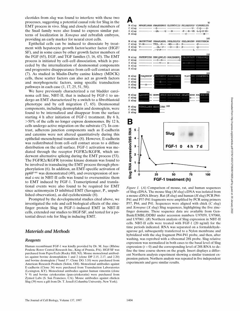
Figure 1. (A) Comparison of mouse, rat, and human sequencesof Slug cDNA. The mouse Slug (M slug) cDNA was isolated froma mouse cDNA library. Rat (R slug) and human (H slug) PCR P64-P41 and P57-P41 fragments were amplified by PCR using primersP57, P64, and P41. Sequences were aligned with chick (C slug)and Xenopus (X slug) Slug sequences, highlighting the five zinc-finger domains. These sequence data are available from Gen-Bank/EMBL/DDBJ under accession numbers U97059, U97060,and U97061. (B) Northern analysis of Slug expression in NBT-IIcells. NBT-II cells were treated with FGF-1 (20 ng/ml) for thetime periods indicated. RNA was separated on a formaldehyde-agarose gel, subsequently transferred to a Nylon membrane andhybridized with the slug fragment P64-P41 probe, and then, afterwashing, was reprobed with a ribosomal 28S probe. Slug relativeexpression was normalized in both cases to the basal level of Slugexpression (t = 0) and the corresponding level of 28S RNA to de-fine the time course shown on the graph. Insert displays a differ-ent Northern analysis experiment showing a similar transient ex-pression pattern. Northern analysis was repeated in five independentexperiments and gave similar results.
Dow
nloaded from http://rupress.org/jcb/article-pdf/137/6/1403/1270639/10829.pdf by guest on 30 January 2022

Savagner et al.
Slug Regulation of Desmosomes
1405
Cell Culture
The rat bladder carcinoma NBT-II cell line was initially obtained fromProf. Marc Mareel (University Hospital, Ghent, Belgium). Cells were cul-tured in DME supplemented with glutamine, antibiotics, and 10% heat-inactivated FCS as previously described (7). Human keratinocyte primaryculture YF29 (newborn foreskin, fourth passage) was grown by A. Rochatand Y. Barrandon (Ecole Normale Supérieure, Paris, France) in DMEsupplemented with glutamine, antibiotics, and 10% heat-inactivated FCS.
RNA Preparation and PCR
Total RNA was extracted from human keratinocytes and NBT-II cellsthat were growth-arrested by serum deprivation and incubated in serum-containing medium for 6 h by the acid guanidinium thiocyanate-phenolmethod (11). Synthesis of cDNA was performed using AMV reverse tran-scriptase as specified by the supplier (Stratagene, La Jolla, CA). The totalvolume for each reaction was 100
m
l for 4
m
g of RNA. PCR reactions in-cluded 5
m
l of cDNA as template, 0.4
m
g of each specific primer, 0.2 mMdNTPs, and 2 U of Taq polymerase in a buffer supplied with the enzyme(Perkin-Elmer Corp., Norwalk, CT). Annealing temperature was 46
8
C.
Primers
Specific primers were designed based on a published chicken Slug se-quence (46) and mouse Snail sequence (45, 55). The primer sequenceswere P41: CTTCGGATGTGCATCTTCAGAC (Mouse Snail bp 744–764), P57: AT(ACT)GA(AG)GC(ACGT)GA(AG)AA(AG)TT(TC)CA(GA)TG (chicken Slug, amino acids 125–131), P64: AAGCCCAACTAT-AGCGAGCTG (mouse Snail bp 46–66), P85: CTTGTAGTCGGATC-
Figure 2. Slug transient transfections. NBT-II cells were cotransfected with vectors containing mouse full-length cDNA for Slug and atruncated (IL-2R) cDNA used as a transfection marker. Alternatively, a control vector pCR 3 (Control) was cotransfected with IL-2RcDNA. After 48 h, cells were fixed and processed for double immunofluorescence using antibodies against IL-2R and antidesmoplakin(A); note that cells expressing the IL-2R transfection marker are positive for desmoplakin in control transfectants and negative in slug-transfected cells. Cells expressing desmoplakin at cell–cell boundaries were counted as desmosome-positive (DP+), i.e., fully epithelial.More than 100 cells expressing IL-2R were analyzed at the same time point for desmosome expression. The number of desmosome-pos-itive cells displaying IL-2R labeling was normalized to the total number of cells expressing IL-2R (B). Bar, 9 mm.
CGTGTGCCACACAGCAGCCAGA (Mouse Slug 3
9
end), and P86:TCGAATTCGCGGTCGCTGTC.
Cloning and Sequencing
The PCR fragments P57-P41 and P64-P41 obtained from human kerati-nocyte cDNA, NBT-II cell cDNA, and a mouse cDNA library derivedfrom the putative neural crest cell line NC15 (a gift from Dr. KarenBrown, National Institute of Dental Research) were cloned in PCR II (In-vitrogen, San Diego, CA) and sequenced using an automated DNA se-quencer. The PCR fragment S64-41, obtained from a mouse cDNA li-brary, was cloned in the PCR II vector and used as a probe for screening amouse cDNA library (kindly provided by Dr. Karen Brown) that wasbased on
l
ZAP II (Stratagene). A clone was excised in pBluescript andsequenced in both directions using an automated DNA sequencer andvarious primers derived from successive sequencing. Slug expression vec-tors were prepared by cloning the mouse PCR product P86-P85 contain-ing the full-length mouse
slug
sequence in a PCR 3 vector (Invitrogen).The interleukin-2 receptor (IL-2R) expression vector was described previ-ously (36). Sequences were analyzed using the Wisconsin package (Genet-ics Computer Group, Madison, WI) and Mac Vector (Oxford MolecularGroup, Campbell, CA) software.
Transfection and Cell Selection
Expression vectors containing full-length mouse
slug
sequence were trans-fected into NBT-II cells plated in 16-well slide chambers (Nunc) with 0.25
m
llipofectamine (Life Technologies, Grand Island, NY). For transient trans-fections in NBT-II cells, 0.1
m
g of IL-2R construct was added during thetransfection to serve as a marker for transfection (20). Wells were washedseveral times after 6 h of incubation. NBT-II cells were cultured for an-
Dow
nloaded from http://rupress.org/jcb/article-pdf/137/6/1403/1270639/10829.pdf by guest on 30 January 2022

The Journal of Cell Biology, Volume 137, 1997 1406
other 48 h before fixation. For stable transfections, NBT-II cells were in-cubated with DNA/lipofectamine for 15 h, washed repeatedly, and thentreated with 400
m
g/ml active G418 (Life Technologies) for 7 d. Survivingcells were cloned individually by limiting dilution.
Immunofluorescence Microscopy
Cells cultured on 16-well multiwell glass slides (Nunc) were treated withFGF-1 for 2 d after cell plating. Heparin (Choay Laboratories, Paris,France) at 10
m
g/ml was added to the culture medium to stabilize the bio-logical activity of FGF-1 (22). Growth factors were renewed every otherday for long term activation. NBT-II cells were processed for immunocy-tochemistry as described previously (8).
Electron Microscopy
Cells were cultured for 72 h before fixation in 2% glutaraldehyde, 2%paraformaldehyde in cacodylate buffer. After postfixation in 1% osmiumtetroxyde, they were progressively dehydrated in ethanol and then liftedfrom the culture dishes before embedding in Epon resin. Ultrathin sec-tions were stained with 2% uranyl acetate and 1% lead citrate before visu-alization with a transmission electron microscope (JEOL; Tokyo, Japan).
Nucleus-to-Nucleus Distance
High-magnification photographs were measured with a ruler to find thedistance from the geometrical center of nuclei from adjacent cells. Morethan 50 measurements were done in each case. Adjacent cells were chosenrandomly, and cells located at the edges of aggregates were excluded.
Motility Assays
Motility assays were performed using cells seeded on plastic tissue culturedishes and cultured for 2 d in standard medium. Dishes were covered witha glass slide, and time-lapse video cinematography was performed over 15 hafter addition of growth factor. The average speed of locomotion was cal-culated from more than 30 distinct cell tracks chosen randomly. For eachcell, total cell migration distance during the time of migration (5–6 h) wasdetermined.
Northern Analysis
After separation on a 1.2% formaldehyde–containing agarose gel, RNAswere transferred overnight to a Nylon membrane (Nytran; Schleicher andSchuell, Keene, NH). The P64-P41
slug
probe was excised from PCR IIconstructs and a ribosomal human 28S probe was excised from HHCD07(American Type Culture Collection, Rockville, MD). Both probes were[
32
P]dCTP-labeled using a Primer II kit (Stratagene) and then incubatedovernight at 42
8
C with filters in hybridization solution (5
3
SSC, 5
3
Den-hardt’s, 50% formamide, 10% dextran sulfate, 50
m
g/ml salmon spermDNA, and 0.4% SDS). After washing twice for 10 min at 42
8
C in 0.25
3
SSC and 0.1% SDS, filters were dried and exposed to Hyperfilm-MP(Amersham Corp., Arlington Heights, IL) for 2 d. Quantitation was per-formed using a PhosphorImager (Molecular Dynamics, Sunnyvale, CA).
Results
Cloning and Sequence Analysis of MammalianSlug cDNA
We cloned by PCR several Slug cDNA fragments usingprimers derived from the chick sequence. We used thefragment P64-P41, amplified with primer set 64/41 to screen
Figure 3. slug
-transfected cells display a modified morphologicalphenotype. After selection and cloning, stable transfectant cloneswere cultured under standard conditions. (
A
) Phenotype expres-sion of stable
slug
transfectants, comparing sense, antisense, andcontrol clones. 37 clones were analyzed by phase-contrast micros-copy for their phenotype and classified according to the appear-ance of cell–cell junctions as tight, intermediate, and loose. (
B
)
Phase-contrast micrographs of sense (S6 and S4) and antisense(AS2.8 and AS2.10)
slug
transfectants were taken after 48–72 hof cell culture. The morphology of the cell junctions in S4 and S6were classified as loose, whereas the other four were classified astight for quantification for
A.
Bar, 10
m
m.
Dow
nloaded from http://rupress.org/jcb/article-pdf/137/6/1403/1270639/10829.pdf by guest on 30 January 2022

Savagner et al.
Slug Regulation of Desmosomes
1407
gether with a Neomycin resistance gene to produce senseand antisense transfectants, as well as transfecting with ex-pression vector alone. Cells were selected in the presenceof Geneticin (G 418) and cloned individually by limitingdilution. A total of 37 independent transfectant clones in-cluding 9 controls, 13 antisense clones, and 15 sense cloneswere established and analyzed.
slug
(sense and antisense)–transfected cells, as well as control-transfected cells, dis-played cell–cell associations classified as tight, intermedi-ate, or loose according to their morphology (Fig. 3
A
).“Tight” cell contacts were defined in this system as sitesvisualized by phase-contrast microscopy as simple phaserefractile (bright) lines, outlining flat apical areas of epi-thelioid cells. “Loose” cell contacts showed a much morecomplex pattern at cell–cell contact areas, with irregularcell–cell boundaries that often showed phase-refractilezones at flattening surfaces of cells that were internal tophase-dark zones at flat sites of variable cell–cell contact.When the cell population phenotype was heterogeneous,cells were considered to display an intermediate morphol-ogy. In subconfluent cultures, the normal pattern of NBT-IIcell distribution, in which clusters of cells organize them-selves into closely packed epithelial islands surrounded byempty spaces, was replaced by a modified phenotype in
slug
-transfected cells, in which cell–cell associations ap-peared much looser. Two sense clones, S4 and S6, areshown in Fig. 3
B.
They show a modification of cell–cellcontact areas that is particularly apparent at higher magni-fication (Fig. 4
A
), with a wider cell–cell junction area orseparations at cell junctions that disrupted the more cuboi-dal epithelial pattern of parental NBT-II cells. The pro-portion of the cell periphery involved in contact with an-other cell was substantially decreased. Cells located withinthe interior of groups of cells had
.
90% of their periph-eral membrane in contact with an adjacent cell, whereascells transfected with the sense
slug
construct had only46% of their membrane in contact, as estimated from pho-tographs. As a consequence, there were spaces of varyingsize in the monolayer even when adjacent cells were incontact (Fig. 4
A
).To study in detail the nature of the cell–cell contact ar-
eas, S6 and untransfected NBT-II cells were examined inelectron microscopy. NBT-II cells were found to exhibitcharacteristic morphological features of epithelial cellssuch as desmosomes (Fig. 4
B
) and putative adherens junc-tions (data not shown). They displayed numerous interdig-itating processes, similar to those reported in bladder epi-thelial cells in vivo (9). In contrast, the Slug sense clonecDNA S6 showed flattened and spread cells devoid of des-mosomes, still in contact with each other through muchmore limited lateral contact areas (Fig. 4
B
). Interdigitat-ing processes similar to those in untransfected cells werefound, as well as putative cell adherens junctions (data notshown).
Phenotype modulation was also characterized by an in-creased spreading apart of the transfected cells on the sub-strate. To quantify this alteration, we calculated the aver-age nucleus-to-nucleus distance in the stable transfectants.We found that sense clone S6 displayed twice the nucleus-to-nucleus distance as compared to the parental NBT-IIcells or to the antisense-transfected cells (Fig. 4
C
). On theother hand, antisense-transfected clones such as AS2.8
a mouse cDNA library. We obtained a full-length mouseSlug cDNA clone; its deduced amino acid sequence isshown in Fig. 1. This clone has 92% amino-acid identitywith the chicken Slug sequence. It includes the “trademark”MPRSFLV K/R K amino-terminal sequence characteristicof the Snail family (42, 46, 61). As in chicken Slug, it con-tains four classic CXXC (12 X) HXXXH zinc-fingers anda fifth one representing a structural variant apparently spe-cific for the Snail family: CXXXC (12 X) HXXXXC (61). Asnoted for
Xenopus
, mouse Slug is very similar (90% iden-tity) to the mouse Snail protein in the carboxy-terminalhalf of the molecule after amino acid 160. This region in-cludes the four zinc-finger domains expressed by the mouseSnail gene.
Amplification products derived from the 57/41 or 64/41primer sets were also cloned and sequenced from humankeratinocytes and rat bladder carcinoma cells (NBT-II).Interestingly, the mouse amino acid sequence in this re-gion, covering the first two zinc-finger domains, was 100%identical to the sequence of the human Slug clone and98% identical to the sequence of the rat clone (Fig. 1).
Slug mRNA Expression Rises Early afterFGF-1 Treatment
RNA was extracted from NBT-II cells at various times af-ter EMT-triggered FGF-1 treatment. The
slug
fragmentS64-41 was labeled with
32
P and used for Northern blot anal-ysis. As shown in Fig. 1, Slug RNA was induced as early as3 h after FGF-1 treatment. Expression reached a peak at 6 hand then decreased after 15 h. Under these conditions,NBT-II cells cultured with FGF-1 for more than 6 d nolonger expressed Slug mRNA.
Slug-transfected Cells Display a Modified Phenotype
The full-length cDNA encoding mouse
slug
was insertedin both orientations in the expression vector PCR 3 underthe control of a cytomegalovirus promoter. Constructs werecotransfected transiently into NBT-II cells using lipofecta-mine together with an expression vector encoding a trun-cated IL-2R described as a marker for DNA uptake andexpression (20, 36). Desmoplakin immunolabeling wasused to localize desmosomes. As described previously (65),untreated parental NBT-II cells have a localization of des-moplakin at punctate sites corresponding to desmosomes,which is restricted to cell–cell contact areas. However, thispattern disappears when cells are induced to undergo EMTwith FGF-1, and cells treated for
.
12 h with FGF-1 are de-void of desmosomes. To examine for similar modulation inthe transiently transfected cells, we counted 100–200 cellsdisplaying IL-2R labeling for each condition. We thencalculated the proportion of desmoplakin-positive cellsamong the cells expressing the IL-2R. Cells cotransfectedwith Slug cDNA were compared to cells cotransfectedwith the control vector PCR 3 (Fig. 2). The proportion ofcells displaying desmosomes after
slug
transfection wasless than half that of control-transfected cells, and this dif-ference was highly significant (
P
,
0.004).Since transient transfections indicated a Slug-mediated
modulation of cell phenotype, we established clones of sta-ble transfectants with Slug cDNA in either orientation to-
Dow
nloaded from http://rupress.org/jcb/article-pdf/137/6/1403/1270639/10829.pdf by guest on 30 January 2022

The Journal of Cell Biology, Volume 137, 1997 1408
and AS2.10 displayed the typical cobblestone epithelialphenotype expressed by parental NBT-II cells (Fig. 3
B
).To check mRNA expression in stable transfectants, North-
ern analysis was performed using RNA isolated from seven
different transfected clones including S3, S4, AS2.8, andAS2.10 clones (Fig. 5). As quantified with a PhosphorIm-ager, transfected clones expressed 10 to 50 times moreSlug RNA than parental NBT-II cells (data not shown).
Dow
nloaded from http://rupress.org/jcb/article-pdf/137/6/1403/1270639/10829.pdf by guest on 30 January 2022

Savagner et al.
Slug Regulation of Desmosomes
1409
Expression of Slug Protein in Parental and Transfected NBT-II Cells
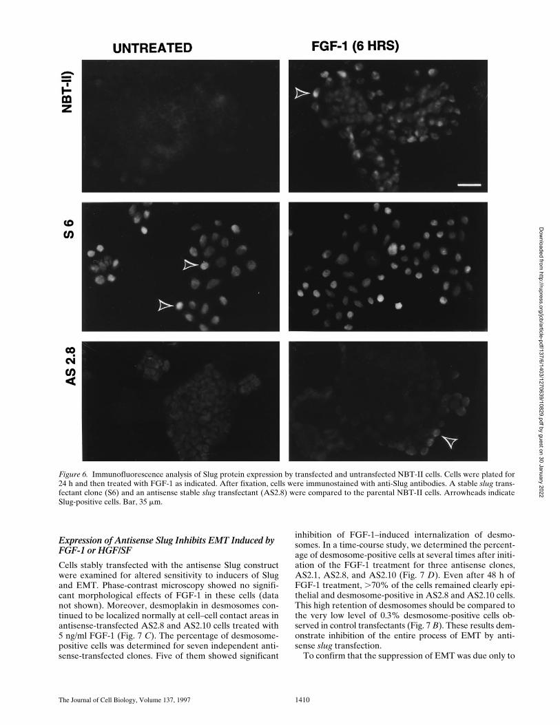
We compared the levels of Slug protein in untransfectedand transfected NBT-II cells by immunofluorescence us-ing anti–chicken Slug antibodies (Fig. 6). The level of Slugprotein expression was very low in parental epithelialNBT-II cells. It was restricted to rare cells that were usu-ally located at the periphery of cell aggregates. After 2 h ofFGF-1 treatment, a substantial subpopulation of cells thatwas also mostly located at the edge of cell aggregates wasclearly positive. This population became predominant af-ter 6 h of FGF-1 treatment (Fig. 6). However, after 48 h ofFGF-1 treatment and full conversion to a mesenchymalphenotype, Slug protein expression was downregulated.
In contrast, sense-transfected cells expressed Slug be-
fore any FGF-1 treatment and maintained it after 6 h oftreatment. Antisense-transfected cells did not express Slugbefore the FGF-1 treatment, and only a few cells locatedat the periphery of cell aggregates expressed Slug even af-ter 6 h of FGF-1 treatment (Fig. 6).
Immunofluorescence Shows Disruption of Desmosomes in slug-transfected Cells
To analyze potential modulation of desmosomal cell–celladhesion structures, we localized desmoplakin by immuno-fluorescence (Fig. 7). To quantitate this modulation, wecalculated the percentage of cells displaying desmoplakinlocalization characteristic of the epithelial phenotype (des-mosome-positive) compared to the total number of cells.This ratio was calculated for untreated cells or at differenttimes after FGF-1 treatment for six independent
slug-
transfected clones, and the results are reported in Fig. 7
B.
Four of these clones, including clones S4 and S6 (Fig. 7
A
),had a low to null percentage of desmosome-positive cellsbefore any FGF-1 treatment. Cytoplasmic immunoreactiv-ity was found with antidesmoplakin antibodies, suggestinga cytoplasmic desmoplakin pool in S4 and S6 cells. Thisimmunoreactivity was no longer found after 48 h of FGF-1treatment (Fig. 7
A
for clone S4; data not shown for S6), aspublished previously for the parental NBT-II cells (7). Incomparison, control clones (C1, C2, and C3) had
.
95%desmosome-positive cells before treatment, a ratio similarto that for the parental NBT-II cells. In all cases, immu-noreactivity was no longer present after prolonged FGF-1treatment, as published previously for the parental NBT-IIcells (7).
Figure 4. Slug transfectant clone exhibits looser cell contact phe-notype. (A) NBT-II and S6 cells were observed at higher magnifi-cation. Arrowheads indicate cell edges, which are often free forS6 cells compared to involvement in tight cell–cell contacts forcontrol NBT-II cells. (B) Electron micrographs of NBT-II cellsand S6 cells. NBT-II cells (a, c, and d) or S6 cells (b) were platedfor 48 h before fixation and processing for electron microscopy.NBT-II cells displayed an epithelial phenotype characterized bynumerous desmosomes (arrow in a, higher magnifications ofother examples in c and d). Conversely, S6 cells appeared flatter,more spread, and less epithelial. No desmosomes could be found.(C) slug-transfected S6 cells are more widely spaced in subconflu-ent cultures than parental NBT-II cells. Cell-to-cell spacing wasevaluated by the average distance between the geometrical cen-ters of nuclei of adjacent cells. Only cells in contact with eachother were used for the calculation. More than 50 distances weremeasured in each case to calculate the average nucleus-to-nucleus distance. Bars: (A) 10 mm; (B, a and b ) 2 mm; (B, c) 1mm; (B, d) 0.2 mm.
Figure 5. Northern analysis of Slug-mRNA expression by stabletransfectants. slug-transfected cells expressed large amounts ofSlug RNA. Northern analysis using slug transfectant clones(sense and antisense) were probed as described for Fig. 2. Thedouble-stranded DNA probe allowed detection of both sense andantisense RNA in the transfectants; Ct 3 was transfected withvector alone.
Dow
nloaded from http://rupress.org/jcb/article-pdf/137/6/1403/1270639/10829.pdf by guest on 30 January 2022
The Journal of Cell Biology, Volume 137, 1997 1410
Expression of Antisense Slug Inhibits EMT Induced by FGF-1 or HGF/SF
Cells stably transfected with the antisense Slug constructwere examined for altered sensitivity to inducers of Slugand EMT. Phase-contrast microscopy showed no signifi-cant morphological effects of FGF-1 in these cells (datanot shown). Moreover, desmoplakin in desmosomes con-tinued to be localized normally at cell–cell contact areas inantisense-transfected AS2.8 and AS2.10 cells treated with5 ng/ml FGF-1 (Fig. 7 C). The percentage of desmosome-positive cells was determined for seven independent anti-sense-transfected clones. Five of them showed significant
inhibition of FGF-1–induced internalization of desmo-somes. In a time-course study, we determined the percent-age of desmosome-positive cells at several times after initi-ation of the FGF-1 treatment for three antisense clones,AS2.1, AS2.8, and AS2.10 (Fig. 7 D). Even after 48 h ofFGF-1 treatment, .70% of the cells remained clearly epi-thelial and desmosome-positive in AS2.8 and AS2.10 cells.This high retention of desmosomes should be compared tothe very low level of 0.3% desmosome-positive cells ob-served in control transfectants (Fig. 7 B). These results dem-onstrate inhibition of the entire process of EMT by anti-sense slug transfection.
To confirm that the suppression of EMT was due only to
Figure 6. Immunofluorescence analysis of Slug protein expression by transfected and untransfected NBT-II cells. Cells were plated for24 h and then treated with FGF-1 as indicated. After fixation, cells were immunostained with anti-Slug antibodies. A stable slug trans-fectant clone (S6) and an antisense stable slug transfectant (AS2.8) were compared to the parental NBT-II cells. Arrowheads indicateSlug-positive cells. Bar, 35 mm.
Dow
nloaded from http://rupress.org/jcb/article-pdf/137/6/1403/1270639/10829.pdf by guest on 30 January 2022

Savagner et al. Slug Regulation of Desmosomes 1411
the antisense Slug, the antisense mouse Slug stable-trans-fectant AS2.8 cell line was cotransfected with mouse SlugcDNA and marker-truncated IL-2R in a “rescue transfec-tion” attempt. After this transient transfection and 48 h ofculture, cells expressing IL-2R (.100) were examined fordesmosome expression as previously described. 85 6 6%of cells cotransfected with the PCR 3 vector alone ex-pressed desmosomes, a proportion similar to that in con-trol AS2.8 cells, whereas only 26 6 14% of the AS2.8 cellscotransfected with sense Slug cDNA expressed desmo-somes, showing a highly significant (P , 0.001) decrease inthe number of cells expressing desmosomes (Fig. 8).
Finally, we examined for the ability of antisense Slug toblock EMT induced by a potent scatter factor, HGF/SF,which was previously described to induce EMT in NBT-IIcells as well as in a variety of other epithelial cells (2, 18,51, 58). Interestingly, the two clones tested, AS2.8 andAS2.10, also resisted HGF/SF action. Double immunoflu-orescence using antibodies against desmoplakin and cy-tokeratin clearly indicated a persistence of desmosomes inantisense-transfected cells with retention of cytokeratinfilament insertion into points of cell–cell contact rich indesmoplakin (Fig. 9 A). Quantitation of desmosome-posi-tive cells showed a pattern of resistance to dissociationsimilar to that obtained with FGF-1 (Fig. 9 B). At higherconcentrations of HGF/SF or of FGF-1, more cell dissocia-tion was observed, suggesting a competitive mechanism.
Modulation of Desmosomal Cadherins inslug-transfected Cells
We examined the distribution of two members of the cad-herin family involved in most cell–cell adhesion structures.We chose desmoglein, which is a cadherin found specifi-cally in desmosomes, and E-cadherin, a ubiquitous cad-herin also present in cell–cell adherens junctions. Desmo-glein was found to disappear from cell–cell contact areasvery similarly to desmoplakin in slug-transfected cells, asreported previously for NBT-II cells treated with FGF-1to induce EMT (8). In contrast, desmoglein localizationpersisted in antisense-transfected clone AS2.8, even afterFGF-1 treatment (Fig. 10 A). E-cadherin immunostainingwas previously described to persist in NBT-II cells under-going EMT, but it became relocalized diffusely over the cellsurface (8). We found a similar distribution in untreatedslug-transfected S4 cells, which showed a partial relocal-ization of E-cadherin to regions not involved in cell–cellcontacts (Fig. 10 B, arrowhead). FGF-1 treatment did notsignificantly change this localization, although E-cadherinimmunoreactivity appeared significantly decreased overallin association with a decrease in total cell surface area in-volved in cell–cell contacts. On the other hand, antisensestable transfectant AS2.8 showed little change in totalstaining compared with untreated NBT-II cells. However,some E-cadherin expression could also be detected in re-gions not involved in cell–cell contact areas (Fig. 10 B).
Slug-transfected Clones Continue to Express Cytokeratin, but Its Organization Is Altered
Cytokeratin protein levels are progressively downregu-lated in NBT-II cells treated with FGF-1 for several days(8). Since desmosomes are linked to a cytokeratin network
in epithelial cells, we studied cytokeratin localization inslug transfectants. Both sense and antisense S4 and AS2.8cells continued to express a dense cytokeratin mesh, indi-cating that this later step in EMT is not triggered by Slug.However, in accord with the absence of desmoplakin anddesmoglein in S4 cells, these slug-transfected cells failed todisplay the typical pattern of cell–cell junctional cytokera-tin filaments anchored tightly in desmosomes as was ob-served in parental NBT-II cells as well as in AS2.8 cells(Figs. 9 and 11). Epithelial NBT-II cells do not express vi-mentin intermediate filaments until they are treated forseveral days with FGF-1 (7). Similarly, ,5% of the slug-transfected S4 cells were found to express vimentin in ab-sence of FGF-1 treatment. This number increased progres-sively after FGF-1 treatment and exceeded 95% after 3–4 din culture, similarly to the alteration observed with paren-tal NBT-II cells. In contrast, very few (,1%) of the anti-sense slug–transfected AS2.8 cells expressed vimentin,even after FGF-1 treatment. Two of the latter rare cellsare shown by double staining for cytokeratin and vimentinin Fig. 11 A and were found to express simultaneously cy-tokeratin and vimentin filaments. In addition, they clearlyexpress typical cell–cell connecting cytokeratin filamentsindicative of functional desmosomes, even though these cellsare also synthesizing vimentin.
Video Time-Lapse Analysis of TransfectantCell Migration
An important characteristic of NBT-II cells expressing vi-mentin filaments associated with the complete mesenchy-mal phenotype achieved after FGF-1 treatment is the ap-pearance of a motile phenotype that can be quantified bytime-lapse video microscopy (65). We quantified the mo-tility of slug transfectants plated on plastic substrates byanalyzing video recordings with or without FGF treatment(Fig. 11 B). Interestingly, even in the absence of desmo-somes, S4 cells did not express a motile phenotype untiltreated with FGF-1. Only after treatment did they beginmigrating similarly to the parental NBT-II cells treatedwith FGF-1. On the other hand, with or without priorFGF-1 treatment, antisense slug–transfected AS2.8 or 2.10cells did not express any significant motility, in accord withthe persistence of desmosomes and cell–cell contacts.
DiscussionIn this study, we tested directly whether the newly de-scribed zinc-finger protein Slug plays a central role in reg-ulating one or more steps in the process of EMT, usingboth gain-of-function and loss-of-function approaches. Wecloned and sequenced a mouse full-length cDNA encod-ing slug, which has known homologues in chicken and Xe-nopus. We also cloned fragments of human and rat slug;sequences from all species were found to be highly con-served. We transfected full-length slug cDNA into NBT-IIcells to generate transient as well as stable transfectants fortesting its function. Slug induced a striking total dissociationof desmosomes in NBT-II epithelial cells incorporatingthe DNA. It also induced increased spreading and separa-tion of these cells, with a doubled average nucleus-to-nucleusdistance and a marked decrease in the proportion of
Dow
nloaded from http://rupress.org/jcb/article-pdf/137/6/1403/1270639/10829.pdf by guest on 30 January 2022

The Journal of Cell Biology, Volume 137, 1997 1412
Figure 7. Desmosome ex-pression by slug-transfectedclones. (A) slug-transfectedNBT-II clones do not ex-press desmosomal structures.Cells were plated for 24 hand then treated with FGF-1(5 ng/ml) for 48 h in the caseof NBT-II cells and S4 cells,and 6 h for S6 cells. After fix-ation, cells were immuno-stained with antidesmoplakinantibodies. Two clones ofstable slug transfectants, S4and S6, were compared tothe parental NBT-II cells.(B) Proportion of epithelialcells among FGF-1–treatedand –untreated slug-trans-fected NBT-II clones. Cellsexpressing desmoplakin at
cell–cell boundaries (DP+) were counted after 0 (nt), 3, 6, or 48 h of treatment with FGF-1. slug-transfected clones (S9, S6, S3, and S4)were compared to control clones C1, C2, and C3 transfected with vector alone. (C) Antisense slug–transfected NBT-II clones resistFGF-1–induced desmosome dissociation. Cells were plated for 24 h and then treated with FGF-1 (5 ng/ml for 24 h) as indicated. Afterfixation, cells were incubated with antidesmoplakin antibodies. Two clones of stable antisense slug transfectants, AS2.8 and AS2.10,were processed for desmoplakin immunolocalization and photographed at two different magnifications, with or without prior FGF-1treatment, as indicated. (D) Proportion of epithelial cells among FGF-1–treated and –untreated antisense slug–transfected NBT-IIclones. Cells expressing desmoplakin at cell–cell boundaries (DP+) were counted after 0 (nt), 3, 6, or 48 h of treatment with FGF-1, asindicated. Antisense slug–transfected clones (AS 2.1, AS2.8, and AS2.10) should be compared to control clones C1, C2, and C3 trans-fected with vector alone in B. Bars, 10 mm.
Dow
nloaded from http://rupress.org/jcb/article-pdf/137/6/1403/1270639/10829.pdf by guest on 30 January 2022

Savagner et al. Slug Regulation of Desmosomes 1413
membrane involved in cell–cell contact. Conversely, wefound that NBT-II cells transfected with antisense SlugcDNA resisted the actions of both FGF-1 or HGF/SF forinducing desmosomal dispersal and for cell scattering inthe EMT process, i.e., antisense slug interferes with a nec-essary Slug expression step in EMT. The specificity of theantisense slug activity was supported by a “rescue” slugtransfection experiment, in which transient overexpressionof a slug sense construct permitted dissociation of stable
antisense transfectants. We conclude from these and otherexperiments that Slug expression is necessary and suffi-cient for the first key steps of EMT involving desmosomaldissociation and cell separation, as determined for FGF-induced EMT in NBT-II cells. The resistance of antisense-transfected cells to EMT mediated by FGF-1 as well asHGF/SF suggests that it is a necessary step on which sev-eral pathways leading to cell–cell dissociation may converge,initiated by distinct factors such as FGFs or HGF/SF.
Dow
nloaded from http://rupress.org/jcb/article-pdf/137/6/1403/1270639/10829.pdf by guest on 30 January 2022

The Journal of Cell Biology, Volume 137, 1997 1414
Sequence and Expression Homologies between Chicken, Human, Rat, and Mouse Slug
The full-length sequence of mouse Slug shows 92% aminoacid sequence identity to the chicken Slug sequence and89% identity to Xenopus Slug. Interestingly, this homol-ogy is much stronger than the homology between mouseand Xenopus Snail proteins (initial members of the Snailtranscription factor family to which Slug belongs), par-tially reflecting the disappearance of a zinc-finger motif inthe mouse Snail protein. In the DNA segments we clonedand sequenced from other species, mouse Slug showed100% identity to human Slug in the deduced amino acidsequence and 98% identity to rat Slug. These data, addedto the similarity of distribution patterns between chickenand Xenopus Slug protein in embryos (42, 46), suggest aconserved function for Slug in vertebrates. Snail is knownto be a transcriptional repressor in Drosophila, probablyacting by direct competition with transcriptional activators(23, 31). A DNA-binding site motif has been defined forthe Drosophila Snail (41), apparently shared by the re-lated escargot gene (14). Escargot was recently describedto play a key role during Drosophila trachea morphogene-sis (60).
It is likely that Slug is also a transcription factor thatmay recognize specific sites in gene regulatory regions.Snail probably downregulates its own expression througha negative regulatory loop mechanism (41). This could alsobe the case for Slug, which could explain the transient pat-tern of expression we observed in NBT-II cells. This tran-sient expression pattern also suggests that Slug initiates agenetic program subsequently dependent on a distinctFGF-1–induced activator. Direct involvement of distincttranscription factors like Ets-1 (66), E1a (21), and c-fos (48)in the induction of cell phenotype modulation, includingEMT phases, has been suggested in several cases. How-ever, these factors do not show functional specificity invivo comparable to that which we describe in the presentstudy of Slug with NBT-II cells.
Slug Targets Desmosomal Proteins
Transient and stable slug transfectants showed a charac-teristic morphological separation of normally tightly ap-posed plasma membranes at cell–cell boundaries, suggest-ing that Slug targets some critical cell–cell adhesionsystem. This separation was accompanied by an increasedspreading of the cells well characterized by electron mi-croscopy, resulting in a doubled nucleus-to-nucleus dis-tance. We therefore compared its effects on desmosomaland cadherin-based adhesion systems. Transient and sta-ble transfectants with slug showed a disappearance of thedesmosomes characteristic of epithelial NBT-II cells. Thiseffect was also found by transient transfections of MDCKcells, a different epithelial cell line normally expressing nu-merous desmosomes at the cell–cell junctions; althoughMDCK cells undergo an EMT-like response to HGF but notFGF, Slug can also lead to losses of desmosomes in thiscell line (Savagner, P., unpublished data). Desmosome dis-sociation was observed in most stable transfectant NBT-IIclones using three markers. First, desmoplakin and desmo-glein, two essential desmosome components, disappearedfrom cell–cell contact areas. The third desmosomal markerwas the focal insertion of cytokeratin filaments, which an-chored the desmosomes to the cytokeratin meshwork. Thestable transfectant line S4 did express a cytokeratin filamentnetwork comparable to parental cells as observed by im-munofluorescence, but they did not express any membrane-anchoring cytokeratin filaments. This altered cytokeratinlocalization pattern was comparable to that observed inepithelial cells transfected with a dominant-negative des-moplakin polypeptide (4). In accord with the immunofluo-rescence results, Western blot analysis confirmed the pres-ence of desmoglein in untreated Slug transfectants, at alevel similar to the untransfected NBT-II cells (Savagner,P., unpublished observation). The three markers weredownregulated after several days of FGF-1 treatment, as forthe untransfected NBT-II cells (Results and Savagner, P.,unpublished observation). Conversely, the three desmo-somal markers were positive in slug antisense–transfectedcells, even after FGF-1 or HGF/SF treatment, demonstrat-ing the continued presence of desmosomes in these cells.Other cell adhesion structures were also investigated. Ad-herens junction components including E-cadherin andb-catenin (Savagner, P., unpublished observation) werefound to be modestly relocalized but still present in S4cells. They were present as normal in cell–cell contact ar-eas, but they were also present sometimes in membraneregions not involved in cell–cell contacts. This relocaliza-tion was mostly present in cells treated with FGF-1. Inter-estingly, a slug antisense stable transfectant exhibited thesame relocalization after FGF-1 treatment in spite of thepersistence of the desmosomes. This relocalization wasvery similar to that previously described for E-cadherin onFGF-1–treated NBT-II cells (8). The mechanism of this re-localization of cadherin membrane and intracytoplasmicadhesion molecules and its functional significance are notclear. NBT-II cells were found previously (8) to aggregatesimilarly via Ca2+- and E-cadherin–dependent mechanismswith or without FGF-1 treatment, suggesting these com-plexes were indeed functional in spite of the motility of themesenchymal NBT-II cells. However, the strength of cell–
Figure 8. Antisense slug stabletransfectants can be reverted bytransient rescue transfection.Antisense slug stable transfec-tant AS2.8 cells were cotrans-fected with Slug cDNA andtruncated IL-2R. After 48 h cul-ture, .100 cells expressing IL-2R were scored for the presenceof desmosomes, as determinedby desmoplakin immunolocaliza-tion. Cells cotransfected withPCR 3 alone (Control) werecompared with cells cotrans-fected with Slug cDNA.
Dow
nloaded from http://rupress.org/jcb/article-pdf/137/6/1403/1270639/10829.pdf by guest on 30 January 2022

Savagner et al. Slug Regulation of Desmosomes 1415
Figure 9. (A) Antisense slug–transfected cells also resist thedissociating activity of HGF/SF.Control cells (Ct 3) and anti-sense slug–transfected AS2.8cells were plated for 24 h andthen treated with HGF/SF at 2ng/ml for 24 h before fixationand double labeling with anti-bodies against desmoplakin orcytokeratin as indicated. (B)Proportion of desmosome-posi-tive (DP+) cells among theAS2.8 cells after HGF/SF treat-ment. Cells were processed fordesmoplakin immunofluores-cence as described above. Des-mosome-positive (DP+) cellswere counted after 3, 6 or 48 h oftreatment with HGF/SF. Anti-sense slug-transfected clones(AS2.8 and AS2.10) can be com-pared to control clone Ct 3,which was transfected with thepCR 3 vector.
cell adhesion might be reduced after such relocalization asin v-src–transfected MDCK cells (59), which would ac-count for the dominant phenotype of Slug-induced cellshape change and cell separation even though componentsof the E-cadherin system are still present.
Interestingly, this relocalization was not described inepithelial cells transfected with a dominant-negative des-
moplakin polypeptide (4), further suggesting that the des-mosomes are not the only target for Slug.
Slug Induces the First Phase in the Process of EMT
Our observations suggest that Slug induction triggers thesteps of desmosomal disruption, cell spreading, and partial
Dow
nloaded from http://rupress.org/jcb/article-pdf/137/6/1403/1270639/10829.pdf by guest on 30 January 2022

The Journal of Cell Biology, Volume 137, 1997 1416
Dow
nloaded from http://rupress.org/jcb/article-pdf/137/6/1403/1270639/10829.pdf by guest on 30 January 2022

Savagner et al. Slug Regulation of Desmosomes 1417
Figure 11. Other processes involved in EMT. (A) slug–trans-fected cells display a modified cytokeratin network and requireFGF-1 treatment to undergo migration. Slug transfectant cloneS4 and antisense clone AS2.8 were processed for immunofluores-cence as described above after 24 h of culture. In addition, AS2.8was treated for 48 h with HGF/SF. Cells were labeled with anti-bodies against cytokeratin or against vimentin as indicated. TheAS2.8 cells were double-labeled, and they generally showed littleor no reactivity with vimentin antibody, even after HGF/SF treat-ment. However, shown here is an unusual example of simulta-neous expression by two cells of cytokeratin intermediate fila-ments (including desmosome-connecting filaments) and vimentinintermediate filaments. (B) Because initiation of motility oftenaccompanies EMT, NBT-II cell migration after slug transfectionwas evaluated by video time-lapse analysis. Cells were plated for24 h on culture dishes before a random field was followed for 5 hby time-lapse microscopy. More than 30 cells were individuallytracked in each case, and the average of the distances traversedby the cells over 5 h was plotted as the motility. Bars, 10 mm.
Figure 10. Cadherin expression by slug transfectants. Stable slug transfectants S4 (sense), AS2.8 (antisense), and parental NBT-II cellswere prepared for immunofluorescence using antibodies against desmoglein (A) or E-cadherin (B). Cells were treated with FGF-1 (5ng/ml) for 24 h as indicated. Bars, 10 mm.
Dow
nloaded from http://rupress.org/jcb/article-pdf/137/6/1403/1270639/10829.pdf by guest on 30 January 2022

The Journal of Cell Biology, Volume 137, 1997 1418
separation at cell–cell borders, which comprise the firstand necessary phase of the EMT process in our NBT-IIcell model, but that Slug cannot trigger the second phase,which includes the induction of cell motility, repression ofthe cytokeratin expression, and activation of vimentin ex-pression (Fig. 12). Our studies therefore provide the firstdemonstration of discrete mechanistic phases in EMT. It isconceivable that the overexpression and the persistence ofSlug expression in transfected clones might interfere witha Slug-triggered induction of this second phase. However,additional FGF treatment of such transfectants readily in-duces this second phase, demonstrating the absence of ablock. Therefore, we propose that a distinct mechanism isresponsible for the induction of motility in NBT-II cellstreated with FGF-1. Such a mechanism is in accord withprevious observations analyzing phases in endothelial cellconversion to mesenchymal cells that differentiate into thevalves and membranous portion of the atrial and ventricu-lar septum during heart morphogenesis (44, 47). Conse-quently, EMT appears to be a sequential set of events thatinvolves at least two necessary and distinct phases that canbe dissected by gain-of-function and loss-of-functiontransfection approaches. Our studies provide a mechanis-tic explanation of the results of previous in vivo experi-ments in which antisense oligonucleotides targeting Slugwere found to interfere with embryonic processes involv-ing EMT as a prelude to subsequent differentiation (46).Slug is essential for initiating growth factor–induced EMT,and its actions include induction of desmosome dissocia-tion and disruption of cell–cell adhesive junction.
We are most grateful to Yoshihiko Yamada (National Institute of DentalResearch) for the help with cloning of mouse slug. We acknowledge the
expert technical assistance of Bill Swain (National Institute of Dental Re-search) for the electron microscopy. The manuscript was improved by crit-ical reading by Brigitte Boyer, Jacqueline Jouanneau, and Ana-MariaVallés (Institut Curie).
This work was supported by the Centre National de la Recherche Sci-entifique, the Association pour la Recherche contre le Cancer (ARC6465), the Ligue Francaise contre le Cancer (National Committee andCommittee of Paris), the National Cancer Institute of the National Insti-tutes of Health (2R01 CA 49417-06), the Philippe Foundation and the Hu-man Frontier Science Program Organization, and the National Institute ofDental Research intramural program.
Received for publication 13 September 1996 and in revised form 18 April1997.
References
1. Behrens, J., K.M. Weidner, U.H. Frixen, J.H. Schipper, M. Sachs, N. Ar-akaki, Y. Daikuhara, and W. Birchmeier. 1991. The role of E-cadherinand scatter factor in tumor invasion and cell motility. EXS. 59:109–126.
2. Bellusci, S., G. Moens, J.P. Thiery, and J. Jouanneau. 1994. A scatter fac-tor-like factor is produced by a metastatic variant of a rat bladder carci-noma cell line. J. Cell Sci. 107:1277–1287.
3. Blay, J., and K.D. Brown. 1985. Epidermal growth factor promotes thechemotactic migration of cultured rat intestinal epithelial cells. J. Cell.Physiol. 124:107–112.
4. Bornslaeger, E.A., C.M. Corcoran, T.S. Stappenbeck, and K.J. Green.1996. Breaking the connection: displacement of the desmosomal plaqueprotein desmoplakin from cell–cell interfaces disrupts anchorage of inter-mediate filament bundles and alters intercellular junction assembly. J.Cell Biol. 134:985–1001.
5. Bouchey, D., W.S. Argraves, and C.D. Little. 1996. Fibulin-1, vitronectin,and fibronectin expression during avian cardiac valve and septa develop-ment. Anat. Rec. 244:540–551.
6. Boyer, B., and J.P. Thiery. 1993. Cyclic AMP distinguishes between twofunctions of acidic FGF in a rat bladder carcinoma cell line. J. Cell Biol.120:767–776.
7. Boyer, B., G.C. Tucker, A.M. Vallés, W.W. Franke, and J.P. Thiery. 1989.Rearrangements of desmosomal and cytoskeletal proteins during thetransition from epithelial to fibroblastoid organization in cultured ratbladder carcinoma cells. J. Cell Biol. 109:1495–1509.
8. Boyer, B., S. Dufour, and J.P. Thiery. 1992. E-cadherin expression duringthe acidic FGF-induced dispersion of a rat bladder carcinoma cell line.Exp. Cell Res. 201:347–357.
9. Carr, K.E., and P.G. Toner. 1982. Cell Structure. An Introduction to Bio-medical Electron Microscopy. Churchill Livingstone, Edinburgh. 388 pp.
10. Cate, R.L., R.J. Mattaliano, C. Hession, R. Tizard, N.M. Farber, A. Cheung,E.G. Ninfa, A.Z. Frey, D.J. Gash, and E.P. Chow. 1986. Isolation of thebovine and human genes for Mullerian inhibiting substance and expres-sion of the human gene in animal cells. Cell. 45:685–698.
11. Chomczynski, P., and N. Sacchi. 1987. Single-step method of RNA isola-tion by acid guanidinium thiocyanate-phenol-chloroform extraction.Anal. Biochem. 162:156–159.
12. Demlehner, M.P., S. Schafer, C. Grund, and W.W. Franke. 1995. Continualassembly of half-desmosomal structures in the absence of cell contactsand their frustrated endocytosis: a coordinated Sisyphus cycle. J. CellBiol. 131:745–760.
13. Fleming, T.P., Q. Javed, J. Collins, and M. Hay. 1993. Biogenesis of struc-tural intercellular junctions during cleavage in the mouse embryo. J. CellSci. 17(Suppl.):119–125.
14. Fuse, N., S. Hirose, and S. Hayashi. 1996. Determination of wing cell fateby the escargot and snail genes in Drosophila. Development (Camb.). 122:1059–1067.
15. Garrod, D.R. 1993. Desmosomes and hemidesmosomes. Curr. Opin. CellBiol. 5:30–40.
16. Geimer, P., and E.G. Bade. 1991. The epidermal growth factor-induced mi-gration of rat liver epithelial cells is associated with a transient inhibitionof DNA synthesis. J. Cell Sci. 100:349–355.
17. Gherardi, E., and M. Stoker. 1991. Hepatocyte growth factor–scatter fac-tor: mitogen, motogen, and met. Cancer Cells. 3:227–232.
18. Gherardi, E., M. Sharpe, K. Lane, A. Sirulnik, and M. Stoker. 1993. Hepa-tocyte growth factor/scatter factor (HGF/SF), the c-met receptor and thebehaviour of epithelial cells. Symp. Soc. Exp. Biol. 47:163–181.
19. Gilbert, S.F. 1994. Developmental Biology. Sinauer Associates, Sunder-land, MA. 894 pp.
20. Giordano, T., T. Howard, H.J. Coleman, K. Sakamoto, and B.H. Howard.1991. Isolation of a population of transiently transfected quiescent andsenescent cells by magnetic affinity cell sorting. Exp. Cell Res. 192:193–197.
21. Gopalakrishnan, S., and M.P. Quinlan. 1995. Modulation of E-cadherin lo-calization in cells expressing wild-type E1A 12S or hypertransformingmutants. Cell Growth Differ. 6:985–998.
22. Gospodarowicz, D., and J. Cheng. 1986. Heparin protects basic and acidic
Figure 12. EMT phases. Slug transfection induced the transitionfrom epithelial cells to an intermediate phenotype stage charac-terized by a drastic modulation of the cell–cell adhesion system,including the dissociation of desmosomes. A second stage is pos-tulated to occur when cells are treated with FGF-1, involving thereplacement of cytokeratin intermediate filaments by vimentinintermediate filaments and the appearance of cell motility. Bothphases are blocked if the first phase is blocked by antisense slugtransfection (¢).
Dow
nloaded from http://rupress.org/jcb/article-pdf/137/6/1403/1270639/10829.pdf by guest on 30 January 2022

Savagner et al. Slug Regulation of Desmosomes 1419
FGF from inactivation. J. Cell. Physiol. 128:475–484.23. Gray, S., P. Szymanski, and M. Levine. 1994. Short-range repression per-
mits multiple enhancers to function autonomously within a complex pro-moter. Genes Dev. 8:1829–1838.
24. Handel, M.A., and L.E. Roth. 1971. Cell shape and morphology of the neu-ral tube: implications for microtubule function. Dev. Biol. 25:78–95.
25. Hartmann, G., L. Naldini, K.M. Weidner, M. Sachs, E. Vigna, P.M. Co-moglio, and W. Birchmeier. 1992. A functional domain in the heavy chainof scatter factor/hepatocyte growth factor binds the c-Met receptor andinduces cell dissociation but not mitogenesis. Proc. Natl. Acad. Sci. USA.89:11574–11578.
26. Hatzfeld, M., G.I. Kristjansson, U. Plessmann, and K. Weber. 1994. Band 6protein, a major constituent of desmosomes from stratified epithelia, is anovel member of the armadillo multigene family. J. Cell Sci. 107:2259–2270.
27. Hay, E.D. 1995. An overview of epithelio-mesenchymal transformation.Acta Anat. 154:8–20.
28. Hay, E.D., and A. Zuk. 1995. Transformations between epithelium andmesenchyme: normal, pathological, and experimentally induced. Am. J.Kidney Dis. 26:678–690.
29. Heid, H.W., A. Schmidt, R. Zimbelmann, S. Schafer, S. Winter-Sima-nowski, S. Stumpp, M. Keith, U. Figge, M. Schnolzer, and W.W. Franke.1994. Cell type-specific desmosomal plaque proteins of the plakoglobinfamily: plakophilin 1 (band 6 protein). Differentiation. 58:113–131.
30. Jackson, B.W., C. Grund, S. Winter, W.W. Franke, and K. Illmensee. 1981.Formation of cytoskeletal elements during mouse morphogenesis. II. Ep-ithelial differentiation and intermediate-sized filaments in early postim-plantation embryos. Differentiation. 20:203–216.
31. Jiang, J., and M. Levine. 1993. Binding affinities and cooperative interac-tions with bHLH activators delimit threshold responses to the dorsal gra-dient morphogen. Cell. 72:741–752.
32. Kimelman, D., and M. Kirschner. 1987. Synergistic induction of mesodermby FGF and TGF-b and the identification of an mRNA coding for FGFin the early Xenopus embryo. Cell. 51:869–877.
33. Klein, G., M. Landegger, R. Timpl, and P. Ekblom. 1988. Role of lamininA chain in the development of epithelial cell polarity. Cell. 55:331–341.
34. Koch, P.J., and W.W. Franke. 1994. Desmosomal cadherins: another grow-ing multigene family of adhesion molecules. Curr. Opin. Cell Biol. 6:682–687.
35. Kouklis, P.D., E. Hutton, and E. Fuchs. 1994. Making a connection: directbinding between keratin intermediate filaments and desmosomal pro-teins. J. Cell Biol. 127:1049–1060.
36. LaFlamme, S.E., L.A. Thomas, S.S. Yamada, and K.M. Yamada. 1994. Sin-gle subunit chimeric integrins as mimics and inhibitors of endogenous in-tegrin functions in receptor localization, cell spreading and migration,and matrix assembly. J. Cell Biol. 126:1287–1298.
37. Lee, H., and G.W. Kalmus. 1976. Effects of cytochalasin B on the morpho-genesis of explanted early chick embryos. Growth. 40:153–162.
38. Leptin, M., and B. Grunewald. 1990. Cell shape changes during gastrula-tion in Drosophila. Development (Camb.). 110:73–84.
39. Liem, K.F., G. Tremmi, H. Roelink, and T.M. Jessel. 1995. Dorsal differen-tiation of neural plate cells induced by BMP-mediated signals from epi-dermal ectoderm. Cell. 82:969–979.
40. Mathur, M., L. Goodwin, and P. Cowin. 1994. Interactions of the cytoplas-mic domain of the desmosomal cadherin Dsg1 with plakoglobin. J. Biol.Chem. 269:14075–14080.
41. Mauhin, V., Y. Lutz, C. Dennefeld, and A. Alberga. 1993. Definition of theDNA-binding site repertoire for the Drosophila transcription factorSNAIL. Nucleic Acids Res. 21:3951–3957.
42. Mayor, R., R. Morgan, and M.G. Sargent. 1995. Induction of the prospec-tive neural crest of Xenopus. Development (Camb.). 121:767–777.
43. Miettinen, P.J., R. Ebner, A.R. Lopez, and R. Derynck. 1994. TGF-b in-duced transdifferentiation of mammary epithelial cells to mesenchymalcells: involvement of type I receptors. J. Cell Biol. 127:2021–2036.
44. Mjaatvedt, C.H., and R.R. Markwald. 1989. Induction of an epithelial-mes-enchymal transition by an in vivo adheron-like complex. Dev. Biol. 136:118–128.
45. Nieto, A.M., M.F. Bennett, M.G. Sargent, and D.G. Wilkinson. 1992. Clon-ing and developmental expression of Sna, a murine homologue of theDrosophila snail gene. Development. 116:227–237.
46. Nieto, A.M., M.G. Sargent, D.G. Wilkinson, and J. Cooke. 1994. Control ofcell behavior during vertebrate development by Slug, a zinc finger gene.Science (Wash. DC). 264:835–839.
47. Potts, J.D., J.M. Dagle, J.A. Walder, D.L. Weeks, and R.B. Runyan. 1991.Epithelial-mesenchymal transformation of embryonic cardiac endothe-lial cells is inhibited by a modified antisense oligodeoxynucleotide totransforming growth factor b3. Proc. Natl. Acad. Sci. USA. 88:1516–1520.
48. Reichmann, E., H. Schwartz, E.M. Deiner, I. Leitner, M. Eilers, J. Berger,
M. Busslinger, and H. Beug. 1992. Activation of an inducible c-FosER fu-sion protein causes loss of epithelial polarity and triggers epithelial-fibro-blastoid cell conversion. Cell. 71:1103–1116.
49. Rodier, J.-M., A.M. Vallés, M. Denoyelle, J.P. Thiery, and B. Boyer. 1995.pp60c-src is a positive regulator of growth factor–induced cell scattering ina rat bladder carcinoma cell line. J. Cell Biol. 131:761–773.
50. Rosa, F., A.B. Roberts, D. Danielpour, L.L. Dart, M.B. Sporn, and I.B.David. 1988. Mesoderm induction in amphibians: the role of TGF-b2-likefactors. Science (Wash. DC). 239:783–785.
51. Rosen, E.M., J. Knesel, and I.D. Goldberg. 1991. Scatter factor and its rela-tionship to hepatocyte growth factor and met. Cell Growth Differ. 2:603–607.
52. Savagner, P., B. Boyer, A.M. Vallés, J. Jouanneau, and J.-P. Thiery. 1994.Modulations of the epithelial phenotype during embryogenesis and can-cer progression. In Mammary Tumorigenesis and Malignant Progression.R. Dickson and M. Lippman, editors. Kluwer Academic, Norwell, MA.229–249.
53. Savagner, P., A.M. Vallés, J. Jouanneau, K.M. Yamada, and J.-P. Thiery.1994. Alternative splicing in fibroblast growth factor receptor 2 is associ-ated with induced epithelial-mesenchymal transition in rat bladder carci-noma cells. Mol. Biol. Cell. 5:851–862.
54. Slack, J.M.W., B.G. Darlington, J.K. Heath, and S.F. Godsave. 1987. Meso-derm induction in early Xenopus embryos by heparin-binding growthfactors. Nature (Lond.). 326:197–200.
55. Smith, D.E., F. Franco del Amo, and T. Gridley. 1992. Isolation of Sna, amouse gene homologous to the Drosophila genes snail and escargot: itsexpression pattern suggests multiple roles during postimplantation devel-opment. Development (Camb.). 116:1033–1039.
56. Stappenbeck, T.S., E.A. Bornslaeger, C.M. Corcoran, H.H. Luu, M.L. Vi-rata, and K.J. Green. 1993. Functional analysis of desmoplakin domains:specification of the interaction with keratin versus vimentin intermediatefilament networks. J. Cell Biol. 123:691–705.
57. Stappenbeck, T.S., J.A. Lamb, C.M. Corcoran, and K.J. Green. 1994. Phos-phorylation of the desmoplakin COOH terminus negatively regulates itsinteraction with keratin intermediate filament networks. J. Biol. Chem.269:29351–29354.
58. Stoker, M., and E. Gherardi. 1989. Scatter factor and other regulators ofcell mobility. Br. Med. Bull. 45:481–491.
59. Takeda, H., A. Nagafuchi, S. Yonemura, S. Tsukita, J. Behrens, W. Birch-meier, and S. Tsukita. 1995. V-Src kinase shifts the cadherin-based cell-adhesion from the strong to the weak state and b catenin is not requiredfor the shift. J. Cell Biol. 131:1839–1847.
60. Tanaka-Matakatsu, M., T. Uemura, H. Oda, M. Takeichi, and S. Hayashi.1996. Cadherin-mediated cell adhesion and cell motility in Drosophilatrachea regulated by the transcription factor Escargot. Development(Camb.). 122:3697–3705.
61. Thisse, C., B. Thisse, and J.H. Postlethwait. 1995. Expression of snail2, asecond member of the zebrafish snail family, in cephalic mesendodermand presumptive neural crest of wild-type and spadetail mutant embryos.Dev. Biol. 172:86–99.
62. Troyanovsky, S.M., L.G. Eshkind, R.B. Troyanovsky, R.E. Leube, andW.W. Franke. 1993. Contributions of cytoplasmic domains of desmo-somal cadherins to desmosome assembly and intermediate filament an-chorage. Cell. 72:561–574.
63. Troyanovsky, S.M., R.B. Troyanovsky, L.G. Eshkind, V.A. Krutovskikh,R.E. Leube, and W.W. Franke. 1994. Identification of the plakoglobin-binding domain in desmoglein and its role in plaque assembly and inter-mediate filament anchorage. J. Cell Biol. 127:151–160.
64. Troyanovsky, S.M., R.B. Troyanovsky, L.G. Eshkind, R.E. Leube, andW.W. Franke. 1994. Identification of amino acid sequence motifs in des-mocollin, a desmosomal glycoprotein, that are required for plakoglobinbinding and plaque formation. Proc. Natl. Acad. Sci. USA. 91:10790–10794.
65. Vallés, A.M., B. Boyer, J. Badet, G.C. Tucker, D. Barritault, and J.P. Thi-ery. 1990. Acidic fibroblast growth factor is a modulator of epithelialplasticity in a rat bladder carcinoma cell line. Proc. Natl. Acad. Sci. USA.87:1124–1128.
66. Vandenbunder, B., C. Queva, X. Desbiens, N. Wernert, and D. Stehelin.1994. Expression of the transcription factor c-Ets1 correlates with the oc-currence of invasive processes during normal and pathological develop-ment. Invasion Metastasis. 14:198–209.
67. Zhang, H.Y., M.L. Chu, T.C. Pan, T. Sasaki, R. Timpl, and P. Ekblom.1995. Extracellular matrix protein fibulin-2 is expressed in the embryonicendocardial cushion tissue and is a prominent component of valves inadult heart. Dev. Biol. 167:18–26.
68. Zhou, X., H. Sasaki, L. Lowe, B.L.M. Hogan, and M.R. Kuehn. 1993.Nodal is a novel TGFb-like gene expressed in the mouse node duringgastrulation. Nature (Lond.). 361:543–546.
Dow
nloaded from http://rupress.org/jcb/article-pdf/137/6/1403/1270639/10829.pdf by guest on 30 January 2022