thearizona group of enterobacteriaceae in animals …
TRANSCRIPT

Bull. Org. mond. Sante 1956, 14, 511-528Bull. Wld Hlth Org.
THE ARIZONA GROUP OF ENTEROBACTERIACEAEIN ANIMALS AND MANOccurrence and Distribution
P. R. EDWARDS, B.S., Ph.D.
ALMA C. McWHORTER, B.S.MARY A. FIFE, B.S.
Bacteriologists, Communicable Disease Center,Public Health Service, United States Department of Health, Education, and Welfare,
Atlanta, Ga.
Manuscript received in February 1955
SYNOPSIS
A summary of the occurrence and distribution of serotypes ofthe Arizona group of Enterobacteriaceae among animals and manis presented. In all, 1308 cultures have now been isolated and 96serotypes identified. In the 75 cultures obtained from man, 21 sero-types have been found. Organisms of the Arizona group have beendiscovered in epizootic infections of animals in which the mortalitywas high. In man they have appeared both in sporadic cases andin well-defined outbreaks of disease. The bacteria have been foundin blood cultures and localized infections as well as in the stools ofpersons affected with gastro-enteritis. The opinion that bacteria ofthe Arizona group are primary excitants of disease is stressed andevidence to support this view is presented.
Introduction
Almost without exception the Arizona group of Enterobacteriaceae haspreviously been referred to as a group of " paracolon " bacteria. It is notthe intention of the writers so to designate the bacteria for several reasons.The term " paracolon " has been applied so indiscriminately to a widevariety of enteric bacteria, including some Salmonella types, that it haslargely lost any definitive meaning it may once have possessed. The Arizonagroup is much more closely related to the genus Salmonella than toEscherichia coli, and it would be more logical to refer to the bacteria as" parasalmonellae " than as " paracolons ". Finally, these bacteria composea well-defined group that deserves recognition as a distinct entity. Probablythis group deserves generic rank and in fact it has been designated a genusby Kauffmann & Edwards'9 and by Kauffmann.18 However, to avoidnomenclative and taxonomic controversies, in the present work the bacteriaare designated merely as a group within the family Enterobacteriaceae.
- 511478

P. R. EDWARDS, A. C. MCWHORTER & M. A. FIFE
The purpose of the present communication is not to report on thedetails of serotyping of the bacteria nor to dwell at length upon theirbiological properties. These phases of the development of the group havebeen described adequately by Peluffo, Edwards & Bruner,23 Edwards,Cherry & Bruner,6 Edwards, West & Bruner,'0 Edwards & West,9 Edwards,Kauffmann & van Oye,8 Edwards, Kauffmann & Fain,7 and Kauffmann,Edwards & van Oye.20 However, the history of the group is traced andattention directed to the growing number of references to the presence ofthe bacteria in diseases of man and animals. In addition, a comprehensivesummary of the Arizona cultures subjected to serotyping since thegroupwas established is presented. Such a presentation seems desirable since thelast published summary 10 included only 382 cultures and 61 serotypes,while now 1308 cultures have been typed and 96 serotypes recognized.
It seems appropriate also briefly to define the group of bacteria. Arizonastrains are motile, Gram-negative bacteria which conform to the definitionof the family Enterobacteriaceae. They produce hydrogen sulfide, fail toform indole, and are methyl-red positive and Voges-Proskauer negative.The organisms utilize citrate and liquefy gelatin. They produce acid andgas promptly from glucose, but fail to ferment sucrose, salicin, dulcitol,inositol, or adonitol. Lactose is fermented with varying rapidity. Withfew exceptions the above-mentioned characteristics apply to all the Arizonastrains which have been examined. One culture fermented sucrose rapidlyand an occasional culture fermented salicin slowly. Cultures of one serotypecharacteristically failed to ferment lactose. Arizona types may be distin-guished from slow lactose-fermenting strains of Escherichia freundii(Bethesda-Ballerup group) by the fact that the former liquefy gelatin andfail to grow in KCN broth (Moeller ;21 Kauffmann 18).
History
The first-described culture, now recognized as a member of the Arizonagroup, was the bacterium isolated from a disease of Gila monsters (Helo-derma suspectum) and of chuckwalla (Sauromalus ater) by Caldwell &Ryerson 4 and designated by them Salmonella sp. (Dar-es-Salaam type var.from Arizona). This name was chosen since the organism seemed to possessthe characteristics of the genus Salmonella and, like Salmonella dar-es-salaam, it liquefied gelatin. The culture was studied later by Kauffmann,17who recognized that it fermented lactose, but, because of the distinctrelationship of its H antigens to those of Salmonella cerro and Salmonelladuesseldorf, classified it as Salmonella arizona and assigned to it the antigenicformula XXXIII: Z4, Z23, Z26- In the meantime, Peluffo, Edwards & Bruner 23studied seven related cultures, including the original Arizona strain, andnoted that all fermented lactose and liquefied gelatin. The bacteria were
512

THE ARIZONA GROUP OF ENTEROBACTERIACEAE
biochemically similar and all were serologically related. It was concludedthat the cultures were Enterobacteriaceae, the biochemical properties ofwhich differed from those of any of the genera then recognized.
Edwards, Cherry & Bruner 6 described 44 cultures isolated from reptiles,fowls, mammals, and man, and found that they were divisible into 15 epi-demiologically significant serological types. The cultures possessed similarbiochemical properties and, directly or indirectly, were related serologicallyto the original Arizona strain. Some of the cultures had H antigens whichwere unrelated either to those of the Arizona strain, or to those of S. cerroor of S. duesseldorf. However, such strains had 0 antigens identical withcultures which did possess flagellar relationships to those organisms. Thus,it became apparent that there existed a group made up of a discontinuousseries of related types much broader in its antigenic constitution than hadpreviously been recognized-the Arizona group. The cultures were derivedlargely from pathological processes, and identical serotypes were demon-strated to be epidemiologically related.
Edwards, West & Bruner 10 presented studies of 382 cultures, isolatedfrom a variety of animals and from commercial egg powder, which weredivisible into 25 0 groups and 61 serological types. Among the culturesfrom animals, strains from turkeys and snakes were predominant. It wasdemonstrated that epidemiological data could be correlated with serologicalproperties and. that, in turkeys, the infections were spread through themedium of eggs. Transmission of given serological types from hatcheryto hatchery and from State to State could be traced with accuracy. Theorganisms were associated with severe infections of young fowls in whichthe mortality was high. Experiments in which normal chicks were exposedto artificially infected birds demonstrated that the bacteria were capable ofinitiating fatal infections in fowls. The bacilli also were apparently patho-genic for reptiles, since they were repeatedly recovered from the internalorgans of snakes under conditions which indicated that they were respon-sible for fatal infections. Only four of the 382 cultures studied by Edwards,West & Bruner 10 were isolated from man-three from the stools of personsaffected with gastro-enteritis, and one from a hepatic abscess at autopsy.The cultures of human origin were so few in number that it was impossibleat that time to judge the role of the bacteria in enteric infections of man.
Since the Arizona group was first established, reports have appearedwhich emphasize its importance in diseases of animals and man. Gophersnakes captured on farms where Arizona infections occurred amongturkeys were found to be carriers of the serotype found in the poults(Hinshaw & McNeil 14). Later the same authors 15 isolated Arizona serotype7: 1,7,8 from a number of outbreaks in poults and showed that all theinfections were traceable to eggs produced in a restricted area in California.Feeding experiments with poults and chicks lent support to the conclusionthat serotype 7: 1,7,8 was responsible for the disease. Hinshaw & McNeil 16
513

P. R. EDWARDS, A. C. MCWHORTER & M. A. FIFE
also reported the isolation of serotype 7: 1,7,8 from a number of rattle-snakes kept in a single pen, and the autopsy findings indicated that thisserotype was responsible for the death of the snakes. Bruner & Peckham'isolated the same serotype from an infection of poults at a Pennsylvaniahatchery which obtained eggs from California. Goetz, Quortrup &Dunsing,13 in an illuminating study, conducted agglutination tests onturkeys in flocks known to harbour serotype 7: 1,7,8. Reactors werefound and eggs from these birds were incubated. Both the eggs and theembryos were found to contain the Arizona serotype 7: 1,7,8. Ryff &Browne 24 isolated a diphasic Arizona type (26: 29-30) from the foetusesin an outbreak of abortion in ewes.
Reports on the presence of the Arizona group in infections of manhave also accumulated in the literature. Seligmann, Saphra & Wassermann 26recorded the isolation of a culture identical with the original Arizonastrain from the faeces of a woman affected with fever, vomiting, anddiarrhoea. Edwards 5 reported the isolation of an Arizona strain belongingto serotype 10: 1,2,5 from the faeces of an 11-month-old baby affectedwith acute colitis.
Ferris, Hertzberg & Atkinson," in Australia, isolated a salmonella-likeorganism from 26 out of 29 cases of a disease characterized by vomiting,diarrhoea, and fever. The infection occurred among the personnel of asingle hospital. Though the bacteria were never identified as to serotype,the description of their biochemical and serological properties leaves nodoubt that they were Arizona strains. So far as is known this is the firstrecorded occurrence of the organisms in a mass infection in man. However,shortly afterwards Verder and her co-workers (unpublished data quoted byMurphy & Morris 22) studied an outbreak of 51 cases of gastro-enteritiswhich occurred among a group of 158 student nurses in a hospital inWashington, D.C. An organism identified by Edwards & West (unpublisheddata) as Arizona serotype 1,2: 1,2,5 (i.e., an organism identical with theoriginal Arizona type) was recovered from 70% of the patients but fromnone of 16 unaffected persons of the group. Many of the cases were severeand required hospitalization for some days. Rises in the agglutinin titrefor the Arizona culture were observed.
Buttiaux & Kesteloot,3 in France, isolated Arizona strains from thefaeces of six persons: three affected with an acute dysentery-like syndrome,one with enteric fever, and two with chronic colitis. One of the cultureswas identical with the original Arizona type, one contained 0 antigen 7,and three contained 0 antigen 12. The authors drew attention to the likeli-hood that the infections were contracted from contaminated eggs or eggpowder. Murphy & Morris22 described two well-defined outbreaks ofinfection in man. The first occurred among six children from three familieswho had eaten a common lunch. Ice-cream was thought to be the vehicleof infection. The children were acutely ill with fever, vomiting, and
514

THE ARIZONA GROUP OF ENTEROBACTERIACEAE
diarrhoea. All of the children developed 0 agglutinins for serotype 10: 1,2,5which was isolated from the stools of all. The second episode, which alsoinvolved serotype 10: 1,2,5, occurred among six patients in a hospital wardfor whom a special beverage had been prepared by an employee who wasjust recovering from diarrhoea of three days' duration. The organism wasrecovered from the stools of three of the six persons affected with fever,vomiting, diarrhoea, and prostration and from the faeces of the employeewho had prepared the food.
Seligmann & Saphra 25 reported the isolation of serotype 5: 1,2,10from the blood and the faeces of an 8-month-old baby affected with aprolonged illness, characterized by fever, diarrhoea, and malaise. Butt &Morris 2 isolated serotype 5: 13,14 from the pus in a case of otitis media.The patient ran a prolonged continuous fever and was hospitalized formore than a month. The blood and stool cultures were negative, but theserum of the patient contained 0 (1-320) and H (1-5120) agglutinins forthe Arizona strain.
From this brief review it is evident that the literature dealing with theArizona group is expanding and that there is an increasing tendency toaccept the bacteria as primary incitants of disease in man and animals indifferent parts of the world.
Observations
A summary of the Arizona cultures studied by the writers since thegroup was established is presented in Table I, which gives the antigenicformulas by which the serotypes are designated. The numbers appearingto the left of the colon in the formulas denote 0 (heat stable, somatic)antigens. Thus an organism designated as 1,2: 1,2,5 belongs to 0 group1,2, whereas an organism possessing the formula 5: 1,2,5 is a member ofO group 5. 0 groups 1,2; 1,3; and 1,4 are related serologically and there-fore a common symbol is used in the formulas. The symbols to the right ofthe colon represent the H (flagellar) antigens of the bacteria. There aremany complex relationships among the H antigens, and the symbols usedby no means indicate all of them. Only those antigens which are importantin distinguishing serotypes are expressed. Here again relationship is ex-pressed by the use of common symbols. For instance, types 5: 1,2,5;5: 1,2,10; and 5: 1,3,1 1 possess identical 0 antigens and related but distinctH antigens. Types 5: 13,14 and 5: 13,15 possess the same 0 antigens as theforegoing types but display H antigens which are related to each other butare not related to those of the foregoing types. Further, as in the genusSalmonella, both monophasic and diphasic forms occur. The H antigensof the monophasic types are expressed by such symbols as 1,2,5 or 1,6,7,9while the diphasic types are indicated by such symbols as 21-23 or 24-28.In the latter H formulas 21 and 24 represent phase I antigens while 23 and 28
515

P. R. EDWARDS, A. C. MCWHORTER & M. A. FIFE
represent phase 2 antigens. The combinations found among the diphasictypes are numerous and sometimes confusing. Two antigens which havebeen considered as phase 1 of different serotypes may occur in combinationin the same organism, in which case one of the antigens must be calledphase 2. This accounts for the apparent contradiction of the same antigenappearing as phase 1 in one diphasic type and as phase 2 in a secondtype.
Table I also gives for each serotype the total number of cultures fromeach source, and lists the number of outbreaks or foci of infection. Inmost instances the number of cultures is greater than the number of out-breaks, since cultures were often obtained from several persons or animalsin a single outbreak of disease. Further, a number of cultures were derivedfrom asymptomatic persons or animals, in which case the cultures wererecorded but no outbreak was set forth. In some instances it was difficultto decide whether an outbreak should be recorded because it was not alwaysclear whether the Arizona culture isolated from a given source was respon-sible for the clinical or pathological conditions observed. In practicallyall instances the opinion of the person who isolated the culture or that ofthe physician or veterinarian concerned was accepted. When insufficientevidence was available upon which to base a logical opinion, no outbreak orfocus of infection was recorded.
It is not intended to describe the histories of the cultures of each serotype,as was done in the earlier report by Edwards, West & Bruner.10 However,some remarks concerning the distribution of the more prominent types anda comparison of their present incidence with that in the last report seemjustified. Types 1,3: 1,2,5 and 1,4: 1,2,5 were previously among the morefrequently occurring types in the group, the latter being the type mostoften recognized. These types were being disseminated by two hatcheriesin California that later replaced their supply-flocks from non-infectedstock. The increase in numbers of these types has not kept pace with thegroup as a whole. Only one culture of type 1,3: 1,2,5 has been recognizedsince 1947 and the number of cultures of type 1,4: 1,2,5 has increased byless than 50% while the group as a whole has increased more than 200%.It is of interest that one culture of this type was isolated from ice-creamsuspected of being responsible for food poisoning, but unfortunately nocultures from the patients in this outbreak were forwarded for typing.
Type 5: 13,14 has increased only moderately in numbers but has beenfound in a greater variety of animals and in man. Type 5: 17,20, whilestill not one of the most frequently occurring types, is represented by31 cultures whereas previously only one culture was found. The majorityof these cultures came from turkey poults hatched in California or frompoults hatched in Minnesota from eggs from a single source in California.
Type 7: 1,2,6 is notable since the number of cultures recognized in-creased from 2 in 1947 to 113 at present. This type is exceptional also
516

517THE ARIZONA GROUP OF ENTEROBACTERIACEAE
TABLE 1. DISTRIBUTION OF ARIZONA SEROTYPES
Turkeys Chickens Rep
Serotype X t o
n)-I cn
0 0 0
1,2: 1,2,5 1 1 11,2:1,3,11 2 31,3: 1,2,5 17 18 11,3: 1,2,6 11,3: 1,2,101,3: 1,3,11 21,3: 1,6,7,9 11,4: 1,2,5 47 90 1 1 31,4: 1,2,61,4:1,3,111,4: 13,141,4:31-335:1,2,55: 1,2,105: 1,3,11 1 1
5:13,14 18 43 5 75: 16,17,185: 17,20 10 26 1 1 16:1,2,5 1 16: 13,147:1,2,57: 1,2,6 27 89 3 3 17: 1,7,8 223 506 6 8 18: 1,7,8 1 4 18: 13,148:17,209 :1,2,59 :1,2,69:1,3,119:1,7,89: 13,149:13,159:16,17,18
tilesMan ~~Other OthertilesMa animals sources Totalsaias (cultures)
0 .0co - U
0.. U a) L.U )Co3 0 _co
=3 C)~~
1
3
3
10
2
7
5
2
1
3
8
1
6
18 19
2
2
5
2
2
3
6
1
62
2
3
310
42
22
9
7
4219
3
54
28
12
49235
2
17
5214
S51
107
3
4
69
31
42
112527
7
21
15
1
111

P. R. EDWARDS, A. C. MCWHORTER & M. A. FIFE
TABLE I. DISTRIBUTION OF ARIZONA SEROTYPES (continued)
Serotype
9: 21-269: 24-3110: 1,2,5 210: 1,2,610: 1,2,1010: 1,3,1110: 13,1410: 17,2010: 33-3011 :1,2,511: 13,1411: 16,17,1812: 1,2,512: 1,6,712: 1,6,7,912: 1,7,812: 21-2313:1,2,5 113: 1,3,1113:1,7,813: 13,1413: 13,1513: 16,17,1813: 17,2014: 1,2,514: 1,6,7,915: 1,2,615: 1,3,11 1215: 13,1415 27-2816: 1,2,716: 13,1416: 21-22 1
Turkeys Chickens
.0 .0-J U -~ U)co P o Pa) a) aD-
:3D.
:M M
O
2 43
2
31
1
65
1 1
518
Reptiles Otheranimals TotalsMan
-v a
U)
0 D
av(D.0
0
Othersources(cultures)
a -a
aO3-a)3U).0a)D0
O3
(na,J3
2
1
Una):3-11
u
1713 21
1 1
cn
a).0
0
60
2
4
2
1
12
12
U)
126
819197
2
51
115
41
222
1
31
5
35
14
1
4
5
7
197
23
5
1
2
32
1 1
1 1
2
4
1 51 7
1 21 1
2
1 1
1
1
1
11

THE ARIZONA GROUP OF ENTEROBACTERIACEAE
TABLE I. DISTRIBUTION OF ARIZONA SEROTYPES (concluded)
Turkeys Chickens Reptiles Man
n
co
5
cna)
0
3
5
a)
CD-O
0
coa)
:3
-Uco a
a)
=3 e
2
1
23
5
32
2
2
2
1
2
a,-o
0
2
an
21
2
22
Otheranimals
cn
.0-0:30
Ia)
0
1 1 2
11
3
1 1
Othersources(cultures)
Wo a) U)
ao() CO 00 o
0
a)
2
1
3
2
2
2
1
Totals
(n
-o-0
22
5
42
12
1
519
Serotype
16: 23-2516: 23-3417: 1,2,517: 13,1517: 29-2518: 13,1419:1,2,519: 1,2,619 21-2220: 1,2,621:1,2,521:1,2,621:1,2,1021:1,3,1121 :17,2022: 16,17.1823: 24-2523: 33-2524: 24-28
24: 26-2525: 27-2825: 29-3126: 23-3026: 2926: 29-3027: 31-2228: 26-2828 32-28
29: 31-3229: 31-33
a1,0
234244
5
37
1562
1
2
31
3
51
32
3
Totals 370 826 61 87 34 96 50 75 23 64 131 29 538 1308
_____________ ___ I~~~~~~~~~~~~~~~~~~~~~~~~~~~~~~~~~~~~~~~~~~~~~~~~~~~~~~~~~~~~~~~~~~~~~~~~~~~~~~~~~~~~~~~~~~~~~~~~~~~~~~~~~~~~~~~~~~~~~~~~~~~~~~~~~~~~
1
1
1

P. R. EDWARDS, A. C. MCWHORTER & M. A. FIFE
because of the frequency of its occurrence and its invasiveness in man.Its occurrence in infections of man will be discussed later.
Type 7: 1,7,8 was found frequently prior to 1947, and now is the mostcommonly isolated serotype, comprising more than one-third of all thecultures studied. The histories of the cultures of this type isolated fromturkeys are most instructive. In 1947, 24 outbreaks of infection due tothis type in poults had been observed. In most of these outbreaks the poultswere hatched from the eggs produced by a single co-operative turkey-breeders' association in California. Up to the time of writing there havebeen 223 recognized outbreaks of this infection, almost without exceptiontraceable, directly or indirectly, to the source mentioned above. Theinfection now seems to be established among turkeys in all sections of thecountry and has caused considerable economic loss. It has been foundinfrequently in other animals, and the fact that it was not found amongcultures from egg powder indicates that this serotype is not widely dis-tributed among chickens. The cultures found in reptiles comprise thosedescribed by Hinshaw & McNeil 16 in an infection occurring in a pen ofrattlesnakes. A capybara in the same zoo suffered an attack of colitis whichwas apparently caused by this serotype. It is most surprising that thisserotype, which is so widely distributed in turkeys and often produces severeinfections in them, has not been found in man. This is in direct contrastto experience with type 7: 1,2,6.
Type 10: 1,2,5 is also worthy of note. Up to 1947 only four culturesof this type had been found, of which one was isolated from a child withacute colitis. It is now represented by 126 cultures from 60 outbreaksof disease, and has continued to appear in man. Of the original fourcultures, one fermented lactose and the other three did not. Of the 122cultures recognized since that time none has fermented lactose, even afterprolonged serial transfer. Otherwise they are typical Arizona cultures.It will be noted that while this type occurred very rarely in turkeys, 50%of the cultures were isolated from chickens. The epizootiology of serotype10: 1,2,5 in chickens is fairly clear. A very large breeding-flock in NorthCarolina harboured this type. Eggs from this flock were sold to hatcheriesin Indiana which used them to establish supply-flocks. Chicks hatchedfrom eggs from these supply-flocks were sold throughout the south-easternStates. Many of these chicks were infected with serotype 10: 1,2,5. Eggsfrom the North Carolina flock sent directly to Georgia also resulted inchicks infected with this type, which is now widely distributed in the south-eastern States and has appeared in man, dogs, and cats as well as inchickens.
It has been difficult to assess the role of Arizona types in diseases ofreptiles. In some instances they appeared to produce disease in snakes,as in the outbreak described by Hinshaw & McNeil."' They have beenisolated repeatedly from organs and tissues which exhibited gross patho-
520

THE ARIZONA GROUP OF ENTEROBACTERIACEAE
logical changes and in which usually no other recognized pathogenicforms were found. Occasionally they have been found in animals infectedwith acid-fast bacilli or fungi, or both. The Arizona types have also beenisolated frequently from the intestines or faeces of normal reptiles. Theyhave been recognized in a variety of snakes, in several species of the lizardfamily, and in tortoises. The organisms have been found in reptiles in thewild state as well as in those kept in captivity. A greater variety of sero-
types has been discovered in cultures from reptiles than in cultures fromany other source. This fact, coupled with the fact that the organismsare frequently found in reptiles in many parts of the world and are often
TABLE II. SEROTYPES FROM ANIMALS OTHER THAN TURKEYS, CHICKENS,REPTILES, AND MAN
Serotype Animal, source of serotype, and disease or symptoms Totals
TTotal 64
1,2: 1,2,5 hog, lymph-node, normal . 1
1,3: 1,2,5 hog, spleen . . . 1
1,3: 1,3,11 hogs, spleen . . 2
1,4:1,2,5 dog, intestine, diarrhoea (1); hog, intestine, colitis (1) 2
1,4: 31-33 duck, cloacal swab, normal . . 1
5: 13,14 dogs, faeces, normal (2); dog, blood (1) . .... . 3
5 :17,20 macaw, liver ..... . . . . . . . . . . . . . . . . . . . 1
7: 1,7,8 capybara, faeces, colitis (1); mink, intestine, colitis (1); pheasant,intestine, enteritis (1); ducks, intestine, enteritis (2); dog,faeces, diarrhoea (1). 6
9:1,2,5 dog, faeces, normal. ................ .. 1
9: 1,3,11 dog, faeces, normal . . 1
10:1,2,5 monkeys, stools, diarrhoea (2); dogs, faeces, normal (14); cat,faeces, normal (1) . . 17
10:1,2,6 hog, intestine, enteritis ................. 1
11 :16,17,18 parrot, liver .
13 :1,2,5 guinea-pigs, livers (inoculated with organs of a rattlesnake) 2
13: 13,14 canaries, livers
14:1,2,5 canary, liver .
16: 21-22 hog, anal swab, normal. 1
17:1,2,5 guinea-pigs, livers. 2
21 :1,2,6 rats, intestine, normal (10); pheasant, liver (1) .11
21 :1,2,10 opossum, spleen (1); dogs, faeces, normal (2). 3
21 :1,3,11 opossum, lung and lymph-node. 1
26: 29-30 ewe, aborted foetus . .... ... ... 1
521

P. R. EDWARDS, A. C. MCWHORTER & M. A. FIFE
present in the absence of any recognized disease, tempts one to speculatethat reptiles may be the normal hosts of the Arizona group.
The zoological distribution of cultures from animals other than turkeys,chickens, reptiles, and man is given in Table II. The organisms werefound both in apparently normal animals and in diseased animals. Theywere frequently discovered in the faeces of animals affected with intestinaldisorders as well as in the internal organs at autopsy. In these cases noother recognized pathogenic forms were reported. In most instances theinfections seemed to be sporadic, but some were from well-defined out-breaks of infection. The 13: 13,14 and 14: 1,2,5 types from canarieswere from two large flocks of birds in both of which there was a highmortality. In neither instance was any other causative agent of diseaserecognized. The 17: 1,2,5 cultures from guinea-pigs were representativeof numerous cultures isolated from a group of animals in which manylosses occurred. Also the 26: 29-30 type isolated from an ovine foetuswas derived from a flock of ewes in which multiple abortions occurred.The organism in question was found in each of the three foetuses presentedfor examination.
In those animals with generalized infections the changes commonlyassociated with bacteraemias were usually found. In many instancesnecrotic foci were found in the liver and an acute enteritis was present.These changes were observed repeatedly in chickens and turkeys, as wellas in other birds, in reptiles, and in mammals.
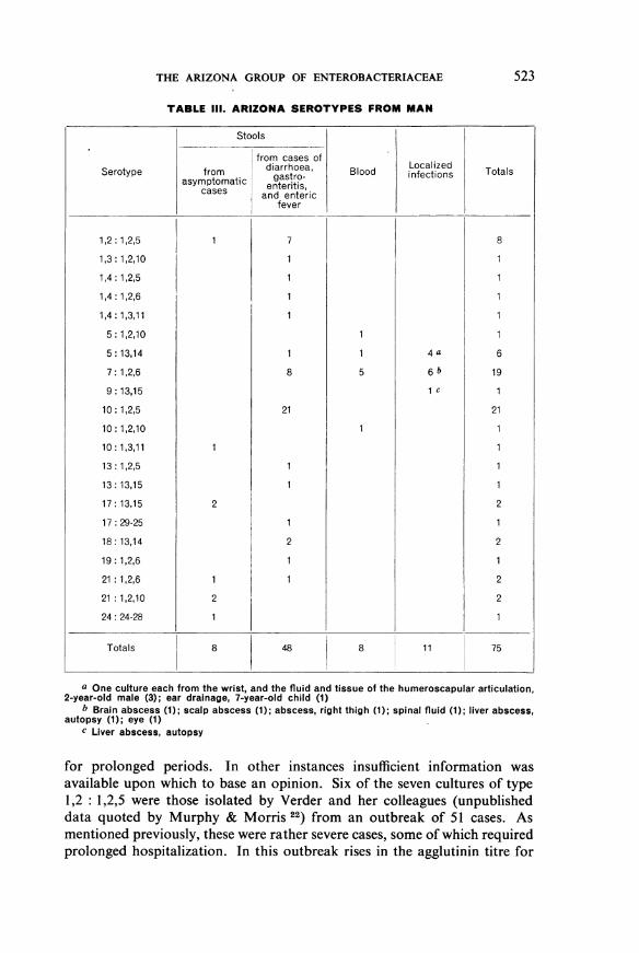
In man the Arizona types apparently produce infections as severe as,if not more severe than, the average Salmonella infection. From the datareported in Table III it is apparent that the bacteria were found withrelative infrequence in normal persons.
The 1,2: 1,2,5 type from an asymptomatic person was isolated froma patient who had previously yielded S. typhi. Since no unusual develop-ments in the course of the disease were noted, the culture was recordedas an adventitious organism. The 10: 1,3,11 culture was isolated from thestool of a 43-year-old midwife, concerning whom no exact informationwas obtainable. The 17: 13,15 cultures were from two nurses on the staffof the same hospital. Again, adequate information was not obtainable.The 21: 1,2,6 type from an asymptomatic person was isolated from thestool of a mother whose child was said to be affected with salmonellosis.The organism from the child was not subjected to serological examination.This culture probably represents a carrier state resulting from contact witha clinical case of Arizona infection. The remaining cultures from asympto-matic individuals were isolated during surveys of food handlers.
No attempt was made to separate the organisms isolated from cases ofdiarrhoea and gastro-enteritis from those isolated from cases with syndromesresembling enteric fever. In some instances the classification would havebeen difficult since persons affected with acute diarrhoea were febrile
522
523THE ARIZONA GROUP OF ENTEROBACTERIACEAE
TABLE III. ARIZONA SEROTYPES FROM MAN
Serotype
1,2:1,2,5
1,3: 1,2,10
1,4: 1,2,5
1,4: 1,2,6
1,4: 1,3,11
5: 1,2,10
5:13,14
7: 1,2,6
9: 13,15
10: 1,2,5
10: 1,2,10
10: 1,3,11
13:1,2,5
13: 13,15
17: 13,15
17 29-25
18: 13,14
19: 1,2,6
21 :1,2,6
21 :1,2,10
24: 24-28
Stools
from cases offrom diarrhoea,
asymptomatic gastro-cases enteritis,cases and enteric
fever
2
2
7
8
21
1
1
21
Blood
5
Localizedinfections
4 a
6 b
1 c
Totals
8
6
19
1
21
2
2
2
21
Totals 8 48 8 11 75
a One culture each from the wrist, and the fluid and tissue of the humeroscapular articulation,2-year-old male (3); ear drainage, 7-year-old child (1)
b Brain abscess (1); scalp abscess (1); abscess, right thigh (1); spinal fluid (1); liver abscess,autopsy (1); eye (1)
c Liver abscess, autopsy
for prolonged periods. In other instances insufficient information wasavailable upon which to base an opinion. Six of the seven cultures of type1,2 :1,2,5 were those isolated by Verder and her colleagues (unpublisheddata quoted by Murphy & Morris 22) from an outbreak of 51 cases. Asmentioned previously, these were rather severe cases, some of which requiredprolonged hospitalization. In this outbreak rises in the agglutinin titre for

P. R. EDWARDS, A. C. MCWHORTER & M. A. FIFE
the Arizona strain were noted. The seventh culture of type 1,2: 1,2,5 wasisolated from the stool of a 58-year-old woman affected with gastro-enteritis. Although the patient died, no autopsy was performed. The1,3: 1,2,10 culture was isolated from the stool of a 5-month-old childaffected with fatal gastro-enteritis. No information concerning the 1,4: 1,2,5culture was available except that it was isolated from a person affected withdiarrhoea. The 1,4: 1,2,6 culture was isolated from the stool of a 25-year-old male affected with diarrhoea but who was afebrile. The 1,4: 1,3,11culture was isolated from the stool of a 3-month-old child affected withgastro-enteritis. The 5: 1,2,10 culture was isolated from the blood of an8-month-old child affected with diarrhoea and fever. This is the culturedescribed by Seligmann & Saphra.25 Of the six 5: 13,14 cultures, one wasisolated from stools, one from blood, and four from localized infections.The culture from stools was derived from a 44-year-old male affected withdiarrhoea who voided 10 to 15 stools per day for 14 days. The culturefrom blood and three of the cultures from localized infections were obtainedfrom a 2-year-old infant. In this child the organisms were known to bepresent for some time since they were isolated at intervals during severalmonths. The organisms localized in the wrist and in the humeroscapularand costochondral articulations; marked destruction of bone was observed.A review of this case was published by Fisher.'2 The fourth culture of5: 13,14 from localized infections was isolated from pus obtained from acase of otitis media. This is the culture reported by Butt & Morris.2
The 7: 1,2,6 type accounted for approximately 25% of the infectionsin man and made up more than 50% of the cultures from blood and localizedinfections, but very little information could be obtained concerning thepersons infected with this serotype. Of the eight cultures from diarrhoealstools, six were said to be from adults. One culture was isolated from a33-year-old male who voided 30 to 40 stools per day. The patient wasfebrile, and pus and blood were present in the stools. No information wasavailable concerning the remaining two cultures. Of the 7: 1,2,6 culturesfrom blood, two were derived from the blood of an adult female at intervalsof four months. The duration of the infection was estimated at eightmonths. The patient ran an intermittent temperature and the organism wasisolated from the blood upon several occasions. Lupus erythematosus wasobserved in this patient. The remaining cultures of this type from bloodwere from infections in adults, but no other information was available.
Of the 7: 1,2,6 cultures from localized infections, one was isolated froma brain abscess in an adult who had been ill for four years with intermittentdiarrhoea, headache, and low-grade fever, and who had also been affectedintermittently with thrombophlebitis. The final outcome of this case is notknown. The second culture of 7: 1,2,6 was isolated from a liver abscess atautopsy. No other information concerning the case was obtained. Of theremaining four cultures of type 7: 1,2,6 from localized infections, only the
524.

THE ARIZONA GROUP OF ENTEROBACTERIACEAE
information contained in the footnotes to Table III could be obtained.From the meagre information available it would appear that serotype7: 1,2,6 is distinctly invasive in man.
No information was forthcoming concerning the 9: 13,15 cultureexcept that it was isolated from a hepatic abscess in an adult at autopsy.
Ten of the 10: 1,2,5 cultures isolated from intestinal infections werederived from the two outbreaks of infection described by Murphy &Morris.22 Of the remaining 11 cultures, six were from infants acutely illwith diarrhoea and enteritis. Two of the infants were siblings who wereill at the same time. Five of the cultures were from four adults, all ofwhomwere -affected with diarrhoea, fever, and vomiting. Two of the cultureswere isolated at an interval of three days from a 36-year-old farmer whowas hospitalized. One of the adults who yielded type 10: 1,2,5 had eatenice-cream from which the organism was isolated.
The 10: 1,2,10 type from blood was isolated from the heart blood ofa 5-month-old infant at autopsy. No details concerning the course ofthe illness were obtained.
The 13: 1,2,5 and 13: 13,15 types were isolated from children withdiarrhoea-the former type, in the Philippine Islands, the latter in Brazil.Nothing is known of the history of the 17: 29-25 type except that it wasisolated from the stool of a patient with diarrhoea. The 18: 13,14 cultureswere isolated from two widely separated sporadic cases of diarrhoea inadults. Both patients were affected with vomiting and fever as well as withdiarrhoea. The 19: 1,2,6 culture was isolated from the stool of an infanthospitalized for two weeks. The child was said to have been dehydratedand to have had a temperature of 103OF (39.40C). The 21:1,2,6 culturefrom a case of diarrhoea was isolated in Israel by Dr W. Hirsch andforwarded to the writers by Dr F. Kauffmann. No further history wasobtained.
The cultures from water, sewage, and miscellaneous sources were derivedlargely from river waters and irrigation waters known to be heavily polluted.S. typhi and other Salmonella types were found in large numbers in thesame waters. Some of the cultures were isolated from the environment inpoultry-processing plants and abattoirs in which surveys for the presenceof salmonellae were being made. Two of the cultures were from foodssuspected of having caused food poisoning and, as mentioned above, in oneinstance the same Arizona type was found in the patient and in the food.
Discussion
From the above data it is evident that bacteria of the Arizona groupcomprise a large number of serotypes, which are distributed widely in manand animals. That they are actually primary incitants of enteric infections
12
525

P. R. EDWARDS, A. C. MCWHORTER & M. A. FIFE
in man and animals there can be little doubt. The epidemiology of theinfections in fowls has been particularly clear-cut and there have beenrepeated instances in which it was possible to trace the transmission of theinfections through eggs. Further, they have been found repeatedly ingeneralized infections in hundreds of flocks of birds in many of which themortality was very high.
In man the organisms occurred with relative frequency in bacteraemiasand in localized infections, at least some of which were probably haemato-genous in origin. One gains the impression from this small series of culturesthat the organisms are as invasive-if not more invasive-in man as arethose Salmonella types not host-adapted to man. One would also gain theimpression from this series of cultures that the organisms occur much morefrequently in fowls than in man. While this is probably true, the differencein the occurrence of the bacteria in fowls and in man may be more apparentthan real. In young fowls the Arizona types, like salmonellae, have amarked tendency to invade the blood and produce fatal infections. In man,and particularly in adults, they usually produce intestinal infections andare found only in the intestinal tract. Cultures from heart blood, liver,spleen, and other internal organs at autopsy are likely to be scrutinizedmuch more carefully than are the lactose-fermenting bacteria isolated fromthe intestinal tract. Therefore, it is possible that many Arizona culturesfrom faeces are discarded. It was once thought that Arizona culturesattacked lactose slowly, and it was not until Solowey & McFarlane 27systematically examined cultures from egg powder that it was recognizedthat many cultures fermented lactose rapidly. Probably the almost completeabsence of rapid lactose-fermenters in the present series, except for a numberof cultures from egg powder, is due to the fact that such cultures wereignored or discarded before they were identified. The small number ofdiphasic types which have been studied have fermented lactose with greaterrapidity than the monophasic types, and their prompt fermentation oflactose may account for the small number of strains forwarded for exam-ination. Since, with the exception of Shigella sonnei, all shigellae andsalmonellae fail to attack lactose, absence of lactose fermentation has becomea fetish in the search for incitants of enteric disease. That such a view isoutmoded is demonstrated not only by study of the Arizona group but bythe ever-increasing acceptance of certain E. coli serotypes as causativeagents in infantile diarrhoea. If the strains of the Arizona group had notfermented lactose they would have been classified as Salmonella serotypesand no questions regarding their role in the production of disease wouldhave arisen. The time has come when the long-held view that primaryincitants of disease among the Enterobacteriaceae fail to ferment lactosemust be abandoned.
526

THE ARIZONA GROUP OF ENTEROBACTERIACEAE 527
RItSUME
Cette etude est consacree 'a certaines enterobacteriacees constituant autrefois legroupe < paracolon) - ai tort selon les auteurs, car elles sont plus proches des Salmo-nellae que des Escherichiae. Elles sont reunies actuellement sous le nom de groupe Arizona,qui comporte 96 types serologiques. Ces bacteries sont mobiles, Gram negatives, liquefientla gelatine et, a quelques exceptions pres, font fermenter le lactose. Elles sont a l'origined'infections intestinales de l'homme et des animaux et leur presence est signalee de plusen plus frequemment. L'etiologie de la maladie qu'elles provoquent chez les oiseauxde basse-cour a pu &re retracee avec precision, en particulier la transmission de l'infec-tion par les ceufs. Chez les jeunes oiseaux, ces bacteries ont tendance, comme les Salmo-nellae, 'a provoquer des bacteri6mies mortelles. Chez l'homme adulte, elles causent desinfections enteriques et n'ont et isolees que de l'intestin.
Jusqu'I maintenant, la recherche des bacteries enteriques avait ete limitee La cellesqui ne font pas fermenter le lactose et il est probable que, de ce fait, un grand nombrede souches pathogenes du groupe Arizona ont echappe aux investigations. Cette exclu-sivite ne se justifie plus aujourd'hui. En effet, la decouverte du r6le joue par les micro-organismes du groupe Arizona dans les infections enteriques, et par certains typesserologiques d'E. coli dans la diarrhee des nourrissons tend a invalider la conceptionselon laquelle les bacteries enteriques pathogEnes ne font pas fermenter le lactose.
Les auteurs decrivent en outre les principales souches correspondant aux principauxtypes serologiques, ainsi que les ph6nomEnes pathologiques qu'elles ont determineschez l'animal et l'homme; ils resument dans des tableaux leur provenance et leurrepartition.
REFERENCES
1. Bruner, D. W. & Peckham, M. C. (1952) Cornell Vet. 42, 222. Butt, E. & Morris, J. F. (1952) J. infect. Dis. 91, 2833. Buttiaux, R. & Kesteloot, A. (1948) Ann. Inst. Pasteur, 75, 3794. Caldwell, M. E. & Ryerson, D. L. (1939), J. infect. Dis. 65, 2425. Edwards, P. R. (1945) J. Bact. 49, 5136. Edwards, P. R., Cherry, W. B. & Bruner, D. W. (1943) J. infect. Dis. 73, 2297. Edwards, P. R., Kauffmann, F. & Fain, A. (1953) Acta path. microbiol. scand. 33, 1918. Edwards, P. R., Kauffmann, F. & Oye, E. van (1952) Acta path. microbiol. scand. 31, 59. Edwards, P. R. & West, M. G. (1950) J. infect. Dis. 87, 184
10. Edwards, P. R., West, M. G. & Bruner, D. W. (1947) Kentucky agric. Exp. Sta.Bull. No. 499
11. Ferris, A. A., Hertzberg, R. & Atkinson, N. (1945) Med. J. Aust. 2, 36812. Fisher, R. H. (1953) J. Bone Jt Surg. 35A, 44513. Goetz, M. E., Quortrup, E. R. & Dunsing, J. W. (1954) J. Amer. vet. med. Ass.
124, 12014. Hinshaw, W. R. & McNeil, E. (1944) Cornell Vet. 34, 24815. Hinshaw, W. R. & McNeil, E. (1946) J. Bact. 51, 28116. Hinshaw, W. R. & McNeil, E. (1946) J. Bact. 51, 39717. Kauffmann, F. (1941) Acta path. microbiol. scand. 18, 35118. Kauffmann, F. (1954) Enterobacteriaceae, 2nd ed., Copenhagen.19. Kauffmann, F. & Edwards, P. R. (1952) Int. Bull. Bact. Nomen. 2, 220. Kauffmann, F., Edwards, P. R. & Oye, E. van (1954) Acta path. microbiol. scand.
35, 15621. Moeller, V. (1954) Acta path. microbiol. scand. 34, 11522. Murphy, W. J. & Morris, J. F. (1950) J. infect. Dis. 86, 255

528 P. R. EDWARDS, A. C. MCWHORTER & M, A. FIFE
23. Peluffo, C. A., Edwards, P. R. & Bruner, D. W. (1942) J. infect. Dis. 70, 18524. Ryff, J. F. & Browne, J. (1952) J. Amer. vet. med. Ass. 121, 26625. Seligmann, E. & Saphra, I. (1951) Publ. Hlth Rep. (Wash.), 66, 136926. Seligmann, E., Saphra, I. & Wassermann, M. (1944) Amer. J. Hyg. 40, 22727. Solowey, M. & McFarlane, V. H. (1947) Amer. J. Publ. Hlth, 37, 971