thermoluminescence properties of olivine · characterized by crystal structure with orthorhombic...
TRANSCRIPT

2011 International Nuclear Atlantic Conference - INAC 2011 Belo Horizonte,MG, Brazil, October 24-28, 2011 ASSOCIAÇÃO BRASILEIRA DE ENERGIA NUCLEAR - ABEN ISBN: 978-85-99141-04-5
THERMOLUMINESCENCE PROPERTIES OF OLIVINE
Shigueo Watanabe1, Nilo F. Cano1,2, Luciana C. Matsushima2 Glauco Veneziane2 José Roberto B. Paião3
1 Instituto de Física, Universidade de São Paulo
Rua do Matão, 187- travessa “R” 05508-090 – São Paulo-SP
2 Instituto de Pesquisas Energéticas e Nucleares (IPEN / CNEN - SP) Av. Professor Lineu Prestes 2242
05508-000 São Paulo, SP [email protected]
3 Universidade São Judas Tadeu
Rua Taquari, 546 – Mooca 03166-000 – São Paulo, SP
ABSTRACT Green and black varieties of olivine of chemical formula, (Mg, Fe)2SiO4, obtained from Luiz Menezes Minerals in Belo Horizonte, MG, have been investigated as to some of their thermoluminescence (TL) properties. The X ray fluorescence analysis indicated that the black sample contains in weight %, SiO4 (44), MgO (33), Fe2O3 (11), Al2O3 (7), Cr2O3 (0.87), MnO (0.17) and other oxides in smaller amount; the green sample, in the same order of oxides, and in w%: 41, 39, 15, 3, 0.07, 0.21. The glow curve of green olivine presented a weak peak at about 150 °C, a stronger at 210 °C, a very intense at 350 °C and less intense at 430 °C, whereas the black one presented small peak at 150 °C, a stronger 200 °C and very broad peak around 350 °C. The optical spectrum of green olivine presented a strong UV absorption, violet and blue light absorption that decreases (in absorbance) gradually up to 500 nm, being minimum the absorbance from 500 to 600 nm (green color of the sample), an absorption band is observed peaking at around 1100 nm, decreasing from 250 nm up to 2500 nm.
1. INTRODUCTION The increasing use of radiation in processes associated with medical and industrial applications has motivated research on new materials with adequate dosimetric properties. There are several studies covering the application of silicates using the thermoluminescent technique for radiation dosimetry. Studies on different kinds of glasses [1,2] and crystal silicate [3] demonstrated their potential application for radiation dosimetry; however, the papers on olivine only address its crystalographical aspects, where synthetic samples were compared to natural ones, or on thermodynamics properties, mainly for olivine. Olivine, (Mg, Fe)2(SiO4), and monticellite, Ca(Mg,Fe)(SiO4) compose the olivine group, characterized by crystal structure with orthorhombic symmetry. The anion (SiO4) is also found in minerals of humite group; however, two of members of this group has monoclinic symmetry. The structure of olivine and monticellite consists of independent (SiO4) tetrahedra linked by divalent atoms in six- fold coordination. In olivine Mg- and Fe- atoms are the
INAC 2011, Belo Horizonte, MG, Brazil.
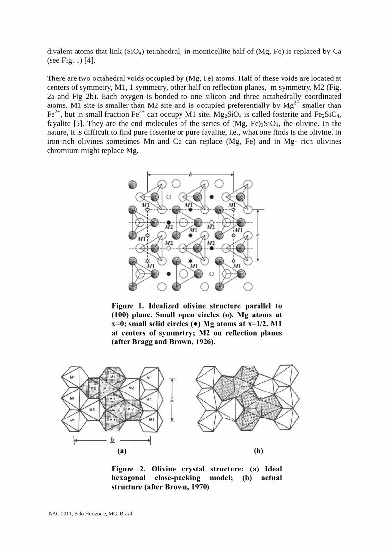
divalent atoms that link (SiO4) tetrahedral; in monticellite half of (Mg, Fe) is replaced by Ca (see Fig. 1) [4]. There are two octahedral voids occupied by (Mg, Fe) atoms. Half of these voids are located at centers of symmetry, M1, 1 symmetry, other half on reflection planes, m symmetry, M2 (Fig. 2a and Fig 2b). Each oxygen is bonded to one silicon and three octahedrally coordinated atoms. M1 site is smaller than M2 site and is occupied preferentially by Mg2+ smaller than Fe2+, but in small fraction Fe2+ can occupy M1 site. Mg2SiO4 is called fosterite and Fe2SiO4, fayalite [5]. They are the end molecules of the series of (Mg, Fe)2SiO4, the olivine. In the nature, it is difficult to find pure fosterite or pure fayalite, i.e., what one finds is the olivine. In iron-rich olivines sometimes Mn and Ca can replace (Mg, Fe) and in Mg- rich olivines chromium might replace Mg.
Figure 1. Idealized olivine structure parallel to (100) plane. Small open circles (o), Mg atoms at x=0; small solid circles (●) Mg atoms at x=1/2. M1 at centers of symmetry; M2 on reflection planes (after Bragg and Brown, 1926).
(a) (b) Figure 2. Olivine crystal structure: (a) Ideal hexagonal close-packing model; (b) actual structure (after Brown, 1970)

INAC 2011, Belo Horizonte, MG, Brazil.
2. MATERIALS AND METHODS
A rock fragments, shown in Fig.3, consists of several small crystals of olivine, from the state of Minas Gerais, Brazil. A closer look enables us to find out that black crystals in lesser number are mixed among greenish crystals.
Figure 3. Fragment of rock with olivine crystals. For the present work more than fifty crystals have been pulled out of the base rock, separating subsequently black ones and green ones. From few largest green crystals slabs with 1.0 mm thickness were cut and polished for optical absorption measurements. Other green and black crystals have been crushed and sieved to retain grains 0.080 to 0.180 mm diameter to be used in TL and EPR measurements. Powder with size smaller than 0.080 mm was used for X-ray fluorescence analysis (XRF). Those between 0.080 and 0.0180 mm diameter are used in TL and EPR measurements. X-ray fluorescence measurements are carried out at the Engineering School of the University of São Paulo, whereas the optical absorption measurements in Carry Spectrophotometer and TL measurements in Daybreak 1100 both at our laboratory. TL read out is carried out with 4 °C/s heating rate.
3. RESULTS AND DISCUSSIONS

INAC 2011, Belo Horizonte, MG, Brazil.
Table 1 list main oxide components in (weight %) of black and green olivines. Four first oxides are components of the crystal, others are impurities. The analysis is from X-ray fluorescence (XRF) measurements.
Table 1. Elements found in black and green olivines in wight % by XRF Compound Black (w. %) Green (w. %)
SiO2 44±2 41±2 MgO 33±2 39±2 Fe2O3 11±1 15±1 Al2O3 7±1 3±1 CaO 3±1 0.79±0.08
Cr2O3 0.87±0.09 0.51±0.05 Na2O 0.32±0.04 0.42±0.04 NiO 0.27±0.03 0.30±0.03 TiO2 0.21±0.02 0.21±0.02 MnO 0.19±0.02 0.07±0.02 K2O 0.10±0.10 0.03±0.01 SO3 0.04±0.01 0.03±0.01
Co2O3 0.02±0.01 0.02±0.01 ZnO <0.01 0.02±0.01 ZrO2 <0.01 0.01±0.01
Glow curves of natural green and black olivine samples are shown in Fig. 4.
Figure 4. Fig. 2. Glow curves of green and black olivine crystal.
Fig. 5(a,b,c) show glow curves of green, black and synthetic olivine irradiated to 10, 30, 50, 75, 100, 150 and 300 Gy gamma rays. The synthetic sample was not irradiated to 300 Gy.

INAC 2011, Belo Horizonte, MG, Brazil.
Figure 5. Glow curves of (a) green natural olivine, (b) black olivine, (c) synthetic sample. All of them irradiated to gamma-doses as indicated.
Fig. 6(a,b) present, respectively, several green and black olivines irradiated to 300 Gy and then their TL read out with heating rate β varying from 1, 2, 4, 8 and 10 °C/s.

INAC 2011, Belo Horizonte, MG, Brazil.
Figure 6. Glow curves of (a) green olivine, (b) black olivine, registered with different heating rates.
Fig. 7 (a) and (b) show, respectively, optical absorption spectra of green olivine, first one in the range 250 to 2500 nm and the second one in the range 375 to 700 nm.
(a) (b)
Figure 7. Optical absorption spectra obtained from natural green olivine. (a) 250 to 2500 nm, (b) 375 to 700 nm
. From Fig. 6 of glow curves of green olivine obtained using following heating rate β for TL read out: 1, 2, 4, 8 and 10 °C/s, we obtain corresponding peak temperature for green olivine.

INAC 2011, Belo Horizonte, MG, Brazil.
Table 2. Peak temperature (Tm) and heating rate (β) of crystal olivine Peak 2 at 150 °C Peak 3 at 330 °C
β Tm (°C) Tm (K) β Tm (°C) Tm (K) 1 184.6 457.6 1 370.1 643.1 2 196.6 469.6 4 401.1 674.2 8 205.6 478.6 8 425.2 698.2 10 217.3 490.3 10 436.6 709.6
At peak temperature Tm Randall-Wilkins equation of TL glow curve has a maximum, therefore the first derivation in Tm of the equation is zero. From this condition, we obtained.
(1) Tm is in absolute temperature scale; k=Boltzman constant. For two β values, say β1, and β2 and taking corresponding values T1m and T2m, from the above equation (2) we obtained:
(2) Using the values in the Table of β´s and T´s and equation (2) we obtained for green olivine E(peak 2) = 1.4 eV and E(peak 3) = 1.6 eV.
4. CONCLUSIONS The crystals in fragment of rock, obtained from a stone dealer in Teofilo Otoni, Minas Gerais State, are olivine mineral of two kinds, one of green color and other one black. X- rays fluorescence nalysis did not show special difference in the impurities why one is green and other, black. Black olivine contains four times more concentration of chromium, however it is difficult to seek for the cause of blackness in this elements. The TL glow curves of both olivines have TL peaks at around 200 °C, 320 °C and 425 °C. The synthetic olivine presented high temperature peak one. It is broad one, hence it is a superposition of 320 and 425 °C peak. This result indicates that these peaks are due to intrinsic defects. From glow curves obtained using different heating rate for TL read out obtained for peak close to 200 °C, E=1.4 eV and for 320 °C peak, E=1.6 eV.
ACKNOWLEDGMENTS This work was supported by FAPESP and CNPq.
REFERENCES 1. A.A. Rodrigues and L.V.E. Caldas, “Commercial plate window glass tested as routine
dosimeter at a gamma irradiation facility” Radiat. Phys. Chem. 63 pp.765-767 (2002).

INAC 2011, Belo Horizonte, MG, Brazil.
2. L.V.E. Caldas and M.I. Texeira, “Commercial glass for high doses using different dosimetric techniques” Radiat. Prot. Dosi. 101 pp.149-152 (2002).
3. D.N. Souza, J.F. Lima, M.E.G. Valerio and L.V.E. Caldas, “Performance of pellets and composites of natural colourless topaz as radiation dosemeters” Radiat. Prot. Dosim., 100 pp. 413-416 (2002).
4. W.L. Gragg and G.B. Brown, “Die Struktur des Olivines” Z. Kristallogr. 63 pp. 538 (1926).
5. G.E. Brown, “Crystal Chemistry of the Olivines” Ph.D. Thesis, Virginia Polytechnic Institute, Blacksburg, Virginia (1970).