thesis kjetil egge combined inhibition of complement and cd14 · combined inhibition of complement...
TRANSCRIPT

Combined inhibition of complement and CD14 in human experimentally induced bacterial inflammation and in porcine
sepsis
PhD Thesis
Kjetil Hagene Egge
2015
Department of Immunology
Institute of Clinical Medicine
Oslo University Hospital Rikshospitalet and
Faculty of Medicine
University of Oslo

Series of dissertations submitted to the Faculty of Medicine, University of Oslo No. 2105

3
TABLE OF CONTENTS
ACKNOWLEDGEMENTS 5
LIST OF PAPERS 7
ABBREVIATIONS 8
1. INTRODUCTION 10
1.1 Sepsis 10
1.2 Innate immunity and inflammation 11
1.3 The complement system 13
1.4 Toll-like receptors 16
1.5 Cross-talk: Complement, TLRs and coagulation 18
1.6 Sepsis: Complement and TLRs 19
1.7 Sepsis treatment 20
2. AIM 24
3. MATERIALS AND METHODS 25
3.1 Bacteria 25
3.2 Inhibitors 25
3.3 Analyses 25
3.4 Models 27
3.5 Statistical considerations 28
4. SUMMARY OF MAIN RESULTS 30
Study I 30
Study II 30
Study III 31
4
Study IV 31
5. DISCUSSION 33
5.1 Upstream versus downstream inhibition 33
5.2 Single versus combined inhibition – redundancy of host defence 34
5.3 Ex vivo studies 35
5.4 In vivo studies and the pig as model animal 36
5.5 The relevance of readouts 38
5.6 Future perspectives 39
6. CONCLUSIONS 41
7. REFERENCES 42
8. PAPERS I-IV 55

5
ACKNOWLEGDGEMENTS
My work with his thesis has been an interesting, sometimes frustrating, but most of all
rewarding experience. During a PhD period one acquires lots of new experiences and skills,
not only applicable for studying a particular scientific field, but in general as a professional
and in life as such. Were it not for the efforts of quite a few great people, my PhD period
would not have been as rewarding; in fact the thesis would not have been finished.
First of all, I would like to thank my supervisor, Professor Tom Eirik Mollnes, for invaluable
scientific guidance. His dedication to science and his hard-working spirit are truly admirable
and constitute standards which are not easily matched. His enthusiasm and positivity for new
results, even when not as exciting as hoped for, are nothing but inspiring.
My gratitude to my co-supervisors, Ebbe Billmann Thorgersen and Andreas Barratt-Due,
cannot be overstated. Their scientific and moral support, also when matters were less bright,
can hardly be fully appreciated. Their thoughtful and thorough feedback and revisions have
been out-most essential.
Special thanks go to the technicians, Anne Pharo, Julie Katrine Lindstad and Margareta
Nilsson. Their steady support and patience in the laboratory were constantly required, and
always served with a smile. I cannot imagine better people to work with in the laboratory.
In my time as PhD student I have been fortunate to be part of a great group of nice, interesting
and hard-working PhD students and postdocs. They have all contributed in different ways,
socially and scientifically, during the process: Albert Castellheim, Søren Erik Pischke, Bernt
Christian Hellerud, Stig Nymo, Per H. Nilsson, Alice Gustavsen, Andrey Sokolov and Hilde
Lang Orrem. In addition, I have been lucky to share laboratory facilities with Professor
Emeritus Morten Harboe, whose wisdom in science and ability to explain complex matters
have been of great help.
6
All the members of the Bodø branch of the Norwegian Complement group deserve special
thanks, for social companionship on conferences and meetings, but also for scientific support.
In particular Professor Erik Waage Nielsen, Espen Waage Scheflo, and the laboratory
technicians Dorte Christiansen and Grethe Bergseth were of important help.
Finally I thank my family for supporting me through the ups and downs of my PhD period. In
particular I thank my wonderful wife, Lise, for listening to frustrations and celebrating
achievements. But most of all, I thank her for being the glue that has held our little family
together. In the long run, that is what really matters.

7
LIST OF PAPERS
Paper I
Egge KH, Barratt-Due A, Nymo S, Lindstad JK, Pharo A, Lau C, Espevik T, Thorgersen
EB, Mollnes TE. The anti-inflammatory effect of combined complement and CD14 inhibition
is preserved during escalating bacterial load. Clin Exp Immunol. 2015 Apr. In press
Paper II
Egge KH, Thorgersen EB, Lindstad JK, Pharo A, Lambris JD, Barratt-Due A, Mollnes TE
Post-challenge inhibition of C3 and CD14 attenuates Escherichia coli-induced inflammation
in human whole blood. Innate Immun. 2014 Jan; 20(1):68-77.
Paper III
Barratt-Due A, Thorgersen EB, Egge K, Pischke S, Sokolov A, Hellerud BC, Lindstad
JK, Pharo A, Bongoni AK, Rieben R, Nunn M, Scott H, Mollnes TE. Combined inhibition of
Complement (C5) and CD14 markedly attenuates inflammation, thrombogenicity and
hemodynamic changes in porcine sepsis. J Immunol. 2013 Jul; 191(2):819-27.
Paper IV
Egge KH, Thorgersen EB, Pischke SE, Lindstad JK, Pharo A, Bongoni AK, Rieben R, Nunn
MA, Barratt-Due A, Mollnes TE. Organ inflammation in porcine Escherichia coli sepsis is
markedly attenuated by combined inhibition of C5 and CD14. Immunobiology. 2015 May. In
press.

8
ABBREVIATIONS
Ab Antibody
ANOVA Analysis of Variance
AU Arbitrary Units
AP Alternative Pathway
BK Bradykinin
C Complement factor
CD Cluster of Differentiation
CLP Cecal Ligation and Puncture
CP Classical Pathway
CR Complement Receptor
CRP C-Reactive Protein
CVP Central venous pressure
C5aR C5a Receptor
DAMP Damage-Associated Molecular Pattern
DIC Disseminated Intravascular Coagulation
E. coli Escherichia coli
EDTA Ethylenediaminetetraacetic acid
ELISA Enzyme-Linked ImmunoSorbent Assay
FITC Fluorescein Isothiocyanate
ICU Intensive Care Unit
Ig Immunoglobulin
IL Interleukin
kPa KiloPascal
LP Lectin Pathway
LPS Lipopolysaccharide
LTB4 Leukotriene B4
MAP Mean Arterial Pressure
MBL Mannose Binding Lectin
MD-2 Myeloid Differentiation factor 2
MFI Median Fluorescence Intensity
mg Milligram
mL Milliliter

9
mM Millimolar
mmol Millimol
MPAP Mean Pulmonary Arterial Pressure
MyD88 Myeloid Differentiation Primary Response Gene 88
NF-ĸB Nuclear Factor kappa-light-chain-enhancer of activated B cells
OmCI Ornithodoros moubata Complement Inhibitor
PAI-1 Plasminogen Activator Inhibitor 1
PAMP Pathogen-Associated Molecular Pattern
PBS Phosphate Buffer Saline
PCR Polymerase Chain Reaction
PRR Pattern Recognition Receptor
SIRS Systemic Inflammatory Response Syndrom
SVRI Systemic Vascular Resistance Index
TAT Thrombin-Antithrombin Complex
TCC Terminal Complement Complex
TF Tissue Factor
TNF Tumor Necrosis Factor
TLR Toll-Like Receptor
TRIF TIR-domain-containing Adapter-inducing Interferon-β
WBC White Blood Cell Count

10
1. INTRODUCTION
1.1 Sepsis
Sepsis is Greek for putrefaction or decay. In clinical medicine it is a term used to describe the
systemic response to an infection, defined as “a systemic inflammatory response syndrome
(SIRS) caused by infection” (1). By the American College of Chest Physicians/Society of
Critical Care Medicine Consensus Conference in August 1991, SIRS was originally defined
by the presence of more than one of the following criteria: (I) a body temperature greater than
38°C or less than 36°C; (II) a heart rate above 90 beats/min; (III) a respiratory rate greater
than 20 breaths/min or hyperventilation defined by PaCO2 less than 4.3 kPa; and (IV) an
alteration in the white blood cell count (WBC) defined by WBC greater than 12 x 109 /L or
WBC less than 4 x 10⁹ /L or more than 10% immature neutrophils in the WBC (1). Later
reviews of the SIRS definition have made slight alterations to the original criteria and taken in
several more criteria including inflammatory, haemodynamic, organ dysfunction and tissue
perfusion variables (2,3). As a number of non-infectious diseases, such as trauma, burns and
pancreatitis may also cause SIRS, a documented or suspected infection is a diagnostic
requirement for sepsis (3).
Based on severity, sepsis is stratified into four subgroups: (I) sepsis; (II) severe sepsis; (III)
septic shock; and (IV) sepsis with multiple organ dysfunction syndrome (MODS) (1). In the
US sepsis and severe sepsis are the most common causes of death among critically ill patients
in non-coronary intensive care units (ICU), and overall one of the leading causes of death (4).
These conditions are public health problems of great cost (5). As growing patient groups such
as elderly patients and patients with pre-existing chronic diseases are at increased risk, the
public health care issues and death tolls may most possibly increase in the future (4). It would
therefore be of great significance to find more effective means to combat the disease.
The leading causes of sepsis in the US and Europe are almost equally divided between Gram-
positive and Gram-negative infections, although with an increase in Gram-positive infections
over the last few decades (6-8). The vast majority of the Gram-positive infections are caused

11
by a variety of Staphylococci species, and in a large European multicentre ICU study from
2006 (the SOAP study), species of Staphylococci and Streptococci accounted for more than
90 % of all Gram-positive infections in septic ICU patients (6). With regards to the Gram-
negative infections in the same study, Escherichia coli and Pseudomonas species were found
to be causative in over 50 % of cases (6). Just as interesting, the same study reports that as
much as 40 % of all septic ICU patients only had clinical signs of infection, and no causative
microbe was found. Furthermore, as much as 18 % of the septic patients had polymicrobial
infections (6). The findings of the SOAP study are analogous to previous and later studies
(7,8).
1.2 Innate immunity and inflammation
The body is under constant threat from microbes in its natural environment. Under
physiological circumstances, the microbes are held outside the body by the skin and mucosal
surfaces as a first line barrier against infections (9). As soon as the microbes trespass the
barrier, the immune system is set in action (9). In the course of evolution, the immune system
has developed several mechanisms in order to cope with invading microbes. These
mechanisms are commonly divided into two main branches: innate and adaptive immunity.
Adaptive immunity serves as the “long term” memory of the immune system. Its main
function is to remember specific microbes with strong affinity in order to detect and destroy
them whenever encountered (9).
Innate immunity on the other hand, is functional even without previous sensitization and
depends on germ-line encoded receptors, referred to as pattern recognition receptors (PRRs)
(10). The cardinal trigger of the immune response is the recognition of conserved molecular
patterns of exogenous as well as endogenous molecules by these PRRs (11,12). The
exogenous conserved patterns from different microbes are referred to as pathogen-associated
molecular patterns (PAMPs), whereas the endogenous conserved patterns, which do not
originate from microbes but rather from damaged host cells, are referred to as damage-
associated molecular patterns (DAMPs) (13-15). Thus, innate immunity is not only structured
to combat invading pathogens, it is also designed to detect, repair or clear damaged cells,

12
representing danger from within the host itself. The main goal of innate immunity, whether it
detects microbes or damaged self, is to re-establish homeostasis (10).
Confusingly, in the ”danger model” proposed by Polly Matzinger, the abbreviation DAMPs is
used as short for danger-associated molecular patterns, a common denomination for PAMPs
and alarmins (15). This model reflects the view that innate immunity is designed to detect
danger, exogenous (PAMPs) and endogenous (alarmins), rather than discriminate self from
non-self, which in the 1990ies was the competing view on innate immunity as stated by
Charles Janeway in his “self-non-self” model (16). In this thesis DAMPs will be used as short
for damage-associated molecular patterns and not as a common denomination for PAMPs and
alarmins.
Upon recognition of PAMPs and DAMPs via PRRs, innate immunity responds by inducing
inflammation (10). The inflammatory mediators are secreted by a number of cells, including
macrophages, monocytes, granulocytes and dendritic cells of the immune system (12,17).
These mediators are commonly denominated cytokines and include a broad variety of locally
and systemically active molecules, which induce chemotaxis, regulate expression of cell
surface receptors and adhesion molecules, increase capillary leakage, trigger coagulation and
fibrinolysis, and activate macrophages and fibroblasts, to mention some of its multifaceted
activities (18). In addition a wide variety of other mediators such as prostaglandins and
leukotrienes directly or indirectly induce a diverse inflammatory response (19).
This thesis is mainly focused on PAMPs. Importantly however, DAMPs and PAMPs appear
largely to depend on the same PRRs to evoke inflammatory responses (9). Therefore, the
translational value of research on pathogen-induced inflammation to sterile-induced
inflammation may prove to be of great significance.
PRRs of innate immunity involved in detection of PAMPs include several groups of receptors:
Intracellular receptors, cell surface bound receptors and soluble receptors (17). Examples of
intracellular PRRs are Nucleotide Oligomerization Domain-like (NOD-like) receptors and

13
Retinoic acid Inducible Gene-like (RIG-like) receptors which recognize intracellular
microbial PAMPs and intracellular viral RNA respectively (20-23). To the family of
exclusively intracellular PRRs belong also the relatively newly discovered AIM-like (Absent
In Melanoma-like) receptors (23). These intracellular PRRs and their effector functions will
not be discussed in this thesis.
Toll-like receptors (TLRs) including its soluble co-receptors and the complement system are
important examples of membrane bound and soluble PRRs (17). They play key roles in the
network of innate immunity functioning as central upstream sensor- and effector-systems,
thus to a large part being responsible for the complex and widespread inflammatory response
(15,24). These systems, their role and the interplay between them in Gram-negative and
Gram-positive induced inflammation, and partly also in sterile induced inflammation will be
described in the following.
1.3 The complement system
The complement system is an important part of innate immunity, where it plays a vital role
defending the host against pathogens and maintaining homeostasis (25). Over the last decades
it has become clear that it serves as a bridge to adaptive immunity involving T and B cells
(26), and also is involved in the pathogenesis of an increasing number of conditions where
microbes are not involved (27). It is constituted by over 30 different plasma and cell surface
proteins forming a cascade system with a plethora of physiological effects (28,29). Activation
of the cascade is triggered via one of three possible pathways; the classical, the lectin and the
alternative pathways. All pathways converge on the central complement factor C3 which is
cleaved to C3a and C3b, thereby launching the terminal pathway as C5 is cleaved to C5a and
C5b and the assembly to form the terminal complement complex is initiated (TCC) (25,30).
The classical pathway (CP) is initiated when immune complexes are formed from C1q
binding to antibodies in complex with pathogenic surface antigens or other non-self antigens
(30). In addition, pentraxins, such as the acute phase protein CRP, may clear pathogens by
directly binding to C1q (31). The complex recruits C1r and C1s, the latter cleaving C4 and C2
to form C4bC2b – the C3 convertase of the CP.

14
Figure 1. The complement system (with permission from the copyright holder T. E. Mollnes).
The lectin pathway (LP) is activated when mannose binding lectin (MBL), ficolins or
collectins are bound to carbohydrate structures on the surface of pathogens (25). In the
circulation, MBL, ficolins and collectins are bound to MBL-associated proteins (MASPs) (32),
which undergo conformational changes upon binding to pathogens, thereby inducing cleavage
of C4 and subsequent recruitment and cleavage of C2 to ultimately form C4bC2b – the C3
convertase of the LP.
The alternative pathway (AP) has a “spontaneous tick over”, as hydrolysis of C3 to form
C3(H2O) goes on continuously (33). In addition, the pathway may be triggered by several
molecular structures, including carbohydrates, proteins and lipids on pathogens and other non-
self surfaces which split C3 into C3b and the anaphylatoxin C3a. Further activation takes
place as C3(H2O) or C3b are bound to factor B, which is further cleaved by factor D to form

15
C3(H2O)Bb or C3bBb respectively – both C3 convertases of the AP. These convertases are
further stabilized when they form complex with Properdin (or factor P) (33). Properdin has
previously also been regarded as a PRR of the AP by recruitment of C3b to induce assembly
of the AP C3 convertase (25,33), but results from later studies seriously question the role of
Properdin as a PRR (34). In addition to being an activation pathway, the AP also functions as
an essential amplification loop for the CP and the LP, maybe responsible for as much as 80%
of the final formation of C5a and TCC (34,35).
Following the cleavage by the C3 convertases, C3b forms complexes with the same C3
convertases C4bC2b and C3bBbP, producing the C5 convertases C4bC2bC3b and
C3bBbPC3b. These convertases cleave C5 to C5a and C5b, initiating the mentioned assembly
of the TCC as C6 and C7 bind to C5b followed by C8 and lastly multiple C9 molecules (25).
When multiple TCCs are inserted into the membrane of certain microbes and cells, each of
the TCCs form a pore, and ultimately lysis may occur. In this setting TCC is often referred to
as the membrane attack complex (MAC). This mechanism is especially important in the
defence against infections with Neisseria (N.) meningitides, one of the microbes responsible
for causing meningitis (36). As importantly, when TCCs are formed in sublytic concentrations
on membranes, they may induce proinflammatory responses, for instance on endothelial cells
and other nuclear cells (37).
Another and important way the complement system defends the host from invading pathogens
is by opsonisation of the microbes by deposition of C3b and C4b fragments to induce
phagocytosis (38). In addition to bactericidal activity, it is also responsible for tissue
regeneration and clearance of apoptotic cells and debris (39,40), but more importantly,
especially in the setting of sepsis, it is one of the main inflammatory triggers.
The small split fragments of the complement cascade C3a and C5a are highly biologically
active anaphylatoxins and the main inflammation inducing complement products (41-43). C3a
and C5a have several biological functions in common such as promotion of vasodilation and

16
increasing capillary leakage (44), and both are associated with pathogenesis in asthma and
acute respiratory distress syndrome (ARDS) (45). C3a also induces mast cell degranulation
and transcription factor activation thereby increasing inflammation (46). Of the two, C5a is a
particularly potent inflammation inducer and a plethora of effects includes upregulation of
adhesion molecules, stimulation of phagocytosis and oxidative burst in neutrophils and
monocytes, induction of granular enzyme release and attraction of neutrophils to the site of
danger (42,44,47). On the other hand, recent studies show that C3a also has several anti-
inflammatory effects partly opposing the proinflammatory effects of C5a, a noteworthy point
in the debate about where in the complement cascade to intervene therapeutically (48).
All of the above mentioned effects are favourable under physiological circumstances, helping
the host to combat infections as part of first line host immune defence mechanisms. However,
when activated systemically, particularly C5a is heavily involved in the pathological
processes causing the clinical syndrome sepsis (42,49).
1.4 Toll-like receptors
The TLR family is constituted by a complex group of PRRs which recognize a wide variety of
evolutionary conserved molecular structures (50). The main function of the TLRs is signalling
to the cells as microbial PAMPs are recognized as exogenous danger (10). Over the last years
it has however become clear that TLRs in addition sense endogenous ligands, which are
cellular components of self-origin such as proteins and peptides, polysaccharides and
proteoglycans, nucleic acids and phospholipids. Accumulating evidence demonstrates
that endogenous ligand-mediated TLR signalling plays a role in several disease processes
including tissue injury, repair and regeneration, autoimmune diseases and tumorigenesis – all
conditions involving sterile inflammation (51).
To date, ten different TLRs have been identified in human (52). Concerning the exogenous
ligands, each TLR has specificity for distinct PAMPs from bacteria, mycobacteria, fungi,
parasites and viruses: Lipoproteins are recognized by TLR1, 2 and 9; double-stranded RNA
by TLR3; LPS by TLR4, flagellin by TLR 5, single-stranded RNA by TLR7 and 8; and DNA

17
by TLR9 (52). Upon recognition of these PAMPs, TLRs recruit a specific set of adaptor
molecules, such as MyD88 and TRIF, to induce downstream signalling (53).
In Gram-negative inflammation, LPS is the main inflammation inducing molecule, and TLR4
is considered to be the only LPS-receptor (54). TLR4 has therefore been regarded a possible
interventional target for modulating the inflammatory response in Gram-negative induced
inflammation. In order to induce downstream intracellular signalling by recognition of LPS,
TLR4 is however dependent on cofactors. One of the cofactors, myeloid differentiation factor
2 (MD-2), is a soluble protein present on the extracellular domain of TLR4 and confers
responsiveness to LPS (55). CD14, another TLR4 cofactor, was actually discovered before
TLR4 itself; it was found to bind the circulating LPS in complex with LPS-binding protein
(56,57). Upon binding with LPS, the CD14/LPS complex binds to the MD2/TLR4-complex
and induces activation of intracellular signalling via the adaptor molecules MyD88 and TRIF
to finally activate the nuclear transcription factor, which is responsible for transcription of a
wide variety of inflammation associated genes and subsequent synthesis and release of several
pro-inflammatory cytokines (53,58,59). Some Gram-negative bacteria such as N. meningitides
induce signalling via both adaptor molecules, whereas E. coli predominantly induce signalling
via MyD88 and Salmonella predominantly via TRIF (60).
As mentioned, TLR2 is the Gram-positive PRR analogue to TLR4 for Gram-negative bacteria
(61,62). However, debate is still ongoing about the Gram-positive TLR2 ligand. Lipoteichoic
acid (LTA) has been claimed to be the Gram-positive LPS analogue (59,60), but later studies
have shown that especially LTA of S. aureus and S. pneumoniae are not ligands for TLR2,
but in fact for the lectin pathway (63). On the other hand, the staphylococcal lipoproteins,
Panton-Valentine toxin and Phenol Soluble Modulins have been identified as potent TLR2
ligands, and little controversy exists over the statement that also TLR2 mediates Gram-
positive inflammation (64). The TLR2 signalling is partly analogous to TLR4 signalling as
TLR2 also uses a MyD88-dependent signalling pathway to translocate NF-ĸB into the nucleus
with subsequent activation of the expression of pro-inflammatory cytokine genes (64). Even
more interestingly, it has been shown that TLR2 signalling by these staphylococcal ligands is
CD14 dependent (65). The true promiscuous role of CD14 as co-receptor for the majority of

18
the TLRs, makes it an important molecule in the TRL family and an utterly interesting target
molecule for anti-inflammatory purposes (50).
TLR2 and TLR4 are transmembrane lipoproteins located on the extracellular surface of the
immune cells, but several other TLRs, including TLR3, 7, 8 and 9, are located on the inside of
the cell in order to detect PAMPs and DAMPs intracellularly. In the setting of an infection,
they are designed to and evoke an inflammatory response upon recognition of intracellular
microorganisms including viruses and certain bacteria and parasites (17,66,67).
TLR stimulation is finally responsible for the production of a wide variety of pro-
inflammatory cytokines, including IL-6 and TNF, which drive the inflammatory response
(66,68).
1.5 Cross-talk: Complement, TLRs and coagulation
There is increasing evidence for a close and widespread cross-talk between complement and
TLRs, but also between innate immunity and coagulation (69-71). Phagocytosis of
mycobacteria and Borrelia burgdorferi has been shown to be enhanced by cross-talk between
complement receptor 3 (CR3) and CD14, and killing of Salmonella has been documented to
be dependent on TLR4 and CR3 (72-74). Several PAMPs, including the TLR4-ligand LPS
and TLR2/6-ligand zymosan have been demonstrated to markedly increase the level of the
pro-inflammatory cytokines IL-6, TNF and IL-1β in a complement dependent manner (75). In
the setting of septic inflammation, especially the proposed ability of C5a and TLRs to
reciprocally promote pro-inflammatory cytokine production is of great relevance (75,76).
Innate immunity and coagulation are intimately connected and may be viewed as partners in
the inflammatory response with the main goal of stabilizing an organism which has
encountered a threat to its homeostasis (71). In settings where the complement system is
activated, such as during an infection, the coagulation system is also tilted towards a pro-
coagulant state (77). Similarly, whenever the coagulation system is triggered, for example

19
during trauma, an associated activation of inflammation will occur as the host is at risk of
infection (78). This simultaneous activation of haemostasis and inflammation is in many
clinical circumstances beneficial. For example, in the instance of an infection, the local
formation of thrombi in the microvasculature draining the site of the infection supplies an
important barrier which prevents the bacteria from entering the circulation.
In fact, not only are these systems concurrently activated. At various levels of inflammation
and haemostasis, members of both systems interact directly or via secondary mediators.
There are several examples of such interactions: C5a may directly trigger tissue factor (TF),
the spark plug of the coagulation cascade; anticoagulation mechanisms are inhibited by
binding of C4b-binding protein to Protein S, an important co-factor for degradation of
coagulation factors Va and VIIIa via activated protein C (79); C3a and C5a promote the
production of TNF and IL-6, which in turn enhance TF expression on monocytes and increase
platelet thrombogenicity; lastly, observations in vitro also suggest that complement
components may be cleaved directly by thrombin, plasmin and factor XIIa (71). Therefore,
when studying inflammation, concurrently recording markers of haemostasis, systemically as
well as in organs, provide important additional information.
1.6 Sepsis: Complement and TLRs
Under physiological conditions complement and TLRs act partly systemically by signalling
danger, but predominantly promote local effects and as part of host defence mechanisms work
to stop microorganisms from entering the circulation and strive after homeostasis. However,
under pathological conditions, such as in sepsis, the tight regulation of these systems fail and
systemic activation occurs promoting SIRS throughout the body, also in organs far from the
site of danger (80-83). The favourable and essential local actions of innate immunity become
the main threat to the host. For example, increased vascular permeability, capillary leakage
and vasodilation are advantageous innate immunity effects in order to allow sequestration of
neutrophils to the site of infection, but when these effects are promoted systemically, the net
result is loss of intravascular volume, hypotension and reduced blood flow to vital organs in
addition to organ inflammation upon sequestration of neutrophils to organs far from the site of
danger (84,85).

20
The mentioned advantageous interplay between coagulation and innate immunity also
becomes harmful in systemic infection-induced inflammation as haemostasis is tilted toward a
more procoagulant state, promoting disseminated intravascular coagulation (DIC) (86).
Hallmarks of DIC are the microvascular depositions of fibrin, platelet consumption and
formation of microthrombi, also far from the site of infection, contributing to organ
dysfunction and failure (86,87). The onset of DIC in sepsis has the clinically devastating
importance of increasing mortality two-fold (88).
The overall myriad of inflammatory responses in sepsis is impossible to fully account for. In
addition to complement, TLRs and coagulation, also the fibrinolytic system and the
kallikrein/kinin system are activated (89,90). The endothelial cells are also important players
in promoting dysfunctional microcirculation as adhesion molecules are upregulated and
platelets and leukocytes adhere to the microvascular lining (91,92).
1.7 Sepsis treatment
To date treatment of severe sepsis and septic shock basically still relies on three arms: Initial
resuscitation, anti-microbial therapy and supportive therapy.
The main goal of the first arm, initial resuscitation, is to re-establish adequate perfusion to all
vital organs. Briefly summarised, haemodynamic support according to Guidelines from 2012
includes (2): initial fluid resuscitation with crystalloids and eventually albumin if needed;
fluid challenge to hypovolemic patients; vasopressor support with noradrenalin and eventually
adrenalin to maintain blood pressure above 65 mmHg.
The main goal of the second arm of treatment, anti-microbial therapy, is obvious and
according to the above mentioned Guidelines from 2012 includes: taking blood cultures or
cultures from the relevant site of infection before administering antibiotics in order to obtain a
specific microbial diagnosis; broad-spectrum antibiotics until the microbe is known; extensive

21
investigations including imaging studies to obtain a certain source of infection; and lastly
reassessment of microbial therapy daily and de-escalation of the microbial therapy according
to microbiological results. Gaining control over the site of infection by surgically revising
infected wounds, abscesses and so on is also of great importance.
The third arm, supportive therapy, is primarily aimed at helping the organs to maintain normal
function and homeostasis, or secondarily replace malfunctioning organs. This includes
mechanic ventilation, dialysis, sedation and administration of blood products and glucose.
Use of corticoid treatment and prophylaxis against deep vein thrombosis and gastric ulcers are
also recommended to specific patients groups when appropriate (2).
Given the elaboration above on innate immunity, inflammation and inflammatory mediators, a
fourth arm of treatment in sepsis is missing and has long been sought: Mediator directed
therapy. Several clinical trials blocking a range of mediators in sepsis including bradykinin,
prostaglandins and platelet activating factor, and furthermore anti-thrombotic treatment using
tissue factor pathway inhibitor and anti-thrombin have all failed to show beneficial effects
(93-97).
IL-1β and TNF are clearly linked to septic inflammation and both have been considered
possible therapeutic targets in patients with severe sepsis (98-100). As the level of IL-6 in
septic patients correlates with severity and mortality, IL-6 was also thought to be a possible
therapeutic target (101,102). However, several large clinical trials show that increased
survival has not been achieved by targeting these downstream cytokines selectively (103-106).
There were also considerable expectations connected to treatment with recombinant human
activated protein C (rhAPC), but promising results were not reproducible, and the ability to
increase survival in a final multicentre study failed (107). Lastly, also the specific TLR4
inhibitor eritoran failed in clinical trials (108). In common, all these trials have tried to combat
the inappropriate inflammation in sepsis by singling out one key downstream mediator. Given
the inexplicably large number of downstream mediators flowing over as part of the
inflammatory response, not to mention the cross-talk and redundancy of innate immunity, it is
not surprising that these trials have failed.
22
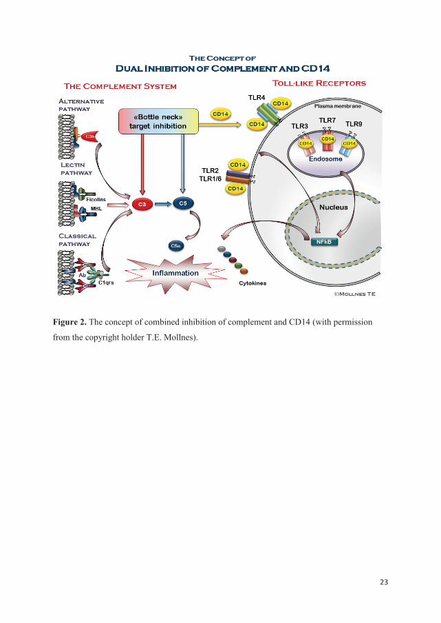
Accordingly, the possibility of dampening the overall inflammatory load in sepsis seems more
likely when targeting upstream. In this context targeting upstream means blocking down or at
least slowing down the cascade mechanisms as close to the interaction between the PRRs and
the PAMPs as possible. However, targeting each specific PAMP or its receptor would demand
specific knowledge about the infecting microbe at the commencement of treatment, which in
practice is impossible. Therefore, targeting unspecific pattern recognition receptors of several
PAMPs may prove to be a possible way forward in the search for mediator directed therapy.
CD14 is one such molecule of the TLR family important for unspecific pattern recognition of
several PAMPs, and the complement factor C3 and to a large degree also C5 another set of
such upstream molecules acting as bottlenecks for downstream effects of the complement
system. Indeed, inhibiting CD14 has been shown to have vast anti-inflammatory effects in
septic pigs, and inhibition of complement factor C3 has also shown promising anti-
inflammatory effects in septic baboons (109,110).
In 2008, following observations of improved anti-inflammatory effects when combining
inhibition of TLRs and a key complement molecule, professor Mollnes and colleagues put
forward the hypothesis that this combined inhibition may be a future strategy to attenuate the
inappropriate inflammatory response seen in sepsis as well as in conditions with sterile
induced inflammation (47,81). Several studies of the effect of the combined inhibition on
sterile and non-sterile induced inflammation have been conducted since (111-115).
23
Figure 2. The concept of combined inhibition of complement and CD14 (with permission
from the copyright holder T.E. Mollnes).

24
2. AIM
In this thesis, dissecting the effects of combined inhibition of a key complement molecule and
CD14 under changing conditions of bacterial induced inflammation and sepsis has been the
main task.
A possible preservation of the anti-inflammatory effects of the combined inhibition of
complement and CD14 was investigated under increasing concentrations of Gram-
negative and Gram-positive bacteria ex vivo (Study I).
The anti-inflammatory effects of the combined inhibition of C3 and CD14 when
introducing a time delay between the onset of Gram-negative induced inflammation
and the addition of the inhibitors were examined in the same ex vivo model (Study II).
The effects on inflammation, haemostasis and haemodynamics of combined inhibition
of C5 and CD14 were examined in a porcine in vivo model of E. coli-induced sepsis
(Study III).
Lastly, the efficacy of the combined inhibition of C5 and CD14 on local organ
responses to septic inflammation was studied in the same in vivo model (Study IV).
25
3. MATERIALS AND METHODS
3.1 Bacteria
E. coli strain LE392 (ATCC 33572) and S. aureus Cowan Strain 1 (ATCC 12598) were
obtained from American Type Culture Collection (Manassas, VA). Heat-inactivated E. coli
was used in paper I and II, whereas live E. coli were used in paper III and IV. S. aureus (heat-
inactivated) was used only in paper I.
3.2 Inhibitors
In paper I, azide-free mouse anti-human CD14 (clone 18D11; F(ab`)2 3118, lot1383) was
used in the E. coli experiments. The recombinant anti-human CD14 IgG2/4 Ab (r18D11) was
used in the S. aureus experiments (116). For complement inhibition, either the complement
C5 inhibitor, eculizumab (Soliris®) or the compstatin analogue Cp40, a complement C3
inhibitor, were used (117).
In paper II, azide-free mouse anti-human CD14 (clone 18D11; F(ab`)2 3118, lot1383) and the
Compstatin analogue AMY-90, a complement C3 inhibitor, were used.
In paper III and IV, recombinant bacterial Ornithodoros moubata Complement Inhibitor
(OmCI), a 16.8 kDa protein also known as coversin (118), and mouse anti-porcine CD14
mAb (clone MIL-2, isotype IgG2b) were used.
3.3 Analyses
ELISA
In paper I and II, the terminal complement complex (TCC) was measured using a sandwich
ELISA (119,120). Briefly, the monoclonal antibody (mAb) aE11 recognizes a neoepitope
exposed in C9 after incorporation in the C5b-9 complex. aE11 was used as capture antibody
binding to the soluble TCC. A biotinylated anti-C6 mAb (clone 9C4) was used as detection
antibody. In paper I, the standard was normal human serum activated with zymosan and
defined to contain 1000 arbitrary units (AU)/mL, whereas in paper II the standard was
International Complement Standard #2 (ICS#2) (121), which is a standardized version of
26
zymosan activated normal human serum. Zymosan activated human serum was used as a
positive control.
The granulocyte activation markers myeloperoxidase (MPO) and lactoferrin (LF) were
quantified using commercially available kits from Hycult Biotech in paper I, whereas in paper
II only MPO was measured.
In paper III Leukotriene B4 (LTB4) from serum was measured using a competitive enzyme
immunoassay from R&D Systems. Citrate plasma was used for measuring thrombin-
antithrombin (TAT) in an immunoassay kit from Dade Behring (122). Plasminogen activator
inhibitor-1 (PAI-1) was measured in citrate plasma by a porcine PAI-1 kit from Molecular
Innovations. The cytokines TNF, IL-1ß, IL-6 and IL-8 were analyzed by Quantikine Porcine
Immunoassay kits from R&D Systems, whereas IL-10 was analyzed by a porcine kit from
BioSource Invitrogen. A Complement System Screen (Wielslab®) from Eurodiagnostica AB
was used to test functional activity of the classical, lectin and alternative complement
pathways.
In paper IV IL-1ß was measured using the same kit as in paper III.
Multiplex analysis
In paper I and II, cytokines TNF, IL-1β, IL-6, IL-8, MIP-1α and MIP-1β were measured
using a multiplex cytokine assay from Bio-Rad Laboratories. In paper III and IV, TCC was
measured using multiplex xMAP technology (123), and in paper IV the same technology was
also used for measuring IL-6, IL-8 and monocyte chemotactic protein-1 (MCP-1). In short,
antibodies for the biomarkers were coupled to carboxylated nonmagnetic beads using a Bio-
Plex amine coupling kit from Bio-Rad. The nonmagnetic beads with the coupled antibodies
were then incubated with organ homogenates or plasma samples and subsequently incubated
with a secondary antibody followed by streptavidin-R-PE from Qiagen. Measurement and
data analysis of all multiplex analyses were performed using Bio-Plex 100 System and Bio-
Plex Manager software version 6.1.
Flow cytometry

27
Flow cytometry was used for measuring CD11b on monocytes and granulocytes in paper I.
The cells were stained with anti-CD11b PE and anti-CD14 FITC. Flow cytometry was also
used in paper III; the procine ortholog to human CD11b, wCD11R3, was measured only on
neutrophils in vivo, the cells stained with mouse anti-porcine wCD11R3 FITC. In the same
study, TF on granulocytes was measured in separate ex vivo experiments, the cells stained
with sheep anti-human TF and donkey anti-sheep IgG-FITC conjugate. In study III,
haematological parameters including leukocyte differential count was measured on a routine
hospital haematological instrument, which in principle is a flow cytometer. This instrument
discriminated neutrophils in porcine blood, but monocytes and lymphocytes were not
discriminated.
Histopathological evaluation
In paper III, two micrometre thick sections from formalin-fixed lung- and liver specimens
were deparaffinised and stained by haematoxylin/eosin (H&E) and saffron. The lung biopsies
were evaluated by morphometry in order to estimate the volume density of interstitial and
alveolar inflammatory changes. In the liver biopsies granulocyte infiltration and oedema in
the portal tracts were evaluated.
Quantitative real-time PCR analyses
In paper IV, homogenised tissue samples from harvested organs were analysed for gene
regulation of C5a Receptor (CD88), TF, PAI-1 and IP-10 using TaqMan qPCR-primers.
3.4 Models
Ex vivo whole blood model
In study I and II a well-established whole blood model was used. The model was developed
in order to maintain all functional effector systems open and able to interact, but without the
blood clotting. The specific thrombin inhibitor lepirudin (Refludan®) leaves all functional
effector systems but thrombin open to interact, and no interference with complement
activation has been discovered (124). Although not equivalent to studying living organisms
undergoing massive inflammation, the whole blood model allows studies of all inflammatory
mediators and cross-talk between these systems.
Whole blood was drawn from healthy volunteering donors, directly anti-coagulated with
lepirudin and further immediately processed. In paper I, all blood was preincubated for five

28
minutes with inhibitors or PBS before receiving increasing concentrations of either E. coli or
S. aureus and then incubated for one hour (for measuring granulocyte activation markers) and
four hours (for measuring cytokines and TCC) at 37 °C. For measuring CD11b, the blood was
incubated for 15 minutes before the cells were fixed using formaldehyde and stained as
described above. In paper II the inhibitors and PBS were added 5 minutes before or 5, 15 or
30 minutes after adding E. coli. The blood was incubated for one and two hours. The
experiments were stopped using EDTA, and plasma was stored at -70 °C until analysed.
In vivo model
The E. coli-induced sepsis model which was used in study III and IV is previously well
described with rapid increase of inflammatory and haemostatic biomarkers (125). The
intravenous infusion of E. coli was conducted with a stepwise increase in bacterial
concentration over four hours, whereby infusing a total of 1.075 x 10⁸ E. coli/kg, which
corresponds to 1.1 x 10⁶ bacteria/mL blood. The pigs were extensively monitored using
continuous ECG recording, artery line, central venous catheter and pulmonary artery catheter.
The monitoring included recording of HR, MAP, MPAP, CVP, SaO2 and urinary output. The
insertion of a PiCCO-catheter was implemented in all experiments and allowed for
measurement of stroke volume variation (SVV). Blood gases were measured regularly, and a
pH of 7.40 was used as guide to adjust the respirator settings. Blood was also drawn regularly;
at the beginning of the experiments, at the time the bacterial infusions were commenced and
every hour thereafter. The blood drawn was used for analyses as mentioned above. At the end
of the experiments, the animals were euthanized and organ samples were harvested
immediately and transported on dry ice to be stored at -70 °C before further processing and
analyses.
3.5 Statistical considerations
Parametric statistics were used in all studies. Whenever two groups were compared a t-test
was applied. In study II parallel data were obtained from the same individuals and a paired t-
test was used to compare each intervention group with the positive control. In study IV, the
collected data were not paired; an independent sample t-test was used to compare the
combined inhibition group with the positive control group.
Whenever multiple groups were compared, ANOVA was used and post-test analyses were
applied. In study I one-way ANOVA followed by Dunnet’s post-test was chosen to compare

29
single and combined inhibition with the positive control group. In study II, the same
statistical analysis was used for comparison of single inhibition of complement and CD14
with combined inhibition. The positive control group was omitted from this testing as the
intention of the analysis was to test whether the combined inhibition gave a significantly
different result from the single inhibitors. In study III a two-way ANOVA followed by
Bonferroni’s correction for multiple comparisons was performed for analysing both
intervention groups.
In study II, one might argue that it would be appropriate to correct for multiple comparison,
as the combined inhibition was added and tested at multiple times after adding the bacteria.
However, correcting for multiple comparisons under such circumstances would imply
decreased power of the analyses for each time-point included in the analysis. Using correction
for multiple comparison would therefore increase the likelihood of making type II errors and
falsely maintain our null-hypothesis simply because to many time points of adding the
combined inhibition were included in the analysis. Before doing any statistical testing, simple
knowledge of the biological cascade systems under investigation tells us that the effect would
be lost sooner or later. It was therefore considered appropriate to run t-tests to investigate at
what time point the anti-inflammatory effect was lost.
A P-value < 0.05 was considered significant in all analyses.

30
4. SUMMARY OF THE MAIN RESULTS.
Study I
Heat-inactivated E. coli and S. aureus were added in increasing concentrations. In the E. coli
experiments with combined complement and CD14 inhibition, IL-6 was significantly reduced
at all bacterial concentrations, whereas the significant effect on IL-1β, IL-8 and MIP-1α was
lost only at the highest bacterial concentration. For TNF and MIP-1β, significant reductions
were observed at the lowest bacterial concentration. Monocyte and granulocyte CD11b
expression were significantly reduced at all bacterial concentrations. Lactoferrin was
significantly reduced at the lowest bacterial concentration.
In the S. aureus experiments with combined complement and CD14 inhibition, IL-6 and TNF
were significantly reduced at the lowest bacterial concentration and IL-8 was significantly
reduced even with twice the bacterial load. Monocyte and granulocyte CD11b expression was
almost completely abolished at all but the highest concentration of S. aureus; at the highest
concentration it was still significantly reduced.
In conclusion, study I demonstrates well preserved anti-inflammatory effects of the combined
inhibition, although with somewhat lesser effects on Gram-positive inflammation.
Study II
Anti-CD14, compstatin (C3 inhibitor) and the combination thereof were added 5 minutes
prior to, or 5, 15 or 30 minutes after adding E. coli. Combined inhibition significantly
attenuated IL-1β, IL-8 and TNF when adding the inhibitors up to 30 minutes delayed. IL-6
was significantly attenuated when adding the inhibitors up to 15 minutes delayed, whereas
MIP-1α and MPO were significantly attenuated when adding the inhibitors 5 minutes delayed.
Finally, as compared to selective inhibition of C3 or CD14, the combined inhibition
significantly more effectively attenuated IL-1β, TNF, IL-8 and MPO when preincubating the
whole blood with the inhibitors. As compared to selective inhibition of C3, but not CD14, the
combined inhibition significantly more effectively attenuated IL-6, MIP-1α and MIP-1β.

31
In conclusion, study II shows that the anti-inflammatory effect is gradually lessened and
eventually lost as the time span between adding the bacteria and applying the inhibitors
increase.
Study III
Thirty pigs were randomly allocated to a negative control group, a positive control group, or
one of two intervention groups receiving either OmCI or OmCI and anti-CD14. In septic pigs,
C5-inhibition completely abrogated complement activation and significantly attenuated the
level of LTB4. C5-inhibition alone significantly attenuated expression of TF on granulocytes,
formation of thrombin-antithrombin complexes (P<0.001), and formation of TNF and IL-6
(P<0.05); the combined inhibition of C5 and CD14 completely abrogated or strongly
inhibited the same biomarkers (P<0.001 for all). The combined inhibition also attenuated the
formation of PAI-1 (P<0.05), IL-1β and IL-8, increased the formation of IL-10, and
completely abrogated the expression of wCD11R3 and the fall in neutrophil cell count
(P<0.001 for all). With regards to haemodynamics, combined inhibition of C5 and anti-CD14
delayed the increase in heart rate by 60 min (P<0.05) and mean pulmonary artery pressure by
30 min (P<0.01).
In conclusion, study III clearly demonstrates that the combined inhibition exerts impressive
and broad systemic anti-inflammatory effects in Gram-negative induced sepsis.
Study IV
Tissues from the same pigs used in study III were examined from the combined treatment
group, the positive control group and the negative control group after four hours of increasing
bacterial infusions. IL-1β and IL-6 were significantly attenuated in liver, kidney, lung and
spleen. IL-8 was significantly attenuated in kidney, lung, spleen and heart, whereas MCP-1
was significantly attenuated in liver, kidney, spleen and heart. The combined inhibition
significantly attenuated tissue factor mRNA upregulation in spleen and IP-10 mRNA
upregulation in heart, liver, spleen and kidney. Finally, C5aR mRNA downregulation was
prevented in heart and kidney. The anti-inflammatory effects in lung and heart correlated with
the delay in hemodynamic changes observed in study III.

32
In conclusion, study IV adds important information about the anti-inflammatory effects on
organ-level, supporting the findings from study III, and indicating preserved organ function
by the combined inhibition.

33
5. DISCUSSION
The four studies constituting this thesis explore and elucidate the efficacy of the combined
inhibition of a key complement molecule and CD14 on bacterial induced inflammation and
sepsis. The different papers demonstrate the efficacy from several perspectives. In paper I, the
preservation of the anti-inflammatory effects is demonstrated over a broad range of Gram-
negative and Gram-positive bacterial concentrations. In paper II, the effect when adding
inhibitors delayed is demonstrated, in addition to statistically proving the superior anti-
inflammatory effect of the combined treatment as compared with single inhibition of either
complement or CD14. In paper III, the systemic effects of the combined inhibition in vivo are
demonstrated exploring haemodynamics, inflammatory biomarkers and haemostasis in
porcine sepsis. In paper IV, the local effects of the combined inhibition are demonstrated
exploring inflammatory and haemostatic biomarkers in septic porcine organs. These findings
are important contributions to “proof of concept” of such combined inhibition.
5.1 Upstream versus downstream inhibition
As mentioned in the introduction, severe sepsis is a state of whole-body inflammation
triggered by an infection, a condition with high morbidity and mortality (103). The initial
triggering of the inflammatory state involves numerous biological systems (80,89,90,126).
Previously, sepsis related research has tried to single out one of several downstream
inflammatory mediators appearing early in the reaction in order to dampen the inflammation.
This has mainly been done on the basis that mediators such as TNF, IL-1β, IL-6 and IL-8 are
correlated with morbidity and mortality in sepsis (101,127-130). Despite promising
experimental and preclinical results, increased survival has proven extremely difficult, not to
say impossible to achieve on this basis (104-106). The inability of this approach to cause an
overall reduction of inflammatory mediators has later been confirmed (131,132).
As downstream inhibition of single inflammatory mediators failed, targeting upstream bottle-
necks of these biological systems in order to block several downstream inflammatory
mediators and their secondary effects emerged as a possibility. The principle of this approach
is to inhibit upstream as close to the interaction between PRRs and their ligands as possible.
Increasing research material over the last decade has proven the principle of this approach, in
particular when targeting complement or CD14, with net attenuation of several downstream

34
inflammatory mediators, such as TNF, IL-6, IL-8 and IL-1β (109,110,114,133,134), but also
with in vivo studies suggesting increased survival (135,136). The concept of upstream
inhibition of inflammation is therefore well founded and has superior anti-inflammatory
potential as compared with downstream inhibition.
The other side of the coin is however that such upstream inhibition of innate immunity may
have adverse effects. One of the functions of the complement system is opsonisation for
phagocytosis and clearance of bacteria (25,38). However, the problem with impaired
opsonisation is solved by inhibiting downstream of C3, as cleavage of this molecule is the key
to this function. In the case of Neisserial infections, the complement system is also
responsible for lysis of the bacteria (38). Therefore, complete inhibition must be done with
great care in order to avoid increasing the risk for new infections, rather than decreasing the
unfavourable hyperinflammatory state observed in sepsis.
Experience with complement inhibition already exists with PNH-patients, and the risk for
Neisserial infections is indeed increased (137). This increase is however shown to be
manageable with vaccinations and under certain circumstances prophylactic use of antibiotics.
Similarly, case reports of C5 inhibition with concurrent use of antibiotics and vaccinations in
patients with catastrophic anti-phospholipid syndrome (CAPS) have proven clinical benefit
without adverse infections (138). Importantly, the PNH-patients receive complement
inhibition over an extended period of time, whereas in the case of proposed sepsis treatment,
the complement inhibition would be applied short term, either prophylactically to patients at
risk for developing sepsis, or to patients with early signs and symptoms of sepsis (81). Both
patient populations should under all circumstances be treated with antibiotics concomitantly.
5.2 Single versus combined inhibition – redundancy of host defence.
Several studies inhibiting either a key complement molecule or TLRs in sepsis prove to have
anti-inflammatory effects, but often with some residue productions of the downstream
inflammatory mediators, such as the cytokines mentioned above (109,115,133,134). Taking
the numerous biological inflammatory systems activated in the course of sepsis, redundancy
of host defence inflammatory mechanisms is a plausible explanation for this, and the using the
potential to increase the anti-inflammatory effect by combining inhibition of two of the major
inflammatory systems is one possible approach to overcome this problem, as set forward in
the hypothesis proposed by professor Mollnes and colleagues (81).

35
In paper I and II, redundancy of host defence inflammatory mechanisms were demonstrated
several times. MPO was the most obvious example, where single inhibition of complement or
CD14 barely affected the level of MPO, whereas the combined inhibition had a striking effect,
thereby demonstrating not only additive but also synergistic anti-inflammatory effects of the
combined inhibition. Although not all readouts displayed such synergistic anti-inflammatory
effects, all readouts in paper II, and most readouts in paper I and III point to an increased
anti-inflammatory effect by the combined inhibition. Especially in paper I, the robustness of
the combined inhibition was tested, and in comparison with selective inhibition of
complement and CD14, the combined inhibition achieved a strikingly more sustainable anti-
inflammatory effect. In paper II, the anti-inflammatory effect of combined inhibition was
tested against single inhibition and also proven to be statistically more effective. These
observations are in line with previous and later research, in Gram-negative, Gram-positive
and polymicrobial induced inflammation (111,112,115,139,140).
In all papers but paper I, E. coli is the sole inflammation inducer, and the model is therefore
relatively LPS-driven and CD14 dependent (56). In paper I, S. aureus is also used for
inducing inflammation, and this model is relatively more complement driven (63,111).
Although only hypothetically, one might assume that complement inhibition in S. aureus
sepsis and CD14 inhibition in E. coli sepsis were sufficient to achieve proper anti-
inflammatory effect. However, acquiring a microbiological diagnosis is always time
demanding and quite often ends up being inconclusive. The combined use of complement and
CD14 inhibition would therefore still be of relevance (6,7). In further support of the combined
inhibition in sepsis the microbiological diagnosis is often polymicrobial and thus both
complement and CD14 should be inhibited. This being said, all data in this thesis indicate a
clear anti-inflammatory benefit from the combined inhibition as compared with single
inhibition, regardless of the causative bacteria being Gram-positive or Gram-negative.
5.3 Ex vivo studies
In the in vitro part of this thesis, one of the goals was trying to find new approaches to test the
combined inhibition of complement and CD14 in an ex vivo sepsis model, simulating aspects
of the clinical setting. Without any argument, ex vivo sepsis studies cannot ever become actual
sepsis studies, but rather studies of bacterial induced inflammation. Although the whole blood
model allows for all the components of the blood but thrombin to interact, the disadvantage is

36
that all other cell systems of the organism, including the sepsis relevant endothelium of the
vascular system, are not part-takers in the induction of inflammation in this model. In addition,
the blood is continuously exposed to air and plastic from the surface of the tubes, the latter
being known to activate the complement system (112).
Nevertheless, paper I and II were attempts to mimic some of the features of live sepsis and
application of possible treatment regimen. In paper I, the gradually increased bacterial load
was meant to resemble the different stages of sepsis, and previous research, especially in
meningococcal disease, show a clear correlation of bacterial load in the patients and the
disease severity (141). From a clinical point of view, although being far from the bedside, we
therefore found it interesting to test to what extent the effect of the inhibitors would be
preserved. The obvious con is of course that the efficacy was tested whilst under otherwise
non-clinical circumstances, the inhibitors were for instance added prior to the bacteria.
In the clinical setting, early onset treatment is of vital importance. To draw a comparison
between antimicrobial treatment and mediator directed anti-inflammatory therapy, the sooner
one may start treatment, the better and faster the response to the treatment. The likelihood of
commencing antimicrobial treatment and anti-inflammatory treatment before the onset of the
infection is small. Most of the research on complement and CD14 inhibition in sepsis has
been conducted without delay between the onset of inflammation and the adding of
complement- or CD14-inhibitors. What are really being tested under such circumstances are
“proof of concept” and the efficacy of the inhibitors when used prophylactically. The main
point of paper II was to test the ability of the combined inhibition to attenuate inflammation,
also when adding the inhibitors after triggering the inflammatory mechanisms. The results
which show that the effect was gradually lost with increasingly delayed addition of inhibitors,
were not unexpected and underscore that in a possible future mediator directed therapy,
aiming for “the sooner the better” would still be the only suitable approach.
5.4 In vivo studies and the pig as model animal
In vivo animal models are substitutes for real patients; however such models approximate live
sepsis as closely as possible in preclinical research. Mice have been, and still are, the most
used model animal for studying sepsis, and mice as model animal for sepsis research are still
of great relevance. However, mice are not men and there are obvious disadvantages with the
use of mice in sepsis research (142). In most aspects pigs are relatively more suitable animals

37
to study human sepsis as they have the advantage of displaying closer physiological,
anatomical, genomic and immunologic resemblance to human (143,144). For these and
practical reasons, the pig was chosen as the animal model in our studies. The pigs used in our
experiments were only a few weeks of age and the size of the pigs was approximately 15 kg.
It is therefore probably more correct to draw lines between this model and paediatric sepsis as
both size and immunologic maturity will influence the inflammatory response (145).
In a porcine sepsis model for human disease, awareness of pig peculiarities is also important;
in particular the resident pulmonary intravascular macrophages are challenging (146). They
resemble human liver Kupffer cells, engulf circulating microparticles including LPS of E. coli
and constitutively express TLR4, which produce large amounts of proinflammatory cytokines
such as IL-1β upon stimulation with LPS (146,147). The immunoreactive features of the
porcine lung may explain the extremely rapid increase in MPAP caused by the intravenous
infusions of the bacteria, a problem not commonly encountered in septic human patients.
Investigating inflammatory mediators and the attenuation of such mediators by intervention is
a common and relevant approach in sepsis research. One of the great advantages of in vivo
studies is the ability to investigate relevant physiological parameters. Normalisation or at least
improvement of cardiac and pulmonary function upon intervention in sepsis gives greater
prospects of actual clinically significant effects as compared to net reduction of a single
cytokine. In study III, the significant delay in haemodynamic changes is therefore of great
interest. Furthermore, the link between reduced haemodynamic changes in study III and
reduced pulmonary and cardiac inflammation observed in study IV strengthen the hypothesis
that the combined inhibition may improve the function of vital organs in sepsis, whereby
reducing the risk for multiple organ failure. As the number of failing organs is correlated to
mortality in sepsis, it is of course tempting to speculate that the anti-inflammatory effect may
also reduce mortality (6).
On the other hand, one might argue that the duration of the delay in haemodynamic changes
in study III was not particularly prolonged. However, the intravenous E. coli sepsis model
used in our study was extremely forceful, and the piglets received bacterial loads leaving them
on the brink of haemodynamic collapse within a very short period of time. The above
described problems with increased MPAP also forced us to use relatively large amounts of

38
norepinephrine, and one might speculate that the delay in haemodynamic changes would have
been more prolonged were it not for the use of norepinephrine.
The model used in the study partly resembles the clinical picture of meningococcal disease
with N. meningitides. However, there is no such thing as a standardized course of sepsis (148),
and the vast majority of septic patients do not display such a rapid development of the disease
(149). A less forceful model, such as the polymicrobial cecal ligation and puncture (CLP)
model, would therefore likely be able to show a more prolonged delay in haemodynamic
changes, and one might argue that a less forceful model would be preferable and maybe better
suited also to possibly demonstrate increased survival (149). In support of this view, a mouse
CLP study demonstrated increased survival by combined inhibition of CD14 and complement
(139), and interestingly survival was also shown to be significantly increased as compared
with the selective inhibition of complement and CD14.
5.5 The relevance of readouts
In sepsis, a “cytokine storm” is released as a response to the initial interaction between
PAMPs and PRRs (150). All readouts in the papers were carefully selected with respect to
their relevance in sepsis. TNF and IL-1β are known to be part of the early inflammatory
response, whereas several other cytokines such as IL-6 and IL-8 are induced slightly later
(18,151). These cytokines are key inflammatory mediators and have been considered possible
therapeutic targets for intervention in sepsis. They have been shown to increase with
morbidity and mortality in sepsis, and a decrease has been shown to correlate with the
resolution of sepsis (98,100,105). Therefore, the broad net reduction of these inflammatory
biomarkers in all papers constituting this thesis is of great relevance trying to explore the anti-
inflammatory potential of the combined inhibition.
The soluble terminal complement complex (TCC) is another biomarker examined in all
papers. When using a complement inhibitor as part of the intervention, the relevance of
detecting soluble TCC is obvious as a massive reduction of TCC confirms that the
complement inhibitor works in the model. Secondly, and especially interesting in the in vivo
sepsis model, TCC has been shown to correlate with mortality in sepsis, at least in
meningococcal disease (152), and changes in TCC therefore may also convey possible clinical
relevance. TCC has readily been detected in vitro using ELISA, so also in paper I and II, but
in porcine plasma samples, we have previously been unable to detect TCC. The explanation

39
for this is probably that the antibody used for detecting TCC in porcine plasma is based on
cross-reactivity, but the sensitivity for porcine TCC is only moderate as compared to human
TCC (120). In study III and IV we were able to access newly developed multiplex
methodology whereby increasing the sensitivity for porcine TCC (123). Detection of TCC in
plasma and organs was therefore an encouraging achievement in itself. The increase of TCC
in plasma and tissues from the positive control group confirms the view that complement
activation contributed to the inflammatory response in the in vivo model. In addition we may
speculate that the significantly reduced levels of TCC in plasma and tissues from the animals
receiving OmCI convey a possible clinical relevance of complement inhibition.
The cross-talk between innate immunity and coagulation and fibrinolysis also makes relevant
the examination of markers of coagulation and fibrinolysis (71,153,154). The convincing
effect on TAT, PAI-1 and TF in study III, and TF and PAI-1 in spleen in study IV opens the
possibility for reducing the risk for developing DIC by the combined inhibition; potentially of
great significance given the clear relationship between development of DIC and mortality in
sepsis (86-88).
5.6 Future perspectives
Paper III showed the effect of combined inhibition of complement and CD14 in vivo, and the
effect was observed also in porcine organs in paper IV. These studies are preclinical “proof of
concept” studies and importantly demonstrated that this kind of intervention is worth further
studies. The prophylactic use of mediator directed inhibitors is so far not on the agenda in
sepsis treatment. It may however be possible to find patients at risk for developing sepsis, and
treat them prophylactically, but this remains a question for the future.
Paper II explored ways to simulate the clinically relevant delay between the initial triggering
of the inflammation and the intervention with complement and CD14 inhibition. More
importantly the study opened the possibility for a therapeutic window of time, and as such this
study constitutes a step towards more clinically relevant testing of the concept. Conducting an
in vivo study, much like study III, but with delayed administration of the combined inhibition
would be a next step in the hunt for possible clinical application of such treatment.
Interestingly, a previous post-challenge study on the effect of an anti-C5a antibody in a mouse
CLP-model demonstrated increased survival (155). Testing the concept of post-challenge
combined inhibition of complement and CD14 in large animals using other sepsis models
40
such as the polymicrobial CLP-model, and also in models using other microbial agents such
as S. aureus, would also be of great interest.
As mentioned in the introduction, DAMPs and PAMPs appear largely to depend on the same
PRRs to evoke inflammatory responses (9). Translational value of research on pathogen-
induced inflammation to sterile-induced inflammation may thus prove to be of significance.
Research from our own group show that treatment with OmCI significantly reduced infarction
size and improved cardiac contractility and function in a porcine model of cardiac ischemia-
reperfusion injury (personal communication with Søren E. Pischke). This finding is supported
by existing research on the role of complement in ischemia-reperfusion injury (156,157).
Even more interestingly, also TLR4 and TLR2 are shown to be implicated in this sterile
inflammatory process (158,159), and expectations are attached to the testing of combined
complement and CD14 inhibition in a similar ischemia-reperfusion model. As ischemia-
reperfusion injuries also occur in several other conditions such as cerebral infarction and
transplantation, the possible clinical value of such research may prove to be substantial.

41
6. CONCLUSIONS
This thesis clearly demonstrates that combined inhibition of complement and CD14
effectively attenuates Gram-negative and Gram-positive bacterial induced inflammation, even
when challenged with relatively high bacterial concentrations. Furthermore, the significance
of delay between the initial triggering of inflammation and intervention is explored, and the
results points to a possible therapeutic window of time. The anti-inflammatory effect of the
combined inhibition of complement and CD14 on Gram-negative induced inflammation is
also shown to be statistically superior to selective inhibition of complement as well as CD14.
The anti-inflammatory effect of the combined treatment is demonstrated in vivo on a broad
range of clinically relevant inflammatory, haemostatic and haemodynamic parameters. Lastly
the anti-inflammatory effect of the combined inhibition is demonstrated also in organs. The
correlation between the anti-inflammatory effects in organs and the delay in haemodynamic
derangement observed, underscores possible future clinical relevance of the combined
inhibition of complement and CD14.
It is difficult to draw hard conclusions upon relatively small amounts of data. However,
altogether the four studies prove the anti-inflammatory potential of the combined inhibition of
complement and CD14, in Gram-negative and Gram-positive induced inflammation. The
results of the in vivo studies in particular point to a potentially clinically relevant application
of the combined inhibition of complement and CD14, although it is known that a lack of
correlation between animal studies and clinical trials is not uncommon.
42
7. REFERENCES
1. Bone, R. C., R. A. Balk, F. B. Cerra, R. P. Dellinger, A. M. Fein, W. A. Knaus, R. M. Schein, and W. J. Sibbald. 1992. Definitions for sepsis and organ failure and guidelines for the use of innovative therapies in sepsis. The ACCP/SCCM Consensus Conference Committee. American College of Chest Physicians/Society of Critical Care Medicine. Chest 101: 1644-1655.
2. Dellinger, R. P., M. M. Levy, A. Rhodes, D. Annane, H. Gerlach, S. M. Opal, J. E. Sevransky, C. L. Sprung, I. S. Douglas, R. Jaeschke, T. M. Osborn, M. E. Nunnally, S. R. Townsend, K. Reinhart, R. M. Kleinpell, D. C. Angus, C. S. Deutschman, F. R. Machado, G. D. Rubenfeld, S. A. Webb, R. J. Beale, J. L. Vincent, and R. Moreno. 2013. Surviving sepsis campaign: international guidelines for management of severe sepsis and septic shock: 2012. Crit Care Med. 41: 580-637.
3. Levy, M. M., M. P. Fink, J. C. Marshall, E. Abraham, D. Angus, D. Cook, J. Cohen, S. M. Opal, J. L. Vincent, and G. Ramsay. 2003. 2001 SCCM/ESICM/ACCP/ATS/SIS International Sepsis Definitions Conference. Crit Care Med. 31: 1250-1256.
4. Angus, D. C., W. T. Linde-Zwirble, J. Lidicker, G. Clermont, J. Carcillo, and M. R. Pinsky. 2001. Epidemiology of severe sepsis in the United States: analysis of incidence, outcome, and associated costs of care. Crit Care Med. 29: 1303-1310.
5. Mayr, F. B., S. Yende, and D. C. Angus. 2014. Epidemiology of severe sepsis. Virulence. 5: 4-11.
6. Vincent, J. L., Y. Sakr, C. L. Sprung, V. M. Ranieri, K. Reinhart, H. Gerlach, R. Moreno, J. Carlet, J. R. Le Gall, and D. Payen. 2006. Sepsis in European intensive care units: results of the SOAP study. Crit Care Med. 34: 344-353.
7. Bernard, G. R., J. L. Vincent, P. F. Laterre, S. P. LaRosa, J. F. Dhainaut, A. Lopez-Rodriguez, J. S. Steingrub, G. E. Garber, J. D. Helterbrand, E. W. Ely, and C. J. Fisher, Jr. 2001. Efficacy and safety of recombinant human activated protein C for severe sepsis. N. Engl. J. Med. 344: 699-709.
8. Annane, D., J. F. Timsit, B. Megarbane, C. Martin, B. Misset, B. Mourvillier, S. Siami, J. L. Chagnon, J. M. Constantin, F. Petitpas, B. Souweine, R. Amathieu, X. Forceville, C. Charpentier, A. Tesniere, J. Chastre, J. Bohe, G. Colin, A. Cariou, A. Renault, C. Brun-Buisson, and E. Bellissant. 2013. Recombinant human activated protein C for adults with septic shock: a randomized controlled trial. Am. J. Respir. Crit Care Med. 187: 1091-1097.
9. Kapetanovic, R., and J. M. Cavaillon. 2007. Early events in innate immunity in the recognition of microbial pathogens. Expert. Opin. Biol. Ther. 7: 907-918.
10. Hoffmann, J., and S. Akira. 2013. Innate immunity. Curr. Opin. Immunol. 25: 1-3.

43
11. Chen, G. Y., and G. Nunez. 2010. Sterile inflammation: sensing and reacting todamage. Nat. Rev. Immunol. 10: 826-837.
12. Medzhitov, R. 2007. Recognition of microorganisms and activation of the immuneresponse. Nature 449: 819-826.
13. Bianchi, M. E. 2007. DAMPs, PAMPs and alarmins: all we need to know aboutdanger. J. Leukoc. Biol. 81: 1-5.
14. Medzhitov, R., and C. A. Janeway, Jr. 2002. Decoding the patterns of self and nonselfby the innate immune system. Science 296: 298-300.
15. Matzinger, P. 2002. The danger model: a renewed sense of self. Science 296: 301-305.
16. Janeway, C. A., Jr. 1989. Approaching the asymptote? Evolution and revolution inimmunology. Cold Spring Harb. Symp. Quant. Biol. 54 Pt 1: 1-13.
17. Janeway, C. A., Jr., and R. Medzhitov. 2002. Innate immune recognition. Annu. Rev.Immunol. 20: 197-216.
18. Cohen, J. 2002. The immunopathogenesis of sepsis. Nature 420: 885-891.
19. Yoshikai, Y. 2001. Roles of prostaglandins and leukotrienes in acute inflammationcaused by bacterial infection. Curr. Opin. Infect. Dis. 14: 257-263.
20. Saleh, M. 2011. The machinery of Nod-like receptors: refining the paths to immunityand cell death. Immunol. Rev. 243: 235-246.
21. Kato, H., K. Takahasi, and T. Fujita. 2011. RIG-I-like receptors: cytoplasmic sensorsfor non-self RNA. Immunol. Rev. 243: 91-98.
22. Gack, M. U. 2014. Mechanisms of RIG-I-like receptor activation and manipulation byviral pathogens. J. Virol. 88: 5213-5216.
23. Hansen, J. D., L. N. Vojtech, and K. J. Laing. 2011. Sensing disease and danger: asurvey of vertebrate PRRs and their origins. Dev. Comp Immunol. 35: 886-897.
24. Kohl, J. 2006. The role of complement in danger sensing and transmission. Immunol.Res. 34: 157-176.
25. Sarma, J. V., and P. A. Ward. 2011. The complement system. Cell Tissue Res. 343:227-235.
26. Dunkelberger, J. R., and W. C. Song. 2010. Complement and its role in innate andadaptive immune responses. Cell Res. 20: 34-50.
27. Wagner, E., and M. M. Frank. 2010. Therapeutic potential of complement modulation.Nat. Rev. Drug Discov. 9: 43-56.
28. Sjoberg, A. P., L. A. Trouw, and A. M. Blom. 2009. Complement activation andinhibition: a delicate balance. Trends Immunol. 30: 83-90.

44
29. Ricklin, D., G. Hajishengallis, K. Yang, and J. D. Lambris. 2010. Complement: a key system for immune surveillance and homeostasis. Nat. Immunol. 11: 785-797.
30. Walport, M. J. 2001. Complement. First of two parts. N. Engl. J. Med. 344: 1058-1066.
31. Bottazzi, B., A. Doni, C. Garlanda, and A. Mantovani. 2010. An integrated view of humoral innate immunity: pentraxins as a paradigm. Annu. Rev. Immunol. 28: 157-183.
32. Wallis, R. 2007. Interactions between mannose-binding lectin and MASPs during complement activation by the lectin pathway. Immunobiology 212: 289-299.
33. Harboe, M., and T. E. Mollnes. 2008. The alternative complement pathway revisited. J. Cell Mol. Med. 12: 1074-1084.
34. Harboe, M., P. Garred, J. K. Lindstad, A. Pharo, F. Muller, G. L. Stahl, J. D. Lambris, and T. E. Mollnes. 2012. The role of properdin in zymosan- and Escherichia coli-induced complement activation. J. Immunol. 189: 2606-2613.
35. Harboe, M., G. Ulvund, L. Vien, M. Fung, and T. E. Mollnes. 2004. The quantitative role of alternative pathway amplification in classical pathway induced terminal complement activation. Clin. Exp. Immunol. 138: 439-446.
36. Sjoholm, A. G., G. Jonsson, J. H. Braconier, G. Sturfelt, and L. Truedsson. 2006. Complement deficiency and disease: an update. Mol. Immunol. 43: 78-85.
37. Kilgore, K. S., E. Schmid, T. P. Shanley, C. M. Flory, V. Maheswari, N. L. Tramontini, H. Cohen, P. A. Ward, H. P. Friedl, and J. S. Warren. 1997. Sublytic concentrations of the membrane attack complex of complement induce endothelial interleukin-8 and monocyte chemoattractant protein-1 through nuclear factor-kappa B activation. Am. J. Pathol. 150: 2019-2031.
38. Granoff, D. M. 2009. Relative importance of complement-mediated bactericidal and opsonic activity for protection against meningococcal disease. Vaccine 27 Suppl 2: B117-B125.
39. Roos, A., W. Xu, G. Castellano, A. J. Nauta, P. Garred, M. R. Daha, and K. C. van. 2004. Mini-review: A pivotal role for innate immunity in the clearance of apoptotic cells. Eur. J. Immunol. 34: 921-929.
40. Mastellos, D., J. C. Papadimitriou, S. Franchini, P. A. Tsonis, and J. D. Lambris. 2001. A novel role of complement: mice deficient in the fifth component of complement (C5) exhibit impaired liver regeneration. J. Immunol. 166: 2479-2486.
41. Guo, R. F., N. C. Riedemann, and P. A. Ward. 2004. Role of C5a-C5aR interaction in sepsis. Shock 21: 1-7.
42. Ward, P. A. 2004. The dark side of C5a in sepsis. Nat. Rev. Immunol. 4: 133-142.
43. Kohl, J. 2001. Anaphylatoxins and infectious and non-infectious inflammatory diseases. Mol. Immunol. 38: 175-187.

45
44. Guo, R. F., and P. A. Ward. 2005. Role of C5a in inflammatory responses. Annu. Rev. Immunol. 23: 821-852.
45. Sarma, V. J., M. Huber-Lang, and P. A. Ward. 2006. Complement in lung disease. Autoimmunity 39: 387-394.
46. Zaidi, A. K., and H. Ali. 2007. C3a receptors signaling in mast cells. Adv. Exp. Med. Biol. 598: 126-140.
47. Brekke, O. L., D. Christiansen, H. Fure, M. Fung, and T. E. Mollnes. 2007. The role of complement C3 opsonization, C5a receptor, and CD14 in E. coli-induced up-regulation of granulocyte and monocyte CD11b/CD18 (CR3), phagocytosis, and oxidative burst in human whole blood. J. Leukoc. Biol. 81: 1404-1413.
48. Coulthard, L. G., and T. M. Woodruff. 2015. Is the Complement Activation Product C3a a Proinflammatory Molecule? Re-evaluating the Evidence and the Myth. J. Immunol. 194: 3542-3548.
49. Klos, A., A. J. Tenner, K. O. Johswich, R. R. Ager, E. S. Reis, and J. Kohl. 2009. The role of the anaphylatoxins in health and disease. Mol. Immunol. 46: 2753-2766.
50. Lee, C. C., A. M. Avalos, and H. L. Ploegh. 2012. Accessory molecules for Toll-like receptors and their function. Nat. Rev. Immunol. 12: 168-179.
51. Yu, L., L. Wang, and S. Chen. 2010. Endogenous toll-like receptor ligands and their biological significance. J. Cell Mol. Med. 14: 2592-2603.
52. Akira, S., S. Uematsu, and O. Takeuchi. 2006. Pathogen recognition and innate immunity. Cell 124: 783-801.
53. Kawai, T., and S. Akira. 2010. The role of pattern-recognition receptors in innate immunity: update on Toll-like receptors. Nat. Immunol. 11: 373-384.
54. Beutler, B., X. Du, and A. Poltorak. 2001. Identification of Toll-like receptor 4 (Tlr4) as the sole conduit for LPS signal transduction: genetic and evolutionary studies. J. Endotoxin. Res. 7: 277-280.
55. Shimazu, R., S. Akashi, H. Ogata, Y. Nagai, K. Fukudome, K. Miyake, and M. Kimoto. 1999. MD-2, a molecule that confers lipopolysaccharide responsiveness on Toll-like receptor 4. J. Exp. Med. 189: 1777-1782.
56. Wright, S. D., R. A. Ramos, P. S. Tobias, R. J. Ulevitch, and J. C. Mathison. 1990. CD14, a receptor for complexes of lipopolysaccharide (LPS) and LPS binding protein. Science 249: 1431-1433.
57. Schumann, R. R., S. R. Leong, G. W. Flaggs, P. W. Gray, S. D. Wright, J. C. Mathison, P. S. Tobias, and R. J. Ulevitch. 1990. Structure and function of lipopolysaccharide binding protein. Science 249: 1429-1431.
58. Lu, Y. C., W. C. Yeh, and P. S. Ohashi. 2008. LPS/TLR4 signal transduction pathway. Cytokine 42: 145-151.

46
59. Rossol, M., H. Heine, U. Meusch, D. Quandt, C. Klein, M. J. Sweet, and S. Hauschildt. 2011. LPS-induced cytokine production in human monocytes and macrophages. Crit Rev. Immunol. 31: 379-446.
60. Zughaier, S. M., S. M. Zimmer, A. Datta, R. W. Carlson, and D. S. Stephens. 2005. Differential induction of the toll-like receptor 4-MyD88-dependent and -independent signaling pathways by endotoxins. Infect. Immun. 73: 2940-2950.
61. Ginsburg, I. 2002. Role of lipoteichoic acid in infection and inflammation. Lancet Infect. Dis. 2: 171-179.
62. Schroder, N. W., S. Morath, C. Alexander, L. Hamann, T. Hartung, U. Zahringer, U. B. Gobel, J. R. Weber, and R. R. Schumann. 2003. Lipoteichoic acid (LTA) of Streptococcus pneumoniae and Staphylococcus aureus activates immune cells via Toll-like receptor (TLR)-2, lipopolysaccharide-binding protein (LBP), and CD14, whereas TLR-4 and MD-2 are not involved. J. Biol. Chem. 278: 15587-15594.
63. Schmidt, R. R., C. M. Pedersen, Y. Qiao, and U. Zahringer. 2011. Chemical synthesis of bacterial lipoteichoic acids: an insight on its biological significance. Org. Biomol. Chem. 9: 2040-2052.
64. Pietrocola, G., C. R. Arciola, S. Rindi, P. A. Di, A. Missineo, L. Montanaro, and P. Speziale. 2011. Toll-like receptors (TLRs) in innate immune defense against Staphylococcus aureus. Int. J. Artif. Organs 34: 799-810.
65. Zivkovic, A., O. Sharif, K. Stich, B. Doninger, M. Biaggio, J. Colinge, M. Bilban, I. Mesteri, P. Hazemi, R. Lemmens-Gruber, and S. Knapp. 2011. TLR 2 and CD14 mediate innate immunity and lung inflammation to staphylococcal Panton-Valentine leukocidin in vivo. J. Immunol. 186: 1608-1617.
66. Takeda, K., and S. Akira. 2005. Toll-like receptors in innate immunity. Int. Immunol. 17: 1-14.
67. Mifsud, E. J., A. C. Tan, and D. C. Jackson. 2014. TLR Agonists as Modulators of the Innate Immune Response and Their Potential as Agents Against Infectious Disease. Front Immunol. 5: 79.
68. Lu, Y. C., W. C. Yeh, and P. S. Ohashi. 2008. LPS/TLR4 signal transduction pathway. Cytokine 42: 145-151.
69. Hajishengallis, G., and J. D. Lambris. 2010. Crosstalk pathways between Toll-like receptors and the complement system. Trends Immunol. 31: 154-163.
70. Hawlisch, H., and J. Kohl. 2006. Complement and Toll-like receptors: key regulators of adaptive immune responses. Mol. Immunol. 43: 13-21.
71. Markiewski, M. M., B. Nilsson, K. N. Ekdahl, T. E. Mollnes, and J. D. Lambris. 2007. Complement and coagulation: strangers or partners in crime? Trends Immunol. 28: 184-192.

47
72. Weingarten, R., L. A. Sklar, J. C. Mathison, S. Omidi, T. Ainsworth, S. Simon, R. J. Ulevitch, and P. S. Tobias. 1993. Interactions of lipopolysaccharide with neutrophils in blood via CD14. J. Leukoc. Biol. 53: 518-524.
73. Zarewych, D. M., A. L. Kindzelskii, R. F. Todd, III, and H. R. Petty. 1996. LPS induces CD14 association with complement receptor type 3, which is reversed by neutrophil adhesion. J. Immunol. 156: 430-433.
74. Hawley, K. L., C. M. Olson, Jr., J. M. Iglesias-Pedraz, N. Navasa, J. L. Cervantes, M. J. Caimano, H. Izadi, R. R. Ingalls, U. Pal, J. C. Salazar, J. D. Radolf, and J. Anguita. 2012. CD14 cooperates with complement receptor 3 to mediate MyD88-independent phagocytosis of Borrelia burgdorferi. Proc. Natl. Acad. Sci. U. S. A 109: 1228-1232.
75. Zhang, X., Y. Kimura, C. Fang, L. Zhou, G. Sfyroera, J. D. Lambris, R. A. Wetsel, T. Miwa, and W. C. Song. 2007. Regulation of Toll-like receptor-mediated inflammatory response by complement in vivo. Blood 110: 228-236.
76. Damman, J., M. R. Daha, W. J. van Son, H. G. Leuvenink, R. J. Ploeg, and M. A. Seelen. 2011. Crosstalk between complement and Toll-like receptor activation in relation to donor brain death and renal ischemia-reperfusion injury. Am. J. Transplant. 11: 660-669.
77. Esmon, C. T. 2004. The impact of the inflammatory response on coagulation. Thromb. Res. 114: 321-327.
78. Keel, M., and O. Trentz. 2005. Pathophysiology of polytrauma. Injury 36: 691-709.
79. Rezende, S. M., R. E. Simmonds, and D. A. Lane. 2004. Coagulation, inflammation, and apoptosis: different roles for protein S and the protein S-C4b binding protein complex. Blood 103: 1192-1201.
80. Ward, P. A., and H. Gao. 2009. Sepsis, complement and the dysregulated inflammatory response. J. Cell Mol. Med. 13: 4154-4160.
81. Mollnes, T. E., D. Christiansen, O. L. Brekke, and T. Espevik. 2008. Hypothesis: combined inhibition of complement and CD14 as treatment regimen to attenuate the inflammatory response. Adv. Exp. Med. Biol. 632: 253-263.
82. Rittirsch, D., M. A. Flierl, B. A. Nadeau, D. E. Day, M. Huber-Lang, C. R. Mackay, F. S. Zetoune, N. P. Gerard, K. Cianflone, J. Kohl, C. Gerard, J. V. Sarma, and P. A. Ward. 2008. Functional roles for C5a receptors in sepsis. Nat. Med. 14: 551-557.
83. Wiersinga, W. J. 2011. Current insights in sepsis: from pathogenesis to new treatment targets. Curr. Opin. Crit Care 17: 480-486.
84. Alves-Filho, J. C., F. A. de, F. Spiller, F. O. Souto, and F. Q. Cunha. 2008. The role of neutrophils in severe sepsis. Shock 30 Suppl 1: 3-9.
85. Brown, K. A., S. D. Brain, J. D. Pearson, J. D. Edgeworth, S. M. Lewis, and D. F. Treacher. 2006. Neutrophils in development of multiple organ failure in sepsis. Lancet 368: 157-169.

48
86. Semeraro, N., C. T. Ammollo, F. Semeraro, and M. Colucci. 2012. Sepsis, thrombosis and organ dysfunction. Thromb. Res. 129: 290-295.
87. Osterud, B., and E. Bjorklid. 2001. The tissue factor pathway in disseminated intravascular coagulation. Semin. Thromb. Hemost. 27: 605-617.
88. Levi, M. 2008. The coagulant response in sepsis. Clin. Chest Med. 29: 627-42, viii.
89. Frick, I. M., L. Bjorck, and H. Herwald. 2007. The dual role of the contact system in bacterial infectious disease. Thromb. Haemost. 98: 497-502.
90. Kidokoro, A., T. Iba, M. Fukunaga, and Y. Yagi. 1996. Alterations in coagulation and fibrinolysis during sepsis. Shock 5: 223-228.
91. Pober, J. S., and W. C. Sessa. 2007. Evolving functions of endothelial cells in inflammation. Nat. Rev. Immunol. 7: 803-815.
92. Sriskandan, S., and D. M. Altmann. 2008. The immunology of sepsis. J. Pathol. 214: 211-223.
93. Opal, S., P. F. Laterre, E. Abraham, B. Francois, X. Wittebole, S. Lowry, J. F. Dhainaut, B. Warren, T. Dugernier, A. Lopez, M. Sanchez, I. Demeyer, L. Jauregui, J. A. Lorente, W. McGee, K. Reinhart, S. Kljucar, S. Souza, and J. Pribble. 2004. Recombinant human platelet-activating factor acetylhydrolase for treatment of severe sepsis: results of a phase III, multicenter, randomized, double-blind, placebo-controlled, clinical trial. Crit Care Med. 32: 332-341.
94. Fein, A. M., G. R. Bernard, G. J. Criner, E. C. Fletcher, J. T. Good, Jr., W. A. Knaus, H. Levy, G. M. Matuschak, H. M. Shanies, R. W. Taylor, and T. C. Rodell. 1997. Treatment of severe systemic inflammatory response syndrome and sepsis with a novel bradykinin antagonist, deltibant (CP-0127). Results of a randomized, double-blind, placebo-controlled trial. CP-0127 SIRS and Sepsis Study Group. JAMA 277: 482-487.
95. Bernard, G. R., A. P. Wheeler, J. A. Russell, R. Schein, W. R. Summer, K. P. Steinberg, W. J. Fulkerson, P. E. Wright, B. W. Christman, W. D. Dupont, S. B. Higgins, and B. B. Swindell. 1997. The effects of ibuprofen on the physiology and survival of patients with sepsis. The Ibuprofen in Sepsis Study Group. N. Engl. J. Med. 336: 912-918.
96. Warren, B. L., A. Eid, P. Singer, S. S. Pillay, P. Carl, I. Novak, P. Chalupa, A. Atherstone, I. Penzes, A. Kubler, S. Knaub, H. O. Keinecke, H. Heinrichs, F. Schindel, M. Juers, R. C. Bone, and S. M. Opal. 2001. Caring for the critically ill patient. High-dose antithrombin III in severe sepsis: a randomized controlled trial. JAMA 286: 1869-1878.
97. Abraham, E., K. Reinhart, S. Opal, I. Demeyer, C. Doig, A. L. Rodriguez, R. Beale, P. Svoboda, P. F. Laterre, S. Simon, B. Light, H. Spapen, J. Stone, A. Seibert, C. Peckelsen, D. C. De, R. Postier, V. Pettila, A. Artigas, S. R. Percell, V. Shu, C. Zwingelstein, J. Tobias, L. Poole, J. C. Stolzenbach, and A. A. Creasey. 2003. Efficacy and safety of tifacogin (recombinant tissue factor pathway inhibitor) in severe sepsis: a randomized controlled trial. JAMA 290: 238-247.

49
98. Dinarello, C. A. 2005. Interleukin-1beta. Crit Care Med. 33: S460-S462.
99. Panacek, E. A., J. C. Marshall, T. E. Albertson, D. H. Johnson, S. Johnson, R. D. MacArthur, M. Miller, W. T. Barchuk, S. Fischkoff, M. Kaul, L. Teoh, M. L. Van, L. Daum, S. Lemeshow, G. Hicklin, and C. Doig. 2004. Efficacy and safety of the monoclonal anti-tumor necrosis factor antibody F(ab')2 fragment afelimomab in patients with severe sepsis and elevated interleukin-6 levels. Crit Care Med. 32: 2173-2182.
100. Reinhart, K., C. Wiegand-Lohnert, F. Grimminger, M. Kaul, S. Withington, D. Treacher, J. Eckart, S. Willatts, C. Bouza, D. Krausch, F. Stockenhuber, J. Eiselstein, L. Daum, and J. Kempeni. 1996. Assessment of the safety and efficacy of the monoclonal anti-tumor necrosis factor antibody-fragment, MAK 195F, in patients with sepsis and septic shock: a multicenter, randomized, placebo-controlled, dose-ranging study. Crit Care Med. 24: 733-742.
101. Wu, H. P., C. K. Chen, K. Chung, J. C. Tseng, C. C. Hua, Y. C. Liu, D. Y. Chuang, and C. H. Yang. 2009. Serial cytokine levels in patients with severe sepsis. Inflamm. Res. 58: 385-393.
102. Kellum, J. A., L. Kong, M. P. Fink, L. A. Weissfeld, D. M. Yealy, M. R. Pinsky, J. Fine, A. Krichevsky, R. L. Delude, and D. C. Angus. 2007. Understanding the inflammatory cytokine response in pneumonia and sepsis: results of the Genetic and Inflammatory Markers of Sepsis (GenIMS) Study. Arch. Intern. Med. 167: 1655-1663.
103. Remick, D. G. 2007. Pathophysiology of sepsis. Am. J. Pathol. 170: 1435-1444.
104. Abraham, E., P. F. Laterre, J. Garbino, S. Pingleton, T. Butler, T. Dugernier, B. Margolis, K. Kudsk, W. Zimmerli, P. Anderson, M. Reynaert, D. Lew, W. Lesslauer, S. Passe, P. Cooper, A. Burdeska, M. Modi, A. Leighton, M. Salgo, and P. Van der Auwera. 2001. Lenercept (p55 tumor necrosis factor receptor fusion protein) in severe sepsis and early septic shock: a randomized, double-blind, placebo-controlled, multicenter phase III trial with 1,342 patients. Crit Care Med. 29: 503-510.
105. Fisher, C. J., Jr., J. F. Dhainaut, S. M. Opal, J. P. Pribble, R. A. Balk, G. J. Slotman, T. J. Iberti, E. C. Rackow, M. J. Shapiro, R. L. Greenman, and . 1994. Recombinant human interleukin 1 receptor antagonist in the treatment of patients with sepsis syndrome. Results from a randomized, double-blind, placebo-controlled trial. Phase III rhIL-1ra Sepsis Syndrome Study Group. JAMA 271: 1836-1843.
106. Opal, S. M., C. J. Fisher, Jr., J. F. Dhainaut, J. L. Vincent, R. Brase, S. F. Lowry, J. C. Sadoff, G. J. Slotman, H. Levy, R. A. Balk, M. P. Shelly, J. P. Pribble, J. F. LaBrecque, J. Lookabaugh, H. Donovan, H. Dubin, R. Baughman, J. Norman, E. DeMaria, K. Matzel, E. Abraham, and M. Seneff. 1997. Confirmatory interleukin-1 receptor antagonist trial in severe sepsis: a phase III, randomized, double-blind, placebo-controlled, multicenter trial. The Interleukin-1 Receptor Antagonist Sepsis Investigator Group. Crit Care Med. 25: 1115-1124.
107. 2011. In brief: Xigris withdrawn. Med. Lett. Drugs Ther. 53: 104.
108. Angus, D. C. 2011. The search for effective therapy for sepsis: back to the drawing board? JAMA 306: 2614-2615.

50
109. Thorgersen, E. B., B. C. Hellerud, E. W. Nielsen, A. Barratt-Due, H. Fure, J. K. Lindstad, A. Pharo, E. Fosse, T. I. Tonnessen, H. T. Johansen, A. Castellheim, and T. E. Mollnes. 2010. CD14 inhibition efficiently attenuates early inflammatory and hemostatic responses in Escherichia coli sepsis in pigs. FASEB J. 24: 712-722.
110. Silasi-Mansat, R., H. Zhu, N. I. Popescu, G. Peer, G. Sfyroera, P. Magotti, L. Ivanciu, C. Lupu, T. E. Mollnes, F. B. Taylor, G. Kinasewitz, J. D. Lambris, and F. Lupu. 2010. Complement inhibition decreases the procoagulant response and confers organ protection in a baboon model of Escherichia coli sepsis. Blood 116: 1002-1010.
111. Skjeflo, E. W., D. Christiansen, T. Espevik, E. W. Nielsen, and T. E. Mollnes. 2014. Combined inhibition of complement and CD14 efficiently attenuated the inflammatory response induced by Staphylococcus aureus in a human whole blood model. J. Immunol. 192: 2857-2864.
112. Brekke, O. L., D. Christiansen, H. Fure, A. Pharo, M. Fung, J. Riesenfeld, and T. E. Mollnes. 2008. Combined inhibition of complement and CD14 abolish E. coli-induced cytokine-, chemokine- and growth factor-synthesis in human whole blood. Mol. Immunol. 45: 3804-3813.
113. Lappegard, K. T., D. Christiansen, A. Pharo, E. B. Thorgersen, B. C. Hellerud, J. Lindstad, E. W. Nielsen, G. Bergseth, D. Fadnes, T. G. Abrahamsen, E. A. Hoiby, L. Schejbel, P. Garred, J. D. Lambris, M. Harboe, and T. E. Mollnes. 2009. Human genetic deficiencies reveal the roles of complement in the inflammatory network: lessons from nature. Proc. Natl. Acad. Sci. U. S. A 106: 15861-15866.
114. Thorgersen, E. B., A. Pharo, K. Haverson, A. K. Axelsen, P. Gaustad, G. J. Kotwal, G. Sfyroera, and T. E. Mollnes. 2009. Inhibition of complement and CD14 attenuates the Escherichia coli-induced inflammatory response in porcine whole blood. Infect. Immun. 77: 725-732.
115. Hellerud, B. C., J. Stenvik, T. Espevik, J. D. Lambris, T. E. Mollnes, and P. Brandtzaeg. 2008. Stages of meningococcal sepsis simulated in vitro, with emphasis on complement and Toll-like receptor activation. Infect. Immun. 76: 4183-4189.
116. Lau, C., K. S. Gunnarsen, L. S. Hoydahl, J. T. Andersen, G. Berntzen, A. Pharo, J. K. Lindstad, J. K. Ludviksen, O. L. Brekke, A. Barratt-Due, E. W. Nielsen, C. R. Stokes, T. Espevik, I. Sandlie, and T. E. Mollnes. 2013. Chimeric anti-CD14 IGG2/4 Hybrid antibodies for therapeutic intervention in pig and human models of inflammation. J. Immunol. 191: 4769-4777.
117. Qu, H., D. Ricklin, H. Bai, H. Chen, E. S. Reis, M. Maciejewski, A. Tzekou, R. A. Deangelis, R. R. Resuello, F. Lupu, P. N. Barlow, and J. D. Lambris. 2012. New analogs of the clinical complement inhibitor compstatin with subnanomolar affinity and enhanced pharmacokinetic properties. Immunobiology.
118. Nunn, M. A., A. Sharma, G. C. Paesen, S. Adamson, O. Lissina, A. C. Willis, and P. A. Nuttall. 2005. Complement inhibitor of C5 activation from the soft tick Ornithodoros moubata. J. Immunol. 174: 2084-2091.
119. Mollnes, T. E., T. Lea, S. S. Froland, and M. Harboe. 1985. Quantification of the terminal complement complex in human plasma by an enzyme-linked immunosorbent
51
assay based on monoclonal antibodies against a neoantigen of the complex. Scand. J. Immunol. 22: 197-202.
120. Mollnes, T. E., H. Redl, K. Hogasen, A. Bengtsson, P. Garred, L. Speilberg, T. Lea, M. Oppermann, O. Gotze, and G. Schlag. 1993. Complement activation in septic baboons detected by neoepitope-specific assays for C3b/iC3b/C3c, C5a and the terminal C5b-9 complement complex (TCC). Clin. Exp. Immunol. 91: 295-300.
121. Bergseth, G., J. K. Ludviksen, M. Kirschfink, P. C. Giclas, B. Nilsson, and T. E. Mollnes. 2013. An international serum standard for application in assays to detect human complement activation products. Mol. Immunol. 56: 232-239.
122. Saetre, T., A. K. Lindgaard, and T. Lyberg. 2000. Systemic activation of coagulation and fibrynolysis in a porcine model of serogroup A streptococcal shock. Blood Coagul. Fibrinolysis 11: 433-438.
123. Bongoni, A. K., J. Lanz, R. Rieben, and Y. Banz. 2013. Development of a bead-based multiplex assay for the simultaneous detection of porcine inflammation markers using xMAP technology. Cytometry A 83: 636-647.
124. Mollnes, T. E., O. L. Brekke, M. Fung, H. Fure, D. Christiansen, G. Bergseth, V. Videm, K. T. Lappegard, J. Kohl, and J. D. Lambris. 2002. Essential role of the C5a receptor in E coli-induced oxidative burst and phagocytosis revealed by a novel lepirudin-based human whole blood model of inflammation. Blood 100: 1869-1877.
125. Castellheim, A., E. B. Thorgersen, B. C. Hellerud, A. Pharo, H. T. Johansen, F. Brosstad, P. Gaustad, H. Brun, E. Fosse, T. I. Tonnessen, E. W. Nielsen, and T. E. Mollnes. 2008. New biomarkers in an acute model of live Escherichia coli-induced sepsis in pigs. Scand. J. Immunol. 68: 75-84.
126. Souza, H. P., T. Lima-Salgado, and L. M. da Cruz Neto. 2010. Toll-like receptors in sepsis: a tale still being told. Endocr. Metab Immune. Disord. Drug Targets. 10: 285-291.
127. Mera, S., D. Tatulescu, C. Cismaru, C. Bondor, A. Slavcovici, V. Zanc, D. Carstina, and M. Oltean. 2011. Multiplex cytokine profiling in patients with sepsis. APMIS 119: 155-163.
128. Cinel, I., and S. M. Opal. 2009. Molecular biology of inflammation and sepsis: a primer. Crit Care Med. 37: 291-304.
129. Waage, A., A. Halstensen, and T. Espevik. 1987. Association between tumour necrosis factor in serum and fatal outcome in patients with meningococcal disease. Lancet 1: 355-357.
130. Debets, J. M., R. Kampmeijer, M. P. van der Linden, W. A. Buurman, and C. J. van der Linden. 1989. Plasma tumor necrosis factor and mortality in critically ill septic patients. Crit Care Med. 17: 489-494.
131. Barratt-Due, A., E. B. Thorgersen, J. K. Lindstad, A. Pharo, O. L. Brekke, D. Christiansen, J. D. Lambris, and T. E. Mollnes. 2010. Selective inhibition of TNF-

52
alpha or IL-1 beta does not affect E. coli-induced inflammation in human whole blood. Mol. Immunol. 47: 1774-1782.
132. Barratt-Due, A., H. T. Johansen, A. Sokolov, E. B. Thorgersen, B. C. Hellerud, J. L. Reubsaet, K. F. Seip, T. I. Tonnessen, J. K. Lindstad, A. Pharo, A. Castellheim, T. E. Mollnes, and E. W. Nielsen. 2011. The role of bradykinin and the effect of the bradykinin receptor antagonist icatibant in porcine sepsis. Shock 36: 517-523.
133. Thorgersen, E. B., S. E. Pischke, A. Barratt-Due, H. Fure, J. K. Lindstad, A. Pharo, B. C. Hellerud, and T. E. Mollnes. 2013. Systemic CD14 inhibition attenuates organ inflammation in porcine Escherichia coli sepsis. Infect. Immun. 81: 3173-3181.
134. Barratt-Due, A., E. B. Thorgersen, J. K. Lindstad, A. Pharo, O. Lissina, J. D. Lambris, M. A. Nunn, and T. E. Mollnes. 2011. Ornithodoros moubata complement inhibitor is an equally effective C5 inhibitor in pigs and humans. J. Immunol. 187: 4913-4919.
135. Hoehlig, K., C. Maasch, N. Shushakova, K. Buchner, M. Huber-Lang, W. G. Purschke, A. Vater, and S. Klussmann. 2013. A novel C5a-neutralizing mirror-image (l-)aptamer prevents organ failure and improves survival in experimental sepsis. Mol. Ther. 21: 2236-2246.
136. Ward, P. A., R. F. Guo, and N. C. Riedemann. 2012. Manipulation of the complement system for benefit in sepsis. Crit Care Res. Pract. 2012: 427607.
137. Keating, G. M., K. A. Lyseng-Williamson, and K. McKeage. 2012. Eculizumab: a guide to its use in paroxysmal nocturnal hemoglobinuria. BioDrugs. 26: 125-130.
138. Shapira, I., D. Andrade, S. L. Allen, and J. E. Salmon. 2012. Brief report: induction of sustained remission in recurrent catastrophic antiphospholipid syndrome via inhibition of terminal complement with eculizumab. Arthritis Rheum. 64: 2719-2723.
139. Huber-Lang, M., A. Barratt-Due, S. E. Pischke, O. Sandanger, P. H. Nilsson, M. A. Nunn, S. Denk, W. Gaus, T. Espevik, and T. E. Mollnes. 2014. Double blockade of CD14 and complement C5 abolishes the cytokine storm and improves morbidity and survival in polymicrobial sepsis in mice. J. Immunol. 192: 5324-5331.
140. Lau, C., S. Nygard, H. Fure, O. K. Olstad, M. Holden, K. T. Lappegard, O. L. Brekke, T. Espevik, E. Hovig, and T. E. Mollnes. 2015. CD14 and complement crosstalk and largely mediate the transcriptional response to Escherichia coli in human whole blood as revealed by DNA microarray. PLoS. One. 10: e0117261.
141. Darton, T., M. Guiver, S. Naylor, D. L. Jack, E. B. Kaczmarski, R. Borrow, and R. C. Read. 2009. Severity of meningococcal disease associated with genomic bacterial load. Clin. Infect. Dis. 48: 587-594.
142. Warren, H. S., R. G. Tompkins, L. L. Moldawer, J. Seok, W. Xu, M. N. Mindrinos, R. V. Maier, W. Xiao, and R. W. Davis. 2015. Mice are not men. Proc. Natl. Acad. Sci. U. S. A 112: E345.
143. Lunney, J. K. 2007. Advances in swine biomedical model genomics. Int. J. Biol. Sci. 3: 179-184.

53
144. Meurens, F., A. Summerfield, H. Nauwynck, L. Saif, and V. Gerdts. 2012. The pig: a model for human infectious diseases. Trends Microbiol. 20: 50-57.
145. Cecilio, C. A., E. H. Costa, P. U. Simioni, D. L. Gabriel, and W. M. Tamashiro. 2011. Aging alters the production of iNOS, arginase and cytokines in murine macrophages. Braz. J. Med. Biol. Res. 44: 671-681.
146. Winkler, G. C. 1988. Pulmonary intravascular macrophages in domestic animal species: review of structural and functional properties. Am. J. Anat. 181: 217-234.
147. Schneberger, D., K. Aharonson-Raz, and B. Singh. 2012. Pulmonary intravascular macrophages and lung health: what are we missing? Am. J. Physiol Lung Cell Mol. Physiol 302: L498-L503.
148. Marshall, J. C. 2008. Sepsis: rethinking the approach to clinical research. J. Leukoc. Biol. 83: 471-482.
149. Dejager, L., I. Pinheiro, E. Dejonckheere, and C. Libert. 2011. Cecal ligation and puncture: the gold standard model for polymicrobial sepsis? Trends Microbiol. 19: 198-208.
150. Ulloa, L., and K. J. Tracey. 2005. The "cytokine profile": a code for sepsis. Trends Mol. Med. 11: 56-63.
151. Klosterhalfen, B., K. Horstmann-Jungemann, P. Vogel, S. Flohe, F. Offner, C. J. Kirkpatrick, and P. C. Heinrich. 1992. Time course of various inflammatory mediators during recurrent endotoxemia. Biochem. Pharmacol. 43: 2103-2109.
152. Brandtzaeg, P., T. E. Mollnes, and P. Kierulf. 1989. Complement activation and endotoxin levels in systemic meningococcal disease. J. Infect. Dis. 160: 58-65.
153. Oikonomopoulou, K., D. Ricklin, P. A. Ward, and J. D. Lambris. 2012. Interactions between coagulation and complement--their role in inflammation. Semin. Immunopathol. 34: 151-165.
154. Ikeda, K., K. Nagasawa, T. Horiuchi, T. Tsuru, H. Nishizaka, and Y. Niho. 1997. C5a induces tissue factor activity on endothelial cells. Thromb. Haemost. 77: 394-398.
155. Huber-Lang, M. S., J. V. Sarma, S. R. McGuire, K. T. Lu, R. F. Guo, V. A. Padgaonkar, E. M. Younkin, I. J. Laudes, N. C. Riedemann, J. G. Younger, and P. A. Ward. 2001. Protective effects of anti-C5a peptide antibodies in experimental sepsis. FASEB J. 15: 568-570.
156. Lappegard, K. T., P. Garred, L. Jonasson, T. Espevik, P. Aukrust, A. Yndestad, T. E. Mollnes, and A. Hovland. 2014. A vital role for complement in heart disease. Mol. Immunol. 61: 126-134.
157. Mueller, M., C. Herzog, J. Larmann, M. Schmitz, D. Hilfiker-Kleiner, J. E. Gessner, and G. Theilmeier. 2013. The receptor for activated complement factor 5 (C5aR) conveys myocardial ischemic damage by mediating neutrophil transmigration. Immunobiology 218: 1131-1138.
54
158. Zhao, P., J. Wang, L. He, H. Ma, X. Zhang, X. Zhu, E. K. Dolence, J. Ren, and J. Li. 2009. Deficiency in TLR4 signal transduction ameliorates cardiac injury and cardiomyocyte contractile dysfunction during ischemia. J. Cell Mol. Med. 13: 1513-1525.
159. Mathur, S., K. R. Walley, Y. Wang, T. Indrambarya, and J. H. Boyd. 2011. Extracellular heat shock protein 70 induces cardiomyocyte inflammation and contractile dysfunction via TLR2. Circ. J. 75: 2445-2452.