theunassembledflavoproteinsubunitsofhumanand ...pkje7 dnak–dnaj–grpe, arab,cmr takara pqe-80l...
TRANSCRIPT

The unassembled flavoprotein subunits of human andbacterial complex II have impaired catalytic activity andgenerate only minor amounts of ROSReceived for publication, January 17, 2018, and in revised form, March 28, 2018 Published, Papers in Press, April 2, 2018, DOI 10.1074/jbc.RA118.001977
Elena Maklashina‡§, Sany Rajagukguk‡, T. M. Iverson¶�**‡‡, and X Gary Cecchini‡§1
From the ‡Molecular Biology Division, San Francisco Veterans Affairs Health Care System, San Francisco, California 94121, the§Department of Biochemistry & Biophysics, University of California, San Francisco, California 94158, and the Departments of¶Pharmacology and �Biochemistry, the **Center for Structural Biology, and the ‡‡Vanderbilt Institute of Chemical Biology,Vanderbilt University, Nashville, Tennessee 37232
Edited by Ruma Banerjee
Complex II (SdhABCD) is a membrane-bound component ofmitochondrial and bacterial electron transport chains, as well asof the TCA cycle. In this capacity, it catalyzes the reversible oxi-dation of succinate. SdhABCD contains the SDHA protein har-boring a covalently bound FAD redox center and the iron–sulfurprotein SDHB, containing three distinct iron–sulfur centers.When assembly of this complex is compromised, the flavopro-tein SDHA may accumulate in the mitochondrial matrix or bac-terial cytoplasm. Whether the unassembled SDHA has anycatalytic activity, for example in succinate oxidation, fumaratereduction, reactive oxygen species (ROS) generation, or otheroff-pathway reactions, is not known. Therefore, here we inves-tigated whether unassembled Escherichia coli SdhA flavopro-tein, its homolog fumarate reductase (FrdA), and the humanSDHA protein have succinate oxidase or fumarate reductaseactivity and can produce ROS. Using recombinant expression inE. coli, we found that the free flavoproteins from these divergentbiological sources have inherently low catalytic activity andgenerate little ROS. These results suggest that the iron–sulfurprotein SDHB in complex II is necessary for robust catalyticactivity. Our findings are consistent with those reported for sin-gle-subunit flavoprotein homologs that are not associated withiron–sulfur or heme partner proteins.
Complex II proteins serve essential cellular functions both aspart of the TCA cycle and in oxidative phosphorylation (1, 2).The complex II soluble domain is a subcomplex comprised ofSdhA (a free flavoprotein subunit of Escherichia coli SQR;�65–70 kDa)2 harboring a covalently bound FAD redox center
and an iron–sulfur protein SdhB (�27 kDa) containing threedistinct iron–sulfur centers. This domain catalyzes the revers-ible oxidation of succinate at the FAD cofactor. Following com-plex II assembly, the SDHAB subcomplex is permanently asso-ciated with the membrane via two transmembrane subunits,SDHC (�15 kDa) and SDHD (�10 –13 kDa). Together, thesemembrane anchor subunits ligate a b-type heme. Further anactive site for quinone oxidoreduction is located at the interfaceof the SDHB, SDHC, and SDHD subunits (3–6).
Critical to the function of complex II is its correct assembly,and alteration of complex II assembly or activity has a numberof clinical manifestation (7). Although the majority of disease-associated mutations of complex II have been found in theSDHB and SDHD genes (8), missense or nonsense mutations ofeither the structural subunits of the four identified assemblyfactors or eukaryotes (9 –12) have been associated with pheo-chromocytoma and paraganglioma tumors, renal cell carcino-mas, and gastrointestinal tumors (13–18). Many of these muta-tions result in severe assembly defects and lead to accumulationof the free SDHA flavoprotein in the mitochondrial matrix(9, 12).
Notably, when electron transport is impaired in intact com-plex II, the FAD cofactor can generate significant quantities ofreactive oxygen species (ROS) (19 –21). In a mouse model witha SDHC mutation, this oxidative stress causes neurologicaldefects and increased tumorigenesis (22). It has been postulatedthat the unassembled flavinylated SDHA subunit may also be asource of ROS (11, 12). Although it has never been directlydemonstrated whether unassembled SDHA has any catalyticactivity or is associated with significant ROS generation, thiscould explain how the diseases associated with complex II mis-assembly might differ from those associated with assembledcomplex II containing missense mutations. Such informationwill be useful to differentiate whether a pathological phenotypeis due to SDHA accumulation or cell wide consequences causedby impaired complex II activity.
This work was supported by National Institutes of Health Grant GM61606 (toG. C. and T. M. I.) and Department of Veterans Affairs Grant BX001077 (toG. C.). The authors declare that they have no conflicts of interest withthe contents of this article. The content is solely the responsibility of theauthors and does not necessarily represent the official views of theNational Institutes of Health.
1 Recipient of Senior Research Career Scientist Award IK6BX004215 from theDepartment of Veterans Affairs. To whom correspondence should beaddressed: Molecular Biology Division (151-S), San Francisco VeteransAffairs Healthcare System, 4150 Clement St., San Francisco, CA 94121. Tel.:415-221-4810, Ext. 24416; E-mail: [email protected].
2 The abbreviations used are: SQR, succinate:ubiquinone oxidoreductase;QFR, quinol:fumarate oxidoreductase; FrdA, free flavoprotein subunit ofE. coli QFR; hSDHA, free flavoprotein of human SQR/complex II; hSDHAF2,
human SDHAF2 covalent flavin assembly factor; BTP, 2-[bis(2-hydroxy-ethyl)amino]-2-(hydroxymethyl)propane-1,3-diol; DCIP, 2,6-dichloroindo-phenol; IPTG, �-D-1-thiogalactopyranoside; ROS, reactive oxygen species;LASPO, L-aspartate oxidase; Ni-NTA, nickel–nitrilotriacetic acid; BV, benzylviologen.
croARTICLE
7754 J. Biol. Chem. (2018) 293(20) 7754 –7765
Published in the U.S.A.
by guest on April 10, 2020
http://ww
w.jbc.org/
Dow
nloaded from
It is well known that gammaproteobacteria such as E. coliencode two complex II homologs that are excellent model sys-tems for the mitochondrial enzyme (2). Aerobically E. coliexpresses succinate:quinone oxidoreductase (SQR, SdhABCD,bacterial complex II), whereas anaerobically a membrane-bound quinol fumarate reductase (QFR, FrdABCD) is utilizedin the anaerobic respiratory chain with fumarate as the terminalelectron acceptor. Many studies have shown that both modelsrecapitulate the catalytic and structural properties of the mam-malian enzyme (1– 6, 23, 24).
Therefore, in the current communication, we have investi-gated the catalytic properties and ROS production by the freeflavoprotein subunit of complex II. This information is lackingfrom either eukaryotic or prokaryotic sources. We have used asmodels for this study the human SDHA (hSDHA) flavoproteinand the E. coli SdhA and FrdA flavoproteins. Our findings sug-gest that the free flavoprotein of complex II is unlikely to be asignificant source of ROS because of its inherently low catalyticactivity.
Results
Isolation of the free flavoprotein subunits of complex II
To express E. coli SdhA or the FrdA flavoproteins in theabsence of their other structural subunits, we took advantage ofE. coli strain RP-2 (Table 1), which harbors a knockout of sdh-CDAB and frdABCD. Plasmids pCA24N-SdhA and pCA24N-FrdA (Table 1) are then used to express and purify the solubleN-terminal His-tagged SdhA or FrdA subunits (Fig. 1A). Toobtain human SDHA (herein noted as hSDHA) that could beused for catalytic analysis, we utilized a synthetic hSDHA genesequence optimized for expression in E. coli. The optimizedhSDHA was cloned into an expression vector (Table 1) andtransformed into E. coli. In agreement with previous studies, wefound that flavinylation of hSDHA is not supported by theE. coli flavinylation assembly factor SdhE and instead requiresthe human homolog of this assembly factor, SDHAF2 (9, 25).Therefore, we used a polycistronic expression vector to coex-press hSDHA and hSDHAF2 (Table 1) as described under“Experimental procedures.” When both proteins are coex-pressed, a fully flavinylated His-tagged hSDHA protein can bevisualized and isolated (Fig. 1A). The hSDHA protein was vali-dated by MS and Western blotting analysis using a mAb againsthSDHA. Interestingly, the isolated hSDHA protein remained
tightly associated with hSDHAF2 (Fig. 1A). This protein com-plex is consistent with observations in Saccharomyces cerevi-siae where a preassembly complex of �90 kDa consisting ofyeast SdhA and Sdh5 (the homolog of SDHAF2) is observed (9).As shown in Fig. 1A, treating the hSDHA–SDHAF2 complexwith 1 M guanidine hydrochloride removed greater than 85% ofthe associated assembly factor from the hSDHA flavoprotein.The detailed purification protocol is outlined under “Experi-mental procedures.”
Previously we had shown in the E. coli system, the binding ofthe covalent flavinylation chaperone SdhE to FrdA had onlyminor effects on the binding of fumarate to the protein (26). Asshown in Fig. 1B, the as-isolated hSDHA–SDHAF2 complexdemonstrates typical spectral characteristics (366 and 458 nmabsorbance) of a flavin containing protein, and the spectra arenot altered by removal of SDHAF2 (Fig. 1C). The insets in Fig. 1(B and C) show difference spectra indicating that the additionof fumarate causes a spectral shift similar to that we previouslyfound for E. coli FrdA or SdhA (26, 27). Also in agreement withour previous studies of FrdA (26), spectral titration of fumaratebinding to hSDHA showed that hSDHA retains its interactionwith substrate when bound to SDHAF2 (Kd � 140 �M; Fig. 1B)or when it is in its free form (Kd � 100 �M; Fig. 1C). Overallthese data suggest that the heterologously expressed hSDHAprotein is flavinylated and binds dicarboxylates similar to itsbacterial counterparts.
Catalytic properties of the flavoprotein subunit of complex II
Succinate/fumarate interconversion by complex II enzymesis considered to be a reversible process (23, 28, 29). Catalysis issuggested to proceed via a hydride transfer between the N5 ofthe flavin and C3 of the substrate and proton exchange at thesubstrate C2 with a catalytic arginine (28, 30). Assembled mam-malian complex II and the E. coli SQR and QFR homologs areall characterized by high rates of succinate oxidation withubiquinone or artificial electron acceptors. Typically, the max-imum dehydrogenase activity of complex II is determined withan electron mediator such as phenazine ethosulfate in the pres-ence of a final electron acceptor like 2,6-dichloroindophenol(DCIP). The steady-state kinetic analysis of the purified mam-malian and bacterial SdhA and FrdA flavoproteins showed a300 –1000-fold lower succinate dehydrogenase activity whencompared with the corresponding fully assembled complexes
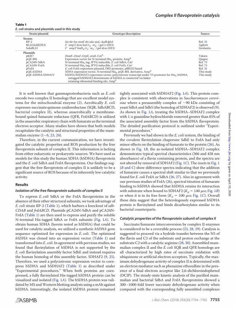
Table 1E. coli strains and plasmids used in this study
Strain/plasmid Genotype/description Source
StrainsRP-2 his thr leu metE thi eda rpsL; �sdh�frd Ref. 35BL21Gold(DE3) F�ompT dcm hsd (rB
�mB�) gal � (DE3) Agilent
SoluBL21 F� ompT hsdSB (rB�mB
�) gal dcm (DE3) GenlantisPlasmids
pKJE7 DnaK–DnaJ–GrpE, arab, CmR TakarapQE-80L Expression vector for N-terminal His6-protein, AmpR QiagenpCA24N-SdhA N-terminal His6 tag, IPTG inducible, E. coli SdhA, Cmr Ref. 73pCA24N-FrdA N-terminal His6 tag, IPTG inducible, E. coli FrdA, Cmr Ref. 73pFrdA E. coli FrdA expression plasmid, FRD promoter, pBR322 based Ref. 35pQE-hSDHA SDHA expression vector, N-terminal His6, pQE-80L derivative, AmpR This studypQE-hSDHA/SDHAF2 hSDHA/hSDHAF2 expression vector, polycistronic transcript under T5 promoter for His6-hSDHA,
untagged hSDHAF2 downstream of hSDHA is connected via linkerretaining ribosomal binding site, AmpR
This study
Complex II flavoprotein catalysis
J. Biol. Chem. (2018) 293(20) 7754 –7765 7755
by guest on April 10, 2020
http://ww
w.jbc.org/
Dow
nloaded from

(Table 2). The steady-state kcatapp of �0.15 s�1 was detected with
DCIP as electron acceptor and was not stimulated in the pres-ence of phenazine ethosulfate. The rates of the reactionsremained the same under either aerobic or anaerobic condi-tions, indicating that there is no significant leak to oxygen dur-ing the reaction.
The rate of FAD reduction by succinate in assembled com-plex II enzymes is much faster than kcat
app and has never beenreliably determined because of instrumental and experimentallimitations (i.e. the mixing time exceeds the rate of FAD reduc-tion and inherent difficulties in removing tightly bound oxalo-acetate from the active site). Direct monitoring of flavin reduc-tion in the unassembled flavoproteins is possible because of thelack of spectral interference from other complex II redox cofac-tors (i.e. iron–sulfur centers and b heme). Therefore we exam-ined the direct reduction of FAD by succinate to investigatewhether the low kcat
app was due to an inherent slow rate of reduc-tion of the FAD or because DCIP is a poor electron acceptor forthe unassembled flavoprotein. As shown in Fig. 2A, both bac-terial and human flavoproteins are redox-active with succinate/fumarate as followed by optical changes of the flavin at 460 nm.The observed monophasic rates of FAD reduction by succinatein the flavoproteins were found to be similar (�0.14 � 0.02 s�1;Fig. 2A, panels a– c). Moreover, the rate constants were notaffected when the reaction was performed under anaerobicconditions (data not shown). Fig. 2B demonstrates the reactionof fumarate reoxidation of the dithionite reduced bacterialFrdA and SdhA. This reaction was performed anaerobically asthe dithionite-reduced form of the flavoproteins, which are freeof dicarboxylates can be reoxidized by oxygen (i.e. when a dicar-boxylate is present at the active site this reoxidation is inhibited(19, 20)). The observed rates of �0.15 � 0.02 s�1 of flavin reoxi-dation by fumarate (Fig. 2, A and B) are also consistent with thesteady-state rate of the BV�-fumarate reductase reaction(Table 2), indicating that in the unassembled flavoprotein the
reaction of succinate-oxidation and fumarate-reduction areequally impaired.
ROS production by isolated complex II flavoproteins
Oxygen is known to react with reduced complex II and pro-duce superoxide and H2O2. With succinate as an electrondonor, mammalian and bacterial SQRs produce mostly super-oxide with maximum rates at �50 �M succinate (20, 21). E. coliQFR emitted predominately superoxide at low and H2O2 athigh succinate concentrations (19). FAD is the most plausiblesite for protein interaction with oxygen in these complexesbased on the ability of high substrate concentrations to sup-press reactive oxygen species formation (19 –21). Thus, there isa high probability that the isolated reduced flavoproteins mightbe especially reactive with oxygen in the absence of the othersubunits. Given that we observed a tight association betweenhSDHA and SDHAF2 (Fig. 1A) and previous observations inyeast (9), it is likely that in vivo hSDHA–SDHAF2 would befound in a complex in the mitochondrial matrix. Nevertheless,when testing whether the proteins could produce ROS, weassayed the purified hSDHA both essentially free of SDHAF2and with tightly associated SDHAF2. We also compared theactivity to what we observe for E. coli SdhA and FrdA. Wefound that there was no detectable reaction observed for eitherthe bacterial or human isolated flavoproteins with acetylatedcytochrome c, indicating a lack of detectable superoxide pro-duction. In contrast, all three proteins had low but detectablelevels of H2O2 production with the highest rates observed at 50�M succinate (Table 3). Higher concentrations of succinatesuppressed this H2O2 production, as would be expected if theflavin is the site of oxygen reactivity (19). The hSDHA withtightly associated SDHAF2 also showed no detectable reactionwith oxygen, which is not surprising because of the dramaticeffect that this association has on FAD reduction by succinate(Fig. 2A, panel d). The unassembled flavoproteins are also
Figure 1. Purification of flavoproteins and spectra of hSdhA. A, SDS-PAGE comparison of purified hSDHA, E. coli SdhA, and FrdA preparations. Proteins wereprepared as described under “Experimental procedures.” The left lane shows that the as-isolated hSDHA contains tightly bound SDHAF2. The identity ofSDHAF2 was determined by MS. The third and fourth lanes show the E. coli SdhA and FrdA preparations, respectively. The top panel (FAD-UV) shows the in gelfluorescence, indicating the similar content of covalent FAD in human and bacterial flavoproteins. B, visible absorbance spectra of the hSDHA/SDHAF2complex. The blue spectrum is the as-isolated complex, and the black spectrum is in the presence of 9 mM fumarate. The spectra were recorded in 50 mM BTP (pH7.0). The concentration of hSDHA was 7.8 �M. The inset shows the fumarate induced difference spectrum. The lower inset shows the optical titration of fumaratebinding using the absorbance at 505– 451 nm as previously described (26). C, visible absorbance spectra of free hSDHA following guanidine HCl treatment. Thespectrum of the ligand free hSDHA (5.2 �M) is shown (blue line) and in the presence of 4 mM fumarate (black line). The inset shows the fumarate induceddifference spectrum (top) and the optical titration of fumarate binding using 508 – 455 nm (bottom).
Complex II flavoprotein catalysis
7756 J. Biol. Chem. (2018) 293(20) 7754 –7765
by guest on April 10, 2020
http://ww
w.jbc.org/
Dow
nloaded from

highly overexpressed (�10 –20 mg protein/L-cell culture)without having any effect on cell growth. We also examinedH2O2 production in E. coli cellular extracts where we added thepurified FrdA or SdhA proteins to see whether there was anendogenous cellular component that might stimulate ROS for-mation. There was, however, no change in H2O2 productionbeyond the low basal H2O2 activity shown in Table 3 even withthe addition of 50 �M to 2 mM succinate. Thus, it appearsunlikely that the isolated flavoprotein subunit would be a majorsource of damaging ROS in situations where assembly iscompromised.
Characterization of the flavoproteins
Because there seems to be no a priori reason for the flavopro-tein subunits to have such a low catalytic activity, we next ruledout several reasons for this loss of catalysis. First, we investi-gated whether the flavoprotein subunits could be bound to aninhibitor molecule. Membrane-bound complex II, as isolated,contains a tightly bound dicarboxylate inhibitor (oxaloacetate)that must be removed to achieve maximal initial activity (31–33). The soluble domain of complex II (SdhAB/FrdAB), how-ever, is isolated free of this metabolic inhibitor and does notrequire preactivation to achieve the full rate of catalysis (34).Each of the free flavoproteins, like soluble SdhAB or FrdAB,however, showed maximal activity as isolated. This would sug-gest that the dicarboxylate-binding site is free of bound inhibi-tor and that bound oxaloacetate is not the reason for the inher-ently lower activity.
Next, we questioned whether aspects of the construct wereinterfering with catalysis. The flavoproteins were constructedwith an N-terminal His tag. Previous work showed that a Histag does not interfere with covalent flavinylation of the proteins(27, 35). This indicates that the proteins are folded correctly forinteraction with the SdhE chaperone and that the His tag doesnot affect active site preorganization for self-catalytic FADattachment (9, 11, 25, 27, 35). Nevertheless, we investigatedwhether the presence of the N-terminal His tag might be thereason for the low activity of the isolated flavoproteins. We thuscompared the kinetic properties of the purified N-terminal His-tagged FrdA and WT untagged FrdA. The rationale for focus-ing on E. coli FrdA for this experiment is that WT FrdA is highlyexpressed anaerobically under control of the natural FRD pro-moter in E. coli RP-2 (Table 2) deleted for both frdABCD and
sdhCDAB. The WT FrdA protein can also be purified by con-ventional ion exchange chromatography (27). Isolated WTFrdA shows the same rates for succinate oxidation and fumar-ate reduction as shown in Table 2 and Fig. 2B as the His-taggedcounterpart. The proteins were also equally stable from multi-ple freeze/thaw cycles and prolonged incubation at ambienttemperature. These data suggest that N-terminal His tag doesnot affect the folding, covalent flavinylation, or catalytic prop-erties of the flavoprotein. As a result, the low activity is likely aninherent property of all complex II flavoproteins in the absenceof the other complex II subunits.
One factor that was important for analysis of catalytic activityin the unassembled flavoproteins was the presence of the chap-erone involved in covalent flavin assembly. Consistent withprevious findings for the effect of SdhE on catalytic activity ofFrdA (26), we found that SDHAF2 inhibited flavin reductionand reoxidation by hSDHA (Fig. 2A, panel d). As noted in Fig.1A, the as-isolated hSDHA protein was in a tight complex withSDHAF2 requiring mild chaotropes to separate the two pro-teins. The association between the hSDHA and SDHAF2appears to be much stronger than binding of SdhE to the bac-terial flavoproteins where this association has Kd values of 0.7�M for bacterial FrdA and 1.5 �M for bacterial SdhA (26). Thus,one major difference between the bacterial and mammalianassembly factors is that the former is readily removed fromthe flavoprotein, whereas the mammalian complex is tightlybound.
Kinetic isotope and pH profiles of isolated FrdA and SdhA
In intact complex II catalysis includes the transfer of twoprotons between the substrate and the protein, as well as protondelivery from the bulk solvent to reprotonate the catalytic argi-nine. Therefore, because of the multiple sites anticipated to beinvolved in proton exchange, we investigated the pH depen-dence and kinetic isotope effects of the succinate dehydroge-nase reaction. We had previously determined the pKa of theintact SQR and QFR complexes to be �7.5 with the rateenhanced as the pH increases (23). We found the same for theisolated SdhA and FrdA subunits, although the pKa had shiftedto �8.0 for both flavoproteins (data not shown).
Previous studies of succinate dehydrogenase and fumaratereductase homologs have observed substrate and solventkinetic isotope effects (36 –38). Therefore, we compared the
Table 2Kinetic parameters of isolated FrdA, SdhA, and membrane-bound SQR and QFR complexes
Protein usedSuccinate-DCIP reductase, kcat
app BV�-fumarate-reductase, activitya
Complex II Flavoprotein Complex II Flavoprotein
s�1 fum/sBovine mitochondrial
Complex IIa 111b 2.7b
hSDHA 0.12 � 0.01 0.12 � 0.02E. coli
SQR 102 � 4c 1.5 � 0.1c
QFR 27 � 0.5c 197 � 3c
SdhA 0.15 � 0.01 0.13 � 0.01FrdA 0.16 � 0.01 0.12 � 0.01
a Activity was determined as described under “Experimental procedures” and corresponds to nmol fumarate/s per nmol flavoprotein (fum/s). Succinate-DCIP reductase andBV�-fumarate reductase activity data were determined at 30 °C and represents three separate independent analyses.
b Calculated for bovine heart complex II based on 6 nmol FAD/mg protein (74).c Succinate:PES-DCIP reductase reaction at pH 8.0 and BV�-fumarate reductase reaction at pH 7.0 was determined as described in Refs. 23, 29, and 31 and under “Experi-
mental procedures.”
Complex II flavoprotein catalysis
J. Biol. Chem. (2018) 293(20) 7754 –7765 7757
by guest on April 10, 2020
http://ww
w.jbc.org/
Dow
nloaded from
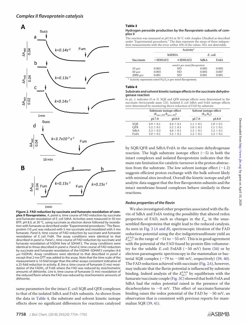
same parameters for the intact E. coli SQR and QFR complexesto that of the isolated SdhA and FrdA subunits. As shown fromthe data in Table 4, the substrate and solvent kinetic isotopeeffects show no significant differences for reactions catalyzed
by SQR/QFR and SdhA/FrdA in the succinate dehydrogenasereaction. The high substrate isotope effect (�5) in both theintact complexes and isolated flavoproteins indicates that themain rate limitation for catalytic turnover is the proton abstrac-tion from the substrate. The low solvent isotope effect (�1.2)suggests efficient proton exchange with the bulk solvent likelywith minimal sites involved. Overall the kinetic isotope and pHanalytic data suggest that the free flavoprotein subunits and theintact membrane-bound complexes behave similarly in thesereactions.
Redox properties of the flavin
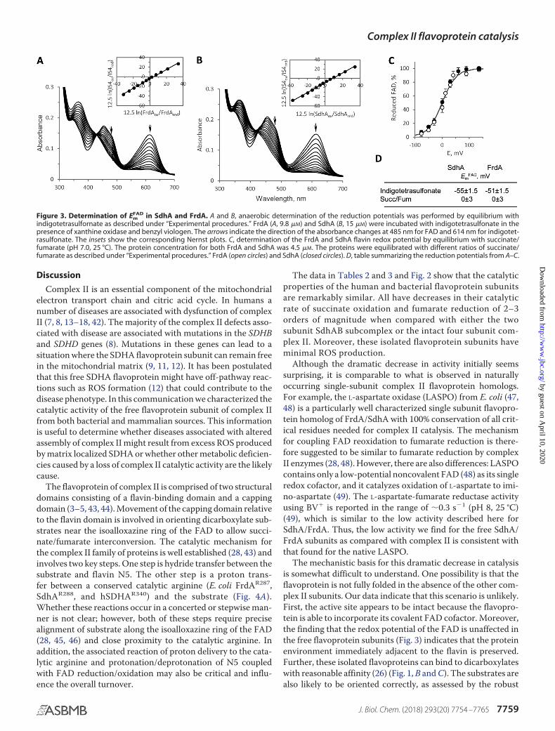
We also investigated other properties associated with the fla-vin of SdhA and FrdA testing the possibility that altered redoxproperties of FAD, such as changes in the Em in the unas-sembled flavoproteins that might lead to the reduced catalysis.As seen in Fig. 3 (A and B), spectroscopic titration of the FADreduction potential using the dye indigotetrasulfonate yield anEm,7
FAD in the range of �51 to �55 mV. This is in good agreementwith the potential of the FAD found by protein film voltamme-try for the soluble E. coli FrdAB (�50 mV) form (34) or byelectron paramagnetic spectroscopy in the mammalian or bac-terial SQR complex (�79 to �100 mV, respectively) (39, 40).The FAD reduction achieved with succinate (Fig. 2A), however,may indicate that the flavin potential is influenced by substratebinding. Indeed analysis of the Em,7
FAD by equilibrium with thefumarate/succinate couple (Fig. 3C) showed that both FrdA andSdhA had the redox potential raised in the presence of thedicarboxylates to �0 mV. This effect of succinate/fumaratebinding raises the redox potential of the FAD by �50 mV, anobservation that is consistent with previous reports for mam-malian SQR (39, 41).
Figure 2. FAD reduction by succinate and fumarate reoxidation of com-plex II flavoproteins. A, panel a, time course of FAD reduction by succinateand fumarate reoxidation of E. coli SdhA. Activities were measured in 50 mM
BTP, pH 8.0, at 30 °C, using succinate as electron donor followed by reoxida-tion with fumarate as described under “Experimental procedures.” The flavo-protein (10 �M) was reduced with 5 mM succinate and reoxidized with 5 mM
fumarate. Panel b, time course of FAD reduction by succinate and fumaratereoxidation of E. coli FrdA. The assay conditions were identical to thatdescribed in panel a. Panel c, time course of FAD reduction by succinate andfumarate reoxidation of hSDHA free of SDHAF2. The assay conditions wereidentical to those described in panel a. Panel d, time course of FAD reductionby succinate and fumarate reoxidation of the hSDHA–SDHAF2 complex (4.6�M hSDHA). Assay conditions were identical to that described in panel aexcept that 2 mM DTT was added to the assay. Note that the time scale of themeasurement is 10-fold longer than the other assays consistent indicative ofa 25-fold reduction in activity. B, line a, time course of fumarate (5 mM) reoxi-dation of the FADH2 of FrdA where the FAD was reduced by stoichiometricamounts of dithionite. Line b, time course of fumarate (5 mM) reoxidation ofthe reduced flavin where the FAD was reduced by stoichiometric amounts ofdithionite.
Table 3Hydrogen peroxide production by the flavoprotein subunits of com-plex IIThe reaction was measured at pH 8.0 at 30 °C with Amplex UltraRed as describedunder “Experimental procedures.” The data represent the mean of three indepen-dent measurements with the error within 10% of the values. ND, not detectable.
Activitya
hSDHA E. coli
Succinate �SDHAF2 �SDHAF2 SdhA FrdA
nmol/s per nmol flavoprotein10 �M 0.002 ND 0.003 0.00550 �M 0.003 ND 0.005 0.0072000 �M 0.001 ND 0.001 0.001
a Activity represents nmol H2O2/s per nmol flavoprotein.
Table 4Substrate and solvent kinetic isotope effects in the succinate dehydro-genase reactionIn pL, L indicates H or D. SQR and QFR isotope effects were determined in thesuccinate–ferricyanide assay (23). Isolated E. coli SdhA and FrdA isotope effectswere determined by monitoring direct reduction of FAD by substrate.
Substrate isotope effect(ksucc-H4/ksucc-D4)
Solvent isotope effect(kH/kD)
pL7.0 pL8.0 pL7.0 pL8.0
SQR 4.9 � 0.1 4.4 � 0.1 1.1 � 0.1 1.0 � 0.1QFR 5.2 � 0.1 5.2 � 0.1 1.0 � 0.1 1.1 � 0.1SdhA 5.3 � 0.2 4.6 � 0.1 1.3 � 0.1 1.2 � 0.1FrdA 5.0 � 0.1 5.2 � 0.1 1.2 � 0.1 1.2 � 0.1
Complex II flavoprotein catalysis
7758 J. Biol. Chem. (2018) 293(20) 7754 –7765
by guest on April 10, 2020
http://ww
w.jbc.org/
Dow
nloaded from
Discussion
Complex II is an essential component of the mitochondrialelectron transport chain and citric acid cycle. In humans anumber of diseases are associated with dysfunction of complexII (7, 8, 13–18, 42). The majority of the complex II defects asso-ciated with disease are associated with mutations in the SDHBand SDHD genes (8). Mutations in these genes can lead to asituation where the SDHA flavoprotein subunit can remain freein the mitochondrial matrix (9, 11, 12). It has been postulatedthat this free SDHA flavoprotein might have off-pathway reac-tions such as ROS formation (12) that could contribute to thedisease phenotype. In this communication we characterized thecatalytic activity of the free flavoprotein subunit of complex IIfrom both bacterial and mammalian sources. This informationis useful to determine whether diseases associated with alteredassembly of complex II might result from excess ROS producedby matrix localized SDHA or whether other metabolic deficien-cies caused by a loss of complex II catalytic activity are the likelycause.
The flavoprotein of complex II is comprised of two structuraldomains consisting of a flavin-binding domain and a cappingdomain (3–5, 43, 44). Movement of the capping domain relativeto the flavin domain is involved in orienting dicarboxylate sub-strates near the isoalloxazine ring of the FAD to allow succi-nate/fumarate interconversion. The catalytic mechanism forthe complex II family of proteins is well established (28, 43) andinvolves two key steps. One step is hydride transfer between thesubstrate and flavin N5. The other step is a proton trans-fer between a conserved catalytic arginine (E. coli FrdAR287,SdhAR288, and hSDHAR340) and the substrate (Fig. 4A).Whether these reactions occur in a concerted or stepwise man-ner is not clear; however, both of these steps require precisealignment of substrate along the isoalloxazine ring of the FAD(28, 45, 46) and close proximity to the catalytic arginine. Inaddition, the associated reaction of proton delivery to the cata-lytic arginine and protonation/deprotonation of N5 coupledwith FAD reduction/oxidation may also be critical and influ-ence the overall turnover.
The data in Tables 2 and 3 and Fig. 2 show that the catalyticproperties of the human and bacterial flavoprotein subunitsare remarkably similar. All have decreases in their catalyticrate of succinate oxidation and fumarate reduction of 2–3orders of magnitude when compared with either the twosubunit SdhAB subcomplex or the intact four subunit com-plex II. Moreover, these isolated flavoprotein subunits haveminimal ROS production.
Although the dramatic decrease in activity initially seemssurprising, it is comparable to what is observed in naturallyoccurring single-subunit complex II flavoprotein homologs.For example, the L-aspartate oxidase (LASPO) from E. coli (47,48) is a particularly well characterized single subunit flavopro-tein homolog of FrdA/SdhA with 100% conservation of all crit-ical residues needed for complex II catalysis. The mechanismfor coupling FAD reoxidation to fumarate reduction is there-fore suggested to be similar to fumarate reduction by complexII enzymes (28, 48). However, there are also differences: LASPOcontains only a low-potential noncovalent FAD (48) as its singleredox cofactor, and it catalyzes oxidation of L-aspartate to imi-no-aspartate (49). The L-aspartate-fumarate reductase activityusing BV� is reported in the range of �0.3 s�1 (pH 8, 25 °C)(49), which is similar to the low activity described here forSdhA/FrdA. Thus, the low activity we find for the free SdhA/FrdA subunits as compared with complex II is consistent withthat found for the native LASPO.
The mechanistic basis for this dramatic decrease in catalysisis somewhat difficult to understand. One possibility is that theflavoprotein is not fully folded in the absence of the other com-plex II subunits. Our data indicate that this scenario is unlikely.First, the active site appears to be intact because the flavopro-tein is able to incorporate its covalent FAD cofactor. Moreover,the finding that the redox potential of the FAD is unaffected inthe free flavoprotein subunits (Fig. 3) indicates that the proteinenvironment immediately adjacent to the flavin is preserved.Further, these isolated flavoproteins can bind to dicarboxylateswith reasonable affinity (26) (Fig. 1, B and C). The substrates arealso likely to be oriented correctly, as assessed by the robust
Figure 3. Determination of EmFAD in SdhA and FrdA. A and B, anaerobic determination of the reduction potentials was performed by equilibrium with
indigotetrasulfornate as described under “Experimental procedures.” FrdA (A, 9.8 �M) and SdhA (B, 15 �M) were incubated with indigotetrasulfonate in thepresence of xanthine oxidase and benzyl viologen. The arrows indicate the direction of the absorbance changes at 485 nm for FAD and 614 nm for indigotet-rasulfonate. The insets show the corresponding Nernst plots. C, determination of the FrdA and SdhA flavin redox potential by equilibrium with succinate/fumarate (pH 7.0, 25 °C). The protein concentration for both FrdA and SdhA was 4.5 �M. The proteins were equilibrated with different ratios of succinate/fumarate as described under “Experimental procedures.” FrdA (open circles) and SdhA (closed circles). D, table summarizing the reduction potentials from A–C.
Complex II flavoprotein catalysis
J. Biol. Chem. (2018) 293(20) 7754 –7765 7759
by guest on April 10, 2020
http://ww
w.jbc.org/
Dow
nloaded from
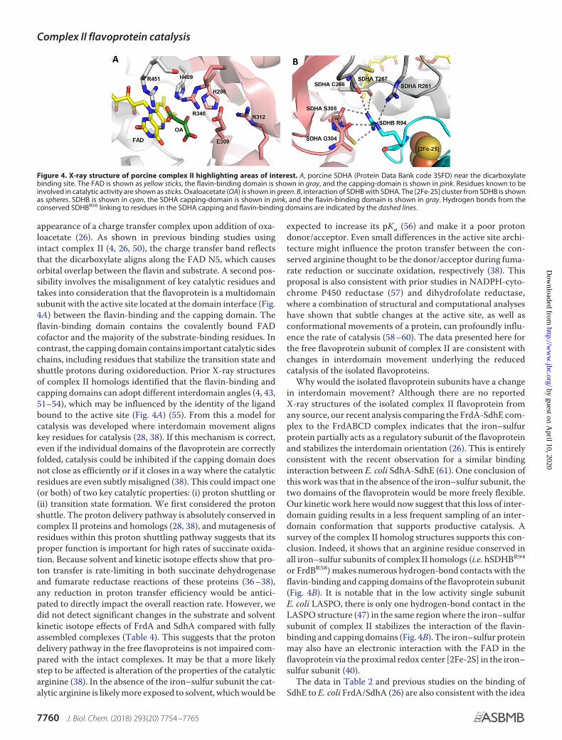
appearance of a charge transfer complex upon addition of oxa-loacetate (26). As shown in previous binding studies usingintact complex II (4, 26, 50), the charge transfer band reflectsthat the dicarboxylate aligns along the FAD N5, which causesorbital overlap between the flavin and substrate. A second pos-sibility involves the misalignment of key catalytic residues andtakes into consideration that the flavoprotein is a multidomainsubunit with the active site located at the domain interface (Fig.4A) between the flavin-binding and the capping domain. Theflavin-binding domain contains the covalently bound FADcofactor and the majority of the substrate-binding residues. Incontrast, the capping domain contains important catalytic sideschains, including residues that stabilize the transition state andshuttle protons during oxidoreduction. Prior X-ray structuresof complex II homologs identified that the flavin-binding andcapping domains can adopt different interdomain angles (4, 43,51–54), which may be influenced by the identity of the ligandbound to the active site (Fig. 4A) (55). From this a model forcatalysis was developed where interdomain movement alignskey residues for catalysis (28, 38). If this mechanism is correct,even if the individual domains of the flavoprotein are correctlyfolded, catalysis could be inhibited if the capping domain doesnot close as efficiently or if it closes in a way where the catalyticresidues are even subtly misaligned (38). This could impact one(or both) of two key catalytic properties: (i) proton shuttling or(ii) transition state formation. We first considered the protonshuttle. The proton delivery pathway is absolutely conserved incomplex II proteins and homologs (28, 38), and mutagenesis ofresidues within this proton shuttling pathway suggests that itsproper function is important for high rates of succinate oxida-tion. Because solvent and kinetic isotope effects show that pro-ton transfer is rate-limiting in both succinate dehydrogenaseand fumarate reductase reactions of these proteins (36 –38),any reduction in proton transfer efficiency would be antici-pated to directly impact the overall reaction rate. However, wedid not detect significant changes in the substrate and solventkinetic isotope effects of FrdA and SdhA compared with fullyassembled complexes (Table 4). This suggests that the protondelivery pathway in the free flavoproteins is not impaired com-pared with the intact complexes. It may be that a more likelystep to be affected is alteration of the properties of the catalyticarginine (38). In the absence of the iron–sulfur subunit the cat-alytic arginine is likely more exposed to solvent, which would be
expected to increase its pKa (56) and make it a poor protondonor/acceptor. Even small differences in the active site archi-tecture might influence the proton transfer between the con-served arginine thought to be the donor/acceptor during fuma-rate reduction or succinate oxidation, respectively (38). Thisproposal is also consistent with prior studies in NADPH-cyto-chrome P450 reductase (57) and dihydrofolate reductase,where a combination of structural and computational analyseshave shown that subtle changes at the active site, as well asconformational movements of a protein, can profoundly influ-ence the rate of catalysis (58 –60). The data presented here forthe free flavoprotein subunit of complex II are consistent withchanges in interdomain movement underlying the reducedcatalysis of the isolated flavoproteins.
Why would the isolated flavoprotein subunits have a changein interdomain movement? Although there are no reportedX-ray structures of the isolated complex II flavoprotein fromany source, our recent analysis comparing the FrdA-SdhE com-plex to the FrdABCD complex indicates that the iron–sulfurprotein partially acts as a regulatory subunit of the flavoproteinand stabilizes the interdomain orientation (26). This is entirelyconsistent with the recent observation for a similar bindinginteraction between E. coli SdhA-SdhE (61). One conclusion ofthis work was that in the absence of the iron–sulfur subunit, thetwo domains of the flavoprotein would be more freely flexible.Our kinetic work here would now suggest that this loss of inter-domain guiding results in a less frequent sampling of an inter-domain conformation that supports productive catalysis. Asurvey of the complex II homolog structures supports this con-clusion. Indeed, it shows that an arginine residue conserved inall iron–sulfur subunits of complex II homologs (i.e. hSDHBR94
or FrdBR58) makes numerous hydrogen-bond contacts with theflavin-binding and capping domains of the flavoprotein subunit(Fig. 4B). It is notable that in the low activity single subunitE. coli LASPO, there is only one hydrogen-bond contact in theLASPO structure (47) in the same region where the iron–sulfursubunit of complex II stabilizes the interaction of the flavin-binding and capping domains (Fig. 4B). The iron–sulfur proteinmay also have an electronic interaction with the FAD in theflavoprotein via the proximal redox center [2Fe-2S] in the iron–sulfur subunit (40).
The data in Table 2 and previous studies on the binding ofSdhE to E. coli FrdA/SdhA (26) are also consistent with the idea
Figure 4. X-ray structure of porcine complex II highlighting areas of interest. A, porcine SDHA (Protein Data Bank code 3SFD) near the dicarboxylatebinding site. The FAD is shown as yellow sticks, the flavin-binding domain is shown in gray, and the capping-domain is shown in pink. Residues known to beinvolved in catalytic activity are shown as sticks. Oxaloacetate (OA) is shown in green. B, interaction of SDHB with SDHA. The [2Fe-2S] cluster from SDHB is shownas spheres. SDHB is shown in cyan, the SDHA capping-domain is shown in pink, and the flavin-binding domain is shown in gray. Hydrogen bonds from theconserved SDHBR56 linking to residues in the SDHA capping and flavin-binding domains are indicated by the dashed lines.
Complex II flavoprotein catalysis
7760 J. Biol. Chem. (2018) 293(20) 7754 –7765
by guest on April 10, 2020
http://ww
w.jbc.org/
Dow
nloaded from
that the catalytic activity of the flavoprotein can be significantlymodulated by binding to a partner protein. For example, succi-nate oxidation in the intact SQR complex occurs at a rate of 100s�1 (Table 2), whereas when SdhA/hSDHA is associated withSdhE/SDHAF2, the rate is diminished to 0.001 s�1. This varia-tion in rate of 5 orders of magnitude may reflect evolutionaryconstraints to inhibit unwanted side reactions that might occurprior to assembly in a mature complex. For example, LASPOand mitochondrial dihydroorotate dehydrogenase can reducefumarate in addition to their normal substrates (48, 62). There-fore, it might be possible that the unassembled flavoproteinscould gain these off-path reactions that intact complex II lacks.We did investigate whether SdhA, hSDHA, and FrdA, in theirfree form or in complex with SdhE or SDHAF2, could react withL-aspartate or dihydroorotate but found no detectable activity(data not shown). Thus, we did not find any evidence for moon-lighting activity of the free flavoproteins. In addition to identi-fying a loss of activity in the isolated flavoproteins, one impactof this work is in the bacterial expression of the flavoproteinssubunit of human complex II. One notable difference betweenthe bacterial and mammalian proteins was in their response totheir covalent FAD assembly factors. In bacteria, SdhE readilydissociates from either SdhA or FrdA once the covalent FADlinkage has formed (35, 63, 64). By contrast, SDHAF2 binds verytightly to hSDHA, and mild chaotropic agents were needed todissociate the tightly bound complex. The reason for the verytight binding between hSDHA and SDHAF2 compared withE. coli SdhA/FrdA–SdhE is not known but is consistent withprior reports on the homologous complex from yeast, SdhA–Sdh5. Bacterial SdhE and yeast Sdh5 have identical structuralcores (65, 66) with this conserved region thought to be suffi-cient for enhancing covalent flavinylation. Here we confirmthat bacterial SdhE cannot enhance the covalent flavinylation ofthe hSDHA, yet prior studies revealed that yeast Sdh5 can com-plement mutations in hSDHAF2 (9). When the assembly ofyeast complex II is perturbed, a complex between SdhA–Sdh5was found in the mitochondrial matrix (9). This is consistentwith the tighter association we also find for the mammalianproteins. Compared with E. coli SdhE, the mature humanSDHAF2 and yeast Sdh5 are �50% larger. For example, thehuman SDHAF2 is extended by 31 amino acids at its N termi-nus and 16 amino acids at its C terminus compared with theshorter bacterial SdhE (88 amino acids total). Whether these N-and C-terminal extensions contribute to the tighter binding ofthe human SDHA–SDHAF2 complex or assist with covalentflavinylation has not yet been determined. This tight associa-tion may be needed to protect hSDHA and SDHAF2 fromLON-mediated proteolysis (67).
In, conclusion the findings are as follows: (i) mammalian andbacterial complex II flavoproteins are catalytically highly simi-lar; (ii) the free flavoprotein subunits of complex II have lowinherent catalytic activity and thus are not likely by themselvesto be an important source of ROS; and (iii) the lower activity ofthe free flavoprotein subunit compared with the intact complexlikely reflects the absence of a partner protein (i.e. the iron–sulfur subunit) whose binding is expected to align active siteresidues to enable more robust catalytic activity.
Experimental procedures
Strains, plasmids, and protein expression
The E. coli strains and the plasmids used in this study aredescribed in Table 1. To express the mature forms of humanSDHA and SDHAF2 lacking the N-terminal mitochondrialtransit peptide their respective DNA sequences were opti-mized for expression in E. coli and synthesized by GenScript(Piscataway, NJ). The synthesized DNA was then cloned intothe 5� BamHI and 3� SalI site of pQE-80L (Qiagen). Theresulting constructs have a N-terminal His6 tag under thecontrol of an isopropyl �-D-1-thiogalactopyranoside (IPTG)-inducible T5 promoter. The resulting N terminus of therecombinant human SDHA flavoprotein including the Histag was MRGSHHHHHHG-43SAKVSDSISA.
Construction of pQE-hSDHA/SDHAF2. hSDHA and humanSDHAF2 were expressed from a single polycistronic transcriptthat includes an N-terminal His6-hSdhA construct under con-trol of a T5 promoter and untagged mature hSDHAF2 thatretains the Shine–Delgarno translational enhancer elementsfrom the original T5 promoter region found in plasmidpQE80L. First, pQN-hSDHAF2 was created noting that thesequence of mature hSDHAF2 starts at amino acid 37 (67). Ourconstruct was cloned into the BglII–SalI site of the pQT vector(pQN-hSDHAF2), and the first two residues are replaced withMet and Arg; thus in our construct SDHAF2 starts with Ser-39.Thus, the N terminus of SDHAF2, which lacks the His tag, wasMR-39SPTDSQKDMI. The SDHAF2 N terminus was designedfrom the work of Bezawork-Galeta et al. (67), in which it wasshown that there is a two-step processing of the SDHAF2 tran-sit peptide.
To make pQE-hSDHA/hSDHAF2, the AatII–Ecl136II frag-ment of pQE-hSDHA harboring the T5-hSDHA was clonedinto the AatII–PsiI site of vector pQN-hSDHAF2. The latterdigestion removes the initial portion of the T5 promoter forSDHAF2 so that expression of both genes is from a single T5promoter. Plasmid pFrdA used to express the tag-free WTE. coli FrdA flavoprotein subunit was constructed as previouslydescribed (35).
DNA manipulations were performed using the QuikChangeII XL site-directed mutagenesis kit (Agilent, Santa Clara, CA)with appropriate primers obtained from Eurofins MWGOperon (Huntsville, AL). All mutations and constructs wereverified by DNA sequencing (Sequetech, Mountain View, CA).
The E. coli SdhA and FrdA subunits containing an N-termi-nal His6 tag were expressed and purified as previously described(35). The hSDHA was expressed from plasmid pQE-hSDHA/SDHAF2. E. coli BL21Gold(DE3) cells transformed with pQE-hSDHA/SDHAF2 and the chaperone containing plasmid pKJE7were maintained in the presence of 100 �g/ml ampicillin and 20�g/ml of chloramphenicol throughout cell growth. A 5-mlstarter culture was grown overnight in 1 ml of LB-broth con-taining the antibiotics at 37 °C with moderate aeration. Thefollowing morning 50 ml of LB medium was inoculated with 1ml of the overnight culture and growth continued for 5 h at30 °C. The entire culture was then used to inoculate 1 liter of LBmedia in a 4-liter flask, and growth was initiated at 25 °C withmoderate aeration. After 1 h the cells were induced with 0.1 mM
Complex II flavoprotein catalysis
J. Biol. Chem. (2018) 293(20) 7754 –7765 7761
by guest on April 10, 2020
http://ww
w.jbc.org/
Dow
nloaded from
IPTG and grown for 17 h. The cells were then harvested bycentrifugation (5,000 g for 10 min), and the cell pellet waswashed one time with 5 mM MgCl2 and then stored at �80 °C.
Protein purification
His-tagged proteins were purified on Ni-NTA resin (Qia-gen). Frozen cells were resuspended in 25 mM HEPES, pH 7.5,with Complete EDTA-free protease inhibitor tablets (RocheApplied Sciences). The cells were disrupted with a Emulsi-Flex-C5 homogenizer (Avestin, Ottawa, Canada), and the celllysate obtained by centrifugation (120,000 g for 40 min). Thelysate was applied to Ni-NTA gravity column. The column waswashed with the equilibration buffer (25 mM HEPES, pH 7.5, 30�M imidazole, 0.5 M NaCl, 10% glycerol), and the His-taggedflavoprotein was eluted with 25 mM HEPES, pH 7.5, 250 mM
imidazole, 0.1 M NaCl, 10% glycerol. The eluted fractions con-taining hSDHA were concentrated on a Centriprep 30-kDa(Millipore) centrifugal filter. The concentrated protein wasthen diluted 10-fold with 25 mM HEPES, pH 7.5, 50 mM NaCl,5% (v/v) glycerol and applied to diethylaminoethyl Sepharosefast flow resin equilibrated with 20 mM HEPES, (pH 7.6) andeluted on an AKTA Explorer (GE Healthcare) with a 20 –120mM NaCl gradient in the same buffer. The eluted fractions con-taining hSDHA were concentrated as described above andstored at �80 °C. The E. coli FrdA/SdhA proteins were purifiedas described (35).
Guanidine HCl treatment
A 3 ml of Ni-NTA (Thermo Scientific) gravity column wasequilibrated with 20 mM HEPES, pH 7.5, 25% glycerol, 0.1 M
NaCl at 4 °C. The purified hSDHA–SDHAF2 complex wasbound to the column and washed with the same equilibrationbuffer used to purify the complex. 20 ml of 1 M guanidine HCl,in the same buffer, was used to wash the column over 40 min.Guanidine HCl was removed by decreasing the concentration(1.0 – 0 M) of guanidine HCl in the washing buffer; in 0.2 M stepincrements and 5 ml for each wash step. Finally the column waswashed with the equilibration buffer and hSDHA eluted with0.25 M imidazole in the buffer. After concentration with Cen-triprep 30-kDa filters and buffer exchange using the same equil-ibration buffer, the resulting Gu-hSDHA which was 85–95%free of SDHAF2 was stored at �80 °C.
Optical and kinetic studies
Spectral and kinetic assays were done using an Agilent 8453UV-visible spectrophotometer. Kinetic studies were performedin 50 mM Bis-Tris-propane (BPT), 0.2 mM EDTA at 30 °C. FADreduction was monitored at 460 nm after manual addition ofsuccinate to the assay cuvette. The succinate dehydrogenasereaction (succinate–DCIP) was determined in the presence of50 �M DCIP, (�600 � 21.8 mM�1 cm�1) and various succinateconcentrations. Superoxide production was monitored in thepresence of 20 �M acetylated cytochrome c at 550 nm. Hydro-gen peroxide formation was detected at 572 nm (resorufin for-mation, �572 � 54 mM�1 cm�1) using 10 �M Amplex UltraRed(Invitrogen) and peroxidase as previously described (68). Cata-lase was added to quench the activity.
Fumarate reductase activity with benzyl viologen (BV)
The reaction was performed anaerobically as described (29)in 50 mM BTP, pH 7.0, at 30 °C. Glucose (10 mM) and glucose/oxidase were added to maintain anaerobic conditions. BV (0.2mM) was substoichiometrically reduced with sodium dithioniteto yield �90 �M BV� (�550 � 7.8 mM�1). Enzyme was added,and the reaction was started with 10 mM fumarate. For themembrane-bound SQR and QFR complexes, 0.05% TritonX-100 was included in the reaction mixture.
Kinetic isotope effects
The effect of solvent deuteration on turnover rates was fol-lowed at 30 °C. For the solvent kinetic isotope effect of the SQRand QFR complexes, the succinate-ferricyanide reductase assaywas used as previously described (23). The solvent kinetic iso-tope effect for the isolated SdhA and FrdA was measured by thedirect monitoring of flavin reduction as described above. Bufferand substrate solutions were prepared in both D2O (CambridgeIsotope Laboratories, Andover, MA) and H2O, and correctionsfor pD � pH meter reading �0.4 were done as described (69).The substrate kinetic isotope effect using succinate deuteratedat positions C2 and C3 (succinic-D4; Sigma–Aldrich) weredone in H2O containing buffer with succinate-H4 or succinate-D4. The protein containing solutions were equilibrated for �1min, and the reaction was started by the addition of either suc-cinate-D4 or succinate-H4. The substrate and solvent isotopeeffects were calculated as a ratio (kH/kD). pL is defined as whereL � H or D.
Determination of FAD reduction potential withindigotetrasulfonate
The reductive titration of SdhA and FrdA were performedspectroscopically according to the method of Massey (70) usingthe reference dye indigotetrasulfonate (Sigma) (Em.7 � �46mV). The reaction was done in 50 mM potassium phosphate, pH7.0, at 25 °C. Anerobic conditions were established in the pres-ence of 10 mM glucose, glucose oxidase/catalase with argonlayered upon the top of the cuvette. Indigotetrasulfonate wasadded to the cuvette to reach A614 � 0.15. Xanthine (0.3 mM)and 20 �M xanthine oxidase were also added to the cuvette.After the addition of the flavoproteins, the reaction was initi-ated by 20 �M benzyl viologen. The spectra were collected overa period of 40 min. The Em values were determined using theNernst equation for n � 2 for FAD and the dye as described(71). The absorbance changes corresponding to the reductionof the dye (614 nm) were plotted against those of the flavinrecorded at 485 nm, the isosbestic point for indigotetrasulfon-ate. Two independent titrations were performed.
FAD redox potential determination by equilibrium withsuccinate/fumarate
The redox potential (Em,7FAD) in the E. coli flavoproteins was
titrated with the succinate/fumarate couple. The steady-statereduction levels of FAD at each succinate/fumarate ratio weredetermined by calculating the residual fraction of oxidized FAD(466 –750 nm) and plotting against the redox potential poisedby the succinate/fumarate couple (Em � �30 mV, n � 2) cal-
Complex II flavoprotein catalysis
7762 J. Biol. Chem. (2018) 293(20) 7754 –7765
by guest on April 10, 2020
http://ww
w.jbc.org/
Dow
nloaded from

culated by the Nernst equation. E. coli SdhA or FrdA, 4.5 �M,were equilibrated in 50 mM potassium phosphate (pH 7.0) at25 °C. 1 M succinate (pH 7.0) and 1 M fumarate (pH 7.0) weremixed in different ratios at 10 mM total final concentration forthe redox titration. A spectrum was recorded when equilibriumhad been reached (�2 min). The fully reduced FAD spectrumwas recorded after the addition of sodium dithionite. 100%oxidized FAD corresponded to the spectrum obtained in thepresence of 10 mM fumarate. The graph (Fig. 3) representssimulation of the Nernst equation with Em � 0 mV. Thepotential measurements with the succinate/fumarate couplewere repeated three times.
In-gel flavin detection
The protein sample was separated by SDS-PAGE using Bio-Rad Any kDa gels. Following separation, the gel was incubatedin 10% acetic acid, 20% methanol for 5 min. FAD fluorescencewas detected using a Safe Imager 2.0 blue-light transilluminator(Thermo Fisher Scientific). The ImageJ (National Institutes ofHealth) program was used for quantification of FAD levels andrelative protein ratios.
Immunodetection of proteins
Proteins were separated by SDS-PAGE and transferred tonitrocellulose membranes using a Trans-Blot Turbo transfersystem (Bio-Rad). Proteins containing a His6 tag were detectedusing murine monoclonal anti-His6 antibody (Aviva SystemsBiology; catalog no. OAE00010) and horseradish peroxidasesecondary antibody (Thermo Fisher). The tag-free SDHAF2protein was detected using a rabbit mAb (Cell Signaling; catalogno. 45849). A mAb against hSDHA (AbCam catalog no.ab14715) was also used to detect the mammalian protein.
Analytical methods
Protein concentration was determined by the BCA proteinassay kit from Pierce. The concentration of the flavoproteinswas determined in 0.5% SDS from the absorbance of the cova-lent FAD cofactor assuming 1 mol FAD (�447 � 12 mM�1 cm�1)per mol flavoprotein (72). Proteins in SDS-PAGE gels werestained with Coomassie Blue G (Sigma–Aldrich).
Author contributions—E. M., T. M. I., and G. C. conceptualization;E. M. and G. C. resources; E. M., S. R., and T. M. I. data curation;E. M. and G. C. formal analysis; E. M. and G. C. supervision; E. M.,T. M. I., and G. C. funding acquisition; E. M., S. R., and T. M. I.investigation; E. M. and S. R. methodology; E. M., S. R., and G. C.writing-original draft; E. M. and G. C. project administration; E. M.,T. M. I., and G. C. writing-review and editing.
Acknowledgment—We thank Izumi Yamakawa for help with analysisof the association of the complex of hSDHA–SDHAF2.
References1. Hägerhäll, C. (1997) Succinate:quinone oxidoreductases: variations on
a conserved theme. Biochim. Biophys. Acta 1320, 107–141 CrossRefMedline
2. Cecchini, G. (2003) Function and structure of complex II of the respiratorychain. Annu. Rev. Biochem. 72, 77–109 CrossRef Medline
3. Yankovskaya, V., Horsefield, R., Törnroth, S., Luna-Chavez, C., Miyoshi,H., Léger, C., Byrne, B., Cecchini, G., and Iwata, S. (2003) Architecture ofsuccinate dehydrogenase and reactive oxygen species generation. Science299, 700 –704 CrossRef Medline
4. Huang, L. S., Shen, J. T., Wang, A. C., and Berry, E. A. (2006) Crystallo-graphic studies of the binding of ligands to the dicarboxylate site of com-plex II, and the identity of the ligand in the “oxaloacetate-inhibited” state.Biochim. Biophys. Acta 1757, 1073–1083 CrossRef Medline
5. Sun, F., Huo, X., Zhai, Y., Wang, A., Xu, J., Su, D., Bartlam, M., and Rao, Z.(2005) Crystal structure of mitochondrial respiratory membrane proteincomplex II. Cell 121, 1043–1057 CrossRef Medline
6. Iverson, T. M., Maklashina, E., and Cecchini, G. (2012) Structural basis formalfunction in complex II. J. Biol. Chem. 287, 35430 –35438 CrossRefMedline
7. Bezawork-Geleta, A., Rohlena, J., Dong, L., Pacak, K., and Neuzil, J. (2017)Mitochondrial complex II: At the crossroads. Trends Biochem. Sci. 42,312–325 CrossRef Medline
8. Bardella, C., Pollard, P. J., and Tomlinson, I. (2011) SDH mutations incancer. Biochim. Biophys. Acta 1807, 1432–1443 CrossRef Medline
9. Hao, H. X., Khalimonchuk, O., Schraders, M., Dephoure, N., Bayley, J. P.,Kunst, H., Devilee, P., Cremers, C. W., Schiffman, J. D., Bentz, B. G., Gygi,S. P., Winge, D. R., Kremer, H., and Rutter, J. (2009) SDH5, a gene requiredfor flavination of succinate dehydrogenase, is mutated in paraganglioma.Science 325, 1139 –1142 CrossRef Medline
10. Na, U., Yu, W., Cox, J., Bricker, D. K., Brockmann, K., Rutter, J., Thummel,C. S., and Winge, D. R. (2014) The LYR factors SDHAF1 and SDHAF3mediate maturation of the iron–sulfur subunit of succinate dehydroge-nase. Cell Metab. 20, 253–266 CrossRef Medline
11. Van Vranken, J. G., Bricker, D. K., Dephoure, N., Gygi, S. P., Cox, J. E.,Thummel, C. S., and Rutter, J. (2014) SDHAF4 promotes mitochondrialsuccinate dehydrogenase activity and prevents neurodegeneration. CellMetab. 20, 241–252 CrossRef Medline
12. Van Vranken, J. G., Na, U., Winge, D. R., and Rutter, J. (2015) Protein-mediated assembly of succinate dehydrogenase and its cofactors. Crit. Rev.Biochem. Mol. Biol. 50, 168 –180 CrossRef Medline
13. Baysal, B. E. (2002) Hereditary paraganglioma targets diverse paraganglia.J. Med. Genet. 39, 617– 622 CrossRef Medline
14. King, K. S., and Pacak, K. (2014) Familial pheochromocytomas and para-gangliomas. Mol. Cell. Endocrinol. 386, 92–100 CrossRef Medline
15. Martucci, V. L., and Pacak, K. (2014) Pheochromocytoma and paragangli-oma: diagnosis, genetics, management, and treatment. Curr. Probl. Cancer38, 7– 41 CrossRef Medline
16. Gill, A. J. (2012) Succinate dehydrogenase (SDH) and mitochondrialdriven neoplasia. Pathology 44, 285–292 CrossRef Medline
17. Yamamoto, H., and Oda, Y. (2015) Gastrointestinal stromal tumor: recentadvances in pathology and genetics. Pathol. Int. 65, 9 –18 CrossRefMedline
18. Ozluk, Y., Taheri, D., Matoso, A., Sanli, O., Berker, N. K., Yakirevich, E.,Balasubramanian, S., Ross, J. S., Ali, S. M., and Netto, G. J. (2015) Renalcarcinoma associated with a novel succinate dehydrogenase A mutation: acase report and review of literature of a rare subtype of renal carcinoma.Hum. Pathol. 46, 1951–1955 CrossRef Medline
19. Imlay, J. A. (1995) A metabolic enzyme that rapidly produces superoxide,fumarate reductase of Escherichia coli. J. Biol. Chem. 270, 19767–19777Medline
20. Messner, K. R., and Imlay, J. A. (2002) Mechanism of superoxide andhydrogen peroxide formation by fumarate reductase, succinate dehydro-genase, and aspartate oxidase. J. Biol. Chem. 277, 42563– 42571 CrossRefMedline
21. Quinlan, C. L., Orr, A. L., Perevoshchikova, I. V., Treberg, J. R., Ackrell,B. A., and Brand, M. D. (2012) Mitochondrial complex II can generatereactive oxygen species at high rates in both the forward and reversereactions. J. Biol. Chem. 287, 27255–27264 CrossRef Medline
22. Ishii, T., Yasuda, K., Akatsuka, A., Hino, O., Hartman, P. S., and Ishii, N.(2005) A mutation in the SDHC gene of complex II increases oxidativestress, resulting in apoptosis and tumorigenesis. Cancer Res. 65, 203–209Medline
Complex II flavoprotein catalysis
J. Biol. Chem. (2018) 293(20) 7754 –7765 7763
by guest on April 10, 2020
http://ww
w.jbc.org/
Dow
nloaded from

23. Maklashina, E., Iverson, T. M., Sher, Y., Kotlyar, V., Andréll, J., Mirza, O.,Hudson, J. M., Armstrong, F. A., Rothery, R. A., Weiner, J. H., and Cec-chini, G. (2006) Fumarate reductase and succinate oxidase activity of Esch-erichia coli complex II homologs are perturbed differently by mutation ofthe flavin binding domain. J. Biol. Chem. 281, 11357–11365 CrossRefMedline
24. Cecchini, G., Schröder, I., Gunsalus, R. P., and Maklashina, E. (2002) Suc-cinate dehydrogenase and fumarate reductase from Escherichia coli.Biochim. Biophys. Acta 1553, 140 –157 CrossRef Medline
25. Zafreen, L., Walker-Kopp, N., Huang, L. S., and Berry, E. (2016) In-vitro,SDH5-dependent flavinylation of immobilized human respiratory com-plex II flavoprotein. Arch. Biochem. Biophys. 604, 47–56 CrossRefMedline
26. Sharma, P., Maklashina, E., Cecchini, G., and Iverson, T. M. (2018) Crystalstructure of an intermediate of the covalent flavinylation process of respi-ratory complex II. Nat. Commun. 9, 274 CrossRef Medline
27. Starbird, C. A., Maklashina, E., Sharma, P., Qualls-Histed, S., Cecchini, G.,and Iverson, T. M. (2017) Structural and biochemical analyses reveal in-sights into covalent flavinylation of the Escherichia coli complex II homo-log quinol:fumarate reductase. J. Biol. Chem. 292, 12921–12933 CrossRefMedline
28. Reid, G. A., Miles, C. S., Moysey, R. K., Pankhurst, K. L., and Chapman,S. K. (2000) Catalysis in fumarate reductase. Biochim. Biophys. Acta 1459,310 –315 CrossRef Medline
29. Ackrell, B. A., Armstrong, F. A., Cochran, B., Sucheta, A., and Yu, T. (1993)Classification of fumarate reductases and succinate dehydrogenases basedupon their contrasting behaviour in the reduced benzylviologen/fumarateassay. FEBS Lett. 326, 92–94 CrossRef Medline
30. Mowat, C. G., Pankhurst, K. L., Miles, C. S., Leys, D., Walkinshaw, M. D.,Reid, G. A., and Chapman, S. K. (2002) Engineering water to act as anactive site acid catalyst in a soluble fumarate reductase. Biochemistry 41,11990 –11996 CrossRef Medline
31. Ackrell, B. A., Kearney, E. B., and Edmondson, D. (1975) Mechanism of thereductive activation of succinate dehydrogenase. J. Biol. Chem. 250,7114 –7119 Medline
32. Kotlyar, A. B., and Vinogradov, A. D. (1984) Interaction of the membrane-bound succinate dehydrogenase with substrate and competitive inhibi-tors. Biochim. Biophys. Acta 784, 24 –34 CrossRef Medline
33. Ackrell, B. A., Cochran, B., and Cecchini, G. (1989) Interactions of oxalo-acetate with Escherichia coli fumarate reductase. Arch. Biochem. Biophys.268, 26 –34 CrossRef Medline
34. Léger, C., Heffron, K., Pershad, H. R., Maklashina, E., Luna-Chavez, C.,Cecchini, G., Ackrell, B. A., and Armstrong, F. A. (2001) Enzyme electro-kinetics: energetics of succinate oxidation by fumarate reductase and suc-cinate dehydrogenase. Biochemistry 40, 11234 –11245 CrossRef Medline
35. Maklashina, E., Rajagukguk, S., Starbird, C. A., McDonald, W. H., Kogan-itsky, A., Eisenbach, M., Iverson, T. M., and Cecchini, G. (2016) Binding ofthe covalent flavin assembly factor to the flavoprotein subunit of complexII. J. Biol. Chem. 291, 2904 –2916 CrossRef Medline
36. Hirst, J., Ackrell, B. A. C., and Armstrong, F. A. (1997) Global observationof hydrogen/deuterium isotope effects on bidirectional catalytic electrontransport in an enzyme: direct measurement by protein film voltammetry.J. Am. Chem. Soc. 119, 7434 –7439 CrossRef
37. Kaczorowski, G. J., Cheung, Y. F., and Walsh, C. (1977) Substrate kineticisotope effects in dehydrogenase coupled active transport in membranevesicles of Escherichia coli. Biochemistry 16, 2619 –2628 CrossRefMedline
38. Pankhurst, K. L., Mowat, C. G., Rothery, E. L., Hudson, J. M., Jones, A. K.,Miles, C. S., Walkinshaw, M. D., Armstrong, F. A., Reid, G. A., and Chap-man, S. K. (2006) A proton delivery pathway in the soluble fumarate re-ductase from Shewanella frigidimarina. J. Biol. Chem. 281, 20589 –20597CrossRef Medline
39. Ohnishi, T., King, T. E., Salerno, J. C., Blum, H., Bowyer, J. R., and Maida,T. (1981) Thermodynamic and electron paramagnetic resonance charac-terization of flavin in succinate dehydrogenase. J. Biol. Chem. 256,5577–5582 Medline
40. Cheng, V. W., Piragasam, R. S., Rothery, R. A., Maklashina, E., Cecchini,G., and Weiner, J. H. (2015) Redox state of flavin adenine dinucleotide
drives substrate binding and product release in Escherichia coli succinatedehydrogenase. Biochemistry 54, 1043–1052 CrossRef Medline
41. Bonomi, F., Pagani, S., Cerletti, P., and Giori, C. (1983) Modification of thethermodynamic properties of the electron-transferring groups in mito-chondrial succinate dehydrogenase upon binding of succinate. Eur.J. Biochem. 134, 439 – 445 CrossRef Medline
42. Rutter, J., Winge, D. R., and Schiffman, J. D. (2010) Succinate dehydroge-nase: assembly, regulation and role in human disease. Mitochondrion 10,393– 401 CrossRef Medline
43. Iverson, T. M., Luna-Chavez, C., Schröder, I., Cecchini, G., and Rees, D. C.(2000) Analyzing your complexes: structure of the quinol-fumarate reduc-tase respiratory complex. Curr. Opin. Struct. Biol. 10, 448 – 455 CrossRefMedline
44. Lancaster, C. R., Kröger, A., Auer, M., and Michel, H. (1999) Structure offumarate reductase from Wolinella succinogenes at 2.2 A resolution. Na-ture 402, 377–385 CrossRef Medline
45. Leys, D., Tsapin, A. S., Nealson, K. H., Meyer, T. E., Cusanovich, M. A., andVan Beeumen, J. J. (1999) Structure and mechanism of the flavocyto-chrome c fumarate reductase of Shewanella putrefaciens MR-1. Nat.Struct. Biol. 6, 1113–1117 CrossRef Medline
46. Mowat, C. G., Moysey, R., Miles, C. S., Leys, D., Doherty, M. K., Taylor, P.,Walkinshaw, M. D., Reid, G. A., and Chapman, S. K. (2001) Kinetic andcrystallographic analysis of the key active site acid/base arginine in asoluble fumarate reductase. Biochemistry 40, 12292–12298 CrossRefMedline
47. Mattevi, A., Tedeschi, G., Bacchella, L., Coda, A., Negri, A., and Ronchi, S.(1999) Structure of L-aspartate oxidase: implications for the succinate de-hydrogenase/fumarate reductase oxidoreductase family. Structure 7,745–756 CrossRef Medline
48. Tedeschi, G., Ronchi, S., Simonic, T., Treu, C., Mattevi, A., and Negri, A.(2001) Probing the active site of L-aspartate oxidase by site-directed mu-tagenesis: role of basic residues in fumarate reduction. Biochemistry 40,4738 – 4744 CrossRef Medline
49. Tedeschi, G., Nonnis, S., Strumbo, B., Cruciani, G., Carosati, E., and Negri,A. (2010) On the catalytic role of the active site residue E121 of E. coliL-aspartate oxidase. Biochimie 92, 1335–1342 CrossRef Medline
50. Dervartanian, D. V., and Veeger, C. (1964) Studies on succinate dehydro-genase: I. spectral properties of the purified enzyme and formation ofenzyme– competitive inhibitor complexes. Biochim. Biophys. Acta 92,233–247 Medline
51. Huang, L. S., Sun, G., Cobessi, D., Wang, A. C., Shen, J. T., Tung, E. Y.,Anderson, V. E., and Berry, E. A. (2006) 3-nitropropionic acid is a suicideinhibitor of mitochondrial respiration that, upon oxidation by complex II,forms a covalent adduct with a catalytic base arginine in the active site ofthe enzyme. J. Biol. Chem. 281, 5965–5972 CrossRef Medline
52. Tomasiak, T. M., Maklashina, E., Cecchini, G., and Iverson, T. M. (2008) Athreonine on the active site loop controls transition state formation inEscherichia coli respiratory complex II. J. Biol. Chem. 283, 15460 –15468CrossRef Medline
53. Bamford, V., Dobbin, P. S., Richardson, D. J., and Hemmings, A. M. (1999)Open conformation of a flavocytochrome c3 fumarate reductase. Nat.Struct. Biol. 6, 1104 –1107 CrossRef Medline
54. Iverson, T. M., Luna-Chavez, C., Cecchini, G., and Rees, D. C. (1999)Structure of the Escherichia coli fumarate reductase respiratory complex.Science 284, 1961–1966 CrossRef Medline
55. Starbird, C. A., Tomasiak, T. M., Singh, P. K., Yankovskaya, V., Mak-lashina, E., Eisenbach, M., Cecchini, G., and Iverson, T. M. (2018) Newcrystal forms of the integral membrane Escherichia coli quinol:fumaratereductase suggest that ligands control domain movement. J. Struct. Biol.202, 100 –104 CrossRef Medline
56. Harms, M. J., Schlessman, J. L., Sue, G. R., and García-Moreno, B. (2011)Arginine residues at internal positions in a protein are always charged.Proc. Natl. Acad. Sci. U.S.A. 108, 18954 –18959 CrossRef Medline
57. Shen, A. L., Sem, D. S., and Kasper, C. B. (1999) Mechanistic studies on thereductive half-reaction of NADPH-cytochrome P450 oxidoreductase.J. Biol. Chem. 274, 5391–5398 CrossRef Medline
Complex II flavoprotein catalysis
7764 J. Biol. Chem. (2018) 293(20) 7754 –7765
by guest on April 10, 2020
http://ww
w.jbc.org/
Dow
nloaded from

58. Boehr, D. D., Dyson, H. J., and Wright, P. E. (2008) Conformational relax-ation following hydride transfer plays a limiting role in dihydrofolate re-ductase catalysis. Biochemistry 47, 9227–9233 CrossRef Medline
59. Arora, K., and Brooks, C. L., 3rd (2013) Multiple intermediates, diverseconformations, and cooperative conformational changes underlie the cat-alytic hydride transfer reaction of dihydrofolate reductase. Top. Curr.Chem. 337, 165–187 CrossRef Medline
60. Hanoian, P., Liu, C. T., Hammes-Schiffer, S., and Benkovic, S. (2015) Per-spectives on electrostatics and conformational motions in enzyme catal-ysis. Acc. Chem. Res. 48, 482– 489 CrossRef Medline
61. Maher, M. J., Herath, A. S., Udagedara, S. R., Dougan, D. A., and Truscott,K. N. (2018) Crystal structure of bacterial succinate:quinone oxidoreduc-tase flavoprotein SdhA in complex with its assembly factor SdhE. Proc.Natl. Acad. Sci. U.S.A. 115, 2982–2987 CrossRef Medline
62. Inaoka, D. K., Sakamoto, K., Shimizu, H., Shiba, T., Kurisu, G., Nara, T.,Aoki, T., Kita, K., and Harada, S. (2008) Structures of Trypanosomacruzi dihydroorotate dehydrogenase complexed with substrates andproducts: atomic resolution insights into mechanisms of dihydrooro-tate oxidation and fumarate reduction. Biochemistry 47, 10881–10891CrossRef Medline
63. McNeil, M. B., Clulow, J. S., Wilf, N. M., Salmond, G. P., and Fineran, P. C.(2012) SdhE is a conserved protein required for flavinylation of succinatedehydrogenase in bacteria. J. Biol. Chem. 287, 18418 –18428 CrossRefMedline
64. McNeil, M. B., Hampton, H. G., Hards, K. J., Watson, B. N., Cook, G. M.,and Fineran, P. C. (2014) The succinate dehydrogenase assembly factor,SdhE, is required for the flavinylation and activation of fumarate reductasein bacteria. FEBS Lett. 588, 414 – 421 CrossRef Medline
65. Eletsky, A., Jeong, M. Y., Kim, H., Lee, H. W., Xiao, R., Pagliarini, D. J.,Prestegard, J. H., Winge, D. R., Montelione, G. T., and Szyperski, T. (2012)Solution NMR structure of yeast succinate dehydrogenase flavinylationfactor Sdh5 reveals a putative Sdh1 binding site. Biochemistry 51,8475– 8477 CrossRef Medline
66. Lim, K., Doseeva, V., Demirkan, E. S., Pullalarevu, S., Krajewski, W.,Galkin, A., Howard, A., and Herzberg, O. (2005) Crystal structure of the
YgfY from Escherichia coli, a protein that may be involved in transcrip-tional regulation. Proteins 58, 759 –763 Medline
67. Bezawork-Geleta, A., Saiyed, T., Dougan, D. A., and Truscott, K. N. (2014)Mitochondrial matrix proteostasis is linked to hereditary paraganglioma:LON-mediated turnover of the human flavinylation factor SDH5 is regu-lated by its interaction with SDHA. FASEB J. 28, 1794 –1804 CrossRefMedline
68. Kareyeva, A. V., Grivennikova, V. G., Cecchini, G., and Vinogradov, A. D.(2011) Molecular identification of the enzyme responsible for the mito-chondrial NADH-supported ammonium-dependent hydrogen peroxideproduction. FEBS Lett. 585, 385–389 CrossRef Medline
69. Rothery, E. L., Mowat, C. G., Miles, C. S., Walkinshaw, M. D., Reid, G. A.,and Chapman, S. K. (2003) Histidine 61: an important heme ligand in thesoluble fumarate reductase from Shewanella frigidimarina. Biochemistry42, 13160 –13169 CrossRef Medline
70. Massey, V. (1991) A simple method for the determination of redox poten-tials. In Flavins and Flavoproteins, pp. 59 – 66, Walter de Gruyter, NewYork
71. Efimov, I., Parkin, G., Millett, E. S., Glenday, J., Chan, C. K., Weedon, H.,Randhawa, H., Basran, J., and Raven, E. L. (2014) A simple method for thedetermination of reduction potentials in heme proteins. FEBS Lett. 588,701–704 CrossRef Medline
72. Cecchini, G., Ackrell, B. A., Deshler, J. O., and Gunsalus, R. P. (1986)Reconstitution of quinone reduction and characterization of Escherichiacoli fumarate reductase activity. J. Biol. Chem. 261, 1808 –1814 Medline
73. Kitagawa, M., Ara, T., Arifuzzaman, M., Ioka-Nakamichi, T., Inamoto,E., Toyonaga, H., and Mori, H. (2005) Complete set of ORF clones ofEscherichia coli ASKA library (a complete set of E. coli K-12 ORF ar-chive): unique resources for biological research. DNA Res 12, 291–299Medline
74. Grivennikova, V. G., Gavrikova, E. V., Timoshin, A. A., and Vinogradov,A. D. (1993) Fumarate reductase activity of bovine heart succinate-ubiqui-none reductase: new assay system and overall properties of the reaction.Biochim. Biophys. Acta 1140, 282–292 CrossRef Medline
Complex II flavoprotein catalysis
J. Biol. Chem. (2018) 293(20) 7754 –7765 7765
by guest on April 10, 2020
http://ww
w.jbc.org/
Dow
nloaded from

Elena Maklashina, Sany Rajagukguk, T. M. Iverson and Gary Cecchiniimpaired catalytic activity and generate only minor amounts of ROS
The unassembled flavoprotein subunits of human and bacterial complex II have
doi: 10.1074/jbc.RA118.001977 originally published online April 2, 20182018, 293:7754-7765.J. Biol. Chem.
10.1074/jbc.RA118.001977Access the most updated version of this article at doi:
Alerts:
When a correction for this article is posted•
When this article is cited•
to choose from all of JBC's e-mail alertsClick here
http://www.jbc.org/content/293/20/7754.full.html#ref-list-1
This article cites 73 references, 22 of which can be accessed free at
by guest on April 10, 2020
http://ww
w.jbc.org/
Dow
nloaded from