thioredoxin and glutaredoxin systems
TRANSCRIPT
Thioredoxin and glutaredoxin
systems
Arne Holmgren
Division of Biochemistry, Department of
Medical Biochemistry and Biophysics,
Karolinska Institute,Stockholm, Sweden
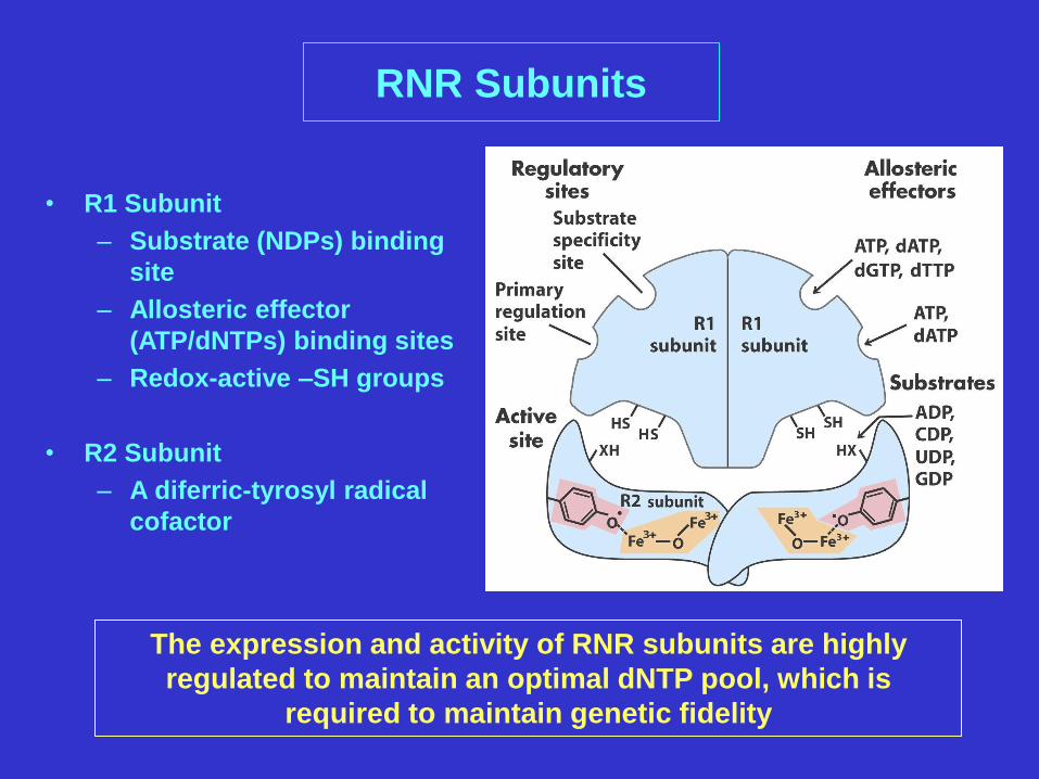
• R1 Subunit
– Substrate (NDPs) binding
site
– Allosteric effector
(ATP/dNTPs) binding sites
– Redox-active –SH groups
• R2 Subunit
– A diferric-tyrosyl radical
cofactor
RNR Subunits
The expression and activity of RNR subunits are highly
regulated to maintain an optimal dNTP pool, which is
required to maintain genetic fidelity
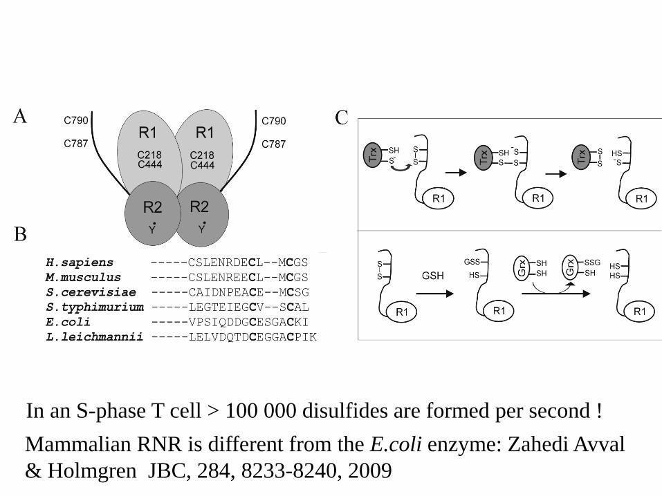
Mammalian RNR is different from the E.coli enzyme: Zahedi Avval
& Holmgren JBC, 284, 8233-8240, 2009
In an S-phase T cell > 100 000 disulfides are formed per second !
Recombinant RNR subunits were used to evaluate/characterize the kinetics of the
electron donors. Further characterization of the kinetics should contribute insights
into the mechanism of mammalian RNR reaction.
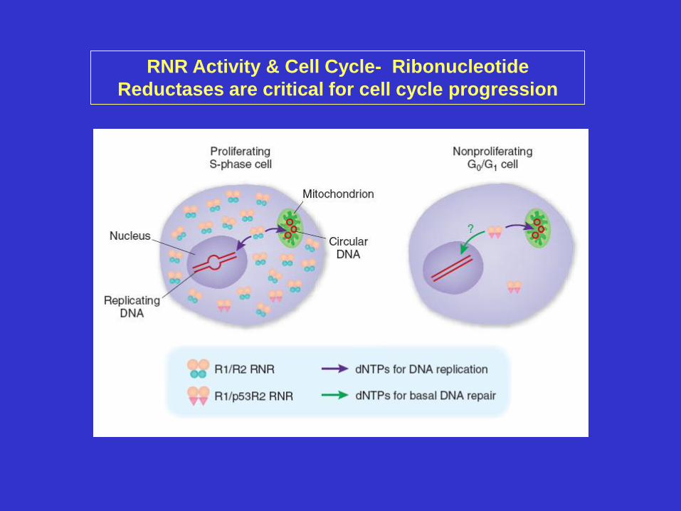
RNR Activity & Cell Cycle- Ribonucleotide
Reductases are critical for cell cycle progression
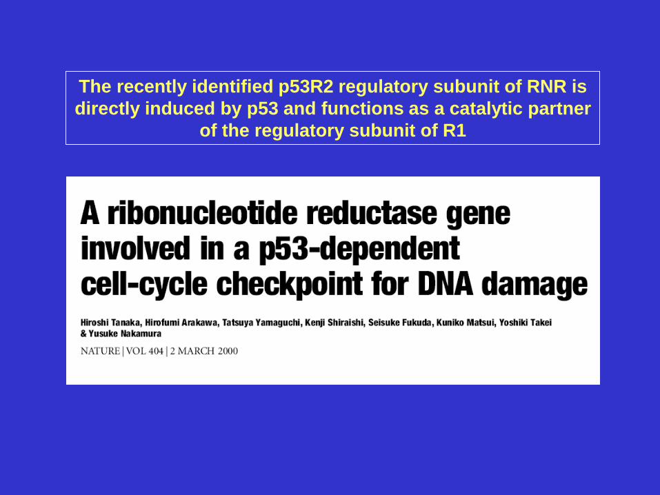
The recently identified p53R2 regulatory subunit of RNR is
directly induced by p53 and functions as a catalytic partner
of the regulatory subunit of R1
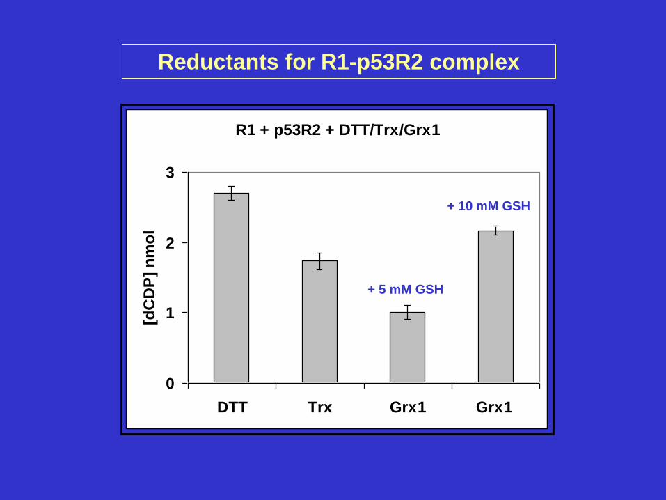
R1 + p53R2 + DTT/Trx/Grx1
0
1
2
3
DTT Trx Grx1 Grx1
[dC
DP
] n
mo
l
Reductants for R1-p53R2 complex
+ 5 mM GSH
+ 10 mM GSH
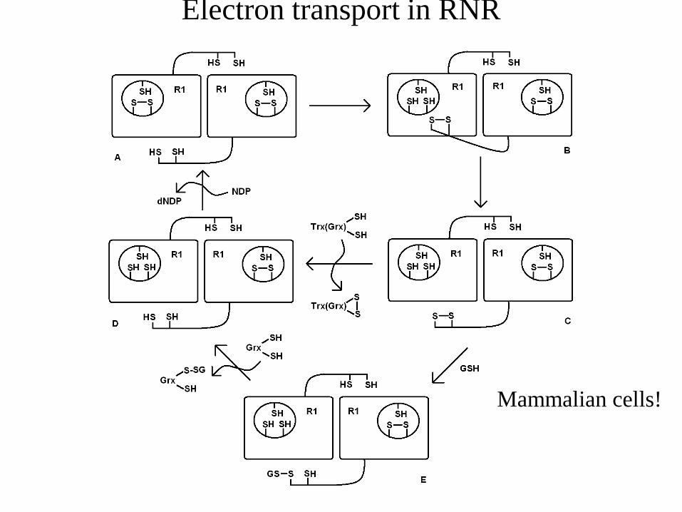
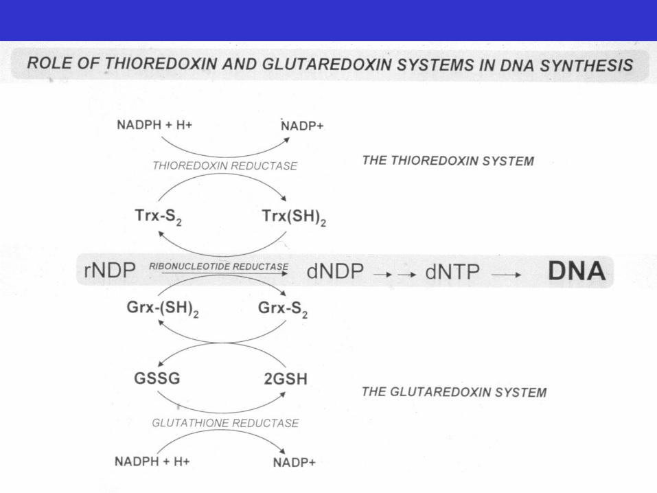
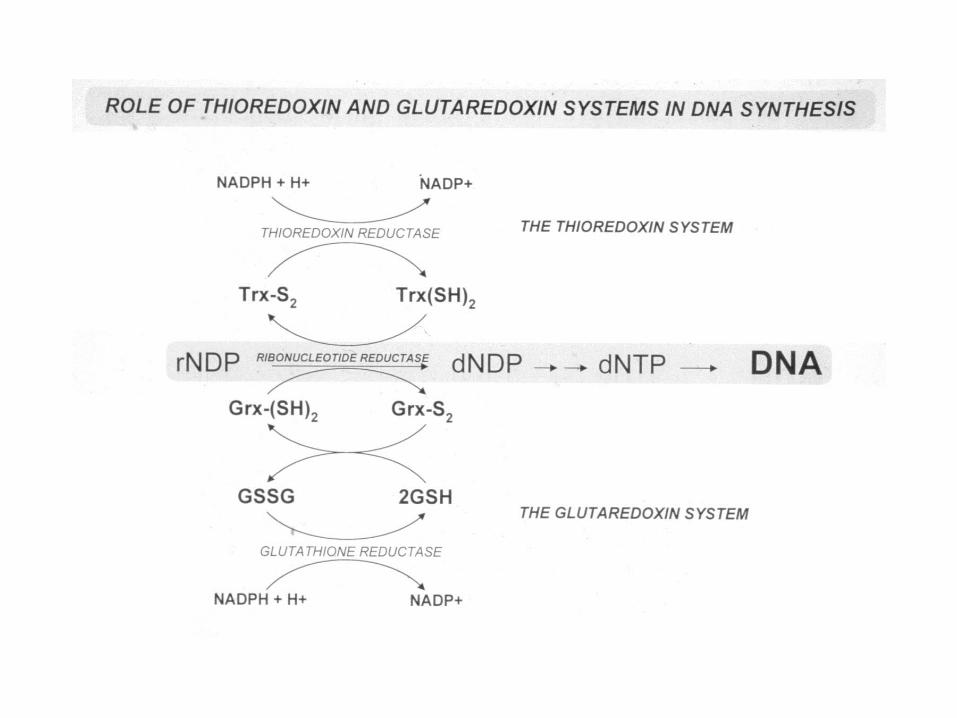
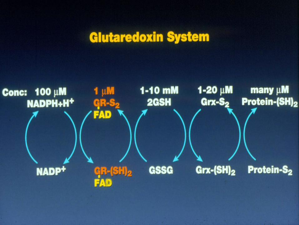
Electron transport in RNR
Mammalian cells!
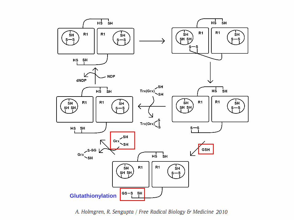
Glutathionylation
Conclusions
The mechanism of human ribonucleotide reductase is
different from that of E.coli and glutaredoxin functions via a
monothiol mechanism and in principle any glutaredoxin
(dithiol) can act together with GSH.
Since GSH is high ( 4-10 mM) in cells this may act to ensure
the supply of dNTPs for DNA repair (BER vith APE-1/Ref1)
in nondividing cells with very low RNR levels.
Thioredoxin system is high capacity for S-phase or when
GSH is low or oxidized. Tumors have often very upregulated
RNR.Unbalanced dNTP pools favour genetic instability?
But if Se is low? TrxR not active- use GSH system plus high
RNR? p53R2 is constitutive for DNA repair and
mitochondrial DNA synthesis in nondividing cells.
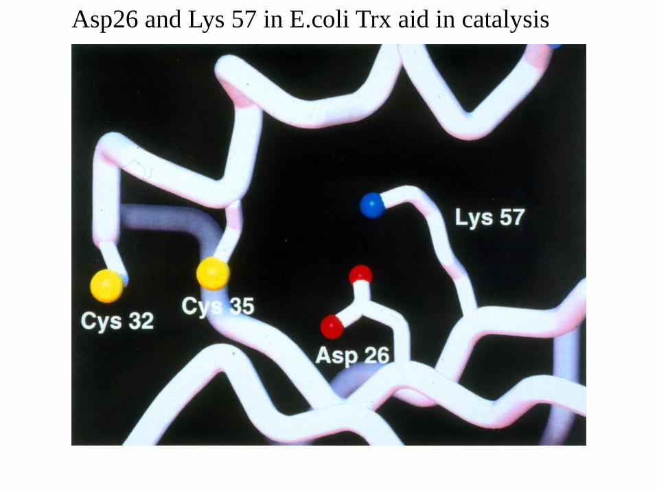
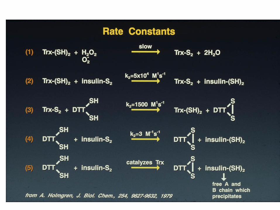
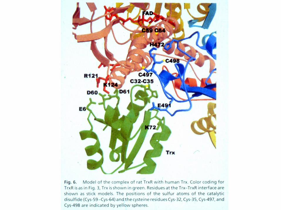
Asp26 and Lys 57 in E.coli Trx aid in catalysis
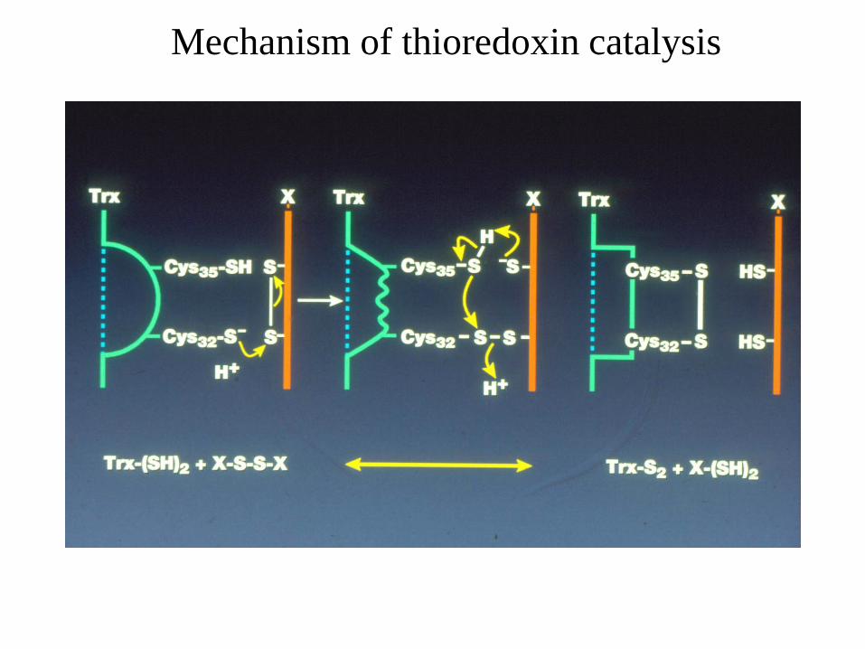
Mechanism of thioredoxin catalysis
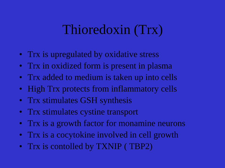
Thioredoxin (Trx)
• Trx is upregulated by oxidative stress
• Trx in oxidized form is present in plasma
• Trx added to medium is taken up into cells
• High Trx protects from inflammatory cells
• Trx stimulates GSH synthesis
• Trx stimulates cystine transport
• Trx is a growth factor for monamine neurons
• Trx is a cocytokine involved in cell growth
• Trx is contolled by TXNIP ( TBP2)
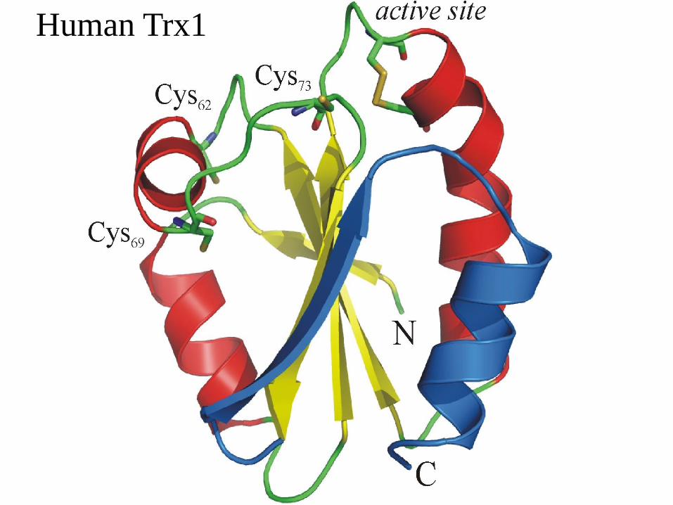
Human Trx1
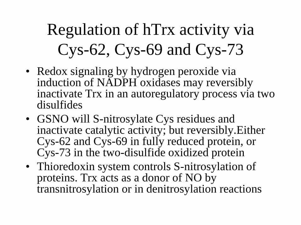
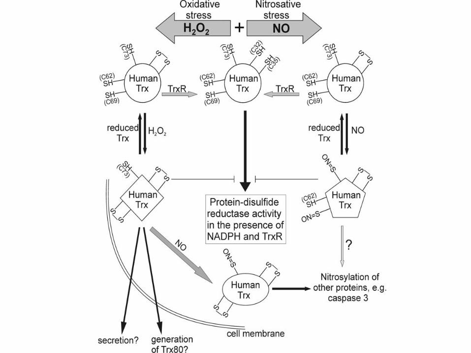
Regulation of hTrx activity via
Cys-62, Cys-69 and Cys-73
• Redox signaling by hydrogen peroxide via induction of NADPH oxidases may reversibly inactivate Trx in an autoregulatory process via two disulfides
• GSNO will S-nitrosylate Cys residues and inactivate catalytic activity; but reversibly.Either Cys-62 and Cys-69 in fully reduced protein, or Cys-73 in the two-disulfide oxidized protein
• Thioredoxin system controls S-nitrosylation of proteins. Trx acts as a donor of NO by transnitrosylation or in denitrosylation reactions

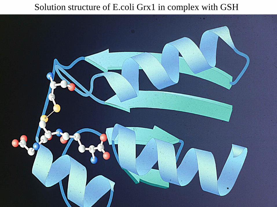
Solution structure of E.coli Grx1 in complex with GSH
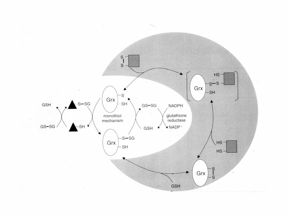
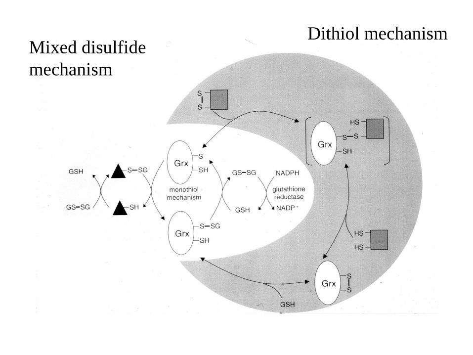
Mixed disulfide
mechanism
Dithiol mechanism
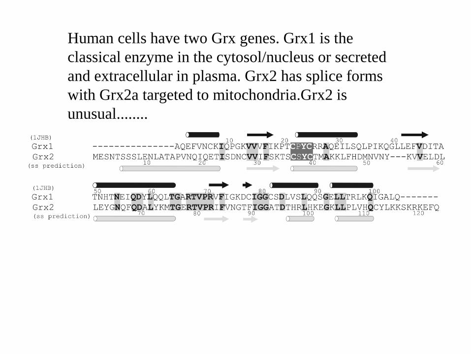
Human cells have two Grx genes. Grx1 is the
classical enzyme in the cytosol/nucleus or secreted
and extracellular in plasma. Grx2 has splice forms
with Grx2a targeted to mitochondria.Grx2 is
unusual........
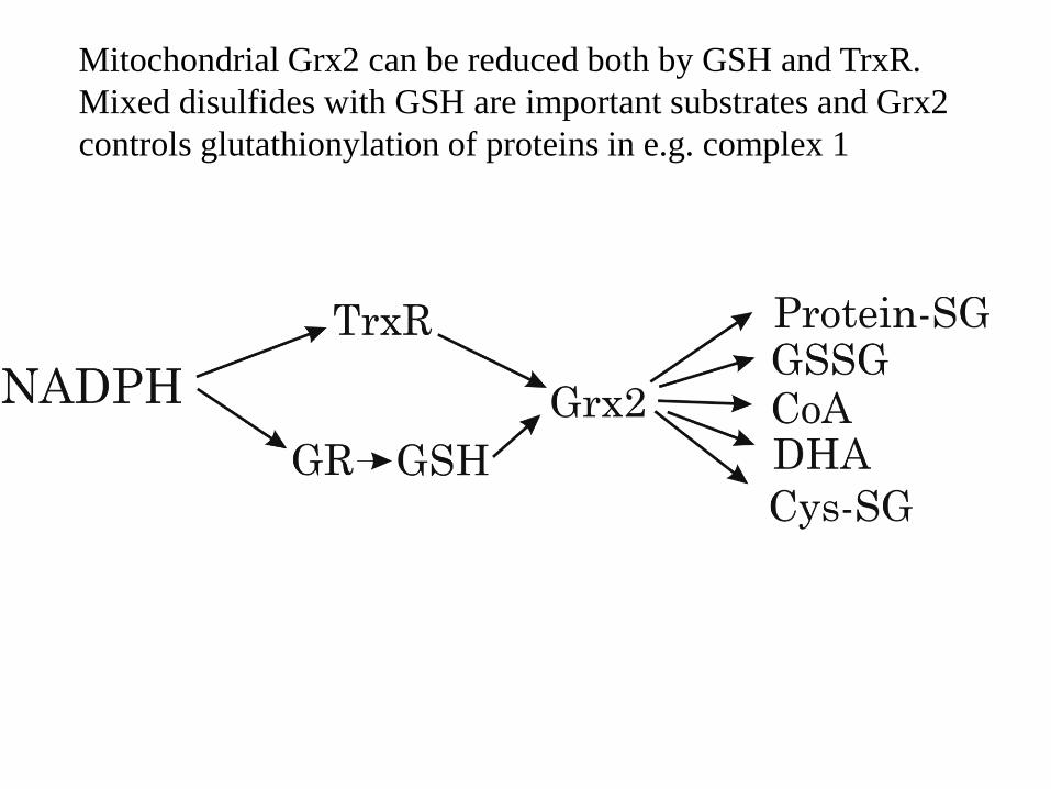
Mitochondrial Grx2 can be reduced both by GSH and TrxR.
Mixed disulfides with GSH are important substrates and Grx2
controls glutathionylation of proteins in e.g. complex 1
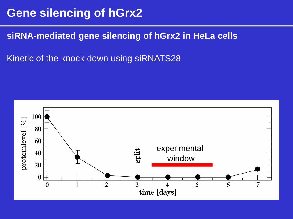
Gene silencing of hGrx2
siRNA-mediated gene silencing of hGrx2 in HeLa cells
Kinetic of the knock down using siRNATS28
experimental
window
Gene silencing of hGrx2
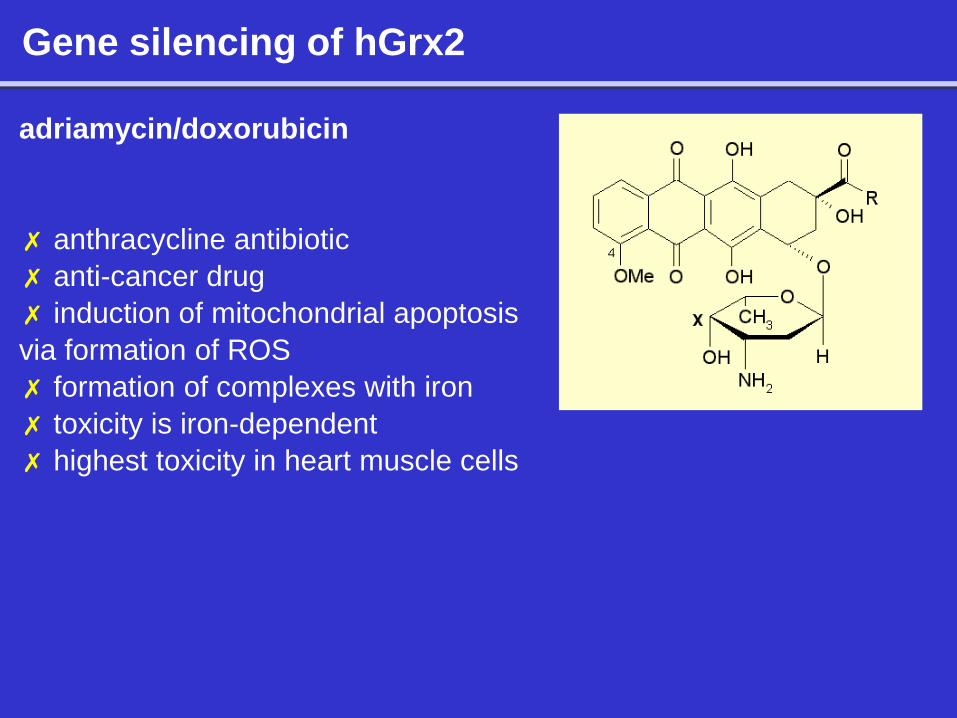
adriamycin/doxorubicin
✗ anthracycline antibiotic
✗ anti-cancer drug
✗ induction of mitochondrial apoptosis
via formation of ROS
✗ formation of complexes with iron
✗ toxicity is iron-dependent
✗ highest toxicity in heart muscle cells
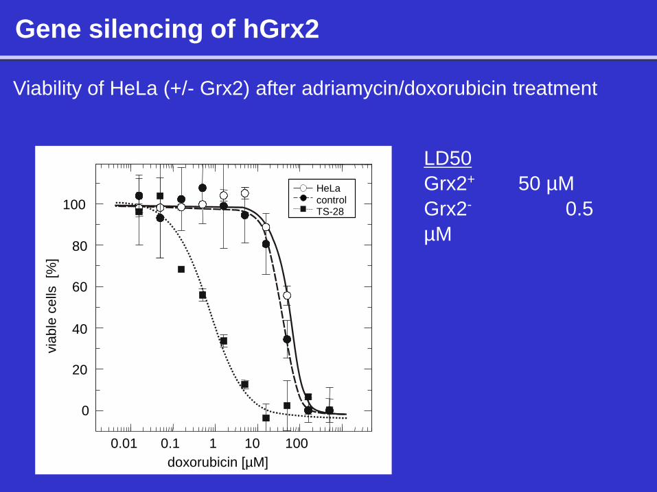
Gene silencing of hGrx2
Viability of HeLa (+/- Grx2) after adriamycin/doxorubicin treatment
LD50
Grx2+ 50 µM
Grx2- 0.5
µM
0
20
40
60
80
100
via
ble
ce
lls [%
]
0.01 1 10 0.1
doxorubicin [µM]
100
HeLa control TS-28
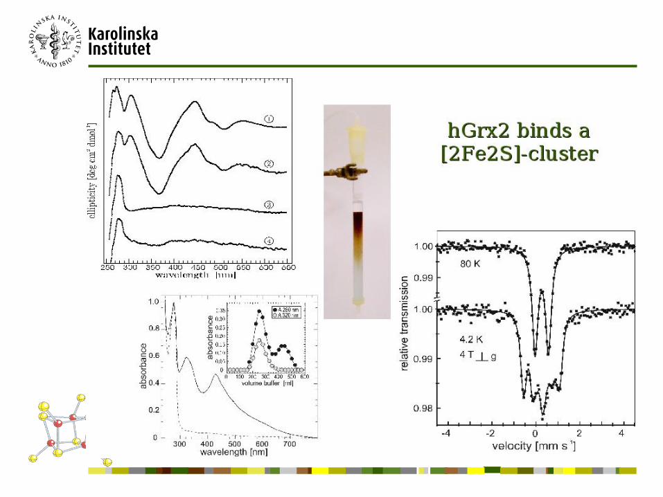
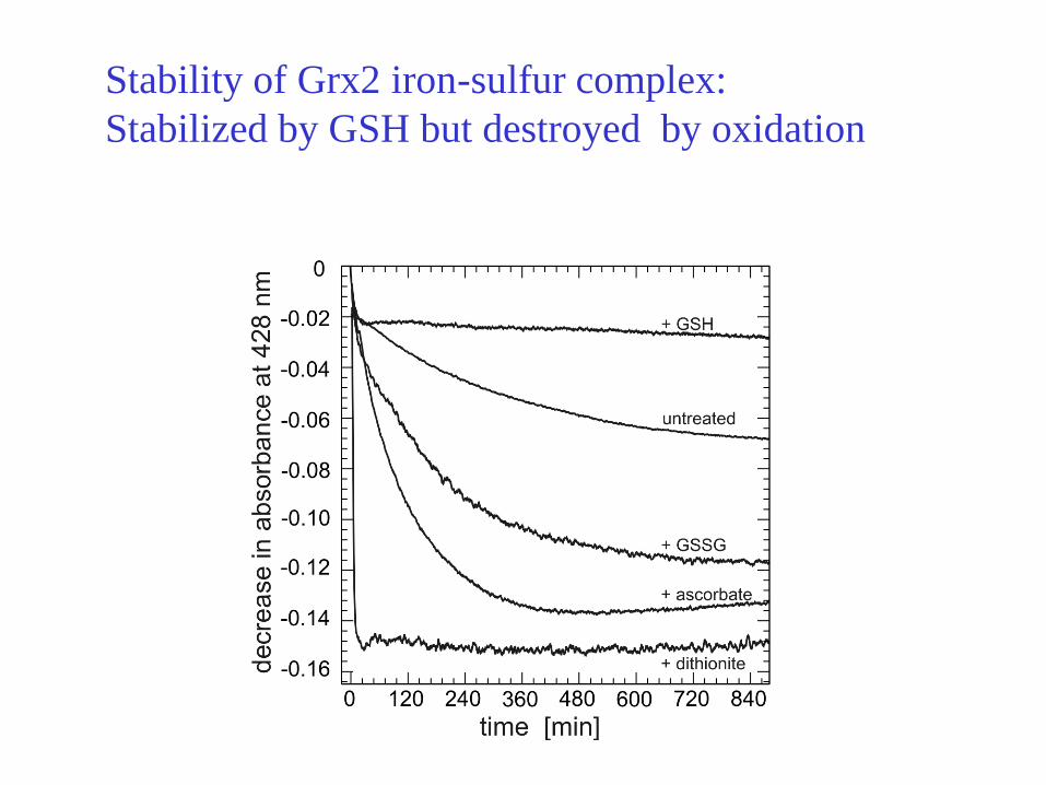
Stability of Grx2 iron-sulfur complex:
Stabilized by GSH but destroyed by oxidation
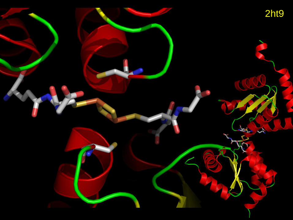
2ht9
Summary I
• Trx, TrxR and NADPH exist in all cells and is a 4 billion years old system as a general protein disulfide reductase.
• Trx has a CXXC active site where the N-terminal C residue is the nucleophilic thiolate operating via a mixed disulfide mechanism.
• Mammalian cells have Trx1 in cytosol with 3 structural Cys residues which can regulate activity by making two disulfides. In contrast mammalian Trx2 in mitochondria is more similar to bacterial Trx.
• TrxRs in bacteria,yeast and plants are smaller specific enzymes with a CXXC active site.
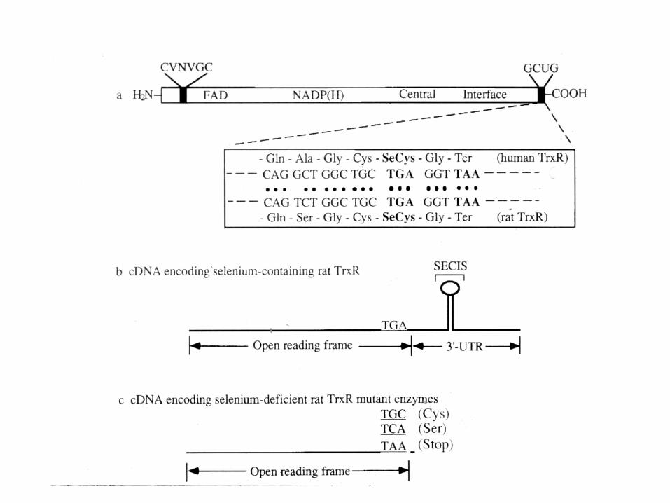
• TrxR in mammalian cells are larger and selenoenzymes with a broad substrate specificity and have a C-terminal GCUG active site where U is a selenocysteine residue.
• TrxR1 is cytosolic, TrxR2 is mitochondrial and TGR is thioredoxin-glutathione reductase with a Grx domain particularly in testes and the only enzyme in some parasitic worms.
Summary II
• Glutathione (GSH) is present in high concentrations in almost all cells (grampositive bacteria have no GSH like Staph. aureus). Made by two enzymes from Glu, Cys and Gly. GSH/GSSG ( >100:1) is very high in the cytosol and particularly during DNA replication when GSH is accumulated in the nucleus.
• Grx catalysis GSH-disulfide-oxidoreductions and is a thioredoxin fold protein with a GSH binding site recognizing GSH for its reduction of the active site and uses GSH-mixed disulfides as substrates. Large number of proteins are regulated by glutathionylation and Grx.
• In contrast nitrosylation and denitrosylation of proteins is catalyzed by thioredoxins if not directly by GSNO and GSH for nonenzymatic nitrosylation and denitrosylation.
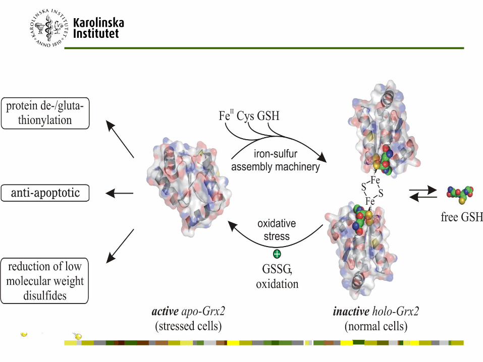
• Grx1 and Grx2 are dithiol glutaredoxins in mammalian cells where Grx2 has splice forms with Grx2a in the mitochondria. Grx2 is also present as an inactive dimeric 2 Fe-2S iron sulfur protein. The monothiol glutaredoxin Grx5 is also a 2Fe2S protein involved in heme synthesis
• Trx and Grx function in a large number of reactions in cells with overlapping functions in some reactions like electron transport to ribonucleotide reductase.