this article was originally published in a journal published by
TRANSCRIPT

This article was originally published in a journal published by Elsevier, and the attached copy is provided by Elsevier for the
author’s benefit and for the benefit of the author’s institution, for non-commercial research and educational use including without
limitation use in instruction at your institution, sending it to specific colleagues that you know, and providing a copy to your institution’s
administrator.
All other uses, reproduction and distribution, including without limitation commercial reprints, selling or licensing copies or access,
or posting on open internet sites, your personal or institution’s website or repository, are prohibited. For exceptions, permission
may be sought for such use through Elsevier’s permissions site at:
http://www.elsevier.com/locate/permissionusematerial
Author's Personal CopyAvailable online at www.sciencedirect.com
Journal of Electromyography and Kinesiology 18 (2008) 116–127
www.elsevier.com/locate/jelekin
Muscular activity during uphill cycling: Effect of slope, posture,hand grip position and constrained bicycle lateral sways
S. Duc a,*, W. Bertucci b, J.N. Pernin a, F. Grappe a
a Laboratoire FEMTO-ST (UMR CNRS 6174), Departement de Mecanique Appliquee, Universite de Franche-Comte,
24 Rue de l’Epitaphe 25000 Besancon, Franceb Laboratoire d’Analyse des Contraintes Mecaniques – EA 3304 LRC CEA/UFR STAPS, Universite de Reims Champagne-Ardenne,
Campus Moulin de la Housse (batiment 6), 51100 Reims, France
Received 6 June 2006; received in revised form 26 September 2006; accepted 26 September 2006
Abstract
Despite the wide use of surface electromyography (EMG) to study pedalling movement, there is a paucity of data concerning the mus-cular activity during uphill cycling, notably in standing posture. The aim of this study was to investigate the muscular activity of eightlower limb muscles and four upper limb muscles across various laboratory pedalling exercises which simulated uphill cycling conditions.Ten trained cyclists rode at 80% of their maximal aerobic power on an inclined motorised treadmill (4%, 7% and 10%) with using twopedalling postures (seated and standing). Two additional rides were made in standing at 4% slope to test the effect of the change of thehand grip position (from brake levers to the drops of the handlebar), and the influence of the lateral sways of the bicycle. For this lastgoal, the bicycle was fixed on a stationary ergometer to prevent the lean of the bicycle side-to-side. EMG was recorded from M. gluteusmaximus (GM), M. vastus medialis (VM), M. rectus femoris (RF), M. biceps femoris (BF), M. semimembranosus (SM), M. gastrocne-mius medialis (GAS), M. soleus (SOL), M. tibialis anterior (TA), M. biceps brachii (BB), M. triceps brachii (TB), M. rectus abdominis(RA) and M. erector spinae (ES). Unlike the slope, the change of pedalling posture in uphill cycling had a significant effect on the EMGactivity, except for the three muscles crossing the ankle’s joint (GAS, SOL and TA). Intensity and duration of GM, VM, RF, BF, BB,TA, RA and ES activity were greater in standing while SM activity showed a slight decrease. In standing, global activity of upper limbwas higher when the hand grip position was changed from brake level to the drops, but lower when the lateral sways of the bicycle wereconstrained. These results seem to be related to (1) the increase of the peak pedal force, (2) the change of the hip and knee joint moments,(3) the need to stabilize pelvic in reference with removing the saddle support, and (4) the shift of the mass centre forward.� 2006 Elsevier Ltd. All rights reserved.
Keywords: EMG; Pedalling–standing–seated-treadmill
1. Introduction
The majority of cycling studies have examined muscularactivity of pedalling with using surface electromyography(EMG), when subjects ride on horizontal surfaces. Up-to-date, there is yet a lack of information concerning musclerecruitment pattern of uphill cycling, especially in the stand-
1050-6411/$ - see front matter � 2006 Elsevier Ltd. All rights reserved.
doi:10.1016/j.jelekin.2006.09.007
* Corresponding author. Tel.: +33 3 81 66 60 37; fax: +33 3 81 66 60 00.E-mail addresses: [email protected] (S. Duc), william.bertucci@
univ-reims.fr (W. Bertucci), [email protected] (J.N. Pernin),[email protected] (F. Grappe).
ing position. Cyclists often switch between seated andstanding posture during mountain climbing, notably todecrease the strain of the lower back muscle. Standing isused by the practitioners to relieve saddle pressure duringflat terrain cycling and to increase power production duringsprinting. From our knowledge, only one study reportedEMG activity of lower limb muscles during standing posi-tion (Li and Caldwell, 1998). The authors showed thatEMG patterns of monoarticular extensor muscles, like M.gluteus maximus (GM) and M. vastus medialis (VM) aremore affected by the transition from seated to standing ped-alling, than the biarticular flexor muscles, i.e. M. biceps
Author's Personal CopyS. Duc et al. / Journal of Electromyography and Kinesiology 18 (2008) 116–127 117
femoris (BF) and M. gastrocnemius (GAS). These resultshave been related to the changes of pedalling kinetics andkinematics, which are due to the removal of the saddle sup-port during standing pedalling and the forward horizontalshift of the total body centre mass (Alvarez and Vinyolas,1999; Caldwell et al., 1998; Soden and Adeyefa, 1979; Stoneand Hull, 1993). Peak pedal force, crank torque and peakankle plantarflexor generated by cyclists are higher andoccur later during the downstroke (Alvarez and Vinyolas,1999; Caldwell et al., 1998). Moreover, while standing, theextensor knee moment is extended longer into the down-stroke (0–180�) whereas the duration of the knee flexormoment is lower. Since the pattern of hip joint moment dis-plays high similarity between the two postures (Caldwellet al., 1999), it has been suggested that changes of GM activ-ity are linked to a decrease of the force moment arm and tothe pelvis stabilization (Li and Caldwell, 1998).
The study of Li and Caldwell (1998) has three major lim-its that should be considered. Firstly, the stationary cyclingergometer (i.e. Velodyne) used by the authors to simulateuphill conditions prevents the lateral sways of the bicyclewhile standing pedalling. Therefore, EMG activity of biar-ticular muscles might to be more altered by change ofcycling posture during ‘‘natural’’ standing pedalling sinceit has been assumed that these muscles play a more complexrole during pedalling compared to monoarticular muscles.Several studies (Raasch et al., 1997; van Ingen Schenauet al., 1992) suggested that biarticular muscles are responsi-ble for the control of the direction of the force applied to thepedal, the transfer of power produced by monoarticularextensors muscles and the regularity of pedalling, notablyduring the flexion-to-extension transition (called top deadcentre, i.e. TDC) and during the extension-to-flexion transi-tion (called bottom dead centre, i.e. BDC).
Secondly, the response of other muscles involved duringpedalling, i.e M. semimembranosus (SM), M. semitendino-sus (ST) and M. soleus (SOL), to the change of posture dur-ing uphill cycling is unknown. It is not sure that SM and STpatterns during standing pedalling are similar to BF patternbecause it has been suggested that these muscles, unlike toBF, work more as knee flexor than knee extensor (Ericson,1988). Moreover, the measure of the EMG activity of SOLcould allow to validate the hypothesis proposed by Li andCaldwell (1998) that the increase of peak plantar flexormoment, observed during standing pedalling, is linked tothe activity of SOL and unrelated to the activity of GAS.This may be caused by the biarticular function of GAS, asit also serves as a knee flexor. With the extended period ofthe knee extensor moment during standing, increasedGAS activity would be contraindicated.
Thirdly, previous authors have not clearly reported theupper body and trunk muscles activity during standingpedalling. It is surprising because these muscular groupsseems to be greatly activated during standing pedalling,notably to support additional weight due to the loss of sad-dle support, to stabilize pelvis and trunk to control bodybalance and to swing the body and the bicycle side-to-side.
During standing pedalling, cyclists can grip the handle-bar on the brake levers (top hand position) or on the dropsof the handlebar (bottom hand position). The top handposition is often used during climbing whereas the bottomhand position is generally employed for sprints. Pedallingbiomechanics can be affected by change of hand grip sincethe trunk is more flexed in the bottom hand position. Savel-berg et al. (2003) observed significant changes of EMGactivity of GM and TA muscles when the trunk is flexed20� to forward during seated pedalling. This effect couldbe increased during standing pedalling because the trunkflexion is higher when the hands are placed on the dropsof the handlebar. At our knowledge, no study has com-pared the effect of the two hand grip positions duringstanding pedalling on muscular activity.
It was suggested that change of road slope or gradient canaffect kinetics and kinematics of pedalling. In the case ofuphill cycling, the orientation of the rider and bicycle withrespect to the gravity force may enhance some modificationsof the pedalling technique. Caldwell et al. (1998) showedthat cyclists produce a greater crank torque during the first120� of the crank cycle and during the first half of theupstroke (180–270�) at 8% slope compared to 0%. Theseforce changes are combined with the alteration of the pedalorientation to a more ‘‘toe-up’’ position. The same authors(Caldwell et al., 1999) have also found that the peak ankleplantarflexor and the peak knee extensor moments arehigher and occur slightly earlier in the crank cycle at 8%slope. However, all these changes are largely explained bythe difference in the pedalling cadence from the 0% slope(82 rpm) to 8% slope (65 rpm) condition. While cadencedecreased, total work done per crank revolution increaseda consequence of holding power output constant. The effectof the slope on muscular activity is ambiguous. Li and Cald-well (1998) have not found differences in EMG activity of sixlower limb muscles between 0% and 8% slope whereasClarys et al. (2001) observed a significant increase in EMGactivity of lower limb (sum of EMG activity of VM, BF,TA, GAS) with increasing slope (2–12%). Differences couldbe due to the cycling experience level of the subjects (stu-dents with 2 years of cycling experience vs professionalcyclists), the experimental context of the two studies (labvs field, respectively) and to the analysis of EMG data (indi-vidual vs global activity, respectively). It is also important toremember that, as throughout studies of Caldwell et al.(1998, 1999), subjects did not use the same pedalling cadencebetween the two slope conditions during the first study. Theeffect of the increase in slope on EMG activity could bemasked by the decrease of pedalling cadence since manystudies have shown that intensity of EMG activity of GM,RF, GAS and BF changes across pedalling cadence (Baumand Li, 2003; Marsh and Martin, 1995; Neptune et al., 1997;Ryschon and Stray-Gundersen, 1991; Sarre et al., 2003).
The purpose of this study is to quantify the influence of(1) the slope (4–7–10%); (2) the pedalling posture (seated–standing); (3) the hand grip position in standing pedalling(on the brake levers–on the drops); and (4) the constrained

Author's Personal Copy118 S. Duc et al. / Journal of Electromyography and Kinesiology 18 (2008) 116–127
lateral sways of bicycle in standing pedalling (ride on a sta-tionary ergometer), on the intensity and the timing ofEMG activity of lower limb, trunk and arm muscles. Morespecifically, four hypotheses were tested. Firstly, muscularactivity of power prime producer muscles (GM, VM),lower back muscles and arm muscles would increase line-arly with the treadmill slope due to the change of rider ori-entation respect to gravity force. Secondly, standingpedalling would affect considerably both intensity and tim-ing of hip extensor and flexor muscles (GM, BF), trunkand arm muscles, since this posture removes the saddlesupport. Thirdly, grip of the handlebar on the drops duringstanding pedalling would also change EMG activity oftrunk and arm muscles since the total body centre massis shifted forward in this position compared to the brakelevers hand grip position. Finally, contrary to Caldwellet al. (1998, 1999) and Li and Caldwell (1998), we hypoth-esized that lateral sways of the bicycle in standing are notinsignificant. Intensity of EMG activity of lower limb mus-cles would increase when cyclists pedal in standing on a sta-tionary ergometer that constrains bicycle tilts.
2. Methods
2.1. Subjects
Ten trained, healthy, male, competitive cyclists of the FrenchCycling Federation volunteered to participate in this study. Theywere classified in national (n = 4), regional (n = 4) or depart-mental (n = 2) category and had regularly competed for at leasttwo years prior the study. Before the experiment, each subjectreceived full explanations concerning the nature and the purposeof the study and gave written informed consent. Age, height, andbody mass of the tested subjects were 28 ± 7 (mean ± SD) yr,1.78 ± 0.07 m, and 71 ± 8 kg, respectively.
2.2. Protocol
Each cyclist performed two test sessions in our laboratory. Thefirst test session was an incremental test to exhaustion to deter-mine maximal aerobic power (MAP), maximal oxygen uptakeð _VO2 maxÞ and maximal heart rate (HRmax). The second test ses-sion consisted of four pedalling sessions of eight randomised trialswith different uphill cycling conditions. Both test sessions wereheld within a period of 1 week and separated by at least 2 days.
Each subject cycled with his own racing bicycle on a largemotorised treadmill (S 1930, HEF Techmachine, Andrezieux-Boutheon, France) of 3.8 m length and 1.8 m wide. Before testing,all the subjects performed several sessions on the motorisedtreadmill to acclimatise themselves to the equipment. Throughoutthe tests, the subjects were attached with a torso harness for theirsafety without hindering the riders motion nor position on thebicycle. All the bicycles were equipped with clipless pedals. Thebicycle tyre pressure was inflated to 700 kPa. The rear wheel wasfitted with the PowerTap hub (professional model, CycleOps,Madison, USA) to measure the power output (PO), the velocityand the pedalling cadence (CAD) during the two test sessions.This system uses the strain gauge technology (eight gauges). Thevalidity and the reproductibility of the PowerTap hubwere showed by Bertucci et al. (2005) and Gardner et al. (2004).
A magnet (Sigma Sport, Neustadt, Germany) was fixed on thebicycle near the bottom bracket in order to isolate each pedalcycle. The magnet signal was then recorded with the amplifierused for the EMG collection.
2.3. Data collection and processing
2.3.1. EMG recording
The EMG activity from eight muscles of the right lower limb(M. gluteus maximus (GM), M. vastus medialis (VM), M. rectusfemoris (RF), M. biceps femoris caput longum (BF), M. semi-membranosus (SM), M. gastrocnemius medialis (GAS), M. soleus(SOL), M. tibialis anterior (TA)), from two muscles of the trunk(M. rectus abdominis (RA), M. erector spinae (ES)) and from twomuscles of the right arm (M. biceps brachii (BB) and M. tricepsbrachii (TB)) were collected during the second test session. Inorder to limit the potential crosstalk between SOL and GASactivity, two surface electrodes for SOL were positioned on thelower third of the calf, just above the Achilles tendon. Data col-lection occurred during the final 15 s of each pedalling trial andlasted for five pedal cycles per collection period. The subjects werekept unaware of the exact timing of data collection.
The EMG sensors were conformed to recommendations of theSENIAM. Recorded sites were shaved and cleaned with an alcoholswab in order to reduce skin impedance to less than 10 kX. Pairs ofsilver/silver-chloride, circular, bipolar, pre-gelled surface electrodes(Control Graphique Medical, Brie-Comte-Robert, France) of20 mm diameter, were applied on the midpoint of the contractedmuscle belly (Clarys, 2001), parallel to the muscle fibbers, with aconstant inter-electrode distance of 30 mm. The reference elec-trodes were placed over electrically neutral sites (scapula andclavicle). All the electrodes and the wires were fixed on the skin withadhesive pads to avoid artifacts. EMG was recorded with a MP30amplifier (Biopac System, Inc., Santa Barbara, USA, commonmode rejection ratio >90 dB, input resistance is in order of 109 X).The EMG signals were amplified (gain = 2500), band pass filtered(50–500 Hz) and analog-to-digital converted at a sampling rate of1000 Hz. We chose a high-pass frequency of the EMG bandpassfilter (50 H) in order to eliminate the ambient noise caused byelectrical-power wires and components of the motorised treadmill.
The raw EMG were expressed in root mean square (RMS)with a time averaging period of 20 ms. The overall activity level ofeach muscle was identified by the mean RMS calculated for fiveconsecutives crank cycles (EMGmean) and normalised to themaximal RMS value measured for each muscle and for eachsubject during all the trials (normalisation to the highest peakactivity in dynamic condition). The EMG signal was also full-wave rectified and smoothed (Butterworth filter, second-order,cut-off frequency of 6 Hz) to create the linear envelope. Using themagnet signal, the linear envelope was then divided into each ofthe five pedal cycles and a mean linear envelope was computed foreach muscle. Finally, the linear envelopes of each muscle werescaled to a percentage of the maximum value found for eachindividual muscle and for each subject.
To analyse the muscle activity pattern, five parameters werecalculated from the linear envelope for each pedalling trial: EMGburst onset (EMGonset), offset (EMGoffset) and peak timing(EMGpeak-timing), EMG burst duration (EMGduration), and peakEMG burst magnitude (EMGpeak). An arbitrary threshold valueof 25% of the maximum value across conditions was chosen todetermine the onset and the offset of EMG burst, like that selectedby Li and Caldwell (1998). Visual inspection determined if this
Author's Personal Copy
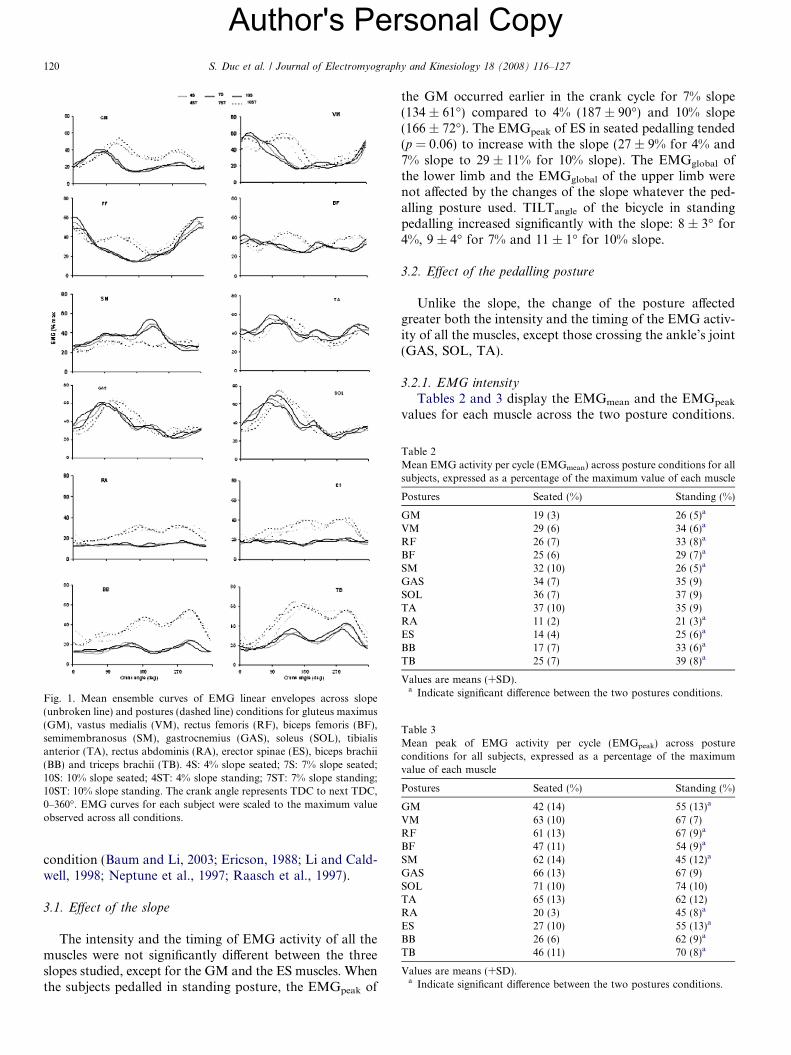
Table 1Physiological characteristics of subjects obtained during the incrementaltest to exhaustion
_VO2 max ðlmin�1Þ _VO2 maxðlmin�1kg�1Þ MAP (W) HRmax (bpm)
4.5 (0.4) 66 (6) 378 (47) 183 (8)
Values are means (+SD). _VO2 max, maximal oxygen uptake; MAP, maxi-mal aerobic power; HRmax, maximal heart rate.
S. Duc et al. / Journal of Electromyography and Kinesiology 18 (2008) 116–127 119
threshold was appropriate. Appropriate thresholds reflected easilyidentifiable onset and offset points and minimal discrepancies inidentifying non-meaningful burst. In the case that 25% was con-sidered inappropriate, the threshold was raised to 35% and more,of the maximum value across conditions. Upon reaching thedetermined threshold, the muscle was considered active, and themuscle burst duration was defined as the duration, in degrees, ofthe crank angle between the onset and offset value. EMGpeak wasthe maximum value from the linear envelope during each pedal-ling trial. EMGpeak-timing was the crank angle at which the peakEMG occurred.
Finally, we have determined the global EMG activity(EMGglobal) of the lower limb and the upper limb with adding theEMGmean of eight lower limb muscles (GM, VM, RF, BF, SM,GAS, SOL, TA) and of the trunk and arm muscles (RA, ES, BB,TB).
2.3.2. Video recording
Bicycle lateral sways were recorded simultaneously with EMGdata at 50 Hz using a JVC video camera (JVC, Yokohama,Japan), with the lens axis oriented parallel to the rear frontal ofthe subject, positioned 4 m behind the rider. The maximal tiltangle (TILTangle) of the bicycle was determined for each pedallingcondition with averaging maximal values measured during 30 s.
2.4. First test session: incremental test
After a brief warm-up period (�5 min), the incremental teststarted at 130 W for 2 min. The treadmill slope at this first stagewas fixed to 1%. The treadmill velocity was determined for eachcyclist with using a mathematical power model, in order to obtainthe initial PO (130 W). The workload was then increased by�30 W every 2 min until the subject became exhausted, byincreasing the treadmill slope by 0.5% during each increment. Thetreadmill velocity was unchanged throughout the incremental test.The cyclists were required to remain in a seated position duringthe entire test, and could choose themselves their CAD byadjusting the bicycle gears. The MAP was determined as the meanPO maintained during the last completed workload stage. A K4b2
breath-by-breath portable gas analyser (Cosmed, Rome, Italy)and a chest belt (Polar, Kempele, Finland) were used to collect themetabolic and the HR data. The Cosmed K4b2 system was cali-brated using the manufacturer’s recommendations. The highestmean _VO2 and HR values obtained during the increment test for10 s were defined, respectively, as the _VO2 max and the HRmax.
2.5. Second test session: uphill conditions
After a short self-selected warm-up period (�10 min), eachcyclist performed six pedalling trials (4S, 7S, 10S, 4ST, 7ST, 10ST)with different slopes (4%, 7% and 10%), and pedalling postures(seated (S) and standing (ST)). For all these pedalling trials, thehands were positioned on the top of the handlebar (on the brakelevers). Two additional pedalling trials were performed in stand-ing position and against the 4% slope, to test the effect of thebottom hand position (4STb) and the effect of the constrainedbicycle lateral sways (4STc). The eight pedalling trials were per-formed in a randomised order. For the 4STc condition, the cyclistswere required to pedal with their bicycle on a stationary AxiomPowerTrain ergometer (Elite, Fontaniva, Italy), which wasmounted on the inclined motorised treadmill. The Axiom Powerergometer has been recently described in detail by Bertucci et al.
(2005). Briefly, the rear wheel of the bicycle was fixed by a quickrelease skewer in the stand of the ergometer. This stand constrainslateral motions of the rear wheel. A roller, which was connectedwith a flywheel in an electromagnetic resistance unit, was broughtin contact with the tyre to provide a resistive force.
The PO (80% of MAP) was kept constant during the eighttrials. The CAD differed between cyclists (range: 60–70 rpm) butnot between the trials. Each cyclist was required to perform fourtimes 8 pedalling trials (4S, 7S, 10S, 4ST, 7ST, 10 ST, 4STb,4STc) since our EMG measurement device can collect only threeEMG signals at the same time. So, in order to minimise themuscular fatigue, we fixed the time of each trial at 1 min. Trialswere separated by 3 min of low active recovery (PO < 40%MAP). The recovery between two-8 pedalling trials due to thechange of the EMG electrodes configuration was higher andpassive (10–15 min).
2.6. Statistical analyses
All data were analysed using the Sigmastat statistical program(Jandel, Germany, version 2.0) for Windows. The data were testedfor normality and homogeneity of variance (Kolmogorov–Smir-nov tests) and turned out to be not normally distributed. Thus, ano-parametric two way (3 slopes · 2 postures) repeated measuresfactorial analysis of variance (ANOVA on ranks) was used todetect significant differences of each dependent variable (EMGmean,EMGonset, EMGoffset, EMGpeak-timing, EMGduration, EMGpeak,EMGglobal, TILTangle). Tukey’s HSD post hoc analysis was per-formed when ANOVA on ranks indicated a significant difference.The Wilcoxon signed rank test was also employed to determinethe effect of the change of hand position (top to bottom) duringstanding pedalling and the influence of the constrained bicyclelateral sway on the same variables. The results were expressed asmeans ± standard deviation (SD). The level of significance was setat p < 0.05.
3. Results
Table 1 displays the results obtained during the incre-mental test to exhaustion. The physiological characteristicsof subjects were common to those obtain with studies usingsimilar cyclists groups (Bertucci et al., 2005; Marsh andMartin, 1995; Millet et al., 2002; Sarre et al., 2003).
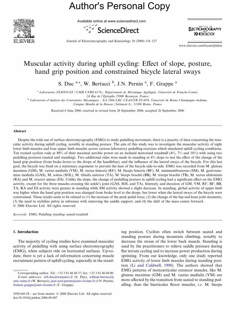
Muscle activity patterns when standing pedalling withgriping the handlebar on the drops or with constrainedbicycle tilts were similar of the 4ST condition (hand onthe brake levers and with bicycle tilting). So we decide torepresent in Fig. 1 only the muscle activity patterns withensemble linear envelopes of the six other cycling condi-tions (3 slopes (4–7–10%) · 2 postures (S, ST)). The patternof EMG activity of lower limb muscles in seated postureagreed with those generally reported in similar cycling
Author's Personal Copy
Fig. 1. Mean ensemble curves of EMG linear envelopes across slope(unbroken line) and postures (dashed line) conditions for gluteus maximus(GM), vastus medialis (VM), rectus femoris (RF), biceps femoris (BF),semimembranosus (SM), gastrocnemius (GAS), soleus (SOL), tibialisanterior (TA), rectus abdominis (RA), erector spinae (ES), biceps brachii(BB) and triceps brachii (TB). 4S: 4% slope seated; 7S: 7% slope seated;10S: 10% slope seated; 4ST: 4% slope standing; 7ST: 7% slope standing;10ST: 10% slope standing. The crank angle represents TDC to next TDC,0–360�. EMG curves for each subject were scaled to the maximum valueobserved across all conditions.
Table 2Mean EMG activity per cycle (EMGmean) across posture conditions for allsubjects, expressed as a percentage of the maximum value of each muscle
Postures Seated (%) Standing (%)
GM 19 (3) 26 (5)a
VM 29 (6) 34 (6)a
RF 26 (7) 33 (8)a
BF 25 (6) 29 (7)a
SM 32 (10) 26 (5)a
GAS 34 (7) 35 (9)SOL 36 (7) 37 (9)TA 37 (10) 35 (9)RA 11 (2) 21 (3)a
ES 14 (4) 25 (6)a
BB 17 (7) 33 (6)a
TB 25 (7) 39 (8)a
Values are means (+SD).a Indicate significant difference between the two postures conditions.
Table 3Mean peak of EMG activity per cycle (EMGpeak) across postureconditions for all subjects, expressed as a percentage of the maximumvalue of each muscle
Postures Seated (%) Standing (%)
GM 42 (14) 55 (13)a
VM 63 (10) 67 (7)RF 61 (13) 67 (9)a
BF 47 (11) 54 (9)a
SM 62 (14) 45 (12)a
GAS 66 (13) 67 (9)SOL 71 (10) 74 (10)TA 65 (13) 62 (12)RA 20 (3) 45 (8)a
ES 27 (10) 55 (13)a
BB 26 (6) 62 (9)a
TB 46 (11) 70 (8)a
Values are means (+SD).a Indicate significant difference between the two postures conditions.
120 S. Duc et al. / Journal of Electromyography and Kinesiology 18 (2008) 116–127
condition (Baum and Li, 2003; Ericson, 1988; Li and Cald-well, 1998; Neptune et al., 1997; Raasch et al., 1997).
3.1. Effect of the slope
The intensity and the timing of EMG activity of all themuscles were not significantly different between the threeslopes studied, except for the GM and the ES muscles. Whenthe subjects pedalled in standing posture, the EMGpeak of
the GM occurred earlier in the crank cycle for 7% slope(134 ± 61�) compared to 4% (187 ± 90�) and 10% slope(166 ± 72�). The EMGpeak of ES in seated pedalling tended(p = 0.06) to increase with the slope (27 ± 9% for 4% and7% slope to 29 ± 11% for 10% slope). The EMGglobal ofthe lower limb and the EMGglobal of the upper limb werenot affected by the changes of the slope whatever the ped-alling posture used. TILTangle of the bicycle in standingpedalling increased significantly with the slope: 8 ± 3� for4%, 9 ± 4� for 7% and 11 ± 1� for 10% slope.
3.2. Effect of the pedalling posture
Unlike the slope, the change of the posture affectedgreater both the intensity and the timing of the EMG activ-ity of all the muscles, except those crossing the ankle’s joint(GAS, SOL, TA).
3.2.1. EMG intensity
Tables 2 and 3 display the EMGmean and the EMGpeak
values for each muscle across the two posture conditions.
Author's Personal CopyS. Duc et al. / Journal of Electromyography and Kinesiology 18 (2008) 116–127 121
The EMG activity of the GM in standing was higherthan in seated condition, notably between 90� and 330�.EMGmean and EMGpeak of GM increased by 41% and31%, respectively. The EMG activity of the quadriceps instanding was also higher than in seated condition, butonly during the second half of the downstroke (90–180�).EMGmean of the VM and RF raised by 18% and 24%,respectively, whereas EMGpeak increased only for the RFby 10%. The effect of the change of pedalling posture onthe EMG activity of the two hamstrings is contrasting:BF activity in standing was higher (between 90� and 180�and between 270� and 360�) while SM activity was lower(notably between 180� and 270�). EMGmean and EMGpeak
of BF increased by 17% and 15%, respectively whereasEMGmean and EMGpeak of SM decreased by 18% and
Table 4Mean crank angle, in degrees, at which the peak EMG activity per cycle(EMGpeak-timing) occurred across posture conditions for all subjects
Postures Seated (�) Standing (�)
GM 74 (23) 162 (75)a
VM 22 (15) 96 (46)a
RF 350 (28) 343 (30)BF 6 (63) 50 (93)a
SM 184 (79) 198 (81)GAS 90 (32) 125 (38)a
SOL 87 (30) 119 (33)a
TA 157 (112) 150 (100)RA 215 (90) 232 (79)ES 234 (94) 227 (71)BB 241 (87) 256 (84)TB 261 (56) 200 (48)
Values are means (+SD).a Indicate significant difference between the two postures conditions.
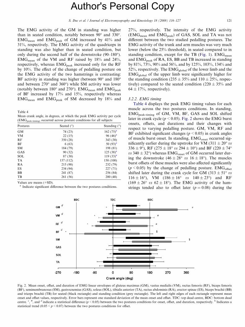
Fig. 2. Mean onset, offset, and duration of EMG linear envelopes of gluteus m(BF), semimembranosus (SM), gastrocnemius (GAS), soleus (SOL), tibialis antand triceps brachii (TB) for seated (black rectangle) and standing condition (onset and offset values, respectively. Error bars represent one standard deviatiocentre. *, #, and d indicate a statistical difference (p < 0.05) between the two postatistical trend (0.05 < p < 0.07) between the two postures conditions for offs
27%, respectively. The intensity of the EMG activity(EMGmean and EMGpeak) of GAS, SOL and TA was notdifferent between the two studied pedalling postures. TheEMG activity of the trunk and arm muscles was very muchlower (below the 25% threshold), in seated compared to instanding condition, except for the TB (Fig. 1). EMGmean
and EMGpeak of RA, ES, BB and TB increased in standingby 81%, 73%, 90% and 56%, and by 125%, 103%, 134% and52%, respectively. The EMGglobal of the lower limb and theEMGglobal of the upper limb were significantly higher forthe standing condition (235 ± 35% and 110 ± 25%, respec-tively) compared to the seated condition (220 ± 35% and64 ± 17%, respectively).
3.2.2. EMG timing
Table 4 displays the peak EMG timing values for eachmuscle across the two postures conditions. In standing,EMGpeak-timing of GM, VM, BF, GAS and SOL shiftedlater in crank cycle (p < 0.05). Fig. 2 shows the EMG burstonsets, offsets, and durations and their changes withrespect to varying pedalling posture. GM, VM, RF andBF exhibited significant changes (p < 0.05) in crank anglesof muscle burst onset. In standing, EMGonset occurred sig-nificantly earlier during the upstroke for VM (311 ± 20� vs
336 ± 9�), RF (275 ± 18� vs 294 ± 10�) and BF (220 ± 74�vs 340 ± 32�) whereas EMGonset of GM occurred later dur-ing the downstroke (46 ± 28� vs 16 ± 18�). The musclesburst offsets of these muscles were also affected significantly(p < 0.05) by the change of pedalling posture. EMGoffset
shifted later during the crank cycle for GM (313 ± 51� vs116 ± 16�), VM (186 ± 16� vs 140 ± 23�) and RF(169 ± 26� vs 62 ± 18�). The EMG activity of the ham-strings tended also to offset later (p = 0.08) during the
aximus (GM), vastus medialis (VM), rectus femoris (RF), biceps femoriserior (TA), rectus abdominis (RA), erector spinae (ES), biceps brachii (BB)grey rectangle). The left and right edges of each rectangle represent meann of the mean onset and offset. TDC: top dead centre, BDC: bottom deadstures conditions for onset, offset, and duration, respectively. $ Indicates aet.
Author's Personal Copy
Table 5Mean crank angle, in degrees, of onset and offset of burst EMG activityfor arm and trunk muscles during standing pedalling for all subjects
Crank angle Onset (�) Offset (�)
RA 113 (64) 293 (71)ES 97 (56) 301 (59)BB 89 (45) 334 (50)TB 70 (16) 354 (21)
Values are means (+SD). In seated pedalling, EMG activity of TB start at123 (69)� and finish at 326 (25)�.
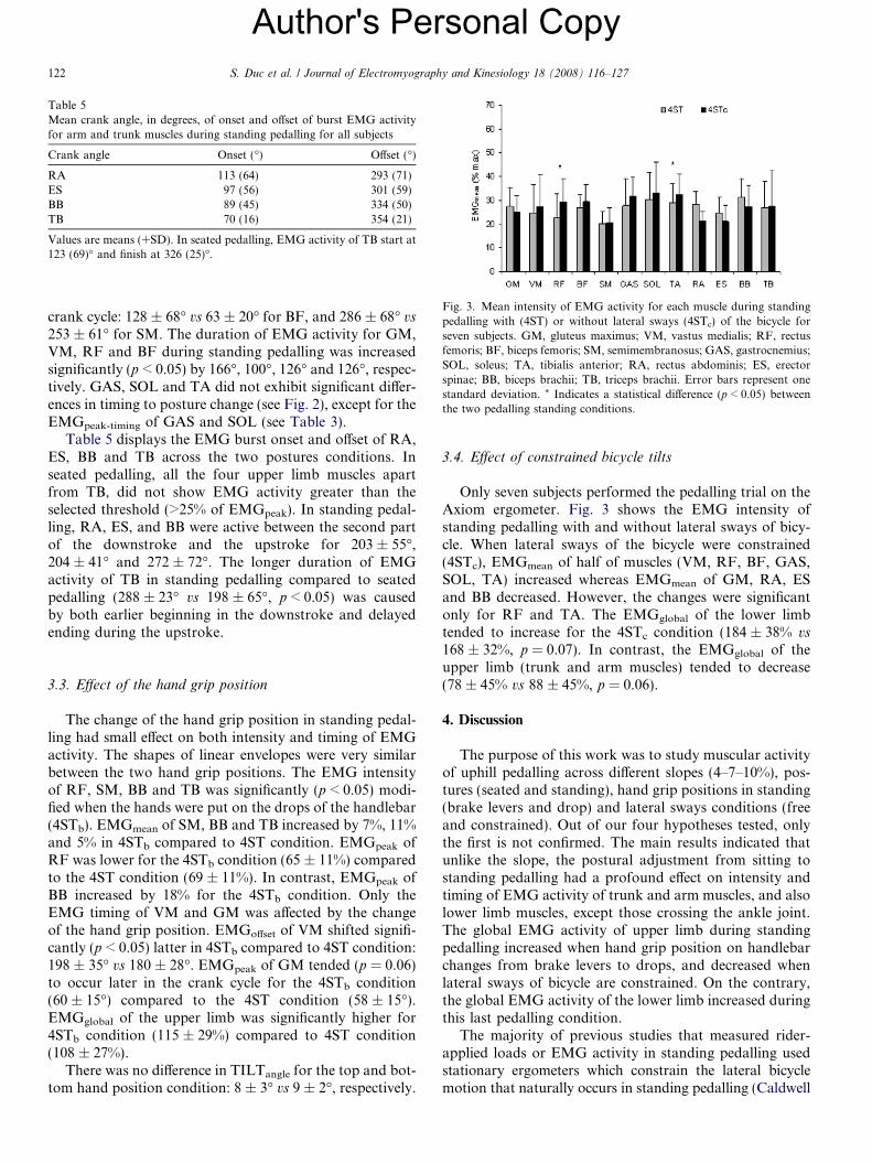
Fig. 3. Mean intensity of EMG activity for each muscle during standingpedalling with (4ST) or without lateral sways (4STc) of the bicycle forseven subjects. GM, gluteus maximus; VM, vastus medialis; RF, rectusfemoris; BF, biceps femoris; SM, semimembranosus; GAS, gastrocnemius;SOL, soleus; TA, tibialis anterior; RA, rectus abdominis; ES, erectorspinae; BB, biceps brachii; TB, triceps brachii. Error bars represent onestandard deviation. * Indicates a statistical difference (p < 0.05) betweenthe two pedalling standing conditions.
122 S. Duc et al. / Journal of Electromyography and Kinesiology 18 (2008) 116–127
crank cycle: 128 ± 68� vs 63 ± 20� for BF, and 286 ± 68� vs
253 ± 61� for SM. The duration of EMG activity for GM,VM, RF and BF during standing pedalling was increasedsignificantly (p < 0.05) by 166�, 100�, 126� and 126�, respec-tively. GAS, SOL and TA did not exhibit significant differ-ences in timing to posture change (see Fig. 2), except for theEMGpeak-timing of GAS and SOL (see Table 3).
Table 5 displays the EMG burst onset and offset of RA,ES, BB and TB across the two postures conditions. Inseated pedalling, all the four upper limb muscles apartfrom TB, did not show EMG activity greater than theselected threshold (>25% of EMGpeak). In standing pedal-ling, RA, ES, and BB were active between the second partof the downstroke and the upstroke for 203 ± 55�,204 ± 41� and 272 ± 72�. The longer duration of EMGactivity of TB in standing pedalling compared to seatedpedalling (288 ± 23� vs 198 ± 65�, p < 0.05) was causedby both earlier beginning in the downstroke and delayedending during the upstroke.
3.3. Effect of the hand grip position
The change of the hand grip position in standing pedal-ling had small effect on both intensity and timing of EMGactivity. The shapes of linear envelopes were very similarbetween the two hand grip positions. The EMG intensityof RF, SM, BB and TB was significantly (p < 0.05) modi-fied when the hands were put on the drops of the handlebar(4STb). EMGmean of SM, BB and TB increased by 7%, 11%and 5% in 4STb compared to 4ST condition. EMGpeak ofRF was lower for the 4STb condition (65 ± 11%) comparedto the 4ST condition (69 ± 11%). In contrast, EMGpeak ofBB increased by 18% for the 4STb condition. Only theEMG timing of VM and GM was affected by the changeof the hand grip position. EMGoffset of VM shifted signifi-cantly (p < 0.05) latter in 4STb compared to 4ST condition:198 ± 35� vs 180 ± 28�. EMGpeak of GM tended (p = 0.06)to occur later in the crank cycle for the 4STb condition(60 ± 15�) compared to the 4ST condition (58 ± 15�).EMGglobal of the upper limb was significantly higher for4STb condition (115 ± 29%) compared to 4ST condition(108 ± 27%).
There was no difference in TILTangle for the top and bot-tom hand position condition: 8 ± 3� vs 9 ± 2�, respectively.
3.4. Effect of constrained bicycle tilts
Only seven subjects performed the pedalling trial on theAxiom ergometer. Fig. 3 shows the EMG intensity ofstanding pedalling with and without lateral sways of bicy-cle. When lateral sways of the bicycle were constrained(4STc), EMGmean of half of muscles (VM, RF, BF, GAS,SOL, TA) increased whereas EMGmean of GM, RA, ESand BB decreased. However, the changes were significantonly for RF and TA. The EMGglobal of the lower limbtended to increase for the 4STc condition (184 ± 38% vs168 ± 32%, p = 0.07). In contrast, the EMGglobal of theupper limb (trunk and arm muscles) tended to decrease(78 ± 45% vs 88 ± 45%, p = 0.06).
4. Discussion
The purpose of this work was to study muscular activityof uphill pedalling across different slopes (4–7–10%), pos-tures (seated and standing), hand grip positions in standing(brake levers and drop) and lateral sways conditions (freeand constrained). Out of our four hypotheses tested, onlythe first is not confirmed. The main results indicated thatunlike the slope, the postural adjustment from sitting tostanding pedalling had a profound effect on intensity andtiming of EMG activity of trunk and arm muscles, and alsolower limb muscles, except those crossing the ankle joint.The global EMG activity of upper limb during standingpedalling increased when hand grip position on handlebarchanges from brake levers to drops, and decreased whenlateral sways of bicycle are constrained. On the contrary,the global EMG activity of the lower limb increased duringthis last pedalling condition.
The majority of previous studies that measured rider-applied loads or EMG activity in standing pedalling usedstationary ergometers which constrain the lateral bicyclemotion that naturally occurs in standing pedalling (Caldwell
Author's Personal CopyS. Duc et al. / Journal of Electromyography and Kinesiology 18 (2008) 116–127 123
et al., 1998, 1999; Juker et al., 1998; Li and Caldwell, 1998;Usabiaga et al., 1997). Because this constraint could poten-tially affect the pedalling movement, two others devices canbe considered: rollers and motorised treadmill. Althoughrollers do not constrain lateral motion of the bicycle, diffi-culties in maintaining balance in the standing position leadto potential effects in pedalling technique. Therefore, wehave used a motorised treadmill in order to simulate betternatural standing pedalling. This device provides a wide, flatsurface upon which subjects can ride naturally withoutrestraint or balance difficulties, after a short training per-iod. Furthermore, the inclination and speed of the tread-mill can be easily adjusted to provide a high constantpower output in order to simulate a steady state hillclimbing.
4.1. Effect of the slope
Our hypothesis, that intensity of EMG activity of powerproducer muscles (GM, VM), lower back muscles (ES) andarm muscles (BB, TB) increase with slope, is not confirmedby the results. Out of the 75 tested variables, only 4% ofthem (three) were influenced by the change of the slope.Our results are in line with Li and Caldwell (1998) whofound no difference in EMG activity for GM, VL, BF,RF, GAS and TA muscles between 0% and 8% slope. How-ever, the effect of the slope could be masked in this last studysince the subjects used a lower pedalling cadence during theuphill condition. Several studies have shown that intensityof EMG activity of GM, RF, GAS and BF are sensitive tovariation of pedalling cadence (Baum and Li, 2003; Marshand Martin, 1995; Neptune et al., 1997; Sanderson et al.,2005; Sarre et al., 2003). Clarys et al. (2001) showed thatthe global muscular intensity of the lower limb, quantifiedby the sum of integrated EMG, increased with increasingroad slope (2–12%), while the global muscular intensity ofthe arm decrease in the same time. We have found no signif-icant changes of global EMG activity of lower and upperlimbs, quantified by the sum of mean RMS, when the slopeof the treadmill increases from 4% to 10%. This differencecan be due to the number of muscles used to quantify mus-cular intensity of the lower limb: only four muscles (VM,BF, GAS and TA) for Clarys et al. (2001). If we calculatethe global muscular activity of the lower limb with usingthe same method, then we find a significant trend(p = 0.06) for global muscular activity of the lower limb toincrease with increasing treadmill slope. Another hypothesisto explain this difference can be the experimental conditions(indoor vs outdoor). In this study, the effect of the slope onmuscular activity was studied by having cyclists ride theirown bicycles on an indoor, motorised treadmill at a constantspeed, grade, and gear ratio. These conditions insured that:(1) air resistance owing to forward movement of the bicycle/rider was eliminated, (2) air resistance to wheel rotation wasconstant, which is unlikely during outdoor cycling owing tovariation in wind and bicycle speeds, (3) pedal speed wasconstant, which is possible in road cycling only if the bicycle
speed and gear ratio are fixed, and (4) the mechanical powerrequirement for each subject was constant, which is difficultto achieve during outdoor cycling due to variations inincline and in factors 1–3.
The tilt of bicycle during standing increased withincreasing treadmill slope: from 8 ± 3� (4% slope) to11 ± 1� (10% slope). To our knowledge, only two studieshave measured bicycle lean during standing pedalling.Soden and Adeyefa (1979) reported a lean of 10� when asubject ride on a 10% slope whereas Hull et al. (1990)observed of lower value (4.8�) but for a lower slope (6%).Besides the difference of the slope, the discrepancy betweenthese two studies can be due to two others factors. The firstfactor is the method used to determine the angle of lean ofthe bicycle. Measurements were made from video film forSoden and Adeyefa (1979) whereas Hull et al. (1990) per-formed 3D goniometric measures. The second factor isthe arm position on the handlebar. During the first study,the subjects gripped the handlebar on the brake leverswhereas Hull et al. (1990) asked to cyclists to place theirhands in the drops position. Since we observed no signifi-cant difference in lean angle between the two hand grippositions during standing pedalling at 4% slope (8 ± 3� vs
9 ± 2�), we can suppose that cyclists increase lateral swaysof the bicycle during standing pedalling with increasingslopes in order to achieve a better balance.
4.2. Effect of the posture
The change of pedalling posture from sitting to standingaffects strongly the intensity and the timing of EMG activ-ity of all muscles but those crossing the ankle joint (GAS,SOL, TA), which is in line with our second hypothesis. Toour knowledge, only one study has measured EMG activityof lower limb muscles during standing pedalling (Li andCaldwell, 1998). The authors showed that EMG activityof GM, RF and TA increased significantly during standingpedalling. The burst duration of GM, VL and RF werealso increased in standing compared to seated pedalling.The EMG activity of BF and GAS did not display signifi-cant alterations with the change of posture.
Our study confirms the results of Li and Caldwell (1998)except for three muscles (GM, RF and BF). The differencescan be due to the materiel used for testing. Unlike thetreadmill, the stationary ergometer (i.e. Velodyne) usedby these authors did not allow cyclists to lean the bicycleduring standing pedalling. We have compared EMG activ-ity of standing pedalling with or without bicycle lean. Inthis last case, subjects had to ride on a stationary ergometerlike a Velodyne (i.e. Axiom). EMG activity of RF and TAincreased significantly when the bicycle lateral sways areconstrained. Moreover, unlike the upper limb, global inten-sity of lower limb was higher. Thus, it is possible that EMGactivity is more affected by the change of pedalling posturewhen lateral sways of the bicycle are not constrained.
We found that mean and peak EMG of GM increaseddramatically in standing pedalling. However, contrary to
Author's Personal Copy124 S. Duc et al. / Journal of Electromyography and Kinesiology 18 (2008) 116–127
Li and Caldwell (1998) we observed a longer duration ofGM activity in standing: 267� vs 160�. These changes areunrelated with hip joint moment since Caldwell et al.(1999) reported only slight modifications of peak extensormoment with alteration in pedalling posture. Therefore, itwas supposed that cyclists activate greater and longerGM in standing to stabilize their pelvis due to the removalof the saddle support (Li and Caldwell, 1998). This factcould be amplified by the lateral sways of the bicycle during‘‘natural’’ standing, which did not occur during the previ-ous study (Li and Caldwell, 1998).
We have observed a significant increase of RF activityduring the second part of the downstroke (between 90�and 180�). Li and Caldwell (1998) also reported this mod-ification, but the increase was lower (cf. fig. 2, p. 929). Thedifference can be explained by the change of the quadri-ceps/hamstrings force ratio. Standing involved a higheractivity of BF. In order to counteract the knee flexor andto increase the period of the knee extensor moment, RFactivity might to be also increased in standing. Anotherpossible explanation is related to the quadriceps musclestrength. Weaker monoarticular knee extensors (VM, VL)may need help from two joint RF muscle to forcefullyextend the knee joint. Moreover, RF can act in synergywith GM to stabilize the pelvis.
The change of VM activity during standing is similarwith the change of VL activity reported by Li and Caldwell(1998). VM is activated earlier in the upward recoveryphase, and the activity lasted longer into the subsequentdownward power phase. In order to explain these changes,it must to be placed within the context of joint momentchanges associated with alteration in posture. Caldwellet al. (1999) showed that the knee extensor moment is pro-longed to the end of the downstroke in standing pedalling,consistent with the greater duration of VM and RF activ-ity. The changes in joint moments from seated to standingposture are related with three factors: the higher pedalforces, the toe down shift in pedal orientation, and themore forward hip and knee positions (Caldwell et al.,1998).
Contrary to the study of Li and Caldwell (1998), wefound that mean and peak EMG of BF was significantlyhigher during standing pedalling. Moreover, the activityof this muscle started earlier in the upstroke and tendedto cease latter in the downstroke. The differences betweenthe two studies might be due to the muscular coordinationused by cyclists to pedal in standing (Li and Caldwell,1998). It seems (cf. Li and Caldwell, 1998, fig. 7, p. 933),that some of them coactivate BF entirely with hip and kneeextension during the downstroke (0–180�) whereas othersstart BF activity well before 0� and cease activity just afterthe middle of the downstroke (�130�). In this last case, BFactivity was associated with hip and knee flexion in laterecovery before top dead centre rather than with hip andknee extension in the early downstroke. The different usageof BF can be related to cyclist specific pedalling technique.The first pattern of BF activity might be employed to trans-
fer the power produced by monoarticular muscles (GM,VM, VL) to the pedal (van Ingen Schenau et al., 1992)whereas the second pattern can reflect the smoothing ofthe pedalling during the flexion-to-extension transition(Raasch et al., 1997). However, it is possible that cyclistsactivate more BF in standing to generate propulsive torqueduring the upstroke (Neptune et al., 1997) or to help GMand RF muscles to stabilize the pelvis.
It is difficult to us to explain why, unlike to BF, EMGactivity of SM decreased during standing pedalling. In fact,it would be expected that SM show similar responses withaltering pedalling posture to BF since these two muscles areagonists. However, it was hypothesized that, contrary toBF, SM acts more in knee flexor than in hip extensor (Eric-son, 1988). Therefore, it is possible that the reduction ofSM activity is linked to the decrease of the peak and theduration of knee flexor moment observed in standingpedalling (Caldwell et al., 1999).
Li and Caldwell (1998) supposed that SOL plays a moreimportant role in the increase of the peak plantar flexormoment because of the biarticular function of GAS, as italso serves as a knee flexor. With the extended period ofthe knee extensor moment in standing, increase GAS activ-ity would be contraindicated. We have not observed signif-icant changes in intensity or timing of EMG activity of theankle plantar flexors (GAS, SOL) and extensor (TA) withaltering pedalling posture. So, the hypothesis of Li andCaldwell (1998) is not supported. It is probable thatincrease of plantar flexor moment during standing isrelated to non-muscular forces. When standing pedalling,the loss of the saddle support results in an increase of grav-itational force to the generated pedal forces as a larger pro-portion of the weight is held by the pedal during thedownstroke. Therefore, with using gravity and with fixingthe ankle in a horizontal position, riders can produce ahigher plantar flexor moment during standing withoutchanging EMG activity of flexor and extensor anklemuscles.
To the best of our knowledge, it is the first time that theinfluence of standing posture on the activity of arm andtrunk muscles is studied in ‘‘natural’’ pedalling condition(with lateral bicycle sways). Moreover, it is also the firsttime that the pattern of EMG activity of arms musclesare described in seated and standing pedalling posture.Mean and peak EMG of BB, TB, RA and ES increaseddramatically in standing. All muscles are recruited betweenthe second part of the downstroke and the upstroke forabout 200–280�. In seated pedalling, only arms muscles,notably TB, are really activated with a double burst occursnear 150� and 300�. A better understanding of these activ-ity changes can be gained by placing them within the con-text of handlebar forces and pelvis motion associated withalteration in posture.
Some studies have taken an interest in the upper bodymuscle work (Juker et al., 1998; Soden and Adeyefa,1979; Stone and Hull, 1993, 1995; Usabiaga et al., 1997).Stone and Hull (1993, 1995) have measured the force
Author's Personal CopyS. Duc et al. / Journal of Electromyography and Kinesiology 18 (2008) 116–127 125
applied on the handlebar in seated and standing positionby using strain gauge dynamometers. Handlebar forceswere also previously determined by Soden and Adeyefa(1979) but they were computed with an equilibrium analy-sis, which involved a number of simplifying assumptions tomake the results debatable. In uphill seated, forces appliedon the handlebar are directed downward and forward sug-gesting that the arms primarily function to passively sup-port the weight of the torso (Stone and Hull, 1993, 1995).In uphill standing, however, handlebar forces are charac-terized by a change of the orientation indicating the activerole played by the arms. Cyclists pull up and back on thehandlebar during the downstroke (between 30� and 160�),and push down and forward during the region of about160� back through 30� (Soden and Adeyefa, 1979). If wesuppose that handlebar forces are symmetric, then pushingdown and forward with the left arm during the downstrokeof the right pedal, occurs simultaneously with the pullingup and back of the right arm. Our results are in line withthis hypothesis. In standing, TB and BB activities showedsimilar patterns with a double EMG burst period(Fig. 1). The high activity of TB (first burst between 135�and 225�) seems to be related with the pushing down andforward action of the right arm, which occurred duringthe downstroke of the right pedal. If there is a symmetryfor EMG activity like handlebar force, we can expect thatthe high activity of BB (second burst between 270� and330�) is linked to the pulling up and forward action occur-ring simultaneously during the downstroke of the left pedal(during the upstroke for the right pedal). However, the cen-tral nervous system coactivates the muscles of the two armsto control the force applied to the handlebar in order tomaintain the equilibrium of the body. Hull et al. (1990)found that the maximum lean of bicycle occurred at about140�, which is close to the transition phase for the directionof the handlebar forces. Therefore, the actions of the armduring standing not only counter the pedal driving forcesbut also act to lean the bicycle from side-to-side. The max-imal power developed by arms in standing was estimated to15 W (Stone and Hull, 1993), which is a small percentage ofthe 1000 W of maximal power generated by the lower limb.Thus, although the arms control the leaning of the bicyclein standing, this control does not result in substantialpower development.
Using a specially designed climbing bicycle with achanging saddle-tube on a simulated road inclinationsfrom 0% to 20%, Antonis et al. (1989) found a decreasein muscular activity with the saddle forward (saddle tubeangle of 80�) on a 20% slope; this mostly resulted fromdecreased activity in the arm muscles (BB, TB). They sug-gested that the cyclist’s position with the saddle forwardseems to offer the best muscular condition for cycling uphillfor the ‘short legged’ athlete. The ‘long legged’ cyclist onthe other hand climbed more economically with the saddlein backward position (saddle-tube angle of 67�). Claryset al. (2001) confirmed these preliminary results by collect-ing data using the same experimental bicycle during field
conditions (‘Kluisberg’ mountain). They observed a signif-icant decrease of EMG of the upper limb (BB, TB) as afunction of increasing slope (2–12%) but mostly with thesaddle in maximal forward position.
Like to arms muscles, flexor (RA) and extensor (ES) ofthe trunk showed a double EMG burst period, whichappeared throughout the bottom dead centre and the halfof the upstroke. Juker et al. (1998) and Usabiaga et al.(1997) have shown that the EMG activity of paravertebrallumbar muscles increased in the more upright position. Theeffect of the posture on the abdominal muscles is not clearsince the results differ between the two studies. It is impor-tant to note that cyclists cannot still lean their bicycle fromside-to-side during these two studies. These changes alter-ing pedalling posture seem to be linked to the removal ofthe saddle support and to the straightening up of the torso.During standing, the pelvis makes simultaneously a verticalelevation and a rotation in rocking. Hull et al. (1990) haveshown that, in standing pedalling, maximal elevation of thepelvis occurred at the middle of the downstroke and theupstroke. The change in pelvis elevation is approximately5 cm at 6% slope. Accordingly, the torso reaches greatestpotential energy just prior the period of propulsive torque(90–160�), and loses potential energy as maximum cranktorque is developed. The rocking angle shows a single cycleduring the crank cycle. The right hip is maximally higherthan the left at 30� of the crank cycle. Recall also thatthe maximal bicycle lean angle occurred in standing justafter the peak of propulsive crank torque. Therefore, lum-bar and abdominal muscles must to be contracted to stabi-lize the pelvis and also torso in order to transfer the workdone by arms and the torso potential energy to the pedal.
4.3. Effect of the hand grip position
The main effect of the change of the hand grip positionfrom brake levers to the drops, in standing, is the increaseof the arm muscles activity. The global activity of the upperlimb (arm and trunk muscles) was also higher when thehands are placed on the drops of the handlebar. The peakEMG activity of RF is lower in this last position. Thesechanges can be explained by the alteration of the cyclist’sposition. When the hands are placed on the drops of thehandlebar, the total body centre mass is shifted further for-ward and the trunk is more flexed. Therefore, cyclists mustactivate greater BB and TB muscles in the bottom handposition (in the drops) because the weight supported bythe arms is higher.
The increase of EMG activity of arm muscles is notlinked to the action of leaning the bike side-to-side sincethe tilt’s angle is not affected by the change of the hand gripposition. Moreover, the change of hand grip position whileseated pedalling does not involve an increase of oxygenuptake at sub-maximal intensity (Grappe et al., 1998;Raasch et al., 1997). Nevertheless, Grappe et al. (1998)observed a higher rating of perceived exertion and ventila-tory response for the drop position than brake lever

Author's Personal Copy126 S. Duc et al. / Journal of Electromyography and Kinesiology 18 (2008) 116–127
position while seated pedalling. These changes were attrib-uted to the difference in mean hip angle (i.e. difference intrunk flexion) which is estimated to 11� between the brakelever position and the drop position (Grappe et al., 1998).The authors suggested that the change in mean hip anglewhich determined mainly changes in the trunk position couldalter alignment and geometry of the upper respiratory tractand therefore the respiratory mechanics (Grappe et al., 1998).
The decrease of the hip flexor activity (RF) seems to beassociated to the reduction of hip angle (angle between thetrunk and the thigh), which is due to the increase of thetrunk flexion. This result agrees with the study of Savelberget al. (2003). These authors showed that the EMG activityof RF is lower for a 22� forward trunk flexion than 20�backward trunk extension.
Juker et al. (1998) observed higher EMG activity of RFand psoas muscle in flexed racing position (hands grip onthe drops) compared to upright normal position (hand gripon the bottom). The EMG activity of abdominal wall andES muscles was not affected by the change of body posi-tion. Our study confirms these results since we have alsonot observed significant difference in EMG activity forabdominal and trunk muscle (RA, ES) when the hand gripposition was changed in standing pedalling.
4.4. What is the advantage of standing pedalling for cycling
performance?
Some cyclists often chose to switch between the two ped-alling postures during climbing whereas others prefer toremain seated for the major time. The first strategy is gener-ally adopted by climbers while the second is often used bytime-trialists and flat-terrain specialists. It is difficult to con-clude what the best strategy to climb is. Through the resultsof the present study, it is clear that standing compared withseated pedalling enhances increased muscular activity of thelower limb, except the muscles crossing the ankle joint.Moreover, the upper body musculature (arm and trunkmuscles) is more involved for pelvic and torso stabilizationand for controling side-to-side leaning of the bicycle. Thisactivity requires energy expenditure greater than that neces-sary to simply propel the bicycle during uphill. Therefore, itis reasonable to believe that this additional energy require-ment results in reduced metabolic efficiency.
Nevertheless, this assumption is not so obvious. Tanakaet al. (1996) and Ryschon and Stray-Gundersen (1991)reported an increase in _VO2 of �6% and �12%, respec-tively, between the uphill seated and standing posture at�50% and �60% of _VO2 max, respectively. However, othersauthors did not observed significant differences for grossefficiency and economy between the two pedalling postureat 75% of MAP or _VO2 max (Millet et al., 2002; Swain andWicox, 1992). Moreover, Tanaka et al. (1996) showed thatat a higher intensity (�83% of ð _VO2 maxÞ, standing andseated induced the same _VO2 response. Consequently, thehypothesis that standing posture is less economic thanseated seems to be valid only when intensity of pedalling
exercise is lower than 75% of _VO2 max. At moderate inten-sity, the extra work of the upper body muscles in the stand-ing posture accounts for a greater proportion of the overallmechanical work produced and therefore leads to a signif-icant increase of energy expended when compared withcycling in seated posture. At higher intensity (>75%_VO2 maxÞ, forces applied on the handlebar with the upperlimbs in seated posture increased significantly (Stone andHull, 1995), explaining partly why the difference in _VO2
between the two pedalling postures disappears.The fact that some cyclists prefer the standing posture in
spite of its higher energy expenditure at moderate power out-put, suggests that the intensity of standing pedalling seems tobe perceived less difficult than the seated pedalling. We canspeculate that the altered perception of exertion may bedue to the redistribution of the workload over a greater mus-cle mass, the alteration of the force–velocity and force–length relationships of power producer muscles, or the avail-ability to generate greater power output with using non-mus-cular force like gravity, in the standing posture. Moreover,the switch between the two pedalling postures in uphill modeenables cyclists to use two distinct muscular chains becausethe muscular coordination of pedalling is different in thestanding compared to the seated position. This fact canexplain why cyclists seem to feel that it is easier to pedal inseated posture just after a short bout of standing pedalling.
Relating to our results, coaches need to advise cyclists tooften train in the standing climbing mode to improve mus-cular coordination of standing pedalling. They can also sug-gest strengthening of the arm and trunk power, notably forthe abdominal wall and lumbar muscles. Finally, we recom-mend often alternating between the seated and the standingposture in long uphills in order to alleviate lower back pain.
5. Conclusion
Our results indicate that the increase of the treadmillslope from 4% to 10% in uphill cycling did not significantlychange the muscular activity of lower and upper limbs. Incontrast, the change of pedalling posture from seated tostanding affected largely the intensity and the timing ofEMG activity of muscles crossing elbow (BB,TB), pelvis(RA, ER), hip (RF, GM) and knee joint (VM, BF, SM).Among all the muscles tested, arm and trunk musclesexhibited the most significant increase of muscle activity.The coordination between antagonist pairs is also alteredby the change of pedalling posture. These changes arestrongly related to the greater peak pedal forces and thesuppression of the saddle support.
Some of these muscles (RF, SM, BB, TB) also showedslight alterations in standing when the hand grip positionchanged from brake levers to the drops. The EMG activityof the arm muscles is increased in the drops positionbecause the total body centre mass is shifted more forwardand the trunk is more flexed. Finally, global activation oflower and upper limbs are modified in standing when thelateral sways of bicycle are constrained.
Author's Personal CopyS. Duc et al. / Journal of Electromyography and Kinesiology 18 (2008) 116–127 127
References
Alvarez G, Vinyolas J. A new bicycle design for on-road measurements ofcycling forces. J Appl Biomech 1999;12:130–42.
Antonis J, Clarys JP, Cabri J, Clijsen L, Welbergen E. EMG of uphillracing – a new qualitative approach of cyclic movements. In:Proceedings of the international congress of biomechanics. LosAngeles (USA): University of California; 1989.
Baum B, Li L. Lower extremity muscle activities during cycling areinfluenced by load and frequency. J Electromyogr Kinesiol2003;13:181–90.
Bertucci W, Duc S, Villerius V, Grappe F. Validity and reliability of theAxiom Powertrain Cycle Ergometer when compared with an SRMpowermeter. Int J Sport Med 2005;26:59–65.
Bertucci W, Duc S, Villerius V, Grappe F. Validity and reliability of thePowerTap mobile cycling powermeter when compared with an SRMdevice. Int J Sport Med 2005;26:868–73.
Caldwell GE, Li L, McCole SD, Hagberg JM. Pedal and crank kinetics inuphill cycling. J Appl Biomech 1998;14:245–59.
Caldwell GE, Hagberg JM, McCole SD, Li L. Lower extremity jointmoments during uphill cycling. J Appl Biomech 1999;15:166–81.
Clarys JP. Electromyography in sports and occupational settings: anupdate of its limits and possibilities. Ergonomics 2001;43:1750–62.
Clarys JP, Alewaeters K, Zinzen E. The influence of geographic variationson the muscular activity in selected sports movements. J ElectromyogrKinesiol 2001;11:451–7.
Ericson MO. Muscle function during ergometer cycling. Scand J RehabilMed 1988;20:35–41.
Gardner AS, Stephens S, Matin DT, Lawton E, Lee H, Jenkins D.Accuracy of SRM and Powertap power monitoring systems forbicycling. Med Sci Sport Exer 2004;36:1252–8.
Grappe F, Candau R, Busso T, Rouillon JD. Effect of cycling position onventilatory and metabolic variables. Int J Sport Med 1998;19:336–41.
Hull ML, Beard A, Varma H. Goniometric measurement of hip motion incycling while standing. J Biomech 1990;23(7):687–703.
Juker D, McGill S, Kropf P. Quantitative intramuscular myoelectricactivity of lumbar portions of psoas and the abdominal wall duringcycling. J Appl Biomech 1998;14:428–38.
Li L, Caldwell GE. Muscle coordination in cycling: effect of surface inclineand posture. J Appl Physiol 1998;85(3):927–34.
Marsh AP, Martin PE. The relationship between cadence and lowerextremity EMG in cyclists and noncyclists. Med Sci Sport Exer1995;27:217–25.
Millet GP, Tronche C, Fuster N, Candau R. Level ground and uphillcycling efficiency in seated and standing positions. Med Sci Sport Exer2002;34(10):1645–52.
Neptune RR, Kautz SA, Hull ML. The effect of pedalling rate oncoordination in cycling. J Biomech 1997;30:1051–8.
Raasch CC, Zajac FE, Ma B, Levine WS. Muscle coordination ofmaximum speed pedalling. J Biomech 1997;6:515–25.
Ryschon TW, Stray-Gundersen J. The effect of body position on theenergy cost of cycling. Med Sci Sport Exer 1991;23(8):949–53.
Sanderson DJ, Martin PE, Honeyman G, Keefer J. Gastrocnemius andsoleus muscle length, velocity, and EMG responses to changes inpedalling cadence. J Electromyogr Kinesiol 2005:21.
Sarre G, Lepers R, Maffiuletti N, Millet G, Martin A. Influence of cyclingcadence on neuromuscular activity of the knee extensors in human.Eur J Appl Physiol 2003;88:476–9.
Savelberg HHCM, Van de Port IGL, Willems PJB. Body configuration incycling affects muscle recruitment and movement pattern. J ApplBiomech 2003;19:310–24.
Soden PD, Adeyefa BA. Force applied to a bicycle during normal cycling.J Biomech 1979;12:527–41.
Stone C, Hull ML. Rider/bicycle interaction loads during standingtreadmill cycling. J Appl Biomech 1993;9:202–18.
Stone C, Hull ML. The effect of rider weight on rider-induced loads duringcommon cycling situations. J Biomech 1995;28(4):365–75.
Swain DP, Wicox JP. Effect of the cadence on the economy of uphillcycling. Med Sci Sport Exer 1992;24:1123–7.
Tanaka H, Bassett DJ, Best S, Baker K. Seated versus standing cycling incompetitive road cyclists: uphill climbing and maximal oxygen uptake.Can J Appl Physiol 1996;21:149–54.
Usabiaga J, Crespo R, Iza I, Aramendi J, Terrados N, Poza JJ.Adaptation of the lumbar spine to different positions in bicycle racing.Spine 1997;22(17):1965–9.
van Ingen Schenau GJ, Boos PJ, de Groot G, Snackers RJ, van WoenselWW. The constrained control of force and position in multi-jointmovements. Neuroscience 1992;46(1):197–207.
Dr. Sebastien Duc is Doctor in Exercise andSport Sciences. He received his Ph.D. degree inexercise biomechanics from Franche-ComteUniversity, in November 2005. His researchtopics concern the biomechanics and thephysiology of cycling. He has published twonational and four international papers in peer-reviewed periodicals. His research interestsfocus on the neuromuscular coordination ped-aling and the biomechanics and physiology ofcycling. He is actually a temporary assistant forteaching and researching (ATER) at the Insti-
tute of Sport Sciences (UFR STAPS) of Font-Romeu, University ofPerpignan, France.
Dr. William Bertucci is a Doctor in Exerciseand Sport Sciences. He received his Ph.D.degree in exercise biomechanics from Franche-Comte University, in December 2003. He hastree national and eight international papers inpeer-reviewed periodicals. His research topicsconcern the biomechanics and the physiologyof cycling. He is actually an assistant professor(MCU) at the Institute of Sport Sciences (UFRSTAPS) of Reims, University of Champagne-Ardenne, France.
Prof. Dr. Jean-Noel Pernin is a Doctor in
Mechanics. He received his Ph.D. degree inphysics from Franche-Comte University, in1977, and his Professor degree in Exercise andSport Sciences in 2003. He has publishedtwenty-three national and international papersin peer reviewed periodicals. His research top- ics concern the biomechanics of sports andmovements (cycling, wheelchair, walking). Heis actually a professor at the Institute of SportSciences (UFR STAPS) of Besancon, Univer-sity of Franche-Comte, France.Prof. Dr. Frederic Grappe is a Doctor in Exerciseand Sport Sciences. He received his Ph.D. degreein exercise biomechanics from Franche-ComteUniversity, in 1994, and his Professor degree in2006. He has published three national andtwenty-two international papers in peer-reviewperiodicals. His research topics concern thebiomechanics and the physiology of cycling. Heis the coach of the French professional teamcycling ‘‘Francaise des Jeux’’ since. He is also atthe Head of the Performance Workgroup of theCycling French Federation. He is actually an
assistant professor (MCU) at the Institute of Sport Sciences (UFR STAPS)of Besancon, University of Franche-Comte, France.