this electronic thesis or dissertation has been downloaded ......cor15a and cor47 transcript...
TRANSCRIPT

This electronic thesis or dissertation has beendownloaded from Explore Bristol Research,http://research-information.bristol.ac.uk
Author:Workman, Sam
Title:The effect of UV-B on freezing tolerance in Arabidopsis thaliana
General rightsAccess to the thesis is subject to the Creative Commons Attribution - NonCommercial-No Derivatives 4.0 International Public License. Acopy of this may be found at https://creativecommons.org/licenses/by-nc-nd/4.0/legalcode This license sets out your rights and therestrictions that apply to your access to the thesis so it is important you read this before proceeding.
Take down policySome pages of this thesis may have been removed for copyright restrictions prior to having it been deposited in Explore Bristol Research.However, if you have discovered material within the thesis that you consider to be unlawful e.g. breaches of copyright (either yours or that ofa third party) or any other law, including but not limited to those relating to patent, trademark, confidentiality, data protection, obscenity,defamation, libel, then please contact [email protected] and include the following information in your message:
•Your contact details•Bibliographic details for the item, including a URL•An outline nature of the complaint
Your claim will be investigated and, where appropriate, the item in question will be removed from public view as soon as possible.

1
A dissertation submitted to the University of Bristol in accordance with the requirements for
the award of the degree of Master of Science in the Faculty of Life Sciences
The effect of UV-B on freezing tolerance
in Arabidopsis thaliana
Samuel Workman
University of Bristol
1325347

2
Abstract
Cold stress causes significant crop losses each year. Some plants can withstand this stress
through the upregulation of genes which result in cold acclimation. This process involves the
C-REPEAT BINDING FACTOR (CBF) regulon and COLD RESPONSE (COR) gene
expression which protect the plant from low temperature damage. The signalling pathway that
leads to cold acclimation is therefore of interest when investigating how to protect plants from
low temperature stress. When multiple signalling pathways converge in a process known as
crosstalk, enhanced resistance to a stress can result. Previous studies have shown that UV-
B can enhance the survivability of Rhododendron at low temperatures and increase
expression of protective enzymes involved in cold resistance in Triticum aestivum. This
phenomenon has not been investigated in the model species, Arabidopsis thaliana. Here, the
interaction of UV-B and cold acclimation in Arabidopsis is explored. An enhancement of
COR15a and COR47 transcript abundance was observed when cold acclimation occurred in
the presence of UV-B, but this was not accompanied by increased plant survival following
freezing stress at -6 oC. The role of flavonoids in cold and UV-B signalling was also analysed
as flavonoids are known to be involved in both plant freezing tolerance and UV-B protection.
Mutants deficient in flavonoid biosynthesis, transparent testa 4 (tt4) and tt7 showed decreased
survival following a freezing stress. This response was further exacerbated by UV-B treatment.
The combination of cold and UV-B resulted in greater levels of CHALCONE SYNTHASE
(CHS) transcript and flavonoid accumulation than either treatment alone, suggesting a
synergistic interaction. Together, these data suggest that crosstalk exists between low
temperature and UV-B signalling and that UV-B could be used conditionally to enhance cold
acclimation and increase freezing tolerance in Arabidopsis.

3
Acknowledgements
I would like to extend thanks to my project supervisor Professor Keara Franklin for her
guidance, support and unrelenting patience throughout this project. Thanks to Dr Donald
Fraser and Dr Ashutosh Sharma for help with protocols, calculations and providing me with
seed stock. I would also like to extend thanks to Mathilda Gustavsson for help with lab
procedures and moral support throughout the project. Finally, I wish to thank my family Linda
Workman, Gerald Workman, Matthew Workman and Parmi Perera for their unwavering
support through the past few years without which this would not have been possible.

4
Author’s declaration
I declare that the work in this dissertation was carried out in accordance with the requirements
of the University's Regulations and Code of Practice for Research Degree Programmes and
that it has not been submitted for any other academic award. Except where indicated by
specific reference in the text, the work is the candidate's own work. Work done in collaboration
with, or with the assistance of, others, is indicated as such. Any views expressed in the
dissertation are those of the author.
SIGNED: DATE: 04/07/2019

5
Table of contents
Title page 1
Abstract 2
Acknowledgments 3
Author’s declaration 4
Table of contents 5
List of figures and tables 7
List of abbreviations 9
CHAPTER 1: INTRODUCTION 12
1.1 Low temperatures 14
1.2 Freezing injury 15
1.3 Cold acclimation 17
1.4 Light signalling 19
1.5 UV-B 20
1.6 Light regulation of plant freezing tolerance 22
1.7 UV-B regulation of plant freezing tolerance 23
1.8 Project aims 24
CHAPTER 2: MATERIALS AND METHODS 25
2.1 Seed stocks and growth conditions 25
2.2 Whole plant freezing tolerance assay 26
2.3 Quantitative polymerase chain reaction 26
2.3.1 RNA extraction 26
2.3.2 cDNA synthesis 26
2.3.3 qPCR 26
2.4 Pigment quantification 27
2.5 Statistics 28
6
2.6 Thin layer chromatography 28
CHAPTER 3: UV-B MAY ENHANCE COLD ACCLIMATION IN ARABIDOPSIS 29
3.1 Introduction 29
3.2 Results 30
3.2.1 UV-B and cold acclimation influence COR15a expression 31
3.2.2 UV-B and cold acclimation influence COR47 expression 31
3.2.3 UV-B has no discernible impact upon freezing tolerance 32
3.3 Discussion 34
CHAPTER 4: THE ROLE OF FLAVONOIDS IN UV-B AND LOW TEMPERATURE
SIGNALLING CROSSTALK 36
4.1 Introduction 36
4.2 Results 37
4.2.1 UV-B and cold reduce survival in tt4 plants 38
4.2.2 UV-B and cold reduce survival in tt7 plants 39
4.2.3 Cold and UV-B may influence CHS transcript abundance 40
4.2.4 Cold and UV-B may enhance flavonoid concentration in WT plants 41
4.3 Discussion 41
CHAPTER 5: DISCUSSION 44
5.1 UV-B enhances cold-induced accumulation of COR gene transcripts, but not whole plant
survival following freezing 44
5.2 UV-B may conditionally enhance freezing tolerance via the up-regulation of flavonoids
46
5.3 Conclusions 48
CHAPTER 6: APPENDIX 49
CHAPTER 7: REFERENCES 56

7
List of figures and tables
Figure 1. Projected crop yield increase and decrease over time due to climate change 12
Figure 2. Revised 2017 projected human population increase 12
Figure 3. Lamellar to hexagonal phase II transition 15
Figure 4. Cold stress signalling pathway 17
Figure 5. Light spectrum and key photoreceptors 19
Figure 6. UV-B light signalling pathway 21
Figure 7. Integration of cold stress and UV-B signalling pathway 23
Figure 8. COR15a transcript abundance in 2-week-old Arabidopsis seedlings 31
Figure 9. COR47 transcript abundance in 2-week-old Arabidopsis seedlings 32
Figure 10. Whole plant freezing tolerance assay using uvr8-6 plants 33
Figure 11. uvr8-6 whole plant freezing tolerance photos 33
Figure 12. Flavonoid biosynthesis pathway 36
Figure 13. tt4 whole plant freezing tolerance assay 38
Figure 14. tt7 whole plant freezing tolerance assay 39
Figure 15. tt4 and tt7 whole plant freezing tolerance photos 40
Figure 16. CHS transcript abundance in 2-week-old Arabidopsis seedlings 40
Figure 17. Thin layer chromatography displaying flavonoid pigments 41
Supplementary figures
Figure S1. Schematic representation of growth cabinet 49
Figure S2. Light spectra from plant growth cabinet without supplementary UV-B 49
Figure S3. Light spectra from plant growth cabinet with supplementary UV-B 50
Figure S4. Schematic representation of the cabinet used for cold acclimation and plant
freezing tolerance assays 50
Figure S5. Light spectra from plant freezing cabinet 51

8
Figure S6. Primer efficiency standard curve 51
Figure S7. Testing control genes 52
Figure S8. Chlorophyll measurement data 53
Figure S9. Flavonoid measurement data 53
Figure S10. Anthocyanin measurement data 54
Figure S11. Thin layer chromatography of flavonoid pigments 56
Tables
Table 1. IPCC predicted temperature increase scenarios for 2090 - 2099 12
Table 2. Primer name and sequence 27

9
List of abbreviations
˙OH - Hydroxyl radical
AP2/ERF - APETALA2/ETHYLENE-RESPONSE ETHYLENE-BINDING FACTOR
bHLH - Beta-helix-loop-helix
CA - Cold acclimated
CAMTA - CALMODULIN-BINDING TRANSCRIPTION ACTIVATOR
CBF - C-REPEAT BINDING FACTOR
CBLs-CIPKs - CALCINEURIN B-LIKE CIPKs
CCA1 - CIRCADIAN CLOCK ASSOCIATED 1
CHI - CHALCONE ISOMERASE
CHS - CHALCONE SYNTHASE
COLD1 - CHILLING-TOLERANCE DIVERGENCE 1
COP1 - CONSTITUTIVELY PHOTOMORPHOGENIC 1
COR - COLD RESPONSE
CPD - Cyclobutene pyrimidine dimers
CRLK 1/2 - CALCIUM/CALMODULIN-REGULATED RECEPTOR-LIKE KINASE
CRLK1/2 - CALCIUM/CALMODULIN- REGULATED RECEPTOR – LIKE KINASE
CRPK1 - COLD RESPONSIVE PROTEIN KINASE
Cry - Cryptochromes
DRE - DEHYDRATION RESPONSE ELEMENT
DREB - DEHYDRATION RESPONSE ELEMENT BINDING PROTEIN
F3’H - FLAVONOID 3-HYDROXYLASE
FHY3 - FAR-RED ELONGATED HYPOCOTYL 3
FLS - FLAVONOL SYNTHASE
GA - Gibberellic acid
GA2ox - GIBBERELLIC ACID 2 OXIDASE

10
H2O2 - Hydrogen peroxide
HOS1 - HIGH EXPRESSION OF OSMOTICALLY RESPONSIVE GENE
HY5 - ELONGATED HYPOCOTYL 5
HYH - HY5 HOMOLOG
ICE1 - INDUCER OF CBF EXPRESSION 1
INAS - Ice nucleation active substance
LEA II - LATE EMBRYOGENESIS ABUNDANT II
LHY - LATE ELONGATED HYPOCOTYL
LOR - Loss of osmotic responsiveness
LTRE - Low temperature responsive element
MAPK - MITOGEN ACTIVATED PROTEIN KINASE
MEKK1 - MITOGEN ACTIVATED PROTEIN KINASE KINASE KINASE 1
MKK2 - MITOGEN ACTIVATED PROTEIN KINASE KINASE 2
MPK4 - MITOGEN ACTIVATED PROTEIN KINASE 4
Phot - Phototropins
Phy - Phytochromes
PIF - PHYTOCHROME INTERACTING FACTOR
qPCR - Quantitative Polymerase Chain Reaction
RGA1 - RICE G-PROTEIN ALPHA SUBUNIT
ROS - Reactive oxygen species
SPA 1-4 - SUPPRESSOR OF PHYTOCHROME A-105
TLC - Thin layer chromatography
TT4 - TRANSPARENT TESTA 4
TT7 - TRANSPARENT TESTA 7
UV-B - ULTRAVIOLET B
UVR8 - UV RESISTANCE LOCUS 8

11
WT - Wild type
ZAT12 - ZINC FINGER OF ARABIDOPSIS THALIANA 12
ZTL - ZEITLUPE

12
CHAPTER 1: INTRODUCTION
The seasonal temperature variation experienced by plants presents a myriad of challenges
which has resulted in the evolution of adaptations to closely monitor seasonal variation. This
monitoring allows plants to adjust their phenology to help them survive through changing
climates. During winter months, a prominent abiotic threat to plant survival is the cold,
through chilling and freeze-damage (Korn et al., 2008). This results in reduced crop yield
and is one of the many contributing factors threatening global food security. Other factors
include heat and drought stress due to global warming. This is predicted to increase the
average temperature over the next century (Table 1) and have a negative impact on crop
yield (Figure 1). The United Nations population growth estimates predict a population
increase of 9.7 billion by 2050 and 11.2 billion by 2100 (Figure 2) (IPCC, 2007; United
nations, 2017). When crop yield losses from global warming projections are taken in
confluence with world population estimates over the coming century then the importance of
food security becomes a problem of paramount importance.
Table 1. Three IPCC predicted temperature increase scenarios for 2090 - 2099 based on
information collected 1980 – 1999 (IPCC, 2007).
IPCC predicted scenario Best estimate Temperature increase (°C)
Likely range temperature increase (°C)
Low 0.6 0.3 – 0.9
Medium 2.4 1.4 – 3.8
High 3.4 2.0 – 5.4
Figure 2. The revised 2017 projected human
population increase based on data collected between
1950 – 2015. Blue dotted line indicates known
population size, red dotted line indicates predicted
population increase. Adapted from United Nations
world population prospects, 2017 revision.
Figure 1. Projected crop yield increase and
decrease over time due to climate change. Data
is based on changes in crop yield from the 20th
century. Adapted from IPCC (2014).
Perc
enta
ge o
f yie
ld

13
As climate change continues to contribute to increases in temperatures around the world
perhaps counter intuitively, sudden frosts are becoming more prominent which are furthering
agricultural losses each year (Warmund et al., 2008). A precocious spring phenology means
plants are germinating earlier in the season (Warmund et al., 2008) thus exposing plants to a
larger range of temperatures than would be expected later in the season. This was
demonstrated by the 2007 Eastern spring freeze in the US (Gu et al., 2008). Immature tissues
are especially susceptible to cold damage and thus the eastern spring freeze was responsible
for widespread agricultural damage across America. Agricultural losses in North Carolina
alone were approximately $111.7 million. During this time, it was noted that the spring freeze
was characterized by seasonally high temperatures followed in quick succession by low
temperatures Gu et al. (2008). Other large frosts such as the Swiss Rhone valley frost in 2012
concur with the paradigm of exceptionally warm temperatures early in the season followed by
a sudden drop in temperature causing plant losses (Meier et al., 2018).
Solutions to thwart agricultural losses from abiotic stresses such as drought and increased
temperature include increasing the geographical range in which crops are grown. Many plant
species have already been demonstrated to be moving poleward or increasing altitude
putatively due to increasing global temperatures (Walther et al., 2002). Moving crops poleward
in hope to reduce the exposure to drought and increased temperatures is a possibility to
reduce yield losses but this brings other inherent risks. Cultivars from the tropics have in
general a less developed cold response compared to cultivars from temperate and poleward
regions and thus the risk of chilling and freezing damage increases the further poleward they
are moved.
One potential solution to reduce agricultural losses may be to increase UV-B supplementation
to plants. Many greenhouse grown plants have reduced UV-B exposure due to greenhouse
materials which attenuate UV-B light. Interesting studies exist which advocate the use of UV-
B to mitigate freezing damage. Wargent et al., (2011) has shown that UV-B treatment of
seedlings increased harvestable yield and photosynthetic output in field-grown Lactuca sativa
(lettuce). These data suggest that UV-B treatment prior to planting may enhance the
robustness of field-grown crops. Dunning et al. (1994) demonstrated that Rhododendron
increased survival during freezing temperatures when treated with supplementary UV-B prior
to exposure of freezing temperatures. Wheat has also been shown to respond to UV-B
increasing its freezing tolerance by enhancing reactive oxygen species (ROS) scavenging
enzymes Yang et al., (2007). In order to safeguard agriculture against crop losses from chilling
and freezing temperatures, a fundamental understanding of the signalling pathways involved
in plant protection is required.

14
Understanding this signalling network would enable the elucidation of new genetic targets
which could be exploited through plant breeding or genetic modification to protect plants
against freezing temperatures. This could be achieved by enhancing the plant’s natural
defences against cold stress through over expression of key genes, or through the
implementation of new initiatives in crop management which exploit naturally occurring
defence mechanisms.
1.1 Low temperatures
Cold-sensitive plants are broadly split into two categories based on their ability to cope with
cold- chilling sensitive and freezing sensitive. Chilling stress refers to plants that become
injured at temperatures below 10 °C but above 0 °C. Plants that suffer chilling injury are
normally endemic to the tropics. Among the injuries that can be expected from chilling stress
are wilting, chlorosis, sterility and occasionally death (Levitt, 1980; Knight and Knight, 2012).
The primary site of injury is the chloroplast in which the organelle swells and disorganisation
occurs (Kimball and Salisbury, 1973). Freezing injury occurs at temperatures below 0 °C and
involves cellular dehydration and membrane injury. As the temperature falls below 0 °C,
extracellular water is prone to freezing as it has a lower solute concentration than the
intracellular water. The higher solute concentration depresses the freezing point inside the
cell. Biological ice nucleation which causes freezing in plants is caused predominantly by an
ice nucleation active substance (INAS) and this process is known as heterogenous ice
nucleation. The presence of an INAS can be external or internal. External freezing occurs
when moisture is present on the leaf surface. When moisture is in contact with an INAS and
at an appropriately low enough temperature, ice nucleation can be initiated. As the leaf cuticle
is a natural ice barrier, for the ice to spread from the external surface of the plant to the
extracellular fluid it must enter through openings in the leaf. Openings occur when the leaf
cuticle becomes damaged or a stomatal pore or hydathode provides access to extracellular
fluid (Pearce and Fuller, 2001). Internal ice nucleation from INAS has been suggested to be
less common but still possible due to bacteria. Some bacterial membranes contain anchored
proteins that act as an INAS (also referred to as INA-bacteria) and this may allow ice formation
to occur without an external ice influence (Lindow et al., 1989; Hirano and Upper, 2000).
Without the presence of INAS, ice nucleation will not occur until approximately -40 °C (also
known as homogenous ice nucleation) (Wisniewski et al., 2014).

15
1.2 Freezing Injury
Once the extracellular spaces become frozen, there is a reduction in water potential (Ψ)
outside the cell. Ψ reduces because the chemical potential of ice is lower than that of water
(Hansen and Beck, 1988). Therefore, osmotically active water exits the cell to address the Ψ
disparity between the inside and outside of the cell. The movement of water molecules
increases the concentration of solutes inside the cell depressing the freezing point by 1.86 °C
per mole of solute dissolved per kg of water (Chang, 1999). The lower the temperature falls
below 0 °C, the more osmotically active water is removed from the cell until at -10 °C, 90% of
the osmotically active water is removed (Thomashow, 1999). The water relocated from inside
the cell to the extracellular space then freezes.
The movement of water to the cell’s extracellular space creates a multitude of problems.
Initially, chemical stress occurs due to the initial movement of water outside the cell and
culminates in cellular dehydration. As membranes are composed of bipolar phospholipids
which rely on a hydrophobic and hydrophilic interactions to maintain a bilayer, dehydration can
disrupt the biological membrane. Dehydration from extracellular freezing can cause lamellar
to hexagonal II phase transition disrupting cellular communication and impair membrane
integrity (Figure 3) (Gordon-Kamm and Steponkus, 1984). The reduction of water in the cell
also increases the density of cytoplasmic components which increases molecular interactions.
This can cause protein denaturation and membrane fusion (Hoekstra et al., 2001).
Mechanical stress can be caused by a phenomenon known as expansion-induced lysis; a
form of freeze-thaw injury to the cell membrane. Plasmolysis occurs upon losing osmotically
active water to extracellular freezing due the disparity in water potential. When the temperature
abates, thawed water travels down its concentration gradient back into the cell. Deplasmolysis
is unable to take place after thawing as the sudden uptake of water occurs too rapidly to allow
re-expansion of the cell and this results in the cell lysing (Dowgert and Steponkus, 1984). If
lysis does not occur there may also be loss of osmotic responsiveness (LOR). This occurs
Figure 3. Schematic representation of the lamella before dehydration (A) and after dehydration (B). When dehydration occurs the lamella transitions from a linear plain (A) to hexagonal phase II (B).

16
after the cell has shrunk and the damage prevents the cell from returning to its hydrated shape
(Yamazakia et al., 2009).
Temperature is also responsible for regulating enzyme function. All enzymes have an optimal
operating temperature where catalytic activity is maximised and a range of temperatures within
which they are able to function. Exceed or drop below the functional threshold temperature
and the enzyme can become denatured or incapacitated. Thus, as enzymes are essential for
metabolic function a wide range of problems can ensue at low temperatures (Privalov, 1990).
To prevent injuries during freezing temperatures, some plants go through a physiological
process called cold acclimation. Cold acclimation is most common among plants from
temperate and polar climates and is an essential survival strategy against freezing
temperatures.

17
1.3 Cold Acclimation
Cold acclimation is a process whereby
low temperatures (<4 °C but above
freezing), can be used to prepare the
plant for below-freezing temperatures.
Cold acclimation reduces freezing injury,
enabling plants to survive lower
temperatures than would be possible
without acclimating. Notably, upon
induction of freezing temperatures,
plants that cold acclimate display rapid
alterations in gene expression (Guy et
al., 1985). Analyses of transcript
changes during cold acclimation have
revealed a substantial number of
important genes among which the C-
REPEAT BINDING FACTOR (CBF) and
COLD RESPONSE (COR) genes have
been paramount in understanding the
cold acclimation process. The CBF
pathway is involved in many
transcriptional responses which protect
against freezing damage (Summarised
in Figure 4). In rice, low temperatures are
putatively sensed by alterations in the
cell membrane which activate
CHILLING-TOLERANCE
DIVERGENCE 1 (COLD1), a
heterotrimeric G protein which spans the
plasma membrane and endoplasmic
reticulum (ER) (Ma et al., 2015; Zhu et
al., 2016). COLD1 interacts with its alpha
subunit RICE G-PROTEIN ALPHA SUBUNIT 1 (RGA1) which upon phosphorylation, induces
conformational changes in calcium ion channels in the plasma membrane. Influxes of calcium
have also been observed in Arabidopsis following cold stress, although no membrane-based
cold sensor has been identified (Knight and Knight, 2012). Calcium activates
Figure 4. Molecular response following perception of cold
stress in Arabidopsis thaliana. Green arrows indicate
upregulation, purple blunt arrows indicate inhibition and
black arrows indicate phosphorylation. For detailed
summary and references of the cold response pathway refer
to the section on cold acclimation. CHILLING-TOLERANCE
DIVERGENCE 1 (COLD1), Rice G-protein α subunit
(RGA1), CALCIUM/CALMODULIN-REGULATED
RECEPTOR-LIKE KINASE (CRLK1/2), CALCINEURIN B-
LIKE-CIPKs (CBLs-CIPKs), MITOGEN ACTIVATED
PROTEIN KINASE KINASE KINASE 1 (MEKK1), MITOGEN
ACTIVATED PROTEIN KINASE KINASE 2 (MKK2),
MITOGEN ACTIVATED PROTEIN KINASE 4 (MPK4),
INDUCER OF CBF EXPRESSION (ICE1/2), C-REPEAT
BINDING FACTOR (CBF), COLD RESPONSE (CORs),
HIGH EXPRESSION OF OSMOTICALLY RESPONSIVE
GENE (HOS1), CALMODULIN BINDING TRANSCRIPTION
ACTIVATOR (CAMTA).

18
CALCIUM/CALMODULIN-REGULATED RECEPTOR-LIKE KINASE (CRLK 1/2) which begins
a kinase cascade where MITOGEN ACTIVATED PROTEIN KINASE KINASE KINASE 1
(MEKK1), MITOGEN ACTIVATED PROTEIN KINASE KINASE 2 (MKK2) and MITOGEN
ACTIVATED PROTEIN KINASE 4 (MPK4) become phosphorylated respectively (Yang et al.,
2010). MPK4 interacts with and inhibits MPK3/6, preventing the degradation of INDUCER OF
CBF EXPRESSION 1 (ICE1) through ubiquitination (Zhu et al., 2016). ICE1 is a constitutively
expressed MYC-like bHLH (beta-helix-loop-helix) transcription factor that binds to the
CANNTG MYC-binding site present in the promoters of CBF 1-3 (Chinnusamy, et al., 2003;
Meshi and Iwabuchi, 1995). Both the calcium -dependent and -independent pathways are
thought to be involved in sumoylation of ICE1 via SIZ proteins while simultaneously inhibiting
ICE1 ubiquitination. The CBF genes encoded by Arabidopsis are CBF1, CBF2 and CBF3, also
referred to as DEHYDRATION RESPONSE ELEMENT BINDING PROTEIN 1,2 and 3
(DREB1, DREB2 and DREB3; Stockinger et al., 1997 and Liu et al., 1998). The CBF genes
are expressed within 15 minutes exposure of 4 °C. This expeditious expression emphasises
their importance in regulating freezing tolerance and has made them a primary target of
investigation for cold acclimation research (Gilmour et al., 1998 and Stockinger et al., 1997).
CBF genes are located in an 8.7 kb region on chromosome 4 in a tandem array and encode
proteins which are closely related to the APETALA2/ETHYLENE-RESPONSE ELEMENT-
BINDING FACTOR (AP2/ERF) family of transcription factors.
CBF proteins bind to a cis-acting element, a conserved CCGAC sequence found in the
promoter region of target genes (COR genes) known as the C-repeat (CRT) or
DEHYDRATION RESPONSE ELEMENT (DRE) (Stockinger et al., 1997). COR genes are
upregulated by CBFs within 2-3 h of cold exposure. Over 100 COR genes have been identified
which make up the CBF regulon, yet the process by which freezing tolerance is achieved has
not been fully elucidated. What is known is that the CBF regulon is responsible for the
proliferation of cryoprotectant proteins which result in resistance to cold related injury (Hughes
et al., 2013).
When the cold acclimation pathway is not active, ICE1 undergoes ubiquitination via HIGH
EXPRESSION OF OSMOTICALLY RESPONSIVE GENE (HOS1) (Dong et al., 2006) and
MPK3/6 which phosphorylates ICE1, resulting in proteasomal degradation. This prevents the
expression of COR genes as CBF transcription is hindered. This was demonstrated using the
mutant ice1 which showed a significant reduction in resistance to freezing temperatures
(Chinnusamy, et al., 2003; Dong et al., 2006 and Knight and Knight, 2012). Calcium-
independent pathways also respond to low temperatures. COLD RESPONSIVE PROTEIN
KINASE (CRPK1) is a plasma membrane- imbedded protein that responds directly to cold
through putative membrane changes and acts as a negative regulator of CBF signalling.

19
CPRK1 interacts with 14-3-3 proteins which in turn inhibit the expression of CBFs (Shi et al.,
2018). Although CBF s have a paramount role in cold induced protection, microarray analyses
have revealed that the CBFs are responsible for regulating only 12% of the cold-responsive
transcriptome in Arabidopsis (Fowler and Thomashow, 2002). CBF- independent pathways
have also been identified which contribute to cold tolerance, including the ZAT12 regulon
(Vogel et al., 2005). Cold responses in plants do not operate in isolation and have been shown
to integrate with a number of other abiotic signalling pathways. Of these, light is particularly
important. The integration of cold and light signalling is discussed below.
1.4 Light signalling
Light plays a crucial role in plant development. Not only is it the primary source of energy
through photosynthesis but light signals also provide an information signal that can direct
morphological and physiological responses. Light responses are governed by specific
wavelengths, from UV-B (280-315 nm) to far red (700-750 nm). Different wavelengths of light
are perceived by photoreceptors. In Arabidopsis, red (600-700 nm) and far red (700-750 nm)
wavelengths are detected by a family of five phytochromes (Phy A-E). Blue light (400-500 nm)
and UV-A (315-400 nm) are detected by phototropins (Phot1 and Phot2; Briggs et al., 2001),
cryptochromes (Cry1 and Cry2; Cashmore et al., 1998) and zeitlupe family members (ZTL,
FKF1 and LKP2; Takemiya et al. 2005; Kami et al., 2010). UV-B (280-315 nm) is detected by
UV RESISTANCE LOCUS 8 (UVR8; Rizzini et al., 2011) (Fig 5).
Figure 5. Light spectrum from UV-B to far red. Key photoreceptor activity ranges are indicated by
horizontal bars

20
1.5 UV-B
UV-B is transmitted through the Earth’s atmosphere and has the highest energy of all
transmitted light at the Earth’s surface. The high energy levels of UV-B make it potentially
damaging and the higher the UV-B fluence, the higher the risk of plant damage. Ambient levels
of UV-B are dependent upon multiple factors, including solar angle (time of day, season and
latitude), ozone cover, altitude, shade and cloud cover (Jenkins, 2009). Since UV-B levels are
so variable, a constant UV-B defence response is not required by plants. Investment in UV-B
defence is therefore initiated and adjusted when necessary, thus optimising resource
allocation.
When UV-B levels are high enough (>1 µM m-2s-1), UV-B radiation can damage plants through
the production of ROS (Tong et al., 2008). This is the consequence of ionizing radiation
interacting with water molecules within the organism, a process known as radiolysis. Free
radicals are produced including ˙OH (Hydroxyl radical) and H2O2 (Hydrogen peroxide) which
are highly reactive and interact with surrounding macromolecules including lipids, proteins and
DNA (Esnault et al., 2010). Free radicals damage DNA through single strand and double
strand breaks. Cyclobutene pyrimidine dimers (CPDs) account for 75% of UV-B-induced DNA
damage (Takahashi et al., 2011). When damaged DNA is repaired, errors can also occur.
These include miscoding and non-coding lesions within genes. If these mutations go unnoticed
or unrepaired then the stability of the genome can be reduced, negatively affecting plant
metabolism and survival (Gill and Tuteja, 2010).
To mitigate the damaging effects of UV-B, plants have evolved a UV-B signalling response
pathway. This is initiated by UVR8 upon the detection of UV-B. UVR8 is the first reported
photoreceptor in plants that is not dependent upon an external chromophore for light detection.
UVR8 encodes a seven-bladed β-propeller protein which exists as a homodimer when inactive
(Kliebenstein et al., 2002). Sensing UV-B is achieved via tryptophan residues located on the
UVR8 molecule (W285 and W233 are the most prominent tryptophan residues involved with
absorbance of UV-B). The residues exist as pairs which bind UVR8 as a homodimer through
salt-bridge interactions between charged amino acids (O’Hara and Jenkins, 2012). When UV-
B is absorbed by tryptophan residues, the interaction between UVR8 homodimers is disrupted.
This allows monomerization to occur, initiating a signalling cascade (Rizzini et al., 2011).
Monomerised UVR8 interacts with the E3 ubiquitin ligase, CONSTITUTIVELY
PHOTOMORPHOGENIC 1 (COP1), in a UV-B- dependent manner (Favory et al., 2009).
Specifically, the UVR8 monomer and COP1 interact through the β-propeller, via the C27
domain (Yin et al., 2015). This interaction sequesters COP1 and prevents degradation of the
basic zipper transcription factor, ELONGATED HYPOCOTYL 5 (HY5; von Arnim and Deng,

21
1994). UVR8 signalling is putatively positively regulated through SUPPRESSOR OF
PHYTOCHROME A-105 (SPA 1-4) proteins which bind to the UVR8-COP1 complex and
enhance the function of COP1 in UV-B (Huang et al., 2013). FAR-RED ELONGATED
HYPOCOTYL 3 (FHY3), a transposase transcription factor, and HY5 are involved in
upregulating COP1 (Huang et al., 2012). HY5 binds to and regulates thousands of genes (Lee
et al., 2007) and, together with its homologue, HY5 HOMOLOG (HYH) promote UV-B
signalling (Ulm et al., 2004). HY5 is known to bind to the promoter of MYB12 which activates
expression of UV-B induced genes. These include CHALCONE SYNTHASE (CHS) and
CHALCONE ISOMERASE (CHI) which are involved in flavonoid biosynthesis (Stracke et al.,
2010, Figure 6).
Figure 6. Plant UV-B response pathway in the presence of light (left) and in the dark (right). Green arrows
indicate upregulation, purple blunt arrows indicate inhibition. For a detailed summary of the pathway and
references refer to the section on light and UV-B signalling. UV RESISTANCE LOCUS 8 (UVR8),
REPRESSOR OF UV-B PHOTOMORPHOGENESIS (RUP1/2), CONSTITUTIVELY
PHOTOMORPHOGENIC 1 (COP1), SUPRESSOR OF PHYTOCHROME A-105 (SPA), ELONGATED
HYPOCOTYL 5 (HY5), Phytochrome A (PhyA), FAR-RED ELONGATED HYPOCOTYL 3 (FHY),
CHALCONE SYNTHASE (CHS), CHALCONE ISOMERASE (CHI), FLAVONE 3 HYDROXYLASE (F3H),
FLAVANOL SYNTHASE (FLS).

22
1.6 Light-regulation of plant freezing tolerance
Different abiotic stresses can cause damage to plants through similar mechanisms. For
example, drought, salinity high temperature and freezing stress are all associated with
dehydration of the plant. It is therefore not surprising that crosstalk exists between abiotic
signalling pathways. Light plays an important role in regulating freezing tolerance in plants.
Over-expression of PhyA in hybrid aspen has been demonstrated to prevent cold acclimation
(Olsen et al., 1997), and in Arabidopsis, PhyB has been shown to be involved in repressing
the CBF pathway in long days, via the transcription factors PHYTOCHROME INTERACTING
FACTOR 3, 4 and 7 (PIF) (Lee and Thomashow, 2012; Jiang et al., 2017). Crosstalk between
light signalling and the circadian clock exists in plant cold signalling. The circadian clock plays
an important role in regulating CBF expression and the phytochrome photoreceptors entrain
the clock (Somers et al., 1998). Kidokoro et al. (2017) have suggested that plants differentiate
between cold temperatures based upon the speed of cold induction. Temperatures that
decrease gradually are associated with seasonal changes in temperate climates and result in
the expression of CBF1 which is modulated by the circadian clock components CIRCADIAN
CLOCK ASSOCIATED 1 (CCA1) and LATE ELONGATED HYPOCOTYL (LHY). Furthermore,
Franklin and Whitelam (2007) demonstrated that low red to far-red ratio (low R:FR) light which
is characteristic of canopy shade was capable of inducing CBF expression and freezing
tolerance at temperatures higher than what would be required for cold acclimation to take
place.
Further crosstalk between light and cold signalling in Arabidopsis may occur through HY5.
This encodes a bZIP TF that acts as a central regulator of light and temperature signalling due
to the thousands of genes it recognises through the Z-box (Catalá et al., 2011; Zhang 2011).
HY5 is positively regulated at the transcriptional level by light through the phytochromes and
in the absence of light HY5 is degraded by COP1. The importance of HY5 in UV-B protection
was highlighted by Brown et al. (2005) who showed that hy5 in Arabidopsis displayed higher
levels of damage in the presence of UV-B than WT controls. HY5 is involved in the
upregulation of flavonoids through MYB12 which protect plants from UV-B damage and may
protect plants from low temperatures (Stracke et al., 2010; Schulz et al., 2016) (Discussed in
chapter 4). Catalá et al. (2011) showed that HY5 acts through the Z-box to mediate the
induction of 10% of all the Arabidopsis cold induced genes. They additionally demonstrated
that both HY5 and COP1 were focal points of crossover between the cold acclimation and light
signalling pathways. As UV-B stabilises and up-regulates HY5 (Ulm et al., 2004), it is possible
that UV-B may enhance freezing tolerance.

23
1.7 UV-B-regulation of plant freezing tolerance
Growth inhibition is a common response to both UV-B and cold exposure and involves
regulation of the growth hormone gibberellic acid (GA; Achard et al., 2008; Hayes et al., 2014).
CBF1 and HY5 are both involved in upregulating GA2ox (GIBBERELLIC ACID 2 oxidase)
which degrades GA (Thomas et al., 1999). GA is thus prevented from degrading growth-
repressing DELLA proteins which are upregulated in the cold (Tyler et al., 2004, Piskurewicz,
and Lopez-Molina, 2009) and UV-B (Hayes et al., 2014). DELLAs repress growth by inhibiting
the accumulation and activity of PIF transcription factors (de Lucas et al., 2008; Feng et al.,
2008; Li et al., 2016) which promote growth via the up-regulation of auxin biosynthesis
(Franklin et al., 2011). Growth inhibition has been suggested to be an adaptive response which
allows resources to be diverted towards protection in adverse conditions. It may therefore
promote survival under cold and UV-B stress (Tong et al., 2008) (Figure 7). Further evidence
of crosstalk between UV-B and low temperature signalling has been provided by Dunning et
al. (1994), who exposed Rhododendron leaf discs to UV-B and recorded increased freezing
tolerance. Yang et aI. (2007), working with Triticum aestivum seedlings, also showed that
exposure to UV-B increased freezing tolerance through upregulation of ROS scavenging
enzymes.
Figure 7. Integration of the Arabidopsis cold stress pathway (indicated by blue boxes) and UV-B
stress pathway (indicated by yellow boxes) showing points of potential crossover between the two
pathways. Green boxes indicate the genetic pathway that leads to growth and the integration of the
circadian rhythm is indicated by orange boxes. Green arrows indicate upregulation, purple blunt
arrows indicate inhibition, bent black arrows indicate phosphorylation, black arrows followed by a
question mark indicate a putative link between stress pathways. Please refer to primary text for
abbreviations and a more in-depth discussion of the pathways.

24
1.8 Project aims
Despite evidence suggesting crosstalk between UV-B and low temperature signalling, the
impact of UV-B on Arabidopsis cold acclimation and freezing tolerance has not been tested.
The aims of this project are therefore to investigate whether UV-B supplementation can
enhance Arabidopsis freezing tolerance and whether crosstalk between cold and UV-B
signalling exists. This will be addressed by the following questions:
1. Does UV-B supplementation affect the survival of Arabidopsis plants subjected to
freezing temperatures?
2. Does UV-B enhance the low temperature-induction of COR genes?
3. Do UV-B and low temperature signals interact to control flavonoid accumulation?

25
CHAPTER 2: MATERIALS AND METHODS
2.1 Seed stocks and Growth Conditions
The mutant lines used in this study in the Columbia-0 (Col-0) background were uvr8-6 (Favory
et al., 2009) tt4 (Winkel-Shirley et al., 1995) and tt7 (Winkel-Shirley et al., 1995). Seed
sterilisation was adapted from the Podar (2013) protocol. Seeds were surface sterilised by
submerging seeds in 70% ethanol (EtOH) for 10 min. They were then washed twice in
deionized water before placing either onto agar or compost.
Plants were grown on compost, 3 parts Compost (Sinclair all-purpose growth medium, William
Sinclair Horticultural Ltd, Lincoln, UK): 1 part Sand (Horticultural Silver Sand, Melcourt Garden
and Landscape, Tetbury, UK). Seeds were sown onto a compost filled petri dish (50 x 18 mm)
or onto agar media (Murashige and Skoog (MS) medium, 1% w/v sucrose, 0.6% w/v agar, pH
5.8) according to Podar (2013) and then transplanted onto soil.
Seeds were stratified for 72 h in the dark at 4 °C. Following this, compost plates were placed
into a tray and covered with perforated clingfilm lids to allow the transmittance of UV-B and
maintenance of moisture and humidity. Trays were then placed under white light (PAR 70
µmol m-2s-1) (White light bulbs used: Philips master TL-D 36W/840, Amsterdam, Netherlands)
in a growth cabinet (MC1600E, Snijders Scientific, Tilburg, Netherlands) maintained at 20 °C,
70% relative humidity (RH) and a light/dark cycle of 16 h/8 h respectively for 72 h to allow
germination. Following germination, plants designated for UV-B treatment were placed under
a mixture of UV-B (1 µmol m-2s-1) and white light (70 µmol m-2s-1) (Appendix, figure S1, S2 and
S3). UV-B was provided by narrowband tubes (TL 40W/01 – RS, Philips, Amsterdam,
Netherlands). UV-B and PAR levels were measured using an Ocean Optics Flame
Spectrometer (Ocean Optics, Oxford, UK) in association with Ocean View 1.6.7 software
Plants were watered using de-ionized water (Purite water system, SUEZ water, Thame, UK)
every 72 h using a wash bottle until the compost was damp. After 14 days growth had
occurred, plants designated for cold acclimation were placed into a separate growth cabinet
(Jumo dTRON 304, Snijders Scientific, Tilburg, Netherlands) fitted with blue and red LEDs at
4 °C for 24 h (Appendix figure S4 and S5). PAR and UV-B levels were replicated in the cold
acclimation chamber and plants received continuous red and blue light for the duration of cold
acclimation period.

26
2.2 Whole plant freezing tolerance assay
The number of plants designated for freezing tolerance assay were counted prior to freezing
to allow percentage survival to be calculated. Following a 24 h cold acclimation treatment, half
of each genotype were subjected to freezing stress; 1 h at 0 °C followed by -6 °C for 24 h in
darkness. All plants were then returned to initial growth conditions and allowed to recover for
a further 14 d. Percentage survival was assessed by counting the number of plants still alive
at this timepoint divided by the total number of plants at the start of the test. Photographs of
plants following recovery were taken using a Samsung Galaxy Note 8 (Samsung, Seoul, South
Korea).
2.3 Quantitative Polymerase Chain Reaction
2.3.1 RNA extraction
The relative transcript abundance of COR15a, COR47 and CHS was recorded following a 24
h cold acclimation treatment. Aerial plant material was removed using scissors and tweezers
sterilised with ethanol and RNase ZAP (Sigma Aldrich, Missouri, USA). Plant samples were
placed into a microcentrifuge tube containing two 3 mm steel ball bearings and submerged in
liquid nitrogen before homogenisation using a tissue lyser (Tissue lyser 2, Qiagen, Hilden,
Germany) for 5 min at 30 Hz. RNA was extracted using a Spectrum™ Plant Total RNA Kit
(STRN250-1KT, Sigma-Aldrich, Missouri, USA) according to the manufacturer’s protocol.
Genomic DNA was removed from each sample using the Amplification Grade DNAse I kit
(AMPD1, Sigma-Aldrich, Missouri, USA), according to the manufacturer’s instructions.
2.3.2 cDNA synthesis
RNA quantity was checked using a Nanodrop ND 1000 spectrophotometer (Thermo Fisher
Scientific, Massachusetts, USA). RNA was then diluted to 1 µg using nuclease free water and
cDNA synthesis performed using the Applied Biosystems High Capacity cDNA Reverse
Transcription kit (4368814, Thermo Fisher Scientific, Massachusetts, USA).
2.3.3 qPCR
A cDNA dilution series was first performed to check primer efficiency (Appendix figure S6).
cDNA results from qPCR were analysed using 2-ΔΔCt algorithm (Pfaffl, 2001), and data was
normalised to PP2A expression. PP2A was selected as a reference gene due to its invariance
under abiotic stress in shoot development (Czechowski et al., 2005). PP2A was further
compared to two other reference genes, UBC21 and PIP41, to check for conformity and
reliability in results between refences genes (Appendix figure S7).

27
qPCR was performed using a 2-step thermal profile and dissociation/melt curve (Agilent
Technologies Stratagene Mx3005P, California, USA) and qPCR analysis was performed using
MxPRO software (Agilent Technologies Stratagene Mx3005P, California, USA).
Table 2. Primer name and sequences
Primer Name Primer Sequence
COR15a Forward GGC CAC AAA GAA AGC TTC AG
COR15a Reverse CTT GTT TGC GGC TTC TTT TC
COR47 Forward AGC TTC ACC GAT CCA ACA GCT CTT C
COR47 Reverse CGG GAT GGT AGT GGA AAC TGG
CHS Forward ATC TTT GAG ATG GTG TCT GC
CHS Reverse CGT CTA GTA TGA AGA GAA CG
PP2A Forward GTT CTC CAC AAC CGC TTG GT
PP2A Reverse TAA CGT GGC CAA AAT GAT GC
2.4 Pigment quantification
Plants used for pigment quantification were grown for 28 days to allow Arabidopsis leaves to
grow large enough for analysis using a Dualex spectrometer (Dualex Scientific, Centre
Universitaire Pans-Sud, France). The Dualex allows the measurement of chlorophyll
anthocyanin and flavonoid levels to be taken over successive days from the same plant unlike
a chemical bioassay and therefore allows a change over time to be measured from the same
leaf. The 1st leaf of the rosette was selected for pigment analysis and a piece of cotton was
tied around the petiole to ensure the same leaf was measured over subsequent days. Plants
were subjected to the same growth conditions except during cold acclimation. A 72 h cold
acclimation period was used to allow time for flavonoid production and measurement. 3
adaxial and 3 abaxial readings were taken at the beginning of the experiment and from the
same leaf every 24 h for 72 h.

28
2.5 Statistics
A three-way ANOVA was used to assess statistically significant differences between
treatments and the interaction between treatments in the freezing tolerance assays. Pairwise
analysis was conducted using a Tukey post-hoc analysis. Kruskal Wallis tests were performed
for all qPCR data sets as data was not normally distributed. For all statistical tests α was set
to 0.05. Normality was assessed using a Levine’s test. To increase normality the percentage
data was arcsine transformed to allow an ANOVA to be performed. All statistical analyses
were performed using SPSS (IBM, New York, USA).
2.6 Thin Layer Chromatography
Flavonol glycosides were extracted and analysed using thin layer chromatography (TLC)
adapted from the protocol by Stracke et al. (2010). 100 mg of aerial leaf tissue was harvested
and placed in microcentrifuge tubes containing two 3 mm steel ball bearings and placed into
liquid nitrogen. Samples were vortexed, homogenising the leaf tissue and 100 µl 80% (v/v) of
MeOH was then added. Tubes were incubated at 70 °C for 15 min and then centrifuged for 10
min at 13200 rpm in an Eppendorf 5415R centrifuge (Eppendorf, Hamburg, Germany).
Supernatants were extracted and 8 µl of each spotted onto HPTLC silica gel 60 glass plates
(Millipore UK ltd, Hertfordshire, UK). Plates were placed in a closed glass container with a
prepared mobile phase (ethyl acetate, formic acid, acetic acid and water, 100:26:6:12 v/v
respectively) for 30 min. These were then removed and air dried for 15 min. 2 ml of 1% (w/v)
2,3-dibromopropanal (DPBA) in MeOH was sprayed onto the plates three times leaving 2 min
between spraying. 2 ml of 5% (w/v) PEG 4000 (AppliChem, Darmstadt, Germany) in MeOH
was then applied similarly. Plates were air dried for 15 min and visualised under UV light (365
nm). Flavonol glycoside-DPBA derivatives fluoresce under UV and the flavonols have been
assigned different colours for qualitative analysis, Quercetin is represented by orange on the
TLC plate and kaempferol is represented by green (Stracke et al., 2010).

29
CHAPTER 3: UV-B MAY ENHANCE COLD ACCLIMATION IN ARABIDOPSIS
3.1 Introduction
Plants have adapted to freezing temperatures through a multitude of physiological and
molecular adaptations. An adaptation widely employed by temperate plants is cold acclimation
(CA). Using light and temperature cues from the environment, plants can monitor seasonal
changes and anticipate the approaching winter. When temperatures drop to around 4 °C,
genes involved in cold acclimation are significantly upregulated. This involves the CBF regulon
which is activated by MYC-like bHLH transcription factors, ICE 1 and ICE 2. The CBF proteins
bind to a cis-acting element, a conserved CCGAC sequence found in the promoter region of
target genes (COR genes) known as the C-repeat (CRT) or DEHYDRATION RESPONSE
ELEMENT (DRE) (Stockinger et al., 1997) (Figure 4). Among the COR genes that are
upregulated during cold acclimation is COR15a, an important marker of cold acclimation. Low
temperatures promote the transcription of COR15a in the nucleus, and the hydrophilic
polypeptide product is then transported to the chloroplast stroma (Lin and Thomashow, 1992;
Nakayama et al., 2007; Candat et al., 2013). The importance of COR15a in protecting plants
during freezing temperatures was demonstrated by Artus et al. (1996) who showed that
chloroplasts and protoplasts were more freezing tolerant in transgenic plants constitutively
expressing COR15a. Complementing this study, Nakayama et al. (2007) demonstrated that
enzymes within the chloroplast are protected by COR15a, which prevents freeze-induced
inactivation. However, contrary to this Thalhammer et al. (2014) analysed COR15a
overexpression lines and found no improvement in the protection of several enzymes at low
temperature. They instead suggest that enzyme protection during low temperatures is due to
other factors involved in cold acclimation and not COR15a. What has been clearly
demonstrated is that COR15a stabilises lipids in the membrane of the chloroplast during low
temperatures preventing solute leakage and thus protecting the chloroplast during low
temperatures (Thalhammer et al., 2014).
Another important COR gene upregulated in low temperatures is COR47. COR47 encodes a
hydrophilic boiling soluble polypeptide which belongs to the LATE EMBRYOGENESIS
ABUNDANT II (LEA II) protein family (Gilmour et al., 1992). These proteins are abundant in
the final stages of seed development allowing seeds to be highly drought resistant. LEA II
proteins also known as dehydrins are proteins that are expressed under environmental
conditions that result in cellular dehydration. This includes low temperatures, high
temperatures, drought and high salinity (Wisniewski et al., 1996; Kosová et aI., 2007).
Freezing temperatures cause cellular dehydration through the formation of ice crystals in the
extracellular space. This reduces water potential outside the cell causing osmotically active

30
water to move from within the cell to the extracellular space where it then freezes making it
biologically unavailable (Hansen and Beck, 1988; Chang, 1999; Thomashow, 1999). Although
the exact role of dehydrins is yet to be confirmed they have been proposed to act as
chaperones that interact with and protect proteins from denaturing during cellular dehydration
(Koag et al., 2003). Evidence also exists that demonstrates their importance in resisting cold
stress. Xing et al. (2011) showed that higher levels of dehydrins correlate to lower levels of
injury during freezing temperatures as measured by electrolyte leakage. Investigating both
COR15a and COR47 transcript abundance may reveal if UV-B can regulate freezing tolerance
through dehydration resistance (COR47), membrane protection (COR15a) or both response
pathways.
As cold acclimation is a very important process in protecting plants against freezing
temperatures, understanding if this process is enhanced by UV-B would be a good indicator
of whether there is potential for UV-B to enhance freezing tolerance. Therefore, if UV-B
enhances COR15a or COR47 transcript abundance, UV-B may be acting to regulate cold
acclimation. This was analysed by qPCR. The uvr8-6 mutant was included to identify whether
any potential effects of UV-B were mediated through UVR8. Whole plant freezing tolerance
assays were also used to identify whether UV-B supplementation could enhance freezing
tolerance (Thorlby et al., 2003).
3.2 Results
Both genotypes (WT and uvr8-6) were grown at 20 °C for 2 weeks following germination. 50%
of the plants were grown with UV-B supplementation (1 µmol m-2s-1). 50% of plants grown
under UV-B and 50% of plants grown in the absence of UV-B were cold acclimated for 24 h
at the end of the 2-week growth phase in continuous red and blue light (treatment groups: UV-
B cold acclimated (CA), UV-B non-cold acclimated (non-CA), no-UV-B CA and no-UV-B non-
CA). Aerial leaf tissue was harvested for RNA extraction and qPCR analysis from Arabidopsis
WT and uvr8-6 plants from which COR15a and COR47 relative transcript abundance were
calculated. For whole plant freezing tolerance assays after the cold acclimation phase all
plants were subjected to a -6 °C freezing treatment for 24 h and a 2-week recovery period.

31
3.2.1 UV-B and cold acclimation
influence COR15a expression
qPCR was used to assess the ability of
Arabidopsis to cold acclimate with and
without supplementary UV-B and with
and without a cold acclimation
treatment (Figure 8). Plants receiving
cold acclimation showed COR15a
expression levels many times greater
than non-cold acclimated treatments.
Cold acclimated WT plants treated with
UV-B displayed over a two-fold
increase in COR15a transcript
abundance compared with UV-B
untreated controls. This trend was not
evident in uvr8-6 mutants as there was
high variation in the data. Together
these data suggest that cold
acclimation treatment was successful
and further show that UV-B
supplementation when combined with
cold acclimation causes a noticeable increase of COR15a transcript abundance. The role of
UVR8 in this process could not be determined due to variation between biological repeats.
UV-B had no noticeable effect on COR15a transcript abundance in non-cold acclimated WT
and uvr8-6 plants. Despite these trends, A Kruskal-Wallis test used to statistically test whether
COR15a transcript abundance differed between groups showed no significant differences.
This may reflect the small replicate numbers used (n = 3).
3.2.2 UV-B and cold acclimation influence COR47 expression
COR47 transcript abundance was analysed using qPCR as it is an indicator of response to
cellular dehydration caused by low temperatures. COR47 transcript abundance was enhanced
by cold acclimation treatment in WT and uvr8-6 plants. Although there is high variation in the
data, UV-B enhanced COR47 transcript abundance in the cold-acclimated WT and uvr8-6
plants. UV-B had no noticeable effect on COR47 transcript levels in the non-cold acclimated
WT and uvr8-6 plants. A Kruskal-Wallis test was used to statistically test whether COR47
Figure 8. Relative transcript abundance of COR15a in
2- week-old Arabidopsis seedlings following cold and
UV-B treatments. 50% of all plants were exposed to
UV-B (1 µmol m-2s-1) during growth phase. Before leaf
tissue was collected for RNA extraction 50% of plants
in each treatment were cold acclimated for 24 h at 4 °C
in continuous light ± UV-B. Non-CA = non-cold-
acclimated, CA = cold-acclimated; striped columns
indicate exposure to UV-B; green columns = WT, blue
columns = uvr8-6. n = 3, SE bars.

32
transcript abundance differed between groups. No significant differences were reported but
this, again may reflect the small numbers of biological replicates used (n = 3) (figure 9).
3.2.3 UV-B has no discernible impact upon freezing tolerance
Percentage survivability was measured in WT and uvr8-6 plants after a -6 °C freezing
treatment for 24 h and then a subsequent 2-week regrowth phase. To investigate the effect of
temperature, UV-B and genotype on survivability, a 3-way ANOVA was performed. This
showed that there was a statistically significant effect on survivability in temperature treatment
[F(1,23) = 3366.750, P <0.001]; UV-B treatment [F(1,23) = 70.083, P <0.001]; and genotype
[F(1,23) =44.083, P <0.001]. Statistically significant interactions between variables were also
identified, temperature and UV-B [F(1,23) = 70.083, P <0.001]; UV-B and genotype [F(1,23)
=52.083, P <0.001]; temperature and genotype [F(1,23) =44.083, P <0.001]; genotype,
temperature and UV-B [F(1,23) = 52.083, P <0.001]. A Tukey post hoc analysis was performed
to analyse which treatments were statistically significant. UV-B had no significant impact on
survivability in WT plants but decreased survivability in uvr8-6 mutants (P <0.001). Cold
acclimated WT plants treated with UV-B displayed significantly higher survivability than uvr8-
6 controls (P <0.001; figures 10 and 11). Collectively, these data show that the cold acclimation
treatment enhances survivability in WT and uvr8-6 plants but UV-B supplementation has no
noticeable impact on survival in this assay.
Figure 9. Relative transcript abundance of COR47 in 2- week-old Arabidopsis seedlings following
cold and UV-B treatments. 50% of all plants were exposed to UV-B (1 µmol m-2s-1) during growth
phase. Before leaf tissue was collected for RNA extraction 50% of plants in each treatment were cold
acclimated for 24 h at 4 °C in the light ± UV-B. Non-CA = non-cold acclimated, CA = cold acclimated;
striped columns indicate exposure to UV-B; green columns = WT, blue columns = uvr8-6. n = 3, SE
bars.

33
Figure 10. Percentage survival of 2-week-old
Arabidopsis seedlings treated with cold and UV-B,
following a 24 h -6 °C freezing treatment. 50% of all
plants were exposed to UV-B (1 µmol m-2s-1) during
the growth phase. Prior to the freezing treatment, 50%
of plants in all treatments were cold acclimated for 24
h at 4 °C in the light ± UV-B. After the freezing
treatment a 2-week regrowth phase was allowed
before calculating percentage survival. Different
letters indicate statistically significant differences
between treatments (P <0.05). Non-CA = non-cold-
acclimated, CA = cold-acclimated; striped columns
indicate exposure to UV-B; green columns = WT, blue
columns = uvr8-6. n = 60, SE bars.
Figure 11. Photograph of cold- and
UV-B-treated plants 2 weeks after
being subjected to a -6° C freezing
treatment. Treatment conditions were
cold-acclimated (CA), non-cold-
acclimated (non-CA) with UV-B
(+UV-B) and without UV-B (-UV-B).
Genotypes used were WT (Col-0),
uvr8-6.
WT uvr8-6
-UV-B
+UV-B
-UV-B
+UV-B
Non-CA
CA

34
3.3 Discussion
As cold acclimation is an extremely important process in protecting plants against freezing
temperatures, transcript abundance of the gene COR15a was selected as a proxy measure
of freezing tolerance potential as it is an indicator of cold acclimation (Artus et al., 1996).
Increased COR15a transcript abundance after 24 h cold acclimation has previously been
described, which is why this timepoint was chosen for analysis (Franklin, and Whitelam, 2007).
Cold-acclimated plants all showed increased transcript abundance compared with the non-
cold- acclimated plants. This suggested that cold acclimation was successful in these
experimental conditions and that low non-freezing temperatures are required to achieve cold
acclimation. The presence of UV-B enhanced COR15a transcript abundance in cold
acclimated WT plants, while trends were less clear in uvr8-6 plants (figure 8). This suggests
that UV-B and cold acclimation are acting synergistically, indicating crosstalk between UV-B
and cold acclimation signalling pathways. However, the mechanism by which UV-B acts to
enhance COR15a expression is currently unknown. The uvr8-6 mutant line was used to
understand if UV-B enhanced transcript abundance through a UVR8-dependent manner.
Although there is an overall increase in COR15a transcript abundance in uvr8-6 cold-
acclimated and UV-B-treated plants compared with UV-B untreated controls there was high
variation in the uvr8-6 data which prevented any conclusions being made regarding the
involvement of UVR8 (figure 8). More replicates would be required to reduce the variation and
increase statistical power.
Similar to COR15a, COR47 is also a marker of cold acclimation but is a dehydrin involved with
preventing cellular dehydration. Cold acclimation treatments in both WT and uvr8-6 plants
increased COR47 transcript abundance when compared with non-cold- acclimated groups.
This result is concurrent with existing literature (Viswanathan and Zhu, 2002) and highlights
the importance of COR47 in the cold acclimation response. Despite high variation between
biological repeats, data from both WT and uvr8-6 plants indicated that in the presence of cold
and UV-B, COR47 transcript abundance was increased when compared to cold treatment
alone (figure 9). Catalá et al. (2011) showed that the transcription factor HY5 binds to the Z
box and is responsible for activating at least 10% of all cold inducible genes. Similarly,
enhanced expression of COR15a and COR47 under UV-B and cold acclimation could be
explained by the activity of a presently unidentified transcription factor produced under these
conditions that binds to an LTRE on the COR genes to enhance COR expression (Yamaguchi-
Shinozaki and Shinozaki, 1994).
Consistent with published reports (Gilmour et al., 1988), cold acclimated groups showed a
statistically significant higher percentage survival than non-cold acclimated groups. The non-

35
cold acclimated groups all had 0% survival, making it impossible to infer the effect of UV-B.
Although transcript abundance analysis results from COR15a and COR47 both suggest a role
for UV-B enhancing cold acclimation (figure 8 and 9), the whole plant freezing tolerance assay
showed no evidence of this (figure 10). Contrary to evidence posited by Dunning et al., (1994)
and Yang et al., (2007), showing that freezing tolerance of Rhododendron and Triticum
aestivum increased in the presence of UV-B, no statistical difference in survivability was found
between UV-B treated and UV-B untreated Arabidopsis in this study. It is possible that with a
more sensitive assay, an enhancement of freezing tolerance may be observed in UV-B-treated
Arabidopsis plants. To assess the effects of UV-B in this assay, some survival in the non-cold
acclimated groups would be required. To resolve this a lower freezing treatment could be
administered to reduce the severity of cold stress. Survivability was very high in the cold
acclimated groups, near 100% in WT plants treated with and without UV-B and near 100% in
the UV-B untreated uvr8-6 plants (figure 10). In combination with adjusting the freezing
treatment, a shorter cold acclimation period could be used to decrease the survivability in the
cold acclimated groups. These changes may increase the sensitivity of the assay and allow
an analysis of any effects UV-B may be having on survivability. Alternatively, an electrolyte
leakage assay could be used, this is an established method for measuring freezing tolerance
in plants (Uemura et al., 2006; Hemsley et al., 2014).
Interestingly, in cold acclimated uvr8-6 mutants, UV-B reduced survivability (figure 10). It is
likely that these plants were exposed to a double stress. The uvr8-6 genotype is deficient in
the UVR8 photoreceptor known to be involved in sensing and responding to UV-B. In
particular, UVR8 up-regulates protective compounds, such as flavonoids, involved in
preventing UV-B-induced damage (Favory et al., 2009; Satio et al., 2013). Although the
fluence rates of UV-B used here are generally considered low and non-stressful (1 µmol m-2s-
1) (Tong et al., 2008) it may be that in combination with receiving a freezing treatment (-6 °C
for 24 h) this level of UV-B becomes stressful.
Together, these data presented in this chapter suggest that UV-B and cold acclimation in
combination enhance the transcript abundance of at least a proportion of COR genes.
Although whole plant freezing tolerance assays were unable to confirm whether UV-B could
enhance plant survival during freezing temperatures this could result from limitations in assay
sensitivity.
36
CHAPTER 4: THE ROLE OF FLAVONOIDS IN UV-B AND LOW TEMPERATURE
SIGNALLING CROSSTALK
4.1 Introduction
At low fluence rates (<1 µmol m-2s-1), UV-B is a photomorphogenic signal which regulates
hypocotyl elongation (Ballaré and Barnes, 1995; Ros and Tevini, 1995; Hayes et al., 2014)
and cotyledon expansion (Boccalandro et al., 2001). At higher fluence rates (>1 µmol m-2s-1),
UV-B can damage plants through the production of ROS. These free radicals can damage
macromolecules such as lipids, DNA and proteins (Esnault et al., 2010). Plants respond to
this stress by depositing flavonoids in their epidermis which are capable of absorbing UV-B
wavelengths (Caldwell et al., 1983). Flavonoids are ubiquitous throughout the plant kingdom
and are considered secondary metabolite pigments which include flavanols, anthocyanins,
isoflavones and flavones, flavanones (Winkel-Shirley, 2002). Flavonoids represent the largest
class of polyphenyols (>8000 metabolites) and share a three-carbon chain in which two
aromatic rings are linked to form a specific class of flavonoid. A minimum of 54 flavonoid
molecules have been identified in Arabidopsis thaliana of which 35 are flavonols,11 are
anthocyanins and 8 are proanthocyanidins (Saito et al., 2013). Flavonols are known to absorb
light between 280-320 nm and the flavonol concentration increases in leaf tissue under UV-B
light (Agati et al., 2011).
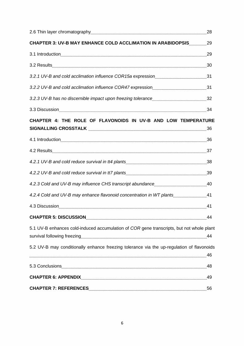
The flavonoid biosynthesis pathway results from the
combination of the phenylpropanoid and polyketide
pathways (Satio et al., 2013). The first enzyme of the
flavonoid biosynthesis pathway is CHS (TT4). CHS
catalyses the reaction between p-coumaroyl CoA and
malonyl-CoA to form a triketide which undergoes
spontaneous cyclization to form naringenin chalcone.
CHI catalyses naringenin chalcone to naringenin. F3H
then catalyses naringenin to form dihydrokaempferol.
Dihydrokaempferol is synthesised into the flavonol
kaempferol through FLS or undergoes an additional
step to form dihydroquercetin through F3’H (TT7). FLS
can synthesise either dihydroquercetin or kaempferol
into the flavonol quercetin (figure 12). These flavonols
are involved in the absorbance of UV-B.
Flavonoids are involved in many biological responses including plant hormone signalling,
pollen tube germination, colouration in angiosperms and protecting plants from UV-B induced
Figure 12. A diagrammatic section of the
Arabidopsis flavonoid biosynthesis
pathway. Enzymes are coloured blue, the
genes for CHS and F3’H are included in
brackets. Figure adapted from Saito et al.
(2013).

37
damage (Li et al., 1993; Mol et al.,1998). The role of flavonoids in protecting plants against
UV-B-induced damage was first demonstrated by Li et al. (1993), using the flavonoid
biosynthesis mutants tt4 (lacking chalcone synthase) and tt5 (lacking chalcone isomerase).
Both mutants were hypersensitive to UV-B induced damage. Flavonoids are sequestered in
vacuoles within the epidermis of a plant to absorb UV-B, thereby acting as a sunscreen
(Stracke et al., 2010). Flavonols absorb light between 280-320 nm making them ideal for
absorbing potentially harmful levels of UV-B. As well as providing plants with an important
defence against UV-B damage, flavonoids have been shown to enhance Arabidopsis cold
acclimation in multiple accessions (Hannah et al., 2006). Korn et al. (2008) further
demonstrated a strong correlation between flavonoid content and freezing tolerance following
crosses between Arabidopsis accessions. The importance of flavonoids during low
temperatures has also been demonstrated using flavonoid biosynthesis mutants. tt4, tt5 and
tt6 (deficient in CHS, CHI and FLS, respectively) all showed a significant reduced survival
when exposed to low temperatures when compared to WT plants. (Schulz et al., 2015).
In addition to absorbing UV-B, phenolics have been suggested to be involved in thickening
the cell wall and preventing ice nucleation within the cell (Chalker-Scott, 1992). Membrane
stabilization has also been proposed as a role of flavonoids during freezing temperatures
(Schulz et al., 2016). Therefore, UV-B-mediated induction of flavonoids may enhance cold
acclimation and whole plant freezing tolerance. In this chapter, the role of UV-B in plant cold
acclimation was investigated in Arabidopsis using whole plant freezing tolerance assays,
qPCR of flavonoid biosynthesis gene transcripts and thin layer chromatography (TLC) of
flavonoids.
4.2 Results
All genotypes (WT, tt4, tt7) were grown at 20 °C for 2 weeks with and without supplementary
UV-B (1 µmol m-2s-1). Half of each treatment group were subsequently cold acclimated under
continuous red and blue light for 24 h, with and without UV-B supplementation (treatment
groups: UV-B CA, UV-B non-CA, no-UV-B CA and no-UV-B non-CA). For qPCR transcript
abundance analysis plant aerial tissue was harvested after cold acclimation. For whole plant
freezing tolerance assays after the cold acclimation phase all plants were subjected to a -6 °C
freezing treatment for 24 h and a 2-week recovery period.

38
4.2.1 UV-B and cold reduce survival in tt4 plants
Percentage survivability was measured in
WT and tt4 plants using a whole plant
freezing tolerance assay. To investigate
the effect of temperature, UV-B and
genotype on freezing tolerance, a 3-way
ANOVA was performed. This analysis
revealed a statistically significant effect on
survivability with temperature treatment
[F(1,23) = 1656.818, P <0.001]; UV-B
treatment [F(1,23) = 20.455, P <0.001];
and genotype [F(1,23) = 596.455, P
<0.001]. Statistically significant
interactions between variables were also
identified between, temperature and UV-B
[F(1,23) = 32.818, P <0.001]; UV-B and
genotype [F(1,23) = 11, P = 0.004];
temperature and genotype [F(1,23) =
720.091, P <0.001]; genotype,
temperature and UV-B [F(1,23) = 20.455,
P <0.001]. A Tukey post hoc analysis was
performed to analyse which treatments
were statistically significant from one
another. All non-cold-acclimated plants showed significantly reduced survivability compared
to the cold-acclimated plants (P <0.001) except cold-acclimated tt4 plants treated with UV-B
which was not significantly different from non-cold-acclimated tt4 treated with UV-B. UV-B did
not significantly effect survivability in non-cold acclimated WT and tt4, or in cold-acclimated
WT plants. However, UV-B treatment significantly reduced survivability in cold acclimated tt4
plants (P <0.001; n = 60; figure 13 and 15). Together, these data suggest that cold acclimation
enhances freezing tolerance in the experimental conditions used here. UV-B supplementation
had no effect on freezing tolerance in WT plants and decreased freezing tolerance in tt4
mutants. The results further show that CHS expression is required for effective cold
acclimation, supporting the importance of flavonoids in this process.
Figure 13. Percentage survival of 2-week-old WT
and tt4 seedlings treated with cold and UV-B after
a 24 h freezing treatment at -6 °C. Plants were
grown for 2 weeks with and without supplementary
UV-B (1 µmol m-2s-1). Half of all plants were cold
acclimated for 24 h at 4 °C. Following freezing
treatment, a 2-week regrowth phase was allowed
before calculating percentage survival. Different
letters indicate statistically significant differences
between treatments (P <0.05). Non-CA = non-cold
acclimated, CA = cold acclimated. Striped columns
indicate exposure to UV-B. Green columns = WT.
Blue columns = tt4. n = 60, SE bars.

39
4.2.2 UV-B and cold reduce survival in tt7 plants
Percentage survivability was also measured in tt7 plants using a whole plant freezing tolerance
assay. A 3-way-ANOVA revealed that there was a statistically significant effect on survivability
with temperature treatment [F(1,23) = 2102.5, P <0.001]; UV-B treatment [F(1,23) = 28.9, P
<0.001]; and genotype [F(1,23) = 624.1, P <0.001]. Statistically significant interactions
between variables were also identified, temperature and UV-B [F(1,23) = 28.9, P <0.001]; UV-
B and genotype [F(1,23) = 16.9, P = 0.001]; temperature and genotype [F(1,23) = 624.1, P
<0.001]; genotype, temperature and UV-B [F(1,23) = 16.9, P = 0.001]. A Tukey post hoc
analysis was performed to analyse which treatments were statistically significant from one
another. 0% survival was recorded in the non-cold-acclimated WT and tt7 plants. Cold-
acclimated WT plants treated with UV-B showed a significantly higher survival rate than cold-
acclimated tt7 plants in the same conditions (P <0.001). Cold-acclimated WT plants grown
without UV-B had a significantly higher survival rate than the cold acclimated tt7 plants grown
in the same conditions (P <0.001). UV-B had no discernible effect on survivability in WT plants.
However, UV-B treatment decreased survivability in cold-acclimated tt7 plants (P <0.001; n =
60; figures 14 and 15). Together, these data support current literature that cold acclimation
enhances freezing tolerance in the experimental conditions used. They further show that F3’H
expression is required for effective cold acclimation, suggesting the importance of flavonoids
in this process. However, UV-B supplementation had no effect on freezing tolerance in WT
plants and decreased freezing tolerance in tt7 mutants.
Figure 14. Percentage survival of 2-week-
old WT and tt7 seedlings treated with cold
and UV-B after a 24 h -6 °C freezing
treatment. 50% of all plants were exposed to
UV-B (1 µmol m-2s-1) during the growth
phase. Prior to freezing treatment 50% of all
plants were cold acclimated for 24 h at 4 °C
in the light ± UV-B. After the freezing
treatment a 2-week regrowth phase was
allowed before calculating percentage
survival. Different letters indicate statistically
significant differences between treatments
(P <0.05). Non-CA = non-cold-acclimated,
CA = cold-acclimated; striped columns
indicate exposure to UV-B; green columns =
WT, blue columns = tt7. n = 60, SE bars.

40
4.2.3 Cold and UV-B may influence CHS transcript abundance
CHS transcript abundance was measured
in WT and uvr8-6 plants to assess the
impact of UV-B and cold acclimation on
flavonoid production. Plants that were cold
acclimated showed higher CHS transcript
abundance than non-cold-acclimated
controls (figure 16). Cold-acclimated WT
and uvr8-6 plants that additionally received
UV-B supplementation showed higher CHS
transcript abundance than plants grown
without UV-B. Cold acclimated uvr8-6
plants grown without UV-B showed higher
CHS transcript abundance than the
corresponding WT controls. When treated
with UV-B, however, this pattern was
reversed. WT plants grown in the presence
of UV-B, without cold acclimation, showed
a 3-fold increase in CHS transcript
abundance compared to No-UV-B controls. No UV-B-induced differences in CHS transcript
abundance were observed in non-cold acclimated uvr8-6 plants (figure 16). Despite visible
trends being observed, a Kruskal-Wallis test determined that no statistically significant
differences existed between any of the treatment groups.
Figure 15. Photograph of cold- and
UV-B-treated WT, tt4 and tt7 mutant
plants, 2 weeks after being subjected
to a -6 °C freezing treatment.
Treatment conditions were cold-
acclimated (CA), non-cold-acclimated
(non-CA) with UV-B (+UV-B) and
without UV-B (-UV-B).
Figure 16. Relative transcript abundance of CHS in
cold-treated WT and uvr8-6 plants grown with and
without UV-B (1 µmol m-2s-1). At 2 weeks, half the
plants were cold acclimated in the light for 24 h at 4
°C (with and without UV-B) and half remained at 20
°C. Non-CA = non-cold acclimated, CA = cold-
acclimated; striped columns indicate exposure to
UV-B; green columns = WT, blue columns = uvr8-
6. n = 3, SE bars.
tt7 tt4 WT
-UV-B
+UV-B
-UV-B
+UV-B
CA
Non-CA

41
4.2.4 Cold and UV-B may enhance flavonoid concentration in WT plants
Qualitative analysis of plant flavonoid content
was performed on WT and uvr8-6 genotypes
using TLC. Higher intensity of the coloured
bands of the silica plate corresponds to higher
concentration of flavonoid pigments. Samples
from WT plants showed greater colour intensity
than uvr8-6, suggesting a lack of flavonoid
production in uvr8-6 mutants (figure 17). The
supplementation of UV-B increased the intensity
of both quercetin and kaempferol bands in WT
plants, with and without cold acclimation (figure
17; quercetin = orange rectangle, kaempferol =
green rectangle). The combination of cold and
UV-B resulted in the highest band intensity,
suggesting the highest accumulation of
flavonoids.
4.3 Discussion
Whole plant freezing tolerance assays were performed using tt4 and WT genotypes to assess
the importance of CHS in protecting Arabidopsis against freezing damage. In both WT and tt4
plants, cold acclimation significantly increased survival (figure 13). These observations
support data showing that cold acclimation enhances Arabidopsis freezing tolerance (Guy et
al., 1985). Cold-acclimated WT plants grown without UV-B showed a significantly higher
percentage survival than tt4 mutants grown in similar conditions. These data suggest that
flavonoid synthesis enhances plant freezing tolerance and support the findings of Schulz et
al. (2015) who showed that all flavonoids were upregulated after cold acclimation. The
supplementation of UV-B had no significant effect on survivability in the cold acclimated WT
plants but interestingly significantly decreased survivability in cold-acclimated tt4 plants.
These observations support the involvement of CHS in protecting plants against UV-B damage
(Li et al.; 1993; Koostra, 1994). Together these findings support published data suggesting
the CHS enzyme is central to both cold and UV-B protection.
Figure 17. Flavonoid pigments in 2-week-old
Arabidopsis leaves (WT and uvr8-6). Qualitative
analysis of flavonoids was performed using TLC.
Plants were grown with and without UV-B (1 µmol
m-2s-1). Half the plants were cold acclimated in the
light for 24 h at 4 °C with and without UV-B before
tissue collection. The flavonoids quercetin and
kaempferol are indicated by orange and green
rectangles, respectively. The intensity of the colour
bands indicates an increase in pigment levels.
Non-CA = non-cold-acclimated, CA = cold-
acclimated; - UV-B received no UV-B treatment, +
UV-B received UV-B treatment.

42
A freezing tolerance assay for tt7 mutants was also performed. Cold acclimated plants showed
significantly increased survival compared to non-cold acclimated plants, however cold
acclimated tt7 plants had significantly reduced survivability compared to the WT control. These
data suggest that for freezing protection, plants are dependent upon the flavonoid quercetin.
The tt7 mutant lacks the key enzyme F3’H (flavonoid 3-hydroxylase) which catalyses the
synthesis of dihydrokaempferol into dihydroquercetin which can then be synthesised to
quercetin by FLS (flavonol synthase), FLS can also synthesise kaempferol into quercetin
(Mehrtens et al., 2005) (figure 12). Data from both the tt4 and tt7 freezing tolerance assays
suggest a strong involvement of flavonoids in protecting plants not only from freezing
conditions but also from UV-B. The effectiveness of cold acclimation in promoting plant
survival following freezing treatment prevented the analysis of whether UV-B could enhance
this process. High levels of survival in cold-acclimated plants (approaching 100%, except in
the UV-B-treated and cold acclimated uvr8-6) and 0% survival in non-cold-acclimated plants
prevented analysis of any effect of UV-B in addition to cold. Further experiments using a more
severe freezing treatment, a reduced cold acclimation period or a more sensitive assay, such
as electrolyte leakage, may elucidate the effect of UV-B on freezing tolerance. The electrolyte
leakage assay would be a primary candidate as it measures damage to the plant through the
leakage of ions and has been successfully used to measure damage induced by low
temperatures (Xing et al., 2011; Hemsley et al., 2014). This may be a more accurate way of
assessing the impact of UV-B in protecting plants against low temperature damage.
qPCR was used to assess if cold acclimation and UV-B could additively upregulate CHS
transcript abundance (figure 16). A Kruskal-Wallis test showed no statistically significant
interactions. This was likely due to a small sample size (n = 3) in combination with variation in
the data. Despite these issues, some trends could be identified. CHS transcript abundance
was increased in WT and uvr8-6 by cold acclimation suggesting that CHS is upregulated
during low temperatures. This complements studies showing flavonoid production during low
temperatures (Schulz et al., 2015). A larger increase in CHS transcript abundance was
observed when cold acclimated WT and uvr8-6 plants received supplementary UV-B
treatment. UV-B is well documented to upregulate CHS (Mehrtens et al., 2005). The data
presented here may suggest that cold acclimation and UV-B act synergistically to upregulate
CHS. Analysis of the TLC plate appears to show there is accumulation of the flavonoids
quercetin and kaempferol in WT plants when receiving cold acclimation and UV-B. This
supports the qPCR data trend that CHS is further upregulated when WT plants are cold
acclimated and treated with UV-B. However, the TLC plate shows comparatively little to no
upregulation of quercetin or kaempferol in uvr8-6 plants. This concurs with the data trends of
the qPCR which showed severely reduced expression of CHS compared with WT plants.
43
Together, the data presented in this chapter suggest that UV-B enhances the production of
flavonoids during cold acclimation and that this occurs, in part, via synergistic up-regulation of
CHS expression by cold and UV-B treatments. This is further supported by the TLC data which
suggest that the flavonoids kaempferol and quercetin accumulate during cold and UV-B in WT
plants. Whole plant survival assays confirmed the importance of flavonoid biosynthesis for
freezing tolerance but were not sensitive enough to determine whether growth in UV-B can
enhance WT cold acclimation.

44
CHAPTER 5: DISCUSSION
5.1 UV-B enhances cold-induced accumulation of COR gene transcripts, but not whole
plant survival following freezing
UV-B supplementation has been demonstrated to guard against freezing-induced damage in
multiple species. Dunning et al. (1994) found that phenolic leakage (a measure of freezing
damage) was reduced in UV-B supplemented Rhododendron plants. Yang et al. (2007)
additionally showed that Triticum aestivum produced more ROS scavenging enzymes under
UV-B supplementation, providing plants with enhanced protection against freezing
temperatures. These findings suggest crosstalk between the low temperature and UV-B
signalling pathways. Crosstalk is a phenomenon in plants where multiple signalling pathways
converge and has been well investigated in the signalling pathways of cold, drought and salt
stress. Since all these stressors cause dehydration, each signalling pathway is involved in
protecting the plant against dehydration regardless of which stress the plant is subjected to
(Seki et al., 2003; Dai et al., 2007). Crosstalk between light and cold has not, however, been
as thoroughly investigated. The integration of light and temperature signals is very important
in the cold acclimation process and it has been demonstrated to be mediated, in part, through
phytochrome photoreceptors (Olsen et al., 1997; Franklin and Whitelam, 2007). Catalá et al.,
(2011) have demonstrated that the bZIP transcription factor HY5 is important for integrating
light and low temperature signals to promote full cold acclimation. HY5 is a key regulator of
plant photomorphogenesis and is repressed by COP1 in the dark (Chen and Xiong, 2008). In
the presence of light, phytochromes and cryptochromes negatively regulate COP1. UV-B
activates UVR8, which monomerises and binds to COP1, stabilising HY5 and promoting the
expression of downstream genes (Heijde and Ulm, 2012) (figure 6). HY5 is required for full
cold acclimation and is responsible for the expression of at least 10% of all cold-inducible
genes (Catalá et al., 2011). This is achieved by HY5 binding to an LTRE in the Z-box which
allows cold inducible genes such as CAB1 to be expressed (Catalá et al., 2011). Knowing the
importance of light signals in the cold acclimation process, UV-B was investigated to
understand if UV-B could enhance freezing tolerance by enhancing cold acclimation. This was
examined by measuring the transcript abundance of two COR genes, COR15a and COR47.
Analyses of transcript abundance showed that when WT plants were treated with a
combination of cold and UV-B, COR15a and COR47 relative transcript abundance increased
(figures 8 and 9). This indicates crosstalk between the signalling pathways of UV-B and cold
acclimation. Although statistical analysis of the results from this study did not reveal any
significant differences between treatment groups, clear trends in the data warrant further
investigation. Time prevented further replication. To confirm COR15a and COR47 trends in

45
WT plants, more replicates would be required. Further replication may also reveal a trend in
the uvr8-6 plants and therefore elude to whether UVR8 activity is required for UV-B-enhanced
expression of COR15a and COR47.
Whole plant freezing tolerance assays were performed to assess if UV-B could directly
increase plant survival following freezing temperatures. In contrast with Dunning et al., (1994),
the freezing tolerance assays conducted here using WT and uvr8-6 plants did not show UV-B
to enhance freezing tolerance. This was due to high levels of survival (approaching 100%) in
the cold acclimated plants (except in the UV-B-treated uvr8-6 mutants) and 0% survival in the
non-cold acclimated plants (figure 10). It is possible that the assay used was not sensitive
enough to detect any impact that UV-B may have had on survivability as previously mentioned
in chapter 3. However, it is also possible that a different outcome compared with Dunning et
al., (1994) was achieved due to measuring whole plant survival instead of leaf disc damage.
Whole plant analysis was chosen over leaf discs as it is more representative of how plants
cope under these stresses in a natural environment. Alternatively, Arabidopsis may respond
differently to cold and UV-B than Rhododendron and Wheat. The uvr8-6 mutant was used to
help elucidate whether any effect UV-B was having in increasing cold tolerance was acting
through the UVR8 photoreceptor. Due to high levels of survival in cold acclimated plants and
low levels of survival in the non-cold acclimated plants, it was not possible to ascertain whether
UV-B impacted survivability and therefore UVR8’s involvement could not be gauged. This
mutant does not appear to be appropriate for the freezing tolerance assay as uvr8-6 plants
exposed to UV-B and freezing temperatures appear to receive a double stress which reduced
survivability (figure 10). As uvr8-6 plants lack the UVR8 photoreceptor, they lack the means
to detect UV-B and activate the flavonoid biosynthesis pathway. Flavonoids absorb UV-B
wavelengths preventing ROS formation, protecting the plant (Kootstra, 1994). The application
of UV-B and a -6 °C freezing treatment may therefore have resulted in considerable tissue
damage in uvr8-6 mutants. In future work, whole plant freezing tolerance assay experiments
could be replaced by electrolyte leakage assays. This would be more informative as damage
can be measured through the leakage of ions instead of the binary results (dead or alive)
collected from the whole plant freezing survival assays. This method has been successfully
used to measure damage induced by low temperatures (Uemura et al., 2006; Hemsley et al.,
2014) and would allow investigation of plant responses to multiple temperatures.
Discrepancies between transcript abundance and freezing survival assays may be explained
if transcripts are prevented from being translated into proteins. To analyse this a quantitative
western blot could be used to ensure that COR transcripts are translated into protein. The
electrolyte leakage assay could also be used to investigate the effects of UV-B
supplementation on cold acclimation in hy5 and hyh mutants. HY5 is known to be involved in

46
regulating the expression of flavonoids through UV-B (Roman et al., 2004). It would therefore
be interesting to assess if HY5 is involved in the integration of UV-B and low temperature
signalling since it has already been linked with phytochrome signalling and low temperature
(Catalá et al., 2011).
5.2 UV-B may conditionally enhance freezing tolerance via the up-regulation of
flavonoids
Flavonoids were investigated in this study as they are known to be involved in protecting plants
from UV-B damage (Li et al., 1993; Stracke et al., 2010) and freezing temperatures (Hannah
et al., 2006; Korn et al., 2008). This makes the flavonoid biosynthesis pathway a point of
potential crosstalk between UV-B and low temperature signalling. To assess this, whole plant
freezing tolerance assays were performed using the flavonoid mutants tt4 and tt7. The TT4
gene encodes chalcone synthase, the first enzyme in the flavonoid biosynthesis pathway and
TT7 encodes F3’H which is a crucial enzyme in the synthesis of quercetin further downstream
(figure 12). The impaired freezing tolerance displayed by tt4 plants suggests that CHS plays
an important role in cold acclimation and freezing tolerance in Arabidopsis (figure 13). Analysis
of CHS transcript abundance showed increased CHS accumulation when plants received both
a cold acclimation treatment and UV-B treatment, although results were not statistically
significant (figure 16). These results may suggest that UV-B and cold act synergistically to
enhance CHS abundance although more replicates would need to be performed to confirm
this. F3’H mutants (tt7) were tested using whole plant freezing tolerance assays to identify if
kaempferol or quercetin were important in protecting plants from freezing temperatures and
UV-B (figure 14). In contrast to the results of Shulz et al. (2016), tt7 mutants showed similar
levels of survival after freezing treatment to tt4 plants. They additionally showed significantly
lower levels of survival than WT plants when cold acclimated and treated with UV-B. tt4 and
tt7 mutants are defective in quercetin biosynthesis. Together, these results suggest that
quercetin is an important component in both UV-B protection and cold acclimation and may
be involved in integrating the two signalling pathways.
To attempt to quantify flavonoid abundance in cold- and UV-B-treated plants, a Dualex
spectrometer was used to measure epidermal pigments (Goulas et al., 2004). Measurements
of chlorophyll accumulation were also taken in parallel to assess the potential impact of cold
and UV-B on photosynthesis. This allowed multiple measurements to be taken without
damaging the plant tissue which could potentially change the concentration of phenolic
compounds. The small leaf size of plants grown in UV-B, did however, prevent reliable
measurements. Plants were grown for an additional 2 weeks (4 weeks total) to increase leaf

47
size area but this failed to resolve the issue, so the experiment was discontinued due to time
constraints (Appendix figure S8, S9 and S10). Adjusting light quality is an option that may
allow greater leaf size and therefore the Dualex spectrophotometer to be used (Casal et al.,
1987). This would allow chlorophyll and anthocyanin measurements to be taken in addition to
flavonoids. To address the failed quantitative analysis of flavonoids, a quantitative
chromogenic assay could be used to enable measurement from plants with small leaves
(Julkunen-Tiitto et al., 2015). A qualitative TLC assay was subsequently used to gain an
understanding of flavonoid accumulation (figure 17). The TLC results support tt7 whole plant
freezing tolerance assay data as coloured bands representing quercetin have a higher
intensity in plants treated with cold acclimation and UV-B (figure 17). Levels of kaempferol and
quercetin were low in uvr8-6 plants suggesting that UV-B-induced flavonoid accumulation
requires functional UVR8 protein. Collectively, these experiments highlight the importance of
flavonoids in protecting plants from low temperatures and UV-B.
Another consideration for future work would be to manipulate the UV-B treatments
administered to plants. Plants acclimate to UV-B over time, reducing differences in transcript
abundance between treated and untreated plants (Jansen et al., 2010). A transient UV-B
treatment may therefore act to enhance the expression of genes involved in UV-B and cold
signalling. To optimise a transient UV-B treatment the time of day UV-B treatment is
administered should be considered. UV-B responses are gated by the circadian clock and
Arabidopsis UV-B induced expression of CHS and HYH have been shown to increase during
approximately midday (Fehér et al., 2011; Horak and Farré, 2015). However, the highest CBF
transcript abundance (an important determinant in cold acclimation) is found 4 h after dawn
(Fowler et al., 2005). Therefore, the most effective time to treat plants with UV-B may lie at
some point between dawn and midday.

48
5.3 Conclusions
Analyses of COR15a and COR47 transcript abundance suggest that UV-B can enhance the
cold acclimation process, although whole plant freezing tolerance assays did not support this
conclusion. Analyses of CHS transcript abundance, whole plant freezing tolerance assays
using flavonoid biosynthesis mutants (tt4 and tt7) and TLC analysis all suggest that UV-B may
enhance cold acclimation by increasing flavonoids. It is likely that a point of crosstalk between
low temperature and UV-B signalling pathways exists at an early point in the flavonoid
biosynthesis pathway. Despite observed crosstalk between UV-B and low temperature in the
regulation of both COR genes and flavonoid synthesis, no evidence exists that suggests the
CBF regulon regulates the flavonoid biosynthesis pathway or vice versa. This would mean
UV-B acts on the flavonoid biosynthesis pathway and CBF pathway separately. The point at
which UV-B is acting on the CBF pathway to enhance COR gene expression remains
unknown. However, phytochrome and the circadian clock are known to be crucial for
regulating freezing tolerance through CBFs. It is therefore possible that UV-B is influencing
the expression COR genes through the CBF regulon.
Understanding how plants respond to environmental stresses is very important to agriculture.
Many greenhouses utilise materials which block UV-B wavelengths. Work from Wargent et al.,
(2011) has shown that UV-B treatment of seedlings increased harvestable yield and
photosynthetic output in field-grown Lactuca sativa (lettuce). These data suggest that UV-B
treatment prior to planting may enhance the robustness of field-grown crops. Increased crop
losses due to low temperatures aggravate problems of food security due to an ever-increasing
world population and opportunities to enhance resistance to environmental stress should be
maximised. The data in this thesis suggest that UV-B may enhance cold acclimation and
therefore freezing tolerance and warrants further investigation into crosstalk between UV-B
and low temperature signalling.

49
CHAPTER 6: APPENDIX
1200 mm
800 mm
Figure S1. Schematic representation of growth cabinet showing where light measurements were
taken (centre, left, right, back, front) and indicating growth cabinet shelf area.
Figure S2. Light spectra from plant growth cabinet without supplementary UV-B. Measurement
taken from: A) back of cabinet B) left of cabinet C) centre of cabinet D) right of cabinet E) front of
cabinet. White light (100 µmol m-2s-1).

50
600 mm
600 mm
Figure S4. Schematic representation of the cabinet used for cold acclimation and plant freezing
tolerance assays. Centre indicates where light measurements were taken from within the cabinet.
Cabinet shelf area also indicated.
Figure S3. Light spectra from plant growth cabinet with supplementary UV-B. Measurement taken
from: A) back of cabinet B) left of cabinet C) centre of cabinet D) right of cabinet E) front of cabinet.
White light (100 µmol m-2s-1) UV-B (1 µmol m-2s-1).

51
Figure S5. Light spectra from plant freezing cabinet A) without supplementary UV-B B) with
supplementary UV-B. White light (70 µmol m-2s-1) UV-B (1 µmol m-2s-1).
Figure S6. Primer efficiency standard curve from MXPro software. Initial quantity shows the dilution
of the starting quantity of primer on the x axis. The Y axis displays the critical threshold (Ct). The
efficiency and R2 values are displayed.

52
Figure S7. Testing of control genes A) UBC21, B) PIP41 and C) PP2A, using relative transcript
abundance of COR15a in 2-week-old Arabidopsis seedlings. 50% of all plants were exposed to
UV-B (1 µmol m-2s-1) during growth phase. Before leaf tissue was collected for RNA extraction
50% of plants in each treatment were cold acclimated for 24 h at 4 °C. Non-CA = non-cold-
acclimated, CA = cold-acclimated; striped columns indicate exposure to UV-B; green columns
= WT, blue columns = uvr8-6. n = 3.

53
Figure S8. Chlorophyll measurement data collected using using a Dualex spectrometer.
Measurements taken from the first leaf of the first rosette from time 0 h, 24 h, 48 h and 72 h. WT
and uvr8-6 plants were grown with and without UV-B (1 µmol m-2s-1) (indicated by -UV-B and
UV-B respectively). Half the plants were cold acclimated in the light for 24 h at 4 °C with and
without UV-B. Plants cold acclimated are indicated in the x axis with ‘4’ and plants grown at 20
°C without cold acclimation are indicated by ‘20’.
Figure S9. Flavonoid measurement data collected using a Dualex spectrometer. Measurements
taken from the first leaf of the first rosette from time 0 h, 24 h, 48 h and 72 h. WT and uvr8-6
Plants were grown with and without UV-B (1 µmol m-2s-1) (indicated by -UV-B and UV-B
respectively). Half the plants were cold acclimated in the light for 24 h at 4 °C with and without
UV-B. Plants cold acclimated are indicated in the x axis with ‘4’ and plants grown at 20 °C without
cold acclimation are indicated by ‘20’.

54
Figure S10. Anthocyanin measurement data collected using a Dualex spectrometer.
Measurements taken from the first leaf of the first rosette from time 0 h, 24 h, 48 h and 72 h.
WT and uvr8-6 Plants were grown with and without UV-B (1 µmol m-2s-1) (indicated by -UV-B
and UV-B respectively). Half the plants were cold acclimated in the light for 24 h at 4 °C with
and without UV-B. Plants cold acclimated are indicated in the x axis with ‘4’ and plants grown
at 20 °C without cold acclimation are indicated by ‘20’.

55
Figure S11. Flavonoid pigments in 2-week-old Arabidopsis leaves (WT and uvr8-6). Qualitative
analysis of flavonoids was performed using TLC. Plants were grown with and without UV-B (1
µmol m-2s-1) (indicated by -UV-B and UV-B respectively). Half the plants were cold acclimated in
the light for 24 h at 4 °C with and without UV-B before tissue collection. Plants cold acclimated
are indicated in the x axis with ‘4’ and plants grown at 20 °C without cold acclimation are indicated
by ‘20’. The flavonoids quercetin and kaempferol are indicated by orange and green bands. The
higher the intensity of the colour bands indicates an increase in pigment levels. Letters A-G
indicate TLC attempts adjusting the polarity of the mobile phase.

56
CHAPTER 7: REFERENCES
Achard, P., Gong, F., Cheminant, S., Alioua, M., Hedden, P. and Genschik, P. (2008). The
cold-inducible CBF1 factor–dependent signaling pathway modulates the accumulation of
the growth-repressing DELLA proteins via its effect on gibberellin metabolism. The Plant
Cell, 20(8), pp.2117-2129.
Achard, P., Renou, J.P., Berthomé, R., Harberd, N.P. and Genschik, P. (2008). Plant
DELLAs restrain growth and promote survival of adversity by reducing the levels of
reactive oxygen species. Current Biology, 18(9), pp.656-660.
Agati, G., Biricolti, S., Guidi, L., Ferrini, F., Fini, A. and Tattini, M. (2011). The biosynthesis
of flavonoids is enhanced similarly by UV radiation and root zone salinity in L. vulgare
leaves. Journal of Plant Physiology, 168(3), pp.204-212.
Alabadı,́ D., Oyama, T., Yanovsky, M.J., Harmon, F.G., Más, P. and Kay, S.A. (2001).
Reciprocal regulation between TOC1 and LHY/CCA1 within the Arabidopsis circadian
clock. Science, 293(5531), pp.880-883.
Ballaré, C.L., Barnes, P.W. and Flint, S.D. (1995). Inhibition of hypocotyl elongation by
ultraviolet‐B radiation in de‐etiolating tomato seedlings. I. The photoreceptor. Physiologia
Plantarum, 93(4), pp.584-592.
Boccalandro, H.E., Mazza, C.A., Mazzella, M.A., Casal, J.J. and Ballaré, C.L. (2001).
Ultraviolet B radiation enhances a phytochrome-B-mediated photomorphogenic response
in Arabidopsis. Plant Physiology, 126(2), pp.780-788.
Bouché, N., Scharlat, A., Snedden, W., Bouchez, D. and Fromm, H. (2002). A novel family
of calmodulin-binding transcription activators in multicellular organisms. Journal of
Biological Chemistry, 277(24), pp.21851-21861.
Briggs, W.R., Beck, C.F., Cashmore, A.R., Christie, J.M., Hughes, J., Jarillo, J.A., Kagawa,
T., Kanegae, H., Liscum, E., Nagatani, A. and Okada, K. (2001). The phototropin family of
photoreceptors. The Plant Cell, 13(5), pp.993-997.
Brown, B.A., Cloix, C., Jiang, G.H., Kaiserli, E., Herzyk, P., Kliebenstein, D.J. and Jenkins,
G.I. (2005). A UV-B-specific signaling component orchestrates plant UV
protection. Proceedings of the National Academy of Sciences, 102(50), pp.18225-18230.
Burke, M.J., Gusta, L.V., Quamme, H.A., Weiser, C.J. and Li, P.H. (1976). Freezing and
injury in plants. Annual Review of Plant Physiology, 27(1), pp.507-528.

57
Caldwell, M.M., Robberecht, R. and Flint, S.D. (1983). Internal filters: prospects for UV‐
acclimation in higher plants. Physiologia Plantarum, 58(3), pp.445-450.
Candat, A., Poupart, P., Andrieu, J.P., Chevrollier, A., Reynier, P., Rogniaux, H.,
Avelange-Macherel, M.H. and Macherel, D. (2013). Experimental determination of
organelle targeting-peptide cleavage sites using transient expression of green fluorescent
protein translational fusions. Analytical Biochemistry, 434(1), pp.44-51.
Casal, J.J., Sanchez, R.A. and Deregibus, V.A. (1987). The effect of light quality on shoot
extension growth in three species of grasses. Annals of Botany, 59(1), pp.1-7.
Catalá, R., Medina, J. and Salinas, J. (2011). Integration of low temperature and light
signaling during cold acclimation response in Arabidopsis. Proceedings of the National
Academy of Sciences, 108(39), pp.16475-16480.
Chalker-Scott, L. (1992). Disruption of an ice-nucleation barrier in cold hardy Azalea buds
by sublethal heat stress. Annals of Botany, 70(5), pp.409-418.
Chalker‐Scott, L. and Scott, J.D. (2004). Elevated Ultraviolet‐B Radiation Induces Cross‐
protection to Cold in Leaves of Rhododendron Under Field Conditions. Photochemistry
and Photobiology, 79(2), pp.199-204.
Chen, H. and Xiong, L. (2008). Role of HY5 in abscisic acid response in seeds and
seedlings. Plant Signaling & Behavior, 3(11), pp.986-988.
Chinnusamy, V., Ohta, M., Kanrar, S., Lee, B.H., Hong, X., Agarwal, M. and Zhu, J.K.
(2003). ICE1: a regulator of cold-induced transcriptome and freezing tolerance in
Arabidopsis. Genes & Development, 17(8), pp.1043-1054.
Close, T.J. (1997). Dehydrins: a commonalty in the response of plants to dehydration and
low temperature. Physiologia Plantarum, 100(2), pp.291-296.
Czechowski, T., Stitt, M., Altmann, T., Udvardi, M.K. and Scheible, W.R. (2005). Genome-
wide identification and testing of superior reference genes for transcript normalization in
Arabidopsis. Plant Physiology, 139(1), pp.5-17.
Dai, X., Xu, Y., Ma, Q., Xu, W., Wang, T., Xue, Y. and Chong, K. (2007). Overexpression
of an R1R2R3 MYB gene, OsMYB3R-2, increases tolerance to freezing, drought, and salt
stress in transgenic Arabidopsis. Plant Physiology, 143(4), pp.1739-1751.
Davière, J.M., De Lucas, M. and Prat, S. (2008). Transcriptional factor interaction: a central
step in DELLA function. Current Opinion in Genetics & Development, 18(4), pp.295-303.

58
DESA. (2015). World population prospects. Economic and social affairs. [PDF file].
Available from:
https://esa.un.org/Unpd/wpp/Publications/Files/Key_Findings_WPP_2015.pdf.
Dong, C.H., Agarwal, M., Zhang, Y., Xie, Q. and Zhu, J.K. (2006). The negative regulator
of plant cold responses, HOS1, is a RING E3 ligase that mediates the ubiquitination and
degradation of ICE1. Proceedings of the National Academy of Sciences, 103(21),
pp.8281-8286.
Doucet, C.J., Byass, L., Elias, L., Worrall, D., Smallwood, M. and Bowles, D.J. (2000).
Distribution and characterization of recrystallization inhibitor activity in plant and lichen
species from the UK and maritime Antarctic. Cryobiology, 40(3), pp.218-227.
Dowgert, M.F. and Steponkus, P.L. (1984). Behavior of the plasma membrane of isolated
protoplasts during a freeze-thaw cycle. Plant Physiology, 75(4), pp.1139-1151.
Dunning CA, Chalker‐Scott L, Scott JD. (1994). Exposure to ultraviolet‐B radiation
increases cold hardiness in Rhododendron. Physiologia Plantarum, 92(3), pp. 516-20.
Esnault, M.A., Legue, F. and Chenal, C. (2010). Ionizing radiation: advances in plant
response. Environmental and Experimental Botany, 68(3), pp.231-237.
Favory, J.-J., Stec, A., Gruber, H., Rizzini, L., Oravecz, A., Funk, M., Albert, A., Cloix, C.,
Jenkins, G. I., Oakeley, E. J., Seidlitz, H. K., Nagy, F., and Ulm, R. (2009). Interaction of
COP1 and UVR8 regulates UV-B-induced photomorphogenesis and stress acclimation in
Arabidopsis. The EMBO Journal, 28(5), pp.591–601.
Fehér, B., Kozma‐Bognár, L., Kevei, É., Hajdu, A., Binkert, M., Davis, S.J., Schäfer, E.,
Ulm, R. and Nagy, F. (2011). Functional interaction of the circadian clock and UV
RESISTANCE LOCUS 8‐controlled UV‐B signaling pathways in Arabidopsis thaliana. The
Plant Journal, 67(1), pp.37-48.
Feng, S., Martinez, C., Gusmaroli, G., Wang, Y., Zhou, J., Wang, F., Chen, L., Yu, L.,
Iglesias-Pedraz, J.M., Kircher, S. and Schäfer, E. (2008). Coordinated regulation of
Arabidopsis thaliana development by light and gibberellins. Nature, 451(7177), p.475.
Fowler, S. and Thomashow, M.F. (2002). Arabidopsis transcriptome profiling indicates that
multiple regulatory pathways are activated during cold acclimation in addition to the CBF
cold response pathway. The Plant Cell, 14(8), pp.1675-1690.

59
Fowler, S.G., Cook, D. and Thomashow, M.F. (2005). Low temperature induction of
Arabidopsis CBF1, 2, and 3 is gated by the circadian clock. Plant Physiology, 137(3),
pp.961-968.
Franklin, K.A. and Whitelam, G.C. (2007). Light-quality regulation of freezing tolerance in
Arabidopsis thaliana. Nature Genetics, 39(11), p.1410.
Franklin, K.A., Lee, S.H., Patel, D., Kumar, S.V., Spartz, A.K., Gu, C., Ye, S., Yu, P., Breen,
G., Cohen, J.D. and Wigge, P.A. (2011). Phytochrome-interacting factor 4 (PIF4) regulates
auxin biosynthesis at high temperature. Proceedings of the National Academy of
Sciences, 108(50), pp.20231-20235.
Gill, S.S. and Tuteja, N. (2010). Reactive oxygen species and antioxidant machinery in
abiotic stress tolerance in crop plants. Plant Physiology and Biochemistry, 48(12), pp.909-
930.
Gilmour, S.J., Artus, N.N. and Thomashow, M.F. (1992). cDNA sequence analysis and
expression of two cold-regulated genes of Arabidopsis thaliana. Plant Molecular
Biology, 18(1), pp.13-21.
Gilmour, S.J., Hajela, R.K. and Thomashow, M.F. (1988). Cold acclimation in Arabidopsis
thaliana. Plant Physiology, 87(3), pp.745-750.
Gilmour, S.J., Zarka, D.G., Stockinger, E.J., Salazar, M.P., Houghton, J.M. and
Thomashow, M.F. (1998). Low temperature regulation of the Arabidopsis CBF family of
AP2 transcriptional activators as an early step in cold‐induced COR gene expression. The
Plant Journal, 16(4), pp.433-442.
Gordon-Kamm, W.J. and Steponkus, P.L. (1984). Lamellar-to-hexagonalII phase
transitions in the plasma membrane of isolated protoplasts after freeze-induced
dehydration. Proceedings of the National Academy of Sciences, 81(20), pp.6373-6377.
Goulas, Y., Cerovic, Z.G., Cartelat, A. and Moya, I. (2004). Dualex: a new instrument for
field measurements of epidermal ultraviolet absorbance by chlorophyll
fluorescence. Applied Optics, 43(23), pp.4488-4496.
Gruber, H., Heijde, M., Heller, W., Albert, A., Seidlitz, H.K. and Ulm, R. (2010). Negative
feedback regulation of UV-B–induced photomorphogenesis and stress acclimation in
Arabidopsis. Proceedings of the National Academy of Sciences, 107(46), pp.20132-
20137.

60
Gu, L., Hanson, P.J., Post, W.M., Kaiser, D.P., Yang, B., Nemani, R., Pallardy, S.G. and
Meyers, T. (2008). The 2007 eastern US spring freeze: increased cold damage in a
warming world?. AIBS Bulletin, 58(3), pp.253-262.
Guy, C.L., Niemi, K.J. and Brambl, R. (1985). Altered gene expression during cold
acclimation of spinach. Proceedings of the National Academy of Sciences, 82(11),
pp.3673-3677.
Han Y, Vimolmangkang S, Soria-Guerra RE, Rosales-Mendoza S, Zheng D, Lygin AV,
Korban SS. (2010). Ectopic expression of apple F3′ H genes contributes to anthocyanin
accumulation in the Arabidopsis tt7 mutant grown under nitrogen stress. Plant Physiology,
153(2), pp.806-20.
Hannah, M.A., Wiese, D., Freund, S., Fiehn, O., Heyer, A.G. and Hincha, D.K. (2006).
Natural genetic variation of freezing tolerance in Arabidopsis. Plant Physiology, 142(1),
pp.98-112.
Hansen, J. and Beck, E. (1988). Evidence for ideal and non‐ideal equilibrium freezing of
leaf water in frosthardy ivy (Hedera helix) and winter barley (Hordeum vulgare). Botanica
Acta, 101(1), pp.76-82.
Hayes, S., Velanis, C.N., Jenkins, G.I. and Franklin, K.A. (2014). UV-B detected by the
UVR8 photoreceptor antagonizes auxin signaling and plant shade
avoidance. Proceedings of the National Academy of Sciences, 111(32), pp.11894-11899.
Heijde, M. and Ulm, R. (2012). UV-B photoreceptor-mediated signalling in plants. Trends
in Plant Science, 17(4), pp.230-237.
Hemsley, P.A., Hurst, C.H., Kaliyadasa, E., Lamb, R., Knight, M.R., De Cothi, E.A., Steele,
J.F. and Knight, H. (2014). The Arabidopsis mediator complex subunits MED16, MED14,
and MED2 regulate mediator and RNA polymerase II recruitment to CBF-responsive cold-
regulated genes. The Plant Cell, 26(1), pp.465-484.
Hirano, S.S., Upper, C.D. (2000). Bacteria in the leaf ecosystem with emphasis on
Pseudomonas syringae - a pathogen, ice nucleus, and epiphyte. Microbiology and
Molecular Biology Review 64, pp.624-653.
Hoekstra, F.A., Golovina, E.A. and Buitink, J. (2001). Mechanisms of plant desiccation
tolerance. Trends in Plant Science, 6(9), pp.431-438.
Horak, E. and Farré, E.M. (2015). The regulation of UV-B responses by the circadian
clock. Plant Signaling & Behavior, 10(5), p.e1000164.

61
Huang, X., Ouyang, X., Yang, P., Lau, O.S., Chen, L., Wei, N. and Deng, X.W. (2013).
Conversion from CUL4-based COP1–SPA E3 apparatus to UVR8–COP1–SPA
complexes underlies a distinct biochemical function of COP1 under UV-B. Proceedings of
the National Academy of Sciences, 110(41), pp.16669-16674.
Huang, X., Ouyang, X., Yang, P., Lau, O.S., Li, G., Li, J., Chen, H. and Deng, X.W. (2012).
Arabidopsis FHY3 and HY5 positively mediate induction of COP1 transcription in response
to photomorphogenic UV-B light. The Plant Cell, pp.tpc-112.
Hughes, S.L., Schart, V., Malcolmson, J., Hogarth, K.A., Martynowicz, D.M., Tralman-
Baker, E., Patel, S.N. and Graether, S. (2013). The importance of size and disorder in the
cryoprotective effects of dehydrins. Plant Physiology, pp-113.
IPCC. (2007). Climate Change 2007: Working Group I: The Physical Science Basis.
[online] Available at: https://www.ipcc.ch/publications_and_data/ar4/wg1/en/spmsspm-
projections-of.html [Accessed 15 Nov. 2018].
IPCC. (2014). Climate Change 2014 Synthesis Report Summary for Policymakers. [PDF
file]. Available from: https://www.ipcc.ch/pdf/assessment-
report/ar5/syr/AR5_SYR_FINAL_SPM.pdf [Accessed 15 Nov, 2018].
Jaenicke, R. (1990). Protein structure and function at low temperatures. Philosphical
Transactions of the Royal Society of. London B, 326(1237), pp.535-553.
Jansen, M.A., Martret, B.L. and Koornneef, M. (2010). Variations in constitutive and
inducible UV‐B tolerance; dissecting photosystem II protection in Arabidopsis thaliana
accessions. Physiologia Plantarum, 138(1), pp.22-34.
Jenkins, G.I. (2009). Signal transduction in responses to UV-B radiation. Annual Review
of Plant Biology, 60, pp.407-431.
Jiang, B., Shi, Y., Zhang, X., Xin, X., Qi, L., Guo, H., Li, J. and Yang, S. (2017). PIF3 is a
negative regulator of the CBF pathway and freezing tolerance in Arabidopsis. Proceedings
of the National Academy of Sciences, 114(32), pp.E6695-E6702.
Jouyban, Z., Hasanzade, R. and Sharafi, S. (2013). Chilling stress in plants. International
Journal of Agriculture and Crop Sciences, 5(24), p.2961.
Julkunen-Tiitto, R., Nenadis, N., Neugart, S., Robson, M., Agati, G., Vepsäläinen, J., Zipoli,
G., Nybakken, L., Winkler, B. and Jansen, M.A. (2015). Assessing the response of plant
flavonoids to UV radiation: an overview of appropriate techniques. Phytochemistry
Reviews, 14(2), pp.273-297.

62
Kami, C., Lorrain, S., Hornitschek, P. and Fankhauser, C., 2010. Light-regulated plant
growth and development. In Current Topics in Developmental Biology, 91, pp.29-66.
Kidokoro, S., Yoneda, K., Takasaki, H., Takahashi, F., Shinozaki, K. and Yamaguchi-
Shinozaki, K. (2017). Different cold-signaling pathways function in the responses to rapid
and gradual decreases in temperature. The Plant Cell, pp.tpc-00669.
Kimball, S.L. and Salisbury, F.B. (1973). Ultrastructural changes of plants exposed to low
temperatures. American Journal of Botany, 60(10), pp.1028-1033.
Kliebenstein, D.J., Lim, J.E., Landry, L.G. and Last, R.L. (2002). Arabidopsis UVR8
regulates ultraviolet-B signal transduction and tolerance and contains sequence similarity
to human regulator of chromatin condensation 1. Plant Physiology, 130(1), pp.234-243.
Knight, M.R. and Knight, H. (2012). Low‐temperature perception leading to gene
expression and cold tolerance in higher plants. New Phytologist, 195(4), pp.737-751.
Koag, M.C., Fenton, R.D., Wilkens, S. and Close, T.J. (2003). The binding of maize DHN1
to lipid vesicles. Gain of structure and lipid specificity. Plant Physiology, 131(1), pp.309-
316.
Kootstra, A. (1994). Protection from UV-B-induced DNA damage by flavonoids. Plant
Molecular Biology, 26(2), pp.771-774.
Korn, M., Peterek, S., MOCK, H.P., Heyer, A.G. and Hincha, D.K. (2008). Heterosis in the
freezing tolerance, and sugar and flavonoid contents of crosses between Arabidopsis
thaliana accessions of widely varying freezing tolerance. Plant, Cell & Environment, 31(6),
pp.813-827.
Kosová, K., Vítámvás, P. and Prášil, I.T. (2007). The role of dehydrins in plant response
to cold. Biologia Plantarum, 51(4), pp.601-617.
Lee J., He K., Stolc V., Lee H., Figueroa P., Gao Y., Tongprasit W., Zhao H.Y., Lee I.,
Deng X.W. (2007). Analysis of transcription factor HY5 genomic binding sites revealed its
hierarchical role in light regulation of development. Plant Cell, 19, pp. 731–749.
Lee, C.M. and Thomashow, M.F. (2012). Photoperiodic regulation of the C-repeat binding
factor (CBF) cold acclimation pathway and freezing tolerance in Arabidopsis
thaliana. Proceedings of the National Academy of Sciences, 109(37), pp.15054-15059.
Lee, J.H. (2016) UV-B signal transduction pathway in Arabidopsis. Journal of Plant
Biology, 59(3), pp.223-230.

63
Levitt, J. (1980). Responses of Plants to Environmental Stress, Volume 1: Chilling,
Freezing, and High Temperature Stresses. Academic Press.
Li, J., Ou-Lee, T.M., Raba, R., Amundson, R.G. and Last, R.L. (1993). Arabidopsis
flavonoid mutants are hypersensitive to UV-B irradiation. The Plant Cell, 5(2), pp.171-179.
Li, M., An, F., Li, W., Ma, M., Feng, Y., Zhang, X. and Guo, H. (2016). DELLA proteins
interact with FLC to repress flowering transition. Journal of Integrative Plant Biology, 58(7),
pp.642-655.
Lin, C. and Thomashow, M.F. (1992). DNA sequence analysis of a complementary DNA
for cold-regulated Arabidopsis gene cor15 and characterization of the COR 15
polypeptide. Plant Physiology, 99(2), pp.519-525.
Lin, C., Guo, W.W., Everson, E. and Thomashow, M.F. (1990). Cold Acclimation in
Arabidopsis and Wheat: A Response Associated with Expression of Related Genes
Encoding Boiling-Stable Polypeptides. Plant Physiology, 94(3), pp.1078-1083.
Lindow, S.E., Lahue, E., Govindarajan, A.G., Panopoulos, N.J. and Gies, D. (1989).
Localization of ice nucleation activity and the iceC gene product in Pseudomonas syringae
and Escherichia coli. Molecular Plant-Microbe Interactions 2(5), pp.262-272.
Liu, Q., Kasuga, M., Sakuma, Y., Abe, H., Miura, S., Yamaguchi-Shinozaki, K. and
Shinozaki, K. (1998). Two transcription factors, DREB1 and DREB2, with an EREBP/AP2
DNA binding domain separate two cellular signal transduction pathways in drought-and
low-temperature-responsive gene expression, respectively, in Arabidopsis. The Plant
Cell, 10(8), pp.1391-1406.
Livak, K.J. and Schmittgen, T.D. (2001). Analysis of relative gene expression data using
real-time quantitative PCR and the 2− ΔΔCT method. Methods, 25(4), pp.402-408.
Ma, Y., Dai, X., Xu, Y., Luo, W., Zheng, X., Zeng, D., Pan, Y., Lin, X., Liu, H., Zhang, D.
and Xiao, J. (2015). COLD1 confers chilling tolerance in rice. Cell, 160(6), pp.1209-1221.
Mehrtens, F., Kranz, H., Bednarek, P. and Weisshaar, B. (2005). The Arabidopsis
transcription factor MYB12 is a flavonol-specific regulator of phenylpropanoid
biosynthesis. Plant Physiology, 138(2), pp.1083-1096.
Meier, M., Fuhrer, J. and Holzkämper, A. (2018). Changing risk of spring frost damage in
grapevines due to climate change? A case study in the Swiss Rhone Valley. International
Journal of Biometeorology, pp.1-12.

64
Meshi, T. and Iwabuchi, M. (1995). Plant transcription factors. Plant and Cell
Physiology, 36(8), pp.1405-1420.
Mol, J., Grotewold, E. and Koes, R. (1998). How genes paint flowers and seeds. Trends
in Plant Science, 3(6), pp.212-217.
Nakayama, K., Okawa, K., Kakizaki, T., Honma, T., Itoh, H. and Inaba, T. (2007).
Arabidopsis Cor15am is a chloroplast stromal protein that has cryoprotective activity and
forms oligomers. Plant Physiology, 144(1), pp.513-523.
O’Hara, A. and Jenkins, G.I. (2012). In vivo function of tryptophans in the Arabidopsis UV-
B photoreceptor UVR8. The Plant Cell, 24, pp.3755–3766
Olsen, J.E., Junttila, O., Nilsen, J., Eriksson, M.E., Martinussen, I., Olsson, O., Sandberg,
G. and Moritz, T. (1997). Ectopic expression of oat phytochrome A in hybrid aspen
changes critical daylength for growth and prevents cold acclimatization. The Plant
Journal, 12(6), pp.1339-1350.
Pearce, R.S. and Fuller, M.P. (2001). Freezing of barley studied by infrared video
thermography. Plant Physiology, 125(1), pp.227-240.
Pfaffl, M. W. (2001). A new mathematical model for relative quantification in real-time RT-
PCR. Nucleic Acids Research, 29(9) p.45.
Piskurewicz, U. and Lopez-Molina, L. (2009). The GA-signaling repressor RGL3 represses
testa rupture in response to changes in GA and ABA levels. Plant Signaling &
Behavior, 4(1), pp.63-65.
Podar, D. (2013). Plant growth and cultivation. In Plant Mineral Nutrients (pp. 23-45).
Humana Press, Totowa, NJ.
Privalov, P.L., (1990). Cold denaturation of protein. Critical Reviews in Biochemistry and
Molecular Biology, 25(4), pp.281-306.
Rizzini, L., Favory, J.J., Cloix, C., Faggionato, D., O’Hara, A., Kaiserli, E., Baumeister, R.,
Schäfer, E., Nagy, F., Jenkins, G.I. and Ulm, R. (2011). Perception of UV-B by the
Arabidopsis UVR8 protein. Science, 332(6025), pp.103-106.
Rogelj, J., Meinshausen, M. and Knutti, R. (2012). Global warming under old and new
scenarios using IPCC climate sensitivity range estimates. Nature Climate Change, 2(4),
p.248.

65
Ros, J. and Tevini, M. (1995). Interaction of UV-radiation and IAA during growth of
seedlings and hypocotyl segments of sunflower. Journal of Plant Physiology, 146(3),
pp.295-302.
Saito, K., Yonekura-Sakakibara, K., Nakabayashi, R., Higashi, Y., Yamazaki, M., Tohge,
T. and Fernie, A.R. (2013). The flavonoid biosynthetic pathway in Arabidopsis: structural
and genetic diversity. Plant Physiology and Biochemistry, 72, pp.21-34.
Salter, M.G., Franklin, K.A. and Whitelam, G.C. (2003). Gating of the rapid shade-
avoidance response by the circadian clock in plants. Nature, 426(6967), p.680.
Schulz, E., Tohge, T., Zuther, E., Fernie, A.R. and Hincha, D.K. (2016). Flavonoids are
determinants of freezing tolerance and cold acclimation in Arabidopsis thaliana. Scientific
Reports, 6, p.34027.
Schulz, E., Tohge, T., Zuther, E., Fernie, A.R. and Hincha, D.K. (2015). Natural variation
in flavonol and anthocyanin metabolism during cold acclimation in Arabidopsis thaliana
accessions. Plant, Cell & Environment, 38(8), pp.1658-1672.
Seki, M., Kamei, A., Yamaguchi-Shinozaki, K. and Shinozaki, K. (2003). Molecular
responses to drought, salinity and frost: common and different paths for plant
protection. Current Opinion in Biotechnology, 14(2), pp.194-199.
Shi, Y., Ding, Y. and Yang, S. (2018). Molecular Regulation of CBF Signaling in Cold
Acclimation. Trends in plant science.
Somers, D.E., Devlin, P.F. and Kay, S.A. (1998). Phytochromes and cryptochromes in the
entrainment of the Arabidopsis circadian clock. Science, 282(5393), pp.1488-1490.
Stockinger, E.J., Gilmour, S.J. and Thomashow, M.F (1997). Arabidopsis thaliana CBF1
encodes an AP2 domain-containing transcriptional activator that binds to the C-
repeat/DRE, a cis-acting DNA regulatory element that stimulates transcription in response
to low temperature and water deficit. Proceedings of the National Academy of
Sciences, 94(3), pp.1035-1040.
Stracke, R., Favory, J.J., Gruber, H., Bartelniewoehner, L., Bartels, S., Binkert, M., Funk,
M., Weisshaar, B. and Ulm, R. (2010). The Arabidopsis bZIP transcription factor HY5
regulates expression of the PFG1/MYB12 gene in response to light and ultraviolet‐B
radiation. Plant, Cell & Environment, 33(1), pp.88-103.
Stracke, R., Jahns, O., Keck, M., Tohge, T., Niehaus, K., Fernie, A.R. and Weisshaar, B.
(2010). Analysis of PRODUCTION OF FLAVONOL GLYCOSIDES‐dependent flavonol

66
glycoside accumulation in Arabidopsis thaliana plants reveals MYB11‐, MYB12‐and
MYB111‐independent flavonol glycoside accumulation. New Phytologist, 188(4), pp.985-
1000.
Suetsugu, N. and Wada, M. (2012). Evolution of three LOV blue light receptor families in
green plants and photosynthetic stramenopiles: phototropin, ZTL/FKF1/LKP2 and
aureochrome. Plant and Cell Physiology, 54(1), pp.8-23.
Takahashi, M., Teranishi, M., Ishida, H., Kawasaki, J., Takeuchi, A., Yamaya, T.,
Watanabe, M., Makino, A. and Hidema, J. (2011). Cyclobutane pyrimidine dimer (CPD)
photolyase repairs ultraviolet‐B‐induced CPDs in rice chloroplast and mitochondrial
DNA. The Plant Journal, 66(3), pp.433-442.
Takemiya, A., Inoue, S.I., Doi, M., Kinoshita, T. and Shimazaki, K.I. (2005). Phototropins
promote plant growth in response to blue light in low light environments. The Plant
Cell, 17(4), pp.1120-1127.
Takeuchi, T., Newton, L., Burkhardt, A., Mason, S. and Farré, E.M. (2014). Light and the
circadian clock mediate time-specific changes in sensitivity to UV-B stress under light/dark
cycles. Journal of Experimental Botany, 65(20), pp.6003-6012.
Thalhammer, A., Bryant, G., Sulpice, R. and Hincha, D.K. (2014). Disordered COR15
proteins protect chloroplast membranes during freezing through binding and folding, but
do not stabilize chloroplast enzymes in-vivo. Plant Physiology, 166(1), pp.190-201.
Thomas, S.G., Phillips, A.L. and Hedden, P. (1999). Molecular cloning and functional
expression of gibberellin 2-oxidases, multifunctional enzymes involved in gibberellin
deactivation. Proceedings of the National Academy of Sciences, 96(8), pp.4698-4703.
Thomashow, M.F. (1999). Plant cold acclimation: freezing tolerance genes and regulatory
mechanisms. Annual Review of Plant Biology, 50(1), pp.571-599.
Thomashow, M.F. (2010). Molecular basis of plant cold acclimation: insights gained from
studying the CBF cold response pathway. Plant Physiology, 154(2), pp.571-577.
Thorlby, G., Fourrier, N. and Warren, G. (2004). The SENSITIVE TO FREEZING2 gene,
required for freezing tolerance in Arabidopsis thaliana, encodes a β-glucosidase. The
Plant Cell, 16(8), pp.2192-2203.
Tong, H., Leasure, C.D., Hou, X., Yuen, G., Briggs, W. and He, Z.H. (2008). Role of root
UV-B sensing in Arabidopsis early seedling development. Proceedings of the National
Academy of Sciences, 105(52), pp.21039-21044.

67
Tyler, L., Thomas, S.G., Hu, J., Dill, A., Alonso, J.M., Ecker, J.R. and Sun, T.P. (2004).
DELLA proteins and gibberellin-regulated seed germination and floral development in
Arabidopsis. Plant Physiology, 135(2), pp.1008-1019.
Uemura, M. and Steponkus, P.L. (1989). Effect of cold acclimation on the incidence of two
forms of freezing injury in protoplasts isolated from rye leaves. Plant physiology, 91(3),
pp.1131-1137.
Uemura, M., Tominaga, Y., Nakagawara, C., Shigematsu, S., Minami, A. and Kawamura,
Y. (2006). Responses of the plasma membrane to low temperatures. Physiologia
Plantarum, 126(1), pp.81-89.
Ulm R., Baumann A., Oravecz A., Máté Z., Adám E., Oakeley E.J., Schäfer E. and Nagy
F. (2004). Genome-wide analysis of gene expression reveals function of the bZIP
transcription factor HY5 in the UV-B response of Arabidopsis. Proc. Natl. Acad. Sci, 101,
pp.1397–1402.
United Nations, Department of Economic and Social Affairs, Population Division (2017).
World Population Prospects: The 2017 Revision.
Viswanathan, C. and Zhu, J.K. (2002). Molecular genetic analysis of cold–regulated gene
transcription. Philosophical Transactions of the Royal Society of London. Series B:
Biological Sciences, 357(1423), pp.877-886.
Vitasse, Y., Schneider, L., Rixen, C., Christen, D. and Rebetez, M. (2018). Increase in the
risk of exposure of forest and fruit trees to spring frosts at higher elevations in Switzerland
over the last four decades. Agricultural and Forest Meteorology, 248, pp.60-69.
Vogel, J.T., Zarka, D.G., Van Buskirk, H.A., Fowler, S.G. and Thomashow, M.F. (2005).
Roles of the CBF2 and ZAT12 transcription factors in configuring the low temperature
transcriptome of Arabidopsis. The Plant Journal, 41(2), pp.195-211.
Von Arnim, A.G. and Deng, X.W. (1994). Light inactivation of Arabidopsis
photomorphogenic repressor COP1 involves a cell-specific regulation of its
nucleocytoplasmic partitioning. Cell, 79(6), pp.1035-1045.
Walther, G.R., Post, E., Convey, P., Menzel, A., Parmesan, C., Beebee, T.J., Fromentin,
J.M., Hoegh-Guldberg, O. and Bairlein, F. (2002). Ecological responses to recent climate
change. Nature, 416(6879), p.389.

68
Wargent, J.J., Elfadly, E.M., Moore, J.P. and Paul, N.D. (2011). Increased exposure to
UV‐B radiation during early development leads to enhanced photoprotection and improved
long‐term performance in Lactuca sativa. Plant, Cell & Environment, 34(8), pp.1401-1413.
Warmund, M.R., Guinan, P. and Fernandez, G. (2008). Temperatures and cold damage
to small fruit crops across the eastern United States associated with the April 2007
freeze. HortScience, 43(6), pp.1643-1647.
Winkel-Shirley, B. (2001). Flavonoid biosynthesis. A colorful model for genetics,
biochemistry, cell biology, and biotechnology. Plant Physiology, 126(2), pp.485-493.
Winkel-Shirley, B. (2002). Biosynthesis of flavonoids and effects of stress. Current Opinion
in Plant Biology, 5(3), pp.218-223.
Winkel-Shirley, B., Kubasek, W. L., Storz, G., Bruggemann, E., Koornneef, M., Ausubel,
F. M., and Goodman, H. M. (1995). Analysis of Arabidopsis mutants deficient in flavonoid
biosynthesis. The Plant Journal, 8(5) pp.659–671.
Wisniewski, M., Close, T.J., Artlip, T. and Arora, R. (1996). Seasonal patterns of dehydrins
and 70‐kDa heat‐shock proteins in bark tissues of eight species of woody
plants. Physiologia Plantarum, 96(3), pp.496-505.
Wisniewski, M., Gusta, L. and Neuner, G. (2014). Adaptive mechanisms of freeze
avoidance in plants: a brief update. Environmental and Experimental Botany, 99, pp.133-
140.
Wu, D., Hu, Q., Yan, Z., Chen, W., Yan, C., Huang, X., Zhang, J., Yang, P., Deng, H.,
Wang, J. and Deng, X. (2012). Structural basis of ultraviolet-B perception by
UVR8. Nature, 484(7393), p.214.
Xing, X., Liu, Y., Kong, X., Liu, Y. and Li, D. (2011). Overexpression of a maize dehydrin
gene, ZmDHN2b, in tobacco enhances tolerance to low temperature. Plant Growth
Regulation, 65(1), pp.109-118.
Yamaguchi-Shinozaki, K. and Shinozaki, K. (1994). A novel cis-acting element in an
Arabidopsis gene is involved in responsiveness to drought, low-temperature, or high-salt
stress. The Plant Cell, 6(2), pp.251-264.
Yamazakia, T., Kawamura, Y. and Uemura, M. (2009). Extracellular freezing-induced
mechanical stress and surface area regulation on the plasma membrane in cold-
acclimated plant cells. Plant Signaling & Behavior, 4(3), pp.231-233.

69
Yang, D.L., Yao, J., Mei, C.S., Tong, X.H., Zeng, L.J., Li, Q., Xiao, L.T., Sun, T.P., Li, J.,
Deng, X.W. and Lee, C.M. (2012). Plant hormone jasmonate prioritizes defense over
growth by interfering with gibberellin signaling cascade. Proceedings of the National
Academy of Sciences, 109(19), pp.1192-1200.
Yang, S.H., Wang, L.J. and Li, S.H. (2007). Ultraviolet-B irradiation-induced freezing
tolerance in relation to antioxidant system in winter wheat (Triticum aestivum L.)
leaves. Environmental and Experimental Botany, 60(3), pp.300-307.
Yang, T., Shad Ali, G., Yang, L., Du, L., Reddy, A.S.N. and Poovaiah, B.W. (2010).
Calcium/calmodulin-regulated receptor-like kinase CRLK1 interacts with MEKK1 in
plants. Plant Signaling & Behavior, 5(8), pp.991-994.
Yin, R. and Ulm, R. (2017). How plants cope with UV-B: from perception to
response. Current Opinion in Plant Biology, 37, pp.42-48.
Yin, R., Arongaus, A.B., Binkert, M. and Ulm, R. (2015). Two distinct domains of the UVR8
photoreceptor interact with COP1 to initiate UV-B signalling in Arabidopsis. The Plant Cell,
27, pp.202–213.
Yu, X.M. and Griffith, M. (1999). Antifreeze proteins in winter rye leaves form oligomeric
complexes. Plant Physiology, 119(4), pp.1361-1370.
Zhang, H., He, H., Wang, X., Wang, X., Yang, X., Li, L. and Deng, X.W. (2011). Genome‐
wide mapping of the HY5‐mediated gene networks in Arabidopsis that involve both
transcriptional and post‐transcriptional regulation. The Plant Journal, 65(3), pp.346-358.
Zhu, J.K. (2016). Abiotic stress signalling and responses in plants. Cell, 167(2), pp.313-
324.

70