this is the author’s version of a work that was accepted
TRANSCRIPT

This is the author’s version of a work that was accepted for publication:
TAYLOR, A., & MAHER, B. (2014). Exposure-dose-response of Tellina deltoidalis to metal
contaminated estuarine sediments 2. Lead spiked sediments. Comparative Biochemistry and
Physiology. Part C: Toxicology Pharmacology, 159, 52-61.
https://doi.org/10.1016/j.cbpc.2013.09.006
This file was downloaded from:
https://researchprofiles.canberra.edu.au/en/publications/exposure-dose-response-of-
tellina-deltoidalis-to-metal-contaminat
©2014 Elsevier
Notice:
This is the author’s version of a work that was accepted for publication in Comparative
Biochemistry and Physiology Part C. Changes resulting from the publishing process, such as
peer review, editing, corrections, structural formatting, and other quality control
mechanisms may not be reflected in this document. Changes may have been made to this
work since it was submitted for publication.
A definitive version was subsequently published at:
http://doi.org/10.1016/j.cbpc.2013.09.006

1
CBP ms.22625 – part C 1
2
Exposure-dose-response of Tellina deltoidalis to metal contaminated 3
estuarine sediments. 2. Lead spiked sediments 4
5
Anne M. Taylor* and William A. Maher 6
7
Ecochemistry Laboratory, Institute for Applied Ecology, University of Canberra, Canberra, 8
ACT 2601, Australia 9
10
*corresponding author: 11
Email: [email protected] 12
Phone: +61 2 62063805 13
14
Tables: 2; Figures: 4; Supplementary Tables and Figures: 7 15
16
Abstract 17
Lead accumulation in estuarine sediments, as a result of activities such as mining and ore 18
smelting, and through urban runoff is a continuing problem in the increasingly developed 19
world. Marine organisms accumulate lead, which is known to be highly toxic to biological 20
processes and to degrade organism and ecosystem health. Here the relationship between lead 21
exposure, dose and response was investigated in the sediment dwelling, deposit feeding, 22
marine bivalve Tellina deltoidalis. Bivalves were exposed in the laboratory to individual lead 23
spiked sediments at 100 and 300 µg/g dry mass, for 28 days and accumulated total tissue lead 24
concentrations of 96 and 430 µg/g, respectively. Subcellular fractionation indicated that 25
around 70% of accumulated lead was detoxified, three quarters of which was converted into 26
metal rich granules. The majority of biologically active lead was associated with the 27
mitochondrial fraction with up to a 128 fold increase in lead burden in exposed organisms 28
compared to controls. This indicates active lead detoxification which at these exposures was 29
unable to prevent significant lead burdens accumulating in sensitive organelles. With 30
increased lead exposure T. deltoidalis showed a suppression in glutathione peroxidase 31
activity, total glutathione concentration and reduced GSH:GSSG ratios, however, these 32
differences were not significant. Lead exposed T. deltoidalis had a significantly reduced total 33
antioxidant capacity which corresponded with increased lipid peroxidation, lysosomal 34

2
destabilisation and micronuclei frequency. The exposure-dose-response relationship 35
demonstrated for lead exposed T. deltoidalis supports its potential for the development of 36
sub-lethal endpoints in lead toxicity assessment. 37
38
Keywords: Biomarkers, subcellular lead, oxidative stress, lysosomes, lipid peroxidation, 39
micronuclei, bivalve. 40
41
1 Introduction 42
43
Lead is released into the environment through a variety of human activities including lead ore 44
mining and smelting, alkyl-lead petroleum combustion, coal burning, waste incineration, 45
production and storage of lead-acid batteries, leaded glass, lead oxide pigments and 46
ferroalloys, metal fabricating industries, cement manufacture and urban runoff (Ãlvarez-47
Iglesias et al., 2012; ARCARP, 2006; Chiaradia et al., 1997; Cook and Gale, 2005; Genaidy 48
et al., 2009; Renberg et al., 2002). In Australia the majority of the population is located 49
along coastlines, the bays and estuaries of which are repositories for sediment bound lead 50
(ABS, 1996; ANZECC and ARMCANZ, 2000; Carroll, 1996; Chiaradia et al., 1997). 51
The accumulation of lead by marine organisms has implications for both ecological and 52
human health at both individual and population levels (Clark, 2001; Gnassia-Barelli and 53
Romeo, 1993). Although it has no known biological function lead is accumulated by marine 54
organisms through similar pathways as essential elements such as calcium, iron and zinc 55
making it one of the more toxic metals in these environments (Company et al., 2011; 2008). 56
The metal exposure route, through either food or water, affects the organism’s metal handling 57
and potentially its toxicity (Rainbow, 2007). Deposit feeding bivalves are of particular 58
interest as biomonitors as they are exposed to contaminants through water and food and 59
directly through ingested fine grain sediment particles containing organic material (Griscom 60
and Fisher, 2004; Lee et al., 2000; 1998). 61
Whole sediment toxicity tests are used worldwide as an integral part of toxicity assessment as 62
they assess contaminant exposure from several exposure routes; including those bound to 63
sediments, from pore waters and in overlying waters (Adams et al., 2005; ASTM, 2010; 64
Ingersoll et al., 2000). In Australia, aquatic toxicity tests have been established (ANZECC 65
and ARMCANZ, 2000) but the use of whole sediment toxicity tests has been hampered by a 66
lack of established test procedures and knowledge of species sensitivity to contaminants. 67
Early investigations of suitable Australian organisms’ sensitivity to metal contamination in 68

3
whole sediment toxicity tests have included polychaetes, amphipods and bivalves, of which, 69
the bivalve Tellina deltoidalis was highly sensitive to zinc and copper (King et al., 2004; 70
2005). T. deltoidalis is a deposit feeding bivalve which satisfies most of the basic 71
requirements to be an effective biomonitor, they are: hardy, accumulate metal and have 72
sufficient tissue for analysis (King et al., 2010; 2004; 2005; Taylor and Maher, 2013); and are 73
widely distributed around Australian coastal estuaries, living in sediments at a depth several 74
times their 15 – 25 mm shell length with siphons extended to the surface to feed (Beesley et 75
al., 1998). 76
The development of Australian guidelines for sediment quality assessment, which includes 77
contaminant exposure, uptake and ecotoxicological effects, identified T. deltoidalis as a 78
suitable species for whole sediment toxicity testing and a protocol for its use is included in 79
the Australian Handbook for Sediment Quality Assessment (Simpson et al., 2005). The 80
assessment of ecotoxicological effects of contaminants is moving from the use of mortality as 81
an endpoint to the use of biomarkers of sub lethal effects and there is a need, in understanding 82
organism sensitivity to contaminants, to develop suitable biomarkers which can be included 83
in routine testing. The use of biomarkers enables links to be made between external exposure 84
and an internal toxicant dose, which has exceeded the organism’s detoxification and repair 85
capacity, allowing ecologically relevant effect concentrations to be determined (Adams et al., 86
2011; Depledge, 1992). Biomarkers of exposure and effect for use in aquatic toxicological 87
assessment have been progressively developed and refined for a range of species and 88
toxicants worldwide (Bergayou et al., 2009; Bocchetti et al., 2008; Cajaraville et al., 2000; 89
Damiens et al., 2007; Domouhtsidou et al., 2004; Durou et al., 2007; Galloway et al., 2004; 90
Lam, 2009; Lam and Gray, 2003; Taylor and Maher, 2010; Viarengo et al., 2007). 91
Lead has a high affinity for sulfhydryl, forming mercaptides with the sulfhydryl group of 92
cysteine and inhibiting several enzymes having functional sulfhydryl groups, thereby 93
initiating oxidative stress responses (Dafre et al., 2004; Ercal et al., 2001; Gurer and Ercal, 94
2000). Changes in the oxidative pathway including increased reactive oxygen by-products 95
and suppression or increased expression of oxygen reduction enzymes have been shown for a 96
number of lead exposed molluscs (Alcutt and Pinto, 1994; Cossu et al., 2000; Dafre et al., 97
2004; Jing et al., 2007; Taylor and Maher, 2012). Lead is also thought to interact with a 98
variety of cellular lipids altering the lipid composition of cellular membranes and increased 99
susceptibility to lipid peroxidation (Campana et al., 2003; Mateo et al., 2003). This can 100
result in perturbations in cell membrane integrity, permeability and function increasing 101
lysosomal destabilisation (Einsporn and Koehler, 2008; Ercal et al., 2001; Gurer and Ercal, 102

4
2000). Lead has been shown to interact directly with DNA by covalent binding of Pb2+ to 103
DNA (Hong, 2007) producing micronuclei, through either the breakage of chromosomes or 104
dysfunction of the mitotic spindle apparatus (Monteiro et al., 2011; Winter, 2007). The 105
micronuclei induction assay which can detect these small masses of cytoplasmic chromatin, 106
present outside the main nucleus, has proven useful in assessing the genotoxic effects of a 107
range of compounds in fish and other aquatic organisms (Bolognesi et al., 2004; Kalpaxis et 108
al., 2004; Udroiu, 2006). 109
The purpose of this study was to examine the exposure-dose-response relationship to lead in 110
T. deltoidalis using a 28 day sediment bioaccumulation test (ASTM, 2010; Ingersoll et al., 111
2000), to develop useful biomarkers of effect, with a view to determining their suitability for 112
laboratory sediment metal toxicity tests using sub lethal endpoints. Two lead sediment 113
exposure concentrations were used, 100 mg/kg based on the high value for lead sediment 114
concentrations from the ANZECC and ARMCANZ (2000) Interim Sediment Quality 115
Guidelines and 300 mg/kg based on concentrations previously measured in lead contaminated 116
Australian estuarine sediments (Roach, 2005). The study examines lead accumulation and 117
subcellular tissue lead distribution. Biomarkers of lead exposure effects were total 118
antioxidant capacity, and the associated oxidative enzymes; total, reduced and oxidised 119
glutathione and glutathione peroxidise. The cellular effects measure lysosomal 120
destabilisation and a genotoxic measure, micronuclei occurrence were also measured in a 121
weight of evidence approach to establishing lead sub lethal toxic effects. 122
123
2 Materials and Methods 124 125 2.1 Organism and sediment collection 126
Sediments were collected from a NSW Department of Environmental and Climate Change 127
reference site in Durras Lake NSW, and stored at 4oC until use. Tellina deltoidalis of 15 – 20 128
mm in size were collected from Durras Lake and Lake Tabourie, NSW in July 2005 and 129
January 2006 and placed in coolers with sediment and water from the collection sites for 130
transportation. Organisms were maintained for a maximum of two weeks at 22oC in 131
uncontaminated sediments, depth 15 cm, in glass aquaria with filtration and aeration to allow 132
acclimation before experimentation. Overlying water used in aquaria was collected from 133
coastal waters near Murramurrang National Park, NSW and adjusted from 35‰ to 28‰ with 134
deionised water to match the salinity of the estuarine water from which organisms were 135
collected. 136
137

5
2.2 Sediment lead spiking 138
Sediments were sieved through a 2 mm stainless steel sieve to remove large pieces of organic 139
matter and organisms prior to the addition of lead. Sub samples of the collected sediments 140
were measured for moisture content and grain size. To produce a sediment matrix which was 141
suitable for organism burrowing and feeding, sediment was mixed with clean beach sand so 142
that the 63 µm fraction was not greater than 20% mass/mass. To ensure added lead was 143
rapidly adsorbed and bound to the sediment particles the procedure developed by (Simpson et 144
al., 2004) for producing metal spiked marine sediments, was followed. Wet sediment was 145
added to mixing containers. PbCl2, (AR grade Sigma-Aldrich), was added to concentrations 146
of 100 mg/kg and 300 mg/kg dry mass of sediment. All containers were topped up with clean 147
deoxygenated sea water and the final mixture was completely deoxygenated by bubbling with 148
nitrogen for 2 hours. Head spaces of containers were filled with nitrogen prior to sealing. 149
Any pH adjustments were made immediately after the addition of lead using 1M NaOH, (AR 150
grade BDH), prepared in seawater. pH was checked weekly and maintained at 7 - 8.2. 151
Sediments were maintained at room temperature 22 - 25oC and mixed on a Cell-production 152
Roller Apparatus (Belco, USA) for several hours each day. The time required for 153
equilibration of added metals is affected by the sediment properties, equilibration pH and the 154
concentration and properties of the metal (Simpson et al., 2004). To determine when the 155
added lead was completely bound to sediment particles, pore waters were collected and 156
acidified to 1% v/v with nitric acid (AristaR, BDH, Australia) and lead measured using an 157
ELAN® 6000 ICP-MS (PerkinElmer SCIEX, USA). Once pore water lead concentrations had 158
fallen below instrument detection limits 0.001 µg/l the sediment was ready for use. Time to 159
full adsorption was 4 - 6 weeks. Unspiked sediments were treated in the same way and used 160
for control treatments. Sediment lead concentrations were measured by ICP-MS after 161
digestion of 0.2 g of lyophilised sediment in 3 ml of nitric acid (AristaR, BDH, Australia) in 162
polyethylene 50 ml centrifuge tubes for 60 minutes at 115oC (Maher et al., 2003). Lead in 163
NRCC Certified Reference Materials, BCSS-1 marine sediment measured along with samples 164
was 21 ± 4 µg/g (n = 10) and in agreement with certified values 22.7 ± 3.4 µg/g. Pre 165
exposure sediment lead concentrations were < 0.001, 100 ± 4.5 and 300 ± 8 µg/g and post 166
exposure were < 0.001, 101 ± 5 and 298 ± 10 µg/g. 167
168
2.3 Microcosm experiment design 169
Procedures for conducting the exposures were adapted from the test method for conducting 170
28 day sediment bioaccumulation tests (Ingersoll et al., 2000). Spiked and control sediments 171

6
(500 g wet wt.) were placed in each of three replicate 770 ml polypropylene containers 172
(Chanrol # 01C30, Australia) per treatment. The containers were filled with fresh seawater 173
adjusted to a salinity of 28‰. Containers were placed in random order on a tray in an 174
incubator set at 22oC with a day / night light cycle of 14 / 10 hours to reflect spring / summer 175
conditions. Aeration was introduced and the treatments were left for 24 hours to allow them 176
settle and the temperature to equilibrate. Fifteen T. deltoidalis were then introduced to each 177
treatment container. Organisms were not given supplementary food and surface water was 178
changed weekly during the 28 day exposure period. Aquaria were continually aerated using 179
an air pump with valves on each line to regulate air flow so oxygen saturation ≈ 100% were 180
maintained in each aquarium but sediments were not agitated. Due to the natural buffering 181
capacity of sea water and associated sediments, pH remained relatively constant at pH 7.8-8.0 182
in all aquaria throughout the 28 days of exposure. This is similar to results of other studies of 183
this type (King et al., 2006; Strom et al., 2011). Total tissue lead bioaccumulation was 184
measured at intervals of 3, 7, 14, 21 and 28 days. A day 0 measurement was made using 185
organisms from the acclimation tanks to give the background lead concentration. All 186
organisms were placed in fresh seawater 28‰ with no sediment for 24 hours to allow 187
depuration of ingested sediment particles, prior to lead analysis. Visual inspection under a 188
dissecting microscope showed gut clearance was achieved in this timeframe. All assays were 189
undertaken on whole tissues of individual organisms. 190
191
2.4 Lead measurements 192
2.4.1 Total lead 193
Lyophilised ground tissue was microwave digested in 1 ml of nitric acid (AristaR BDH, 194
Australia) in a 630 watt oven (CEM MDS-2000, USA) for two min at 630 W, two min 0 W, 195
and 45 min at 315 W (Baldwin et al., 1994). Prior to analysis samples were diluted with 196
deionised water to 1% v/v HNO3, and an ICP-MS mixed 7־element internal standard (EM 197
Science) was added to monitor for variations due to instrument drift and/or matrix effects. 198
Lead was measured using an ELAN® 6000 ICP-MS (PerkinElmer, SCIEX) following the 199
method of Maher et al. (2001). NRCC Certified Reference Material, NIST 1566a oyster 200
tissue and acid blanks were routinely digested and diluted in the same way as the samples and 201
analysed along with them to verify accuracy and precision of lead analysis. The measured 202
CRM mean lead value; 0.36 ± 0.02 µg/g (n = 50) was not significantly different from the 203
certified value 0.37 ± 0.014 µg/g. 204
205

7
2.4.2 Subcellular lead 206
The subcellular tissue lead distribution was examined in tissues of day 28 exposed organisms 207
using a procedure adapted from Sokolova et al. (2005) and Wallace et al. (2003). The 208
dissected tissues were placed in polypropylene vials, snap frozen in liquid nitrogen and stored 209
at -80oC until processed. The tissue was thawed and minced on ice with a blade. A sub 210
sample, approximately 0.1 g wet wt., was taken for total tissue lead analysis. The remainder, 211
approximately 0.5 g wet wt., was homogenised in Ca 2+ / Mg2+ free saline buffer pH 7.35 on 212
ice using an IKA® Labortechnick Ultra-turrax-T25 homogeniser equipped with an S25-UT 213
dispersing tool at 9,500 rpmin-1 (Janke & Kunkel, Germany). Homogenised tissue was 214
subjected to differential centrifugation and tissue digestion procedures according to the 215
protocol outlined in Taylor and Maher (2010; 2013), using an Eppendorf 5804R centrifuge 216
and a Himac CP90WX preparative ultracentrifuge (Hitachi, Japan). The mitochondria (P3), 217
lysosomes+microsomes (P4) and heat sensitive protein (P5) pellets were grouped as 218
biologically active lead fractions while the metal rich granule (P2) and heat stable 219
metallothionein like proteins (S5) were grouped as biologically detoxified lead fractions 220
(Supplementary Figure 1). The supernatant from the granule pellet isolation (S2) contained 221
the nuclei and cellular debris (Supplementary Figure 1). To determine the mitochondrial and 222
lysosomal content of the fractions obtained the activity of enzymes specific for these 223
organelles, cytochrome c oxidase and acid phosphatase, respectively, were measured in each 224
of the total tissue homogenate, mitochondrial (P3) and lysosome+microsome (P4) pellets 225
using commercial colorimetric assays (CYTOC-OX1 Sigma-Aldrich, USA and CS0740 226
Sigma-Aldrich, USA, respectively). In the total homogenate both the mitochondrial enzyme 227
cytochrome c oxidase and the lysosomal enzyme acid phosphatase showed an increase in 228
concentration with increased lead exposure (Supplementary Figure 2). The mitochondrial 229
fractions had higher cytochrome c oxidase concentrations than the lysosome fractions 230
indicating mitochondrial enrichment of this fraction (Supplementary Figures 1 & 2). The 231
lysosome+microsome fractions had slightly higher acid phosphatase concentrations than the 232
mitochondrial fractions indicating some enrichment of lysosomes in this fraction with some 233
carryover of mitochondria. All fractions were acidified to 10% v/v with nitric acid (AristaR 234
BDH, Australia) and placed in a water bath at 80oC for 4 hours. NIST CRM 1566a oyster 235
tissue, buffer and acid blanks were digested and diluted in the same way as the samples and 236
analysed along with them. Analysis of lead was as previously described above. The 237
measured CRM lead value; 0.38 ± 0.05 µg/g (n = 5) was in good agreement with certified 238
value 0.37 ± 0.014 µg/g. 239

8
240
2.5 Biomarker measurements 241
2.5.1 Total antioxidant capacity and lipid peroxidation 242
Tissues were homogenised on ice in a 5 mM potassium phosphate buffer containing 0.9% 243
(w/v) sodium chloride and 0.1% (w/v) glucose, pH 7.4 (1:5 w/v) using a motorised 244
microcentrifuge pellet pestle, sonicated on ice for 15 seconds at 40 V (VibraCell™ Sonics 245
Materials, USA) and centrifuged, in a 5804R centrifuge (Eppendorf, Germany), at 10,000 x g 246
for 15 minutes at 4oC (Cayman, 2011). The supernatant was stored at -80oC until analysis. 247
Total antioxidant capacity was measured using an assay based on the ability of the tissue 248
lysate antioxidant system to inhibit the oxidation of ABTS (2,2’-azino-di-[3-249
ethylbenzthiazoline sulphonate]) to ABTS •+ by metmyoglobin in the presence of hydrogen 250
peroxide. This was compared with the antioxidant capacity of a standard, Trolox (Cayman, 251
2011). The amount of ABTS•+ produced was measured by the suppression of absorbance at 252
750 nm and is proportional to the final total antioxidant capacity concentration, expressed in 253
millimolar Trolox equivalents. Samples were pipetted into a 96 well plate with 254
metmyoglobin and ABTS. The reactions were initiated with a 441 µl solution of hydrogen 255
peroxide. The plate was shaken for 5 minutes at 25oC and absorbance was read at 750 nm on 256
a BioRad Benchmark Plus microplate spectrophotometer. The Thiobarbituric Acid Reactive 257
Substances (TBARS) assay was used to measure lipid peroxidation by measuring the 258
malondialdehyde (MDA) concentration in each tissue lysate. The end product of lipid 259
peroxidation, MDA, forms a 1:2 adduct with TBARS and produces a colour reaction that can 260
be read spectrophotometrically at 532 nm and compared to an MDA standard curve 261
(ZepoMetrix, 2011). The samples were incubated in a solution of sodium dodecyl sulphate, 262
thiobarbituric acid and sodium hydroxide dissolved in acetic acid at 95oC for 60 minutes. 263
After cooling on ice and centrifuging at 3000 rpm for 10 minutes at room temperature, the 264
colour reaction was measured, on a BioRad Benchmark Plus microplate spectrophotometer at 265
532 nm. 266
267
2.5.2 Reduced:oxidised glutathione ratio and glutathione peroxidase 268
Tissue lysates were produced by homogenisation on ice in a 50 mM Tris-HCl buffer 269
containing 5 mM EDTA and 1 mM DTT, pH 7.5 (1:5 w/v) using the technique outlined 270
above. A thiol scavenging agent 1-methyl-2-vinyl-pyridium trifluoromethane sulfonate in 271
HCl (Calbiochem®, Merck, Germany) was added to GSSG tissue homogenates to remove 272
GSH, prior to the addition of buffer and production of the final supernatant. The remaining 273

9
GSSG is then reduced to GSH and determined by the reaction with Ellman’s reagent 274
(Calbiochem, 2004). Supernatants were stored at -80oC until analysis of reduced glutathione 275
(GSH), glutathione peroxidise (GPx) and protein (Calbiochem, 2004). The ratio of reduced 276
to oxidised glutathione (GSH:GSSG) was measured using an enzymatic method based on one 277
developed by (Tietze, 1969). The method uses Ellman’s reagent (5,5’-dithiobis-(2278־
nitrobenzoic acid) (DTNB) which reacts with GSH to form a colour which is detected at 412 279
nm (Calbiochem®, Merck, Germany). The samples were acidified by the addition of a 5% 280
solution of metaphosphoric acid, vortexed for 15 seconds and centrifuged at 1000 x g for 10 281
minutes at room temperature. The metaphosphoric acid extracts were diluted with a sodium 282
phosphate buffer and mixed at room temperature in 1 ml cuvettes with DTNB and glutathione 283
reductase enzyme at (1:1:1 v/v/v). The reaction was initiated with ß ˉ nicotinamide adenine 284
dinucleotide phosphate (NADPH) and absorbance read at 412 nm for 3 minutes at intervals of 285
15 seconds on a Unicam Helios Gamma UV-Vis spectrophotometer (Spectronic, UK). 286
Absorbance rates were calculated and GSH and GSSG concentrations calculated using a 6 287
point GSH calibration curve. A GSSG buffer blank was run for interference correction. 288
Glutathione peroxidise activity (GPx) was measured using a coupled reaction with 289
glutathione reductase (GR) (Cayman Chemicals, USA). The oxidation of NADPH to NADP+ 290
is accompanied by a decrease in absorbance at 340 nm. Under conditions where GPx activity 291
is rate limiting, the rate of decrease in the A340 is directly proportional to the GPx activity in 292
the sample. Assay buffer 50 mM Tris-HCl, pH 7.6, 5 mM EDTA was added to sample wells 293
of a flat bottomed 96 well plate with a co-substrate mixture NADPH, glutathione and GR (2:1 294
v/v). Samples were added to each well and the reaction was initiated by the addition of 295
cumene hydroperoxide. The plate was shaken briefly and the decrease in absorbance read at 296
340 nm for 5 minutes at intervals of 30 seconds at 25oC on a BioRad Benchmark Plus 297
microplate spectrophotometer. Rates were calculated and samples were compared with a 298
bovine erythrocyte GPx positive control. Buffer blanks run with the samples were used to 299
correct for interferences and GPx activity was calculated using the NADPH extinction 300
coefficient, adjusted for the pathlength of the solution, of 0.00373 µM-1. One unit is defined 301
as the amount of enzyme that will cause oxidation of 1.0 nmol of NADPH to NADP+ per 302
minute at 25oC. 303
304
2.5.3 Protein 305
All tissue lysates used for enzymatic assays were analysed for protein concentration and 306
enzyme concentration / activity is expressed as mg-1 of protein in the sample. The 307

10
FluoroProfile™ (Sigma #FP0010, Sigma-Aldrich, USA) protein assay used is a fluorescent 308
assay based on Epiccoconone, a biodegradable natural product. The fluorescence intensity 309
was read at 485 nm excitation and 620 nm emission, on a Luminoskan Ascent Fluorescence 310
Plate Reader (Thermo Electrical Corp., USA). Bovine serum (BSA) calibration curve 311
standards used were made up in sample buffer. 312
313
2.6 Cellular and Genotoxic Biomarkers 314
2.6.1 Lysosomal Stability 315
Lysosomal stability was assessed using a method developed by (Ringwood et al., 2003) for 316
oysters. The assay uses neutral red (NR) dye retention to assess the integrity of the lysosomal 317
membrane. Cells incubated in neutral red accumulate the lipophilic dye in the lysosomes. 318
Healthy cells retain the dye in the lysosomes whereas in cells with damaged lysosomal 319
membranes it leaks out into the cytoplasm. Minced tissue was shaken in CMFS buffer pH 320
7.35 salinity 30‰ on a reciprocating shaker at 100 rpm for 20 minutes. Trypsin (T4799 321
Sigma, USA), 325 µl at 1 mg/ml in CMFS buffer, was added and samples shaken for a 322
further 20 minutes. Cells were then collected by centrifuging samples through a 20 µm 323
screen 250 - 500 x g at 15oC for 5 - 15 minutes. Cells were incubated in neutral red (Sigma, 324
USA), 0.04 mg/ml in CMFS for 1 hour and one hundred cells per slide were counted using a 325
light microscope with 40x lens and scored as stable or unstable. Two slides per sample were 326
counted. 327
328
2.6.2 Micronuclei Frequency 329
The micronuclei assay used was based on a technique developed on the mussel Mytilus 330
galloprovincialis (Gorbi et al., 2008). The assay uses DAPI (4’,6-diamidine-2’-phenylindole 331
dihydrochloride), a fluorescent dye specific for nucleic material, to stain the nuclei. 332
Micronuclei are defined as small round structures less than one third the diameter and in the 333
same optical plan as the main nucleus, with a boundary distinct from the nuclear boundary. 334
Tissue preparation for the collection of cells was the same as that used for the neutral red 335
retention assay. The rinsed cells were fixed in Carnoy’s solution (methanol:glacial acetic 336
acid 3:1) and stored at 4oC until counted. A drop of the fixed cell suspension was placed on a 337
slide and air dried. A drop of the DAPI (# 32670 Sigma, USA) working solution was added 338
to each slide and a cover-slip added. Slides were incubated in the dark for 5 minutes and 339
observed under an inverted epifluorescent microscope (Nikon, Eclipse TE 300, Japan) with 340

11
the appropriate filter for DAPI, excitation wavelength 350 nm magnification 40x. Two slides 341
per sample were counted with 1000 cells per slide scored as micronuclei present or absent. 342
343
2.7 Statistical analyses 344
A Mixed Linear Model analysis of variance (ANOVA) (SPSS v 14.0) was used to 345
simultaneously analyse the effects of time (day) and treatment (lead exposure concentration) 346
on organism tissue lead accumulation. A Mixed Linear Model ANOVA was used to analyse 347
the effects of treatment (lead exposure concentration) on the effect measurement variables 348
antioxidant capacity, total glutathione, GSH:GSSG ratio, glutathione peroxidase, lipid 349
peroxidation, lysosomal stability and micronuclei frequency (Supplementary Tables 1 – 3). 350
Regressions of sediment lead and mean tissue lead concentrations and means of effects 351
variables antioxidant capacity, lipid peroxidation, lysosomal stability and micronuclei 352
frequency were calculated using EXCEL™ v 2003 (Supplementary Table 4). 353
354
3 Results 355 356 3.1 Lead accumulation 357
Lead accumulation by Tellina deltoidalis was dependent on time and sediment lead 358
concentration, (p < 0.001; Supplementary Tables 1 & 2). Lead tissue concentrations were in 359
the order 300 µg/g > 100 µg/g > control for each analysis time and at day 28 were equal to or 360
above that of the sediment concentrations in both treatments (Figure 1). The 100 µg/g 361
treatment organisms had the highest lead concentration at day 28, while the 300 µg/g 362
treatment organisms had the highest lead concentration at day 21 and with a small but not 363
significant decrease to day 28 (Figure 1). The regression between sediment lead 364
concentration and organism tissue lead concentration after 28 days showed a significant (r2 = 365
0.72; p ≤ 0.0001; n = 41) positive relationship (Supplementary Table 4). 366
367
3.1.1 Subcellular tissue lead 368
Between 72 and 92% of the total lead was recovered in the fractions (Table 1). Of the lead 369
recovered around 70% was in the biologically detoxified metal fractions for all treatments 370
(Table 1). Within the biologically detoxified fraction, the lead was fairly equally distributed 371
between the metallothionein like protein fraction and the metal rich granule fraction in the 372
control organisms, while in the lead exposed organisms 74% was in the metal rich granule 373
fraction (Figure 2; Table 2). The percentage of lead recovered in the biologically active 374
metal fractions of each of the 100 and 300 µg/g lead treatments was about half that of the 375

12
control, however, the total lead burden (µg) within these fractions was 28 and 100 times, 376
respectively, greater in the lead exposed organisms (Table 1). The percentage of biologically 377
active lead in the mitochondrial fraction increased to from 50 to 67% with increased lead 378
exposure. The remainder of the biologically active lead in the exposed organisms was fairly 379
equally distributed between the lysosome+microsome and heat sensitive protein fractions 380
with the balance towards the lysosomal fraction, while the control organisms had 3.5 times 381
more lead in the heat sensitive protein fraction than the lysosomal fraction (Figure 2; Table 382
2). 383
384
3.2 Biomarkers 385
The antioxidant capacity (TAOC) was significantly reduced (p ≤ 0.01; Supplementary Table 386
3a) in lead exposed organisms compared to that of unexposed organisms, however, the 387
antioxidant capacity of each of the high and low lead treatments were not significantly 388
different to each other (Figure 3A; Supplementary Table 3b). The activity of the glutathione 389
peroxidase enzyme, the total glutathione concentrations and ratio of reduced and oxidised 390
glutathione were reduced in both lead treatments compared to the control organisms (Figure 391
3B), however, the difference was not significant (p > 0.05; Supplementary Table 3b) 392
Thiobarbituric acid reactive substances (TBARS) were higher in lead exposed organisms than 393
in unexposed organisms (Figure 4A). The 100 µg/g lead exposed organisms did not have 394
significantly higher TBARS than the controls while the 300 µg/g lead exposed organisms had 395
significantly higher concentrations than both the controls (p ≤ 0.001) and the 100 µg/g lead 396
exposed organisms (p ≤ 0.05; Supplementary Table 3b). Lysosomal stability decreased and 397
micronuclei frequency increased significantly (p ≤ 0.001 Supplementary Tables 3a & 3b) 398
with exposure to increased lead concentrations (Figure 4B & C). Regression analysis showed 399
that when increased lead exposure reduced TAOC within cells there was a corresponding 400
increase in the other effects measures; TBARS (r2 = 0.57; p ≤ 0.0001; n = 29), lysosomal 401
destabilisation (r2 = 0.54; p ≤ 0.001; n = 18) and micronuclei frequency (r2 = 0.52; p ≤ 0.001; 402
n = 18) (Supplementary Table 4). As TBARS increased there was a corresponding increase 403
in lysosomal destabilisation (r2 = 0.55; p ≤ 0.001; n = 18) and micronuclei frequency (r2 = 404
0.51; p ≤ 0.001; n = 18) (Supplementary Table 4). 405
406
4 Discussion 407 408

13
4.1 Lead Accumulation and Subcellular Distribution 409
4.1.1 Whole organism 410
Lead as Pb2+ acts as a Ca2+ analogue, and is taken up presumably through high affinity Ca 411
uptake mechanisms (Macdonald et al., 2002). The T. deltoidalis exposed to 100 µg/g lead 412
showed a rapid accumulation of lead in the first three days of exposure followed by small 413
increases over the remainder of the 28 day exposure to reach a final tissue concentration 414
equal to that of the spiked sediment (Figure 1). Mussels Mytilus galloprovincialis 415
transplanted in a lead polluted area also reached a steady state of tissue lead after four weeks 416
exposure (Regoli and Orlando, 1993). The organisms exposed to 300 µg/g of lead also 417
accumulated lead rapidly over the first 3 days and reached a tissue concentration very close to 418
that of the spiked sediment in this time (Figure 1). The subsequent drop in tissue lead 419
between day seven and day fourteen (Figure 1) in these organisms suggests a regulatory 420
response involving excretion. By day 21, the lead tissue concentration was almost double the 421
exposure concentration but had again decreased to a concentration which was 1.5 times 422
greater than the exposure concentration by day 28 (Figure 1). Rapid accumulation of lead 423
and slow excretion has been observed in mussels, Mytilus galloprovincialis (Regoli and 424
Orlando, 1994) and clams, Ruditapes philippinarum (Blasco and Puppo, 1999). This pattern 425
of pulses of uptake and loss could be due to binding sites being temporarily saturated, 426
following which lead is bound to metallothioneins and transferred into lysosomes with some 427
excretion then occurring, this in turn would ‘free up’ binding sites for further lead 428
accumulation (Marigómez et al., 2002; Rainbow, 2002; Rainbow, 2007). A study by 429
Atkinson et al. (2007) which exposed T. deltoidalis to sediment containing concentrations of 430
314 µg/g of lead for 21 days achieved a final lead tissue concentration of only 55 µg/g dry 431
mass. The zinc, copper and cadmium which the sediment also contained may have competed 432
for binding sites, with preferential binding of zinc and copper reducing the lead binding 433
(Rainbow, 2007). Alternatively, despite their observation that bioturbation by T. deltoidalis 434
increased lead bioavailability compared to undisturbed sediments, lead in their experiment 435
may have been less bioavailable than in the present study due to different physicochemical 436
properties of the sediment and overlying waters used. 437
438
4.1.2 Subcellular lead distribution 439
Around 70% of the accumulated lead was in the biologically detoxified fractions of 440
organisms from all treatments (Figure 2, Table 1). With lead exposure the proportion of the 441
detoxified metal in the metal rich granules increased from 47% to nearly three quarters of the 442

14
total (Table 2). A high proportion of lead was found to be associated with the metal rich 443
granules of lead exposed mussels, Mytilus galloprovincialis (Regoli and Orlando, 1994), and 444
the variegated scallop, Chlamys varia (Bustamante and Miramand, 2005). Simkiss and 445
Taylor, (1989) describe two pathways of granule formation in marine organisms, one based 446
on orthophosphate and the other on pyrophosphate. They suggest that these are derived from 447
adenosine triphosphate during normal cell metabolism and that many marine organisms have 448
the ability to switch between the two metabolic pathways of granule production which is 449
related to the physical properties of the cell membranes. Some metals, such as lead, appear to 450
enter the deposits by displacing calcium into the cytoplasm which in extreme conditions can 451
result in cell death, but in less severe treatments it is presumed that small amounts of calcium 452
are released inducing exocytosis and excretion of the granules. This mechanism may explain 453
the fluctuation in lead tissue concentrations over the course of the exposure (Figure 1). 454
Metallothioneins are a class of inducible proteins which have a high metal binding capacity 455
and can detoxify excess metals that have entered cells (Viarengo and Nott, 1993). The 456
turnover rate of metallothioneins in the oyster Crassostrea virginica is about 4 - 5 days 457
(Roesijadi and Robinson, 1994). It has been shown in mussels that metallothionein binding 458
of cellular metal can reduce interaction of metals with lysosomal membranes thus protecting 459
them from destabilisation (Viarengo et al., 1987), however, it has also been demonstrated that 460
metallothionein bound metals can be internalised into the lysosomes (Viarengo and Nott, 461
1993). It is likely that a combination of the two transfer routes of metallothioneins and 462
lysosomes to granules are operating in T. deltoidalis as seen in the change in the distribution 463
of lead in these three fractions with increasing lead exposure (Figure 2, Table 2). The activity 464
of mitochondrial and lysosomal marker enzymes cytochrome c oxidase and acid phosphatase, 465
in the total tissue homogenates shows an increase in activity of both organelles with increased 466
lead exposure (Supplementary Figure 2). This is also seen in the mitochondria (P3) and 467
lysosomal (P4) fractions (Supplementary Figures 1 & 2) and suggests a response in both 468
organelles to the accumulation of lead within the cells. The percentage of lead in the 469
biologically active metal compartment of the exposed organisms was quite low, 7% in the 470
100 µg/g and 9% in the 300 µg/g compared to 17% in the controls (Table 1), a further clear 471
indication that detoxification processes were operating at these lead exposures. Within the 472
biologically active metal compartment the percentage of lead in the mitochondrial fraction 473
increased from 50% to 67% with exposure (Table 2), which in the 100 µg/g and 300 µg/g 474
lead exposed organisms corresponded to a 29 and 128 fold increase, respectively, in 475
mitochondrial lead burden, compared to the control organisms. Lead is known to accumulate 476

15
in mitochondria causing swelling indicative of nephrotoxicity. It is thought to inhibit the 477
synthesis of a variety of enzymes in the mitochondria and may cause cell death by 478
impairment of energy production in this organelle system (Fowler and Mahaffey, 1978). The 479
percentage of lead in the lysosomal+microsomal fraction of the 100 µg/g lead exposed 480
organisms was close to three times that of the controls while the 300 µg/g lead exposed 481
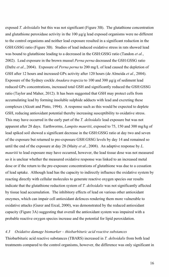
organisms had only a slightly higher percentage than that of the controls (Table 2). The lead 482
associated with them, however, was 63 and 141, respectively, times greater than that of the 483
controls. It is thought that metals in the lysosomes may bind to lipofuscins, which are lipid 484
peroxidation end products in the lysosomes, as insoluble lipoprotein granules (George, 485
1983a; George, 1983b). In most cells of marine invertebrates these lipofuscin granules are 486
usually excreted by exocytosis (George, 1983b). Thus, lead associated with lysosomes may 487
be in the process of detoxification, however, unbound lead associated with this fraction can 488
lead to the formation of oxygen derived free radicals in the cells which if not fully detoxified 489
can start a sequence of lipid peroxidation reactions (Viarengo and Nott, 1993). The 490
percentage of lead associated with the heat sensitive protein fraction and nuclei and cell 491
debris in the lead exposed organisms (Table 2) is indicative of a detoxification system 492
struggling with the excess lead. The high percentage of lead in the biologically detoxified 493
metal fraction is clear evidence that an efficient system of lead binding and detoxification 494
was present and active; however, the increased lead burden associated with the biologically 495
active metal fractions indicates that the metal sensitive organelles were exposed to potentially 496
toxic concentrations of lead as the tissue concentrations increased. 497
498
4.2 Enzymatic biomarkers – oxidative enzymes 499
The antioxidant capacity of the lead exposed T. deltoidalis was significantly reduced in both 500
treatments compared to the control organisms (Figure 3A; Supplementary Table 3b). While 501
the 300 µg/g lead exposed organisms had a slightly higher antioxidant capacity than the 100 502
µg/g lead exposed organisms, the difference was not significant (Figure 3A; Supplementary 503
Table 3b). This is a surprising result as the 300 µg/g lead exposed organisms had four times 504
the lead burden of the 100 µg/g lead exposed organisms in the biologically active metal 505
fraction, of which 67% was associated with the mitochondria (Table 2; Figure 2). Mytilus 506
edulis exposed to lead accumulated it in granules in the lysosomes and nucleus causing 507
impairment of lysosomal function, a reduction of the mitochondrial cristae and deformation 508
of the nuclear envelope (Einsporn and Koehler, 2008). There was some suppression of the 509
total glutathione concentration and glutathione peroxidase activity in the 300 µg/g lead 510
16
exposed T. deltoidalis but this was not significant (Figure 3B). The glutathione concentration 511
and glutathione peroxidase activity in the 100 µg/g lead exposed organisms were no different 512
to the control organisms and neither lead exposure resulted in a significant reduction in the 513
GSH:GSSG ratio (Figure 3B). Studies of lead induced oxidative stress in rats showed lead 514
was bound to glutathione leading to a decreased in the GSH:GSSG ratio (Tandon et al., 515
2002). Lead exposure in the brown mussel Perna perna decreased the GSH:GSSG ratio 516
(Dafre et al., 2004). Exposure of Perna perna to 200 mg/L of lead caused the depletion of 517
GSH after 12 hours and increased GPx activity after 120 hours (de Almeida et al., 2004). 518
Exposure of the Sydney cockle Anadara trapezia to 100 and 300 µg/g of sediment lead 519
reduced GPx concentrations, increased total GSH and significantly reduced the GSH:GSSG 520
ratio (Taylor and Maher, 2012). It has been suggested that GSH may protect cells from 521
accumulating lead by forming insoluble sulphide adducts with lead and excreting these 522
complexes (Alcutt and Pinto, 1994). A response such as this would be expected to deplete 523
GSH, reducing antioxidant potential thereby increasing susceptibility to oxidative stress. 524
This may have occurred in the early part of the T. deltoidalis lead exposure but was not 525
apparent after 28 days. Earthworms, Lampito mauritii, exposed to 75, 150 and 300 mg/kg of 526
lead spiked soil showed a significant decrease in the GSH:GSSG ratio at day two and seven 527
of the exposure but returned to pre-exposure GSH:GSSG levels by day 14 and remained so 528
until the end of the exposure at day 28 (Maity et al., 2008). An adaptive response by L. 529
mauritii to lead exposure may have occurred, however, the lead tissue dose was not measured 530
so it is unclear whether the measured oxidative response was linked to an increased metal 531
dose or if the return to the pre-exposure concentrations of glutathione was due to a cessation 532
of lead uptake. Although lead has the capacity to indirectly influence the oxidative system by 533
reacting directly with cellular molecules to generate reactive oxygen species our results 534
indicate that the glutathione reduction system of T. deltoidalis was not significantly affected 535
by tissue lead accumulation. The inhibitory effects of lead on various other antioxidant 536
enzymes, which can impair cell antioxidant defences rendering them more vulnerable to 537
oxidative attacks (Gurer and Ercal, 2000), was demonstrated by the reduced antioxidant 538
capacity (Figure 3A) suggesting that overall the antioxidant system was impaired with a 539
probable reactive oxygen species increase and the potential for lipid peroxidation. 540
541
4.3 Oxidative damage biomarker – thiobarbituric acid reactive substances 542
Thiobarbituric acid reactive substances (TBARS) increased in T. deltoidalis from both lead 543
treatments compared to the control organisms, however, the difference was only significant in 544

17
the 300 µg/g lead exposed organisms which had significantly higher lipid peroxidation than 545
both the control and the 100 µg/g lead exposed organisms (Figure 4A; Supplementary Table 546
3b). The marine bivalve Perna viridis exposed to 13 µg/l of lead over 30 days had a linear 547
accumulation of lead into the tissues and enhanced TBARS concentrations relative to controls 548
were seen in all tissues but this response was not linear over the 30 days (Prakash and Rao, 549
1995). The brown mussel Perna perna exposed to 200 mg/l of lead showed no significant 550
increase in TBARS over 120 hours despite significant changes to antioxidant enzymes (de 551
Almeida et al., 2004). De Almeida et al. (2004) suggest that this may be due to elevated 552
concentrations of the enzyme phospholipid hydroperoxide glutathione peroxidase, which is 553
highly reactive towards oxidised phospholipids, having a protective effect against lipid 554
peroxidation. T. deltoidalis lipid peroxidation was highly correlated with the antioxidant 555
capacity (r2 = 0.57; p ≤ 0.0001). Ercal et al. (2001) suggest that as lead cannot initiate lipid 556
peroxidation on membrane lipids directly it might induce oxidative stress by interacting with 557
oxyhaemoglobin, leading to peroxidative hemolysis in red blood cell membranes. As T. 558
deltoidalis do not have haemoglobin, this pathway for lipid peroxidation is not available 559
thereby reducing the total lipid peroxidation potential of lead in these organisms. It appears 560
from the range of different TBARS responses seen for lead exposures that lipid peroxidation 561
alone may not be strong indicator for lead toxicity in the majority of marine invertebrates, 562
however, when viewed together with the antioxidant capacity a pattern of lead induced 563
oxidative perturbation in T. deltoidalis is apparent. 564
565
4.4 Cellular biomarker – lysosomal stability 566
The lead exposed T. deltoidalis had significantly higher lysosomal destabilisation than the 567
control organisms (Figure 4B; Supplementary Table 3a). The 100 µg/g lead exposed T. 568
deltoidalis had 40% and the 300 µg/g 48% lysosomal destabilisation which, using the 569
Ringwood et al. (2003) criteria, would class them as stressed. Ercal et al. (2001) suggests 570
that lead alters the lipid composition of cellular membranes which may alter membrane 571
integrity, permeability and function. Mytilus galloprovincialis transplanted in a lead 572
contaminated area of the North Tyrrhenian Sea accumulated lead over time and showed a 573
severe disturbance in lysosomal membrane stability with increased tissue lead (Regoli and 574
Orlando, 1993). A. trapezia exposed to the same sediment lead concentrations as the T. 575
deltoidalis in this experiment were highly stressed, with significant lysosomal destabilisation, 576
up to 61% (Taylor and Maher, 2012). The T. deltoidalis lysosomal destabilisation was 577
negatively correlated with antioxidant capacity, (r2 = 0.54; p ≤ 0.001) and positively 578

18
correlated, (r2 = 0.55; p ≤ 0.001) with lipid peroxidation. Despite the limited increase in lipid 579
peroxidation of the 100 µg/g lead exposed T. deltoidalis the lysosomal destabilisation of 580
organisms from both treatments was at a similar level (Figures 4A & B), suggesting that lead 581
interaction with the lysosomal membrane leading to its destabilisation was probably 582
occurring via more than just the lipid peroxidation pathway. 583
584
4.5 Genotoxic biomarker – micronuclei frequency 585
T. deltoidalis exposed to lead had a significantly higher micronuclei frequency than the 586
control organisms but they were not significantly different to each other (Figure 4C; 587
Supplementary Table 3b). The frequency of micronuclei in the lead exposed T. deltoidalis 588
was much lower than that of these organisms when exposed to cadmium, which is generally 589
in keeping with the pattern seen for the other stress indices (Taylor and Maher, 2013). This 590
suggests that cadmium is a genotoxic agent exerting a direct influence on the induction of 591
micronuclei, perhaps in concert with ROS, while lead had a more indirect influence perhaps 592
via ROS alone. Lead, for example, has been shown to affect the expression of 2 nuclear 593
transcription factors: NF-kB and HIF-1 which may lead to cell cycle arrest, apoptosis or 594
interruption to gene transcription, while cadmium affects 3: NF-kB, and AP-1 and p53 which 595
help protect cells from carcinogenesis (Leonard et al., 2004). The frequency of micronuclei 596
in the lead exposed T. deltoidalis was negatively correlated with antioxidant capacity (r2 = 597
0.52; p ≤ 0.001) and positively correlated with lipid peroxidation (r2 = 0.51; p ≤ 0.001) 598
indicating that an increase in ROS likely contributed to an increase in genotoxic damage. 599
600
5 Conclusions 601
602
The response of T. deltoidalis to lead exposure differed between sediment lead exposure 603
concentrations. Accumulation was rapid at both exposures in the first three days, however, 604
the 100 µg/g lead exposed organisms continued to accumulate slowly over the remaining 605
time reaching an equilibrium tissue with sediment lead concentration, while the 300 µg/g lead 606
exposed organisms had a pattern of pulses of uptake and loss over the remainder of the 607
exposure suggesting saturation of binding sites may have occurred. In lead exposed 608
organisms around 70% of accumulated lead was detoxified with 74% converted to metal rich 609
granules and the remainder in the metallothionein like protein fraction. The exposure – dose 610
showed a strong relationship for lead with significant increases in biologically active lead 611
burdens. The dose-response relationships were mixed, with no significant changes in the 612

19
glutathione enzymes measured but a significant reduction in the total antioxidant capacity. 613
There were strong correlations between antioxidant capacity and lipid peroxidation, 614
lysosomal destabilisation and increased micronuclei and between lipid peroxidation and 615
lysosomal destabilisation and increased micronuclei frequency. The mixed response to lead 616
observed for these effects biomarkers supports the multibiomarker approach, to both 617
investigate the mechanisms of toxicological effect and by using interlinked biomarkers to 618
demonstrate effects. The exposure-dose-response relationships demonstrated in this 619
experiment for lead in T. deltoidalis indicates that lead exposure leads to significant tissue 620
lead accumulation and increased biologically active lead burdens with associated cellular 621
perturbations. These results show T. deltoidalis is sensitive to lead toxicity and would be a 622
suitable organism for use in marine sediment lead toxicity assessment. 623
624
References 625
626 ABS (1996) Australian Bureau of Statistics-Census Data http:// www.abs.gov.au/census In. 627 Adams WJ, Blust R, Borgmann U, Brix KV, DeForest DK, Green AS, Meyer JS, McGeer JC, Paquin 628
PR, Rainbow PS, Wood CM (2011) Utility of Tissue Residues for Predicting Effects of Metals 629 on Aquatic Organisms. Integrated Environmental Assessment and Management 7, 75-98. 630
Adams WJ, Green AS, Ahlf W, Brown SS, Burton GA, Chadwick B, Crane M, Gouget R, Ho KT, 631 Hogstand C, Reynoldson TB, Ringwood AH, Savitz JD, Sibley PK (2005) Using sediment 632 assessment tools and a weight-of evidence approach. In 'Use of Sediment Quality Guidelines 633 and Related Tools for the Assessment of Contaminated Sediments.'. (Eds RJ Wenning, GE 634 Batley, CG Ingersoll and DW Moore). (SETAC Press: Pensacola, FL.) 635
Alcutt F, Pinto JT (1994) Glutathione concentrations in the hard clam, Mercenaria mercenaria, 636 following laboratory exposure to lead (a potential model system for evaluating exposure to 637 carcinogens and toxins) Comparative Biochemistry and Physiology 107C, 347-352. 638
Ãlvarez-Iglesias P, Rubio B, Millos J (2012) Isotopic identification of natural vs. anthropogenic lead 639 sources in marine sediments from the inner Ria de Vigo (NW Spain). Science of The Total 640 Environment 437, 22-35. 641
ANZECC, ARMCANZ (2000) Australian guidelines for water quality monitoring and reporting. 642 National Water Quality Management Strategy Paper No 4. In. (Australian and New Zealand 643 Environment and Conservation Council / Agriculture and Resource Management Council of 644 Australia and New Zealand Canberra, ACT) 645
ARCARP (2006) 'Trace Elements in Coal.' Australian Coal Association Research Program. 646 ASTM (2010) ASTM E1688 - 10 Standard Guide for Determination of the Bioaccumulation of 647
Sediment-Associated Contaminants by Benthic Invertebrates. 648 Atkinson CA, Jolley DF, Simpson SL (2007) Effect of overlying water pH, dissolved oxygen, salinity 649
and sediment disturbances on metal release and sequestration from metal contaminated marine 650 sediments. Chemosphere 69, 1428-1437. 651
Baldwin S, Deaker M, Maher W (1994) Low-volume microwave digestion of marine biological 652 tissues for the measurement of trace elements. Analyst 119, 1701-1704. 653

20
Beesley PL, Ross GJB, Wells A (Eds) (1998) 'Mollusca: The Southern Synthesis ' Fauna of Australia 654 Vol. 5 (CSIRO Publishing: Melbourne) 655
Bergayou H, Mouneyrac C, Pellerin J, Moukrim A (2009) Oxidative stress responses in bivalves 656 (Scrobicularia plana, Cerastoderma edule) from the Oued Souss estuary (Morocco). 657 Ecotoxicology and Environmental Safety 72, 765-769. 658
Blasco J, Puppo J (1999) Effect of heavy metals (Cu, Cd and Pb) on aspartate and alanine 659 aminotransferase in Ruditapes philippinarum (Mollusca: Bivalvia). Comparative Biochemistry 660 and Physiology Part C: Pharmacology, Toxicology and Endocrinology 122, 253-263. 661
Bocchetti R, Fattorini D, Pisanelli B, Macchia S, Oliviero L, Pilato F, Pellegrini D, Regoli F (2008) 662 Contaminant accumulation and biomarker responses in caged mussels, Mytilus 663 galloprovincialis, to evaluate bioavailability and toxicological effects of remobilized chemicals 664 during dredging and disposal operations in harbour areas. Aquatic Toxicology 89, 257-266. 665
Bolognesi C, Frenzilli G, Lasagna C, Perrone E, Roggieri P (2004) Genotoxicity biomarkers in 666 Mytilus galloprovincialis: wild versus caged mussels. Mutation Research/Fundamental and 667 Molecular Mechanisms of Mutagenesis 552, 153-162. 668
Bustamante P, Miramand P (2005) Subcellular and body distributions of 17 trace elements in the 669 variegated scallop Chlamys varia from the French coast of the Bay of Biscay. Science of The 670 Total Environment 337, 59-73. 671
Cajaraville MP, Bebianno MJ, Blasco J, Porte C, Sarasquete C, Viarengo A (2000) The use of 672 biomarkers to assess the impact of pollution in coastal environments of the Iberian Peninsula: a 673 practical approach. The Science of The Total Environment 247, 295-311. 674
Calbiochem (2004) GSH/GSSG Ratio Assay Kit User Protocol 371757. 675 Campana O, Sarasquete C, Blasco J (2003) Effect of lead on ALA-D activity, metallothionein levels, 676
and lipid peroxidation in blood, kidney, and liver of the toadfish Halobatrachus didactylus. 677 Ecotoxicology and Environmental Safety 55, 116-125. 678
Carroll B (1996) 'A Review of Selenium and Heavy Metal contamination of Lake Macquarie, N.S.W. 679 due to Power Generation and Lead/Zinc Smelting. A Review Submitted in Consideration for 680 the AMEEF Excellence Awards Review Prize.' University of Sydney, Sydney. 681
Cayman (2011) Antioxidant Assay Kit User Protocol 709001. 682 Chiaradia M, Chenhall BE, Depers AM, Gulson BL, Jones BG (1997) Identification of historical lead 683
sources in roof dusts and recent lake sediments from an industrialized area: indications from 684 lead isotopes. Science of The Total Environment 205, 107-128. 685
Clark RB (2001) 'Marine Pollution 5th Edition.' (Oxford University Press) 686 Company R, Serafim A, Lopes B, Cravo A, Kalman J, Riba I, DelValls TA, Blasco J, Delgado J, 687
Sarmiento AM, Nieto JM, Shepherd TJ, Nowell G, Bebianno MJ (2011) Source and impact of 688 lead contamination on δ-aminolevulinic acid dehydratase activity in several marine bivalve 689 species along the Gulf of Cadiz. Aquatic Toxicology 101, 146-154. 690
Company R, Serafim A, Lopes B, Cravo A, Shepherd TJ, Pearson G, Bebianno MJ (2008) Using 691 biochemical and isotope geochemistry to understand the environmental and public health 692 implications of lead pollution in the lower Guadiana River, Iberia: A freshwater bivalve study. 693 Science of The Total Environment 405, 109-119. 694
Cook DE, Gale SJ (2005) The curious case of the date of introduction of leaded fuel to Australia: 695 Implications for the history of Southern Hemisphere atmospheric lead pollution. Atmospheric 696 Environment 39, 2553-2557. 697

21
Cossu C, Doyotte A, Babut M, Exinger A, Vasseur P (2000) Antioxidant Biomarkers in Freshwater 698 Bivalves, Unio tumidus, in Response to Different Contamination Profiles of Aquatic Sediments. 699 Ecotoxicology and Environmental Safety 45, 106-121. 700
Dafre AL, Medeiros ID, MUller IC, Ventura EC, Bainy ACD (2004) Antioxidant enzymes and 701 thiol/disulfide status in the digestive gland of the brown mussel Perna perna exposed to lead 702 and paraquat. Chemico-Biological Interactions 149, 97-105. 703
Damiens G, Gnassia-Barelli M, Loquès F, Roméo M, Salbert V (2007) Integrated biomarker response 704 index as a useful tool for environmental assessment evaluated using transplanted mussels. 705 Chemosphere 66, 574-583. 706
de Almeida EA, Miyamoto S, Bainy ACD, de Medeiros MHG, Di Mascio P (2004) Protective effect 707 of phospholipid hydroperoxide glutathione peroxidase (PHGPx) against lipid peroxidation in 708 mussels Perna perna exposed to different metals. Marine Pollution Bulletin 49, 386-392. 709
Depledge MH, Amaral-Mendes, J.J., Daniel, B., Halbrook, R.S., Kloepper-Sams, P., Moore, M.N., 710 Peakall, D,B. (1992) The conceptual basis of the biomarker approach. In 'Biomarkers: Research 711 and application in the assessment of environmental health'. (Ed. DB Peakall, & Shugart, L.R.) 712 pp. 15 - 29. (NATO ASI Series H: Cell Biology: Springer, Berlin, Germany) 713
Domouhtsidou GP, Dailianis S, Kaloyianni M, Dimitriadis VK (2004) Lysosomal membrane stability 714 and metallothionein content in Mytilus galloprovincialis (L.), as biomarkers: Combination with 715 trace metal concentrations. Marine Pollution Bulletin 48, 572-586. 716
Durou C, Poirier L, Amiard J-C, Budzinski H, Gnassia-Barelli M, Lemenach K, Peluhet L, 717 Mouneyrac C, Roméo M, Amiard-Triquet C (2007) Biomonitoring in a clean and a multi-718 contaminated estuary based on biomarkers and chemical analyses in the endobenthic worm 719 Nereis diversicolor. Environmental Pollution 148, 445-458. 720
Einsporn S, Koehler A (2008) Immuno-localisations (GSSP) of subcellular accumulation sites of 721 phenanthrene, aroclor 1254 and lead (Pb) in relation to cytopathologies in the gills and 722 digestive gland of the mussel Mytilus edulis. Marine Environmental Research 66, 185-186. 723
Ercal N, Gurer-Orhan H, Aykin-Burns N (2001) Toxic metals and oxidative stress part 1: mechanisms 724 involved in metal induced oxidative damage. Current Topics in Medicinal Chemistry 1, 529-725 539. 726
Fowler BA, Mahaffey KR (1978) Interactions among lead, cadmium, and arsenic in relation to 727 porphyrin excretion patterns. Environmental Health Perspectives 25, 87-90. 728
Galloway TS, Brown RJ, Browne MA, Dissanayake A, Lowe D, Jones MB, Depledge MH (2004) 729 Ecosystem management bioindicators: the ECOMAN project - a multi-biomarker approach to 730 ecosystem management. Marine Environmental Research 58, 233-237. 731
Genaidy AM, Sequeira R, Tolaymat T, Kohler J, Rinder M (2009) Evidence-based integrated 732 environmental solutions for secondary lead smelters: Pollution prevention and waste 733 minimization technologies and practices. Science of The Total Environment 407, 3239-3268. 734
George S (1983a) Heavy metal detoxication in Mytilus kidney - an in vitro study of Cd- and Zn-735 binding to isolated tertiary lysosomes. Comparative Biochemistry and Physiology 76C, 59-65. 736
George S (1983b) Heavy metal detoxication in the mussel Mytilus edulis - composition of Cd-737 containing kidney granules (tertiary lysosomes). Comparative Biochemistry and Physiology 738 76C, 53-57. 739
Gnassia-Barelli M, Romeo M (1993) Some aspects of lead ecotoxicology in the marine environment. 740 Aquatic Toxicology 26, 163-170. 741

22
Gorbi S, Virno Lamberti C, Notti A, Benedetti M, Fattorini D, Moltedo G, Regoli F (2008) An 742 ecotoxicological protocol with caged mussels, Mytilus galloprovincialis, for monitoring the 743 impact of an offshore platform in the Adriatic sea. Marine Environmental Research 65, 34-39. 744
Griscom SB, Fisher NS (2004) Bioavailability of Sediment-Bound Metals to Marine Bivalve 745 Molluscs: An Overview. Estuaries 27, 826-838. 746
Gurer H, Ercal N (2000) Can antioxidants be beneficial in the treatment of lead poisoning? Free 747 Radical Biology and Medicine 29, 927-945. 748
Hong F, Wu, C., Liu, C., Wang, L., Gao, F., Yang, F., Xu, J., Liu, T., Xie, Y., Li, X. (2007) Direct 749 evidence for interaction between lead ions and kidney DNA from silver crucian carp. 750 Chemosphere 68, 1442-1446. 751
Ingersoll CG, Burton GA, Dawson TD, Dwyer FJ, Ireland DS, Kemble NE, Mount DR, Norburg-752 King TJ, Sibley PK, Stahl L (2000) Methods for Measuring the Toxicity and Bioaccumulation 753 of Sediment-associated Contaminants with Freshwater Invertebrates In. (Ed. SaTW Offices of 754 Research and Development Mid-Continent Ecology Department). (U.S. Environmental 755 Protection Agency) 756
Jing G, Li Y, Xie L, Zhang R (2007) Different effects of Pb2+ and Cu2+ on immune and antioxidant 757 enzyme activities in the mantle of Pinctada fucata. Environmental Toxicology and 758 Pharmacology 24, 122-128. 759
Kalpaxis DL, Theos C, Xaplanteri MA, Dinos GP, Catsiki AV, Leotsinidis M (2004) Biomonitoring 760 of Gulf of Patras, N. Peloponnesus, Greece. Application of a biomarker suite including 761 evaluation of translation efficiency in Mytilus galloprovincialis cells. Environmental Research 762 94, 211-220. 763
King CK, Dowse MC, Simpson SL (2010) Toxicity of Metals to the Bivalve Tellina deltoidalis and 764 Relationships Between Metal Bioaccumulation and Metal Partitioning Between Seawater and 765 Marine Sediments. Archives of Environmental Contamination and Toxicology 58, 657-665. 766
King CK, Dowse MC, Simpson SL, Jolley DF (2004) An assessment of five Australian polychaetes 767 and bivalves for use in whole-sediment toxicity tests: Toxicity and accumulation of copper and 768 zinc from water and sediment. Archives of Environmental Contamination and Toxicology 47, 769 314-323. 770
King CK, Gale SA, Hyne RV, Stauber JL, Simpson SL, Hickey CW (2006) Sensitivities of Australian 771 and New Zealand amphipods to copper and zinc in waters and metal-spiked sediments. 772 Chemosphere 63, 1466-1476. 773
King CK, Simpson SL, Smith SV, Stauber JL, Bately GE (2005) Short-term accumulation of Cd and 774 Cu from water, sediment and algae by the amphipod Melita plumulosa and the bivalve Tellina 775 deltoidalis. Marine Ecology Progress Series 287, 177-188. 776
Lam PKS (2009) Use of biomarkers in environmental monitoring. Ocean & Coastal Management 52, 777 348-354. 778
Lam PKS, Gray JS (2003) The use of biomarkers in environmental monitoring programmes. Marine 779 Pollution Bulletin 46, 182-186. 780
Lee BG, Griscom SB, Lee JS, Choi HJ, Koh CH, Luoma SN, Fisher NS (2000) Influences of dietary 781 uptake and reactive sulfides on metal bioavailability from aquatic sediments. Science 287, 282 - 782 284. 783
Lee BG, Wallace WG, Luoma SN (1998) Uptake and loss kinetics of Cd, Cr and Zn in the bivalves 784 Potamocorbula amurensis and Macoma balthica: effects of size and salinity. Marine Ecology 785 Progress Series 175, 177-189. 786

23
Leonard SS, Harris GK, Shi X (2004) Metal-induced oxidative stress and signal transduction. Free 787 Radical Biology and Medicine 37, 1921-1942. 788
Macdonald A, Silk L, Schwartz M, Playle RC (2002) A lead-gill binding model to predict acute lead 789 toxicity to rainbow trout (Oncorhynchus mykiss). Comparative Biochemistry and Physiology 790 Part C: Toxicology & Pharmacology 133, 227-242. 791
Maher W, Forster S, Krikowa F, Snitch P, Chapple G, Craig P (2001) Measurement of trace elements 792 and phosphorus in marine animal and plant tissues by low-volume microwave digestion and 793 ICP-MS. Atomic Spectroscopy 22, 361-370. 794
Maher W, Krikowa F, Kirby J, Townsend AT, Snitch P (2003) Measurement of trace elements in 795 marine environmental samples using solution ICP-MS. Current and future applications. 796 Australian Journal of Chemistry 56, 103-116. 797
Maity S, Roy S, Chaudhury S, Bhattacharya S (2008) Antioxidant responses of the earthworm 798 Lampito mauritii exposed to Pb and Zn contaminated soil. Environmental Pollution 151, 1-7. 799
Marigómez I, Soto M, Cajaraville MP, Angulo E, Giamberini L (2002) Cellular and subcellular 800 distribution of metals in molluscs. Microscopy Research and Technique 56, 358-392. 801
Mateo R, Beyer WN, Spann JW, Hoffman DJ (2003) Relation of fatty acid composition in lead-802 exposed mallards to fat mobilization, lipid peroxidation and alkaline phosphatase activity. 803 Comparative Biochemistry and Physiology Part C: Toxicology & Pharmacology 135, 451-458. 804
Monteiro V, Cavalcante DGSM, Vilela MBFA, Sofia SH, Martinez CBR (2011) In vivo and in vitro 805 exposures for the evaluation of the genotoxic effects of lead on the Neotropical freshwater fish 806 Prochilodus lineatus. Aquatic Toxicology 104, 291-298. 807
Prakash NT, Rao KSJ (1995) Modulations in antioxidant enzymes in different tissues of marine 808 bivalve Perna viridis during heavy metal exposure. Molecular and Cellular Biochemistry 146, 809 107-113. 810
Rainbow PS (2002) Trace metal concentrations in aquatic invertebrates: why and so what? 811 Environmental Pollution 120, 497-507. 812
Rainbow PS (2007) Trace metal bioaccumulation: Models, metabolic availability and toxicity. 813 Environment International 33, 576-582. 814
Regoli F, Orlando E (1993) Mytilus galloprovincialis as a bioindicator of lead pollution: biological 815 variables and cellular responses The Science of The Total Environment Supplement 2, 1283-816 1292. 817
Regoli F, Orlando E (1994) Accumulation and subcellular distribution of metals (Cu, Fe, Mn, Pb and 818 Zn) in the Mediterranean mussel Mytilus galloprovincialis during a field transplant experiment. 819 Marine Pollution Bulletin 28, 592-600. 820
Renberg I, Brannvall M-L, Bindler R, Emteryd O (2002) Stable lead isotopes and lake sediments - a 821 useful combination for the study of atmospheric lead pollution history. Science of The Total 822 Environment 292, 45-54. 823
Ringwood AH, Hoguet J, Keppler CJ, Gielazyn ML, Ward CH, Rourk AR (2003) 'Cellular 824 Biomarkers (Lysosomal Destabilization, Glutathione & Lipid Peroxidation) in Three Common 825 Estuarine Species: A Methods Handbook.' (Marine Resources Research Institute, Sth. Carolina 826 Dept. Nat. Res. Charleston) 827
Roach A (2005) Assessment of metals in sediments from Lake Macquarie, New South Wales, 828 Australia, using normalisation models and sediment quality guidelines. Marine Environmental 829 Research 59, 453-472. 830

24
Roesijadi G, Robinson WE (1994) Metal Regulation in Aquatic Animals: Mechanisms of Uptake, 831 Accumulation, and Release. In 'Aquatic Toxicology: Molecular, Biochemical, and Cellular 832 Perspectives'. (Eds DC Malins and GK Ostrander) pp. 387-419. (Lewis: Boca Raton, Florida) 833
Simkiss K, Taylor MG (1989) Convergence of cellular systems of metal detoxification. Marine 834 Environmental Research 28, 211-214. 835
Simpson SL, Angel BM, Jolley DF (2004) Metal equilibration in laboratory-contaminated (spiked) 836 sediments used for the development of whole-sediment toxicity tests. Chemosphere 54, 597-837 609. 838
Simpson SL, Batley GE, Chariton AA, Stauber JL, King CK, Chapman JC, Hyne RV, Gale SA, 839 Roach AC, Maher WA (2005) 'Handbook for sediment quality assessment.' (CSIRO: Bangor, 840 NSW) 841
Sokolova IM, Ringwood AH, Johnson C (2005) Tissue-specific accumulation of cadmium in 842 subcellular compartments of eastern oysters Crassostrea virginica Gmelin (Bivalvia: 843 Ostreidae). Aquatic Toxicology 74, 218-228. 844
Strom D, Simpson SL, Bately GE, Jolley DF (2011) The influence of sediment particle size and 845 organic carbon on toxicity of copper to benthic invertebrates in oxic/suboxic surface sediments. 846 Environmental Toxicology and Chemistry 30, 1599-1610. 847
Tandon SK, Singh S, Prasad S, Srivastava S, Siddiqui MKJ (2002) Reversal of lead-induced oxidative 848 stress by chelating agent, antioxidant, or their combination in the rat. Environmental Research 849 90, 61-66. 850
Taylor AM, Maher WA (2010) Establishing Metal Exposure - Dose - Response Relationships in 851 Marine Organisms: Illustrated with a Case Study of Cadmium Toxicity in Tellina deltoidalis. 852 In 'New Oceanography Research Developments: Marine Chemistry, Ocean Floor Analyses and 853 Marine Phytoplankton'. (Ed. K Puopolo, Martorino, L.) pp. 1-57. (Nova Science: New York) 854
Taylor AM, Maher WA (2012) Exposure-dose-response of Anadara trapezia to metal contaminated 855 estuarine sediments 2. Lead spiked sediments. Aquatic Toxicology 116-117, 79-89. 856
Taylor AM, Maher WA (2013) Exposure-dose-response of Tellina deltoidalis to metal-contaminated 857 estuarine sediments: 1. Cadmium spiked sediments. Comparative Biochemistry and Physiology, 858 Part C 158, 44-55. 859
Tietze F (1969) Enzymatic method for quantitative determination of nanogram amounts of total and 860 oxidised glutathione: Applications to mammalian blood and other tissues. Analytical 861 Biochemistry 27, 502-522. 862
Udroiu I (2006) The micronucleus test in piscine erythrocytes. Aquatic Toxicology 79, 201-204. 863 Viarengo A, Lowe D, Bolognesi C, Fabbri E, Koehler A (2007) The use of biomarkers in 864
biomonitoring: A 2-tier approach assessing the level of pollutant-induced stress syndrome in 865 sentinel organisms. Comparative Biochemistry and Physiology Part C: Toxicology & 866 Pharmacology 146, 281-300. 867
Viarengo A, Moore MN, Mancinelli G, Mazzucotelli A, Pipe RK, Farrar SV (1987) Metallothioneins 868 and lysosomes in metal toxicity and accumulation in marine mussels: the effect of cadmium in 869 the presence and absence of phenanthrene. Marine Biology 94, 251-257. 870
Viarengo A, Nott JA (1993) Mechanisms of heavy metal cation homeostasis in marine invertebrates. 871 Comparative Biochemistry and Physiology Part C: Comparative Pharmacology 104, 355-372. 872
Wallace WG, Lee BG, Luoma SN (2003) Subcellular compartmentalization of Cd and Zn in two 873 bivalves. I. Significance of metal-sensitive fractions (MSF) and biologically detoxified metal 874 (BDM). Marine Ecology Progress Series 249, 183-197. 875

25
Winter MJ, Ellis, L.C.J., Hutchinson, T.H. (2007) Formation of micronuclei in erythrocytes of the 876 fathead minnow (Pimephales promelas) after acute treatment with mitomycin C or 877 cyclophosphamide. Mutation Research/Genetic Toxicology and Environmental Mutagenesis 878 629, 89-99. 879
ZepoMetrix (2011) Oxitek TBARS Assay Kit User Protocol ZMC Catalog # 0801192. 880 881 Figure Captions 882 883 Figure 1: Tissue lead concentrations (µg/g dry mass) of T. deltoidalis exposed to lead spiked 884 sediments of 0 (control), Pb 100 and 300µg/g dry mass. Mean ± SE, n = 12. Day 0 are 885 unexposed organisms n = 6. 886 887 Figure 2: Distribution (%) of lead in each of the subcellular fractions of T. deltoidalis 888 following 28 days exposure to lead spiked sediments. Subcellular fractions are: 889 nuclei+cellular debris, metal rich granules (MRG); heat stable, metallothionein like proteins 890 (MTLP); mitochondria (Mit); lysosomes+ microsomes (Lys & Mic); heat sensitive proteins 891 (HSP). Stippled fractions ( ) make up the biologically active lead (BAM), dashed 892 fractions ( ) make up the biologically detoxified lead (BDM), n = 2. 893 894 Figure 3: Antioxidant enzyme biomarkers of T. deltoidalis after 28 days exposure to lead 895 spiked sediments of 0 (control), Pb100 and Pb 300µg/g dry mass. Mean ± SE, n = 12. 3A: 896 TAOC (total antioxidant capacity); 3B: GPx (glutathione peroxidise); GSH+2GSSG (total 897 glutathione); GSH/GSSG (ratio of reduced to oxidised glutathione). Different letters indicate 898 significant differences between means (Bonferroni test; p < 0.05). 899 900 Figure 4: Changes in oxidative damage biomarkers: 4A: MDA (lipid peroxidation); 4B: 901 cellular (lysosomal destabilisation); and 4C: genotoxic (micronuclei) of T. deltoidalis after 28 902 days exposure to lead spiked sediments, 0 (control), Pb 100 and Pb 300µg/g dry mass. Mean 903 ± SE n = 12. Different letters indicate significant differences between means (Bonferroni 904 test; p < 0.05). 905 906

26
Figure 1 907
908 909
0
100
200
300
400
500
600
700
Pb Control Pb 100 Pb 300
Tis
sue
Pb µ
g/g
Sediment Pb µg/g
Day 0 Day 3 Day 7 Day 14 Day 21 Day 28

27
Figure 2 910
911
Nuclei-Cell debris
MRG
MTLP
Mit
Lys & MicHSP
Control
BAM = 0.04 µgBDM = 0.17 µg
Nuclei-Cell debris
MRG
MTLP
Mit
HSPPb 100 µg/g
BAM = 1.14 µgBDM = 12.56 µg
Lys & Mic
Nuclei-Cell debris
MRG
MTLP
Mit
Lys & Mic HSPPb 300 µg/g
BAM = 3.99 µgBDM = 29.24 µg

28
Figure 3 912
913
0
50
100
150
200
250
control Pb 100 Pb 300
Sediment Lead (µg/g)
GPx µmol/min/mg proteinGSH+2GSSG µmol/mg proteinGSH/GSSG ratio
a
b
ca
b
c a
b
c
B
0
5
10
15
20
25
control Pb 100 Pb 300
Ant
ioxi
dant
mm
ol/m
g pr
otei
n
Sediment Lead (µg/g)
a
b
b
A

29
Figure 4 914
915 916 917
0
10
20
30
40
50
60
control Pb 100 Pb 300
% u
nsta
ble
lyso
som
es
Sediment Lead (µg/g)
bb
a
B
0
0.1
0.2
0.3
0.4
0.5
0.6
0.7
0.8
0.9
control Pb 100 Pb 300
% m
icro
nucl
ei
Sediment Lead (µg/g)
b
b
a
C
0
10
20
30
40
50
60
70
control Pb 100 Pb 300
MD
A n
mol
/mg
prot
ein
Sediment Lead (µg/g)
b
a
a
A

30
Table 1: Total lead concentrations (µg wet mass) in whole tissue and subcellular fractions 918 with the percentage of total lead recovered in all fractions of T. deltoidalis after 28 days 919 exposure to lead spiked sediments. Lead subcellular concentrations (µg wet mass) and 920 percentage distribution of total recovered lead fractions are grouped as debris and 921 biologically active and biologically detoxified lead (Fig. 3). 922 Sediment Treatments (µg/g) control Pb 100 Pb 300 Total Tissue Lead (µg) 0.34 ± 0.08 20 ± 3 46 ± 6 Total Recovered Lead (µg) 0.25 ± 0.05 17 ± 7 42 ± 5 Proportion of total recovered in fractions (%) 72 ± 0.2 89 ± 20 92 ± 4 Lead Subcellular Distribution Nuclei + Cellular debris (µg) 0.032 ± 0.01 3.7 ± 1.5 9.1 ± 1 Nuclei + Cellular debris (%) 13 ± 3 21 ± 0.5 21 ± 1 Biologically Active Metal (µg) 0.04 ± 0.01 1.1 ± 0.6 4 ± 0.3 Biologically Active Metal (%) 17 ± 1.7 7 ± 0.5 9 ± 0.4 Biologically Detoxified Metal (µg) 0.17 ± 0.03 12.6 ± 5.3 29 ± 4 Biologically Detoxified Metal (%) 70 ± 5 72 ± 0.02 70 ± 0.6
Mean ± SD, n = 2 923 924 925 Table 2: Mean percentage of lead in the debris, biologically detoxified metal (BDM) and 926 biologically active metal (BAM) with the percentage of lead each of the fractions contributes 927 to BDM or BAM of T. deltoidalis subcellular fractions after 28 days exposure to lead spiked 928 sediments, n = 2. 929 Sediment Treatments (µg/g) control Pb 100 Pb 300 Nuclei + Cellular debris % of total 13 21 21 Biologically Detoxified Metal % of total 70 72 70 Metal Rich Granules % of BDM 47 74 74 Heat Stable MT Like Proteins % of BDM 53 26 26 Biologically Active Metal % of total 17 7 9 Mitochondria % of BAM 50 54 67 Lysosomes + Microsomes % of BAM 11 27 18 Heat Sensitive Proteins % of BAM 39 19 15
930 931

31
Supplementary Figure 1: Procedure for subcellular fractionation of bivalve tissues by 932 differential centrifugation. The shaded boxes show details of the centrifugation and digestion 933 / heating steps used to obtain the specific fractions. The final fractions, four pellets P2, P3, 934 P4 & P5 and two supernatants S2 & S5 are grouped as: biologically detoxified metal (BDM) 935 P2 & S5; biologically active metal (BAM) P3, P4 & P5 metals or S2 which contains metal 936 associated with dissolved tissues. 937
938 939
80oC x 10min4oC x 60min
10min @ 50,000g, 4oC
TissueHomogenate
10 min @ 1,000g, 4oC
P1Nuclei, unbroken cells,
cell membranes & granules
+ 0.5ml 1N Na OH 60oC x 60min10 min @ 10,000g, 20oC
P2Granules
S2Nuclei +Cellulardebris
S1supernatant
(Total Fraction)
S3Supernatant
S4supernatant(CytosolicFraction)
60 min @100,000g, 4oC
30 min @ 10,000g, 4oC
P3Heavy
OrganelleFraction
(Mitochondria)
P4 Light
Organelle & ParticulateFraction
(Lysosomes &Microsomes)
P
S
P
S
P
S5Heat stable
MT & MT-likeProteins
S
Resuspend in 0.5ml H2O
100oC x 2 min
Biologically A
ctive Metal (B
AM
)
Biologically Detoxified Metal (BDM)
P5Heat sensitive
Proteins(Enzymes)
S P
P
S

32
Supplementary Figure 2: Activity of specific marker enzymes for lysosomes (acid 940 phosphatase) and mitochondria (cytochrome c oxidase) in whole tissue; and mitochondria 941 and lysosome+microsome fractions following subcellular fractionation of whole tissue of T. 942 deltoidalis exposed to lead spiked sediments at 0 (Control), 100 and 300 µg/g dry mass after 943 28 days. Mean ± SD, n = 2. 944 945
946
Statistical Analysis 947 A Mixed Linear Model analysis of variance (ANOVA) (SPSS v 14.0) was used to 948 simultaneously analyse the effects of time (day) and treatment (lead exposure concentration) 949 on whole organism tissue lead accumulation. Multiple pair-wise comparisons were also 950 performed to determine where the significant differences lay between and within the 951 treatments on each of the analysis days. As a high number of tests were performed, a 952 Bonferroni correction for experiment wise error rate was applied to all calculations to 953 determine the significance level (Sokal and Rohlf, 1995). A preliminary investigation of the 954 data was done to examine the distribution of residuals using a non-parametric Kolmogorov-955 Smirnov Z Test. Where the population distribution was found to be non-normal a log10 956 transformation was performed prior to analysis. A Mixed Linear Model ANOVA (SPSS v 957 14.0) was used analyse the effect of treatment (lead exposure concentration) on the effect 958 measurement variables total antioxidant capacity (TAOC), glutathione peroxidase, total 959 glutathione, reduced and oxidised glutathione ratio, thiobarbituric acid reactive substances 960 (TBARS), lysosomal stability and micronuclei frequency. Tank(treatment) was run as a 961 covariance parameter in all analysis to determine whether the tank replicates were 962 contributing to the treatment variance, in all cases the variance estimate was found to be ≤ 963 0.01. Arsine transformation was used for lysosomal stability and micronuclei frequency data 964 as these values were percentages. 965
0
2
4
6
8
10
12
14
16
U m
g-1pr
otei
n
Acid Phosphatase
Cytochrome c oxidase

33
Supplementary Table 1: Mixed linear model ANOVA of tissue lead accumulation for 966 collection day and treatment. Lead log10 for T. deltoidalis whole tissue. 967 968 Lead Source df F p Treatment 2 301 *** Day 4 6 *** Day*Treatment 8 2 ns
*** p < 0.0005 ns p > 0.05 969 970 Supplementary Table 2: Pair-wise comparisons, with Bonferroni adjustment for multiple 971 comparisons, showing significant differences (p) between treatments. Lead log10 for T. 972 deltoidalis whole tissue. None = day 0 unexposed organisms 973 974 Treatments Lead p none - control ns none - Pb 100 *** none - Pb 300 *** control - Pb 100 *** control - Pb 300 *** Pb 100 - Pb 300 ***
*** p ≤ 0.001, ns p > 0.05 975 976 Supplementary Table 3a: Mixed linear model ANOVA of effects for treatment of T. 977 deltoidalis tissue. Lysosomes and micronuclei were calculated on arsine transformed data, all 978 other effects are calculated on log10 transformed data. TAOC: antioxidant capacity; GPx: 979 glutathione peroxidase; GSH+2GSSG: total glutathione; GSH:GSSG: ratio of reduced to 980 oxidised glutathione; TBARS: total thiobarbituric acid reactive substances; lysosomes: % 981 unstable lysosomes; micronuclei: % micronucleus frequency. 982 983 Source Lead df F p TAOC 2 9.8 ** GPx 2 2.4 ns GSH+2GSSG 2 1.5 ns GSH:GSSG 2 3.4 * TBARS 2 11 *** Lysosomes 2 53 *** Micronuclei 2 17 ***
*** p ≤ 0.001, ** p ≤0.01, * p ≤0.05, ns p > 0.05 984 985

34
Supplementary Table 3b: Pair-wise comparisons, with Bonferroni adjustment for multiple 986 comparisons, of effects for treatment of T. deltoidalis tissue. Lysosomes and micronuclei 987 were calculated on arsine transformed data, all other effects are calculated on log10 988 transformed data. TAOC: antioxidant capacity; GPx: glutathione peroxidase; GSH+2GSSG: 989 total glutathione; GSH:GSSG: ratio of reduced to oxidised glutathione; TBARS: total 990 thiobarbituric acid reactive substances; lysosomes: % unstable lysosomes; micronuclei: % 991 micronuclei frequency. 992 993 Effects Measures Treatments TAOC GPx GSH+2GSSG GSH:GSSG TBARS Lysosomes Micronuclei
Lead p p p p p p p
Control - Pb 100 * ns ns ns ns *** ** Control - Pb 300 * ns ns ns *** *** *** Pb 100 - Pb 300 ns ns ns ns * ns ns
*** p ≤ 0.001; ** p ≤0.01; * p ≤ 0.05; ns p > 0.05 994 995 Supplementary Table 4: Regression analysis of relationships between sediment and tissue 996 lead, between the effects measure total antioxidant capacity and TBARS, Lysosomal stability 997 and micronuclei frequency and between TBARS and Lysosomal stability and micronuclei 998 frequency 999 1000 Regression Lead N df F R2 p p Sediment lead vs Tissue lead 41 39 99.84 0.72 0.0000 **** TAOC vs TBARS 29 27 35.9 0.57 0.0000 **** TAOC vs lysosomal stability 18 16 18.6 0.54 0.0005 *** TAOC vs micronuclei frequency 18 16 17.3 0.52 0.0007 *** TBARS vs lysosomal stability 18 16 19.9 0.55 0.0004 *** TBARS vs micronuclei frequency 18 16 16.7 0.51 0.0009 ***
**** p ≤ 0.0001; *** p ≤0.001 1001 1002 References 1003 1004 Sokal RR, Rohlf FJ (1995) 'Biometry, The Principles and Practice of Statistics in Biological 1005 Research.' (W. H. Freeman and Company: New York) 1006 1007 1008