thoracic anesthesia: an update and · pdf file2/23/11 1 thoracic anesthesia: an update and...
TRANSCRIPT

2/23/11
1
THORACIC ANESTHESIA: AN UPDATE AND REVIEW
LOUIS M. GUZZI, M.D.,FCCM FLORIDA HOSPITAL ORLANDO, FLORIDA
THORACIC ANESTHESIA OBJECTIVES
ANATOMY REVIEW PHYSIOLOGY CHEST DYNAMICS
SPONTANEOUS LATERAL
CHEST CRISIS OPTIONS FOR LUNG ISOLATION
Diagram of Thoracic Area

2/23/11
2
The Larynx
hyoid bone
thyroid cartilage
cricoid cartilage trachea
epiglottis
TRACHEA & BRONCHI

2/23/11
3
LUNG ANTERIOR
LUNG POSTERIOR
LUNG LEFT SIDE

2/23/11
4
LUNG RIGHT SIDE
Tracheobronchial Tree
THE BRONCHUS TO
BRONCHIOLE BREAKDOWN

2/23/11
5
BRONCHIAL DIAGRAM
DYNAMICS OF PULMONARY BLOOD FLOW
• Blood flow is greatest in dependent parts of lung
• Hypoxic Pulmonary Vasoconstriction (HPV) redistributes blood away from poorly ventilated alveoli
SPONTANEOUS VENTILATION
Perfusion greatest at bases

2/23/11
6
DYNAMIC BLOOD FLOW IN THE LATERAL DECUBITUS POSITION
Gravity pulls blood flow to bases
Dynamics of Spontaneous Breathing
• Diaphragm descends causing a negative intrathoracic pressure
• Gas flows from higher pressure to lower pressure
• Greatest gas flow in spontaneous ventilation is to bases
SPONTANEOUS VENTILATION
Ventilation greatest at bases

2/23/11
7
Dynamics of Spontaneous Breathing
• Apex alveoli already distended from greater NEGATIVE pleural pressure thus they have less compliance to expand and receive volume increases
• Apex ribs short and expand minimally • Base alveoli have greatest gas flow due to greater
change in thoracic pressures during insp.- exp. Phases d/t insp. diaphragmatic downward movement d/t pail handle effect
• Abdominal contents pushing up and gravity pulling lungs down lessens the negative pleural pressure in bases (REMEMBER MO/ABDOMINAL PRESSURES)
*Greater negative pressure in apex during end expiration- small change during inspiration
LUNG
PLEURAL SPACE
CHEST WALL
pale handle effect
diaphragm moves down
lung follows
*
PAIL HANDLE EFFECT • Internal
intercostals, pull downward, aid expiration
• External intercostal, elevate ribs, aid inspiration.
• Pneumonic; In-Ex, Ex-In

2/23/11
8
INTERCOSTALS
Note; internal and external intercostal muscles
LUNGS WANT TO RECOIL, THORACIC CAGE WANTS TO EXPAND
Thus, the pleural cavity has a vacuum ( a negative pressure)

2/23/11
9
SPONTANEOUS VENTILATION
Ventilation(V) to Perfusion(Q) well matched in spontaneous ventilating patients
Decreasing intra-pleural pressure during inspiration draws inspired gas into bases of lung where there is the most blood flow
Pleural pressure end exp. –5 cm H2O Pleural pressure during insp. –7.5 H2O Pleural pressure change 2.5 cm H2O
Thoracic Pressure Differences
Driving pressure- Pressure difference between two points in a tube or vessel (force)
Trans airway pressure-Barometric pressure difference between the mouth pressure and alveolar pressure
Trans pulmonary pressure- The pressure difference between alveolar pressure and pleural pressure
Trans thoracic pressure- The difference between alveolar pressure and the body surface pressure
Pleural pressure- The primarily negative pressure in the pleura
Changes in lung volume, alveolar pressure, pleural pressure, and trans pulmonary
pressure during normal breathing

2/23/11
10
Ventilation/Perfusion V/Q
Ventilation is closely matched to perfusion Normal V/Q matching is 0.8 Causes of mismatching include; Physiologic shunt Hypoventilation Dead space Pneumonic process
Pressure Dynamics within lung units: Alveolar (A) arterial (a) venous (v)
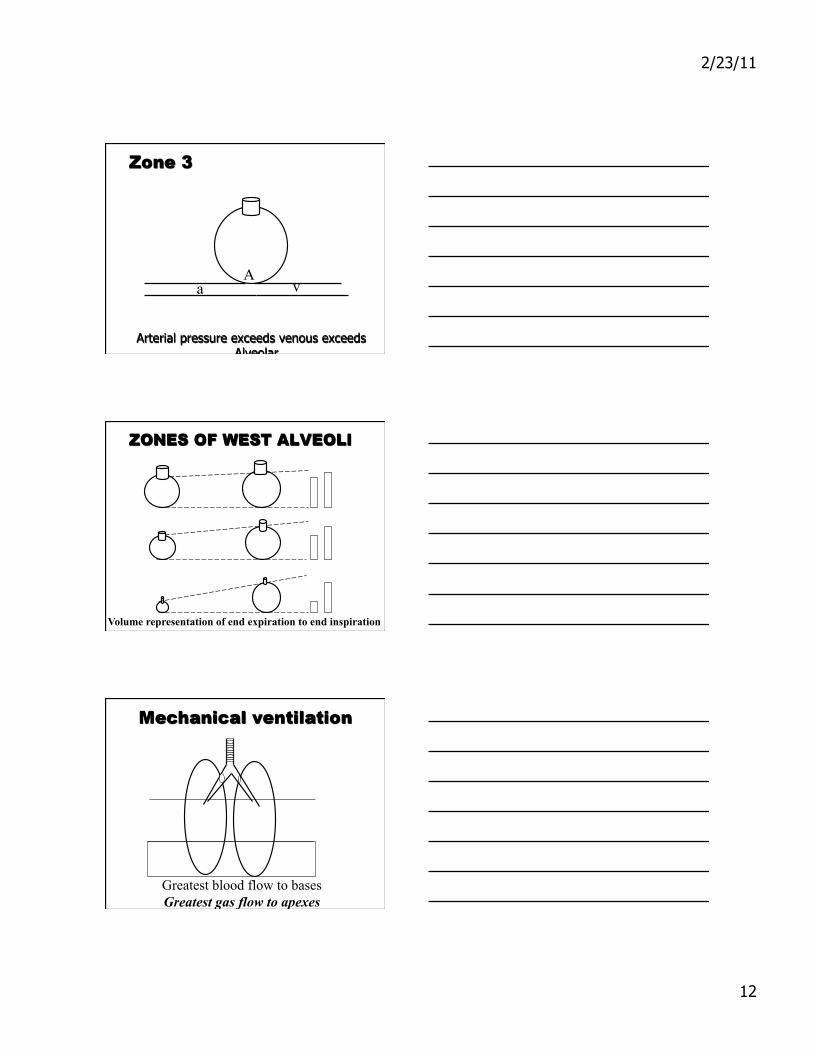
Zones of West

2/23/11
11
Zones of West
1
2
3
PA>Pa>Pv
Pa>PA>Pv
Pa>Pv>PA
Zone 1
Alveolar pressure exceeds arterial exceeds venous
A v a
Zone 2
Arterial pressure exceeds Alveolar exceeds venous
A v a
2/23/11
12
Zone 3
Arterial pressure exceeds venous exceeds Alveolar
A v a
ZONES OF WEST ALVEOLI
Volume representation of end expiration to end inspiration
Mechanical ventilation
Greatest blood flow to bases Greatest gas flow to apexes

2/23/11
13
Mechanical ventilation
Greatest gas flow to apices of lung
MECHANICAL VENTILATION
Ventilation(V) to Perfusion(Q) poorly matched in mechanically ventilated patients
Positive pressure ventilation pushes gas into apexes of lung. Path of least resistance. Blood perfuses primarily the dependent parts of lung again due in part to the pull of gravity
Hypoxic Pulmonary Vasoconstriction (HPV)
HPV effectively redirects blood flow away from hypoxic or poorly ventilated lung units
Pulmonary vascular endothelium release potent vasoconstrictor peptides called endothelins
Volatile anesthetics above 1 MAC and nitrous oxide block HPV

2/23/11
14
MECHANICAL VENTILATION
Gas flow to apex and blood flow to bases = V/Q mismatching
Poorly ventilated alveoli are prone to atelectasis and collapse
Intravasculor volume, Increased pressures, Pleural Effusions, Mucous plugging all causes.
ATELECTASIS Atelectasis is essentially collapse of pulmonary
tissue that prevents O2 & CO2 exchange. Primary causes: obstruction of airway and lack of
surfactant Absorption atelectasis is caused by occlusion of an
airway with resultant absorption of trapped gas and collapse of alveoli. higher [O2] worsens due to removal of N as an inert stabilizer
Hypoventilation during positive pressure ventilation is often primary cause of absorption atelectasis
FACTORS THAT AFFECT ONE LUNG(OLV) AND THORACIC ANESTHESIA
General anesthetics above 1 MAC block HPV Mechanical ventilation alters gas flow
dynamics Paralysis increases resistance to gas flow Absorption atelectasis frequently seen to
varying degrees

2/23/11
15
Worsening V/Q mismatch
THE V/Q MISMATCH IS A COMBINATION OF SO MANY PHYSIOLOGIC VARIABLES!
spontaneous spontaneous positive pressure positive pressure ventilation ventilation ventilation ventilation
anesthetized anesthetized anesthetizedparalyzed
V/Q 0.8 V/Q 0.7 V/Q 0.5 V/Q 0.4
Open Chest Ventilation Dynamics
Paradoxical ventilation Closed (simple) pneumothorax Communicating pneumothorax Tension pneumothorax Hemothorax
CLOSED(SIMPLE) PNEUMOTHORAX
No atmospheric communication Treatment based on size and severity-catheter aspiration,
thoracostomy, observation

2/23/11
16
COMMUNICATING PNEUMOTHORAX
Affected lung collapses on inspiration and slightly expands on expiration
Treatment: O2,thoracostomy tube, intubation, mech. vent.
“sucking chest wound”
TENSION PNEUMOTHORAX
Air progressively accumulates under pressure within pleural cavity. Compressing other lung, great vessels
Treatment; Immediate needle decompression
HEMOTHORAX
Accumulation of blood in pleural space Treatment; Airway management, support hemodynamics,
evacuation

2/23/11
17
Lung Isolation Tubes/ Techniques Single-Lumen Endobronchial Tubes Endobronchial Blockers Double-Lumen Endobronchial Tubes
Indications for Lung Isolation
Control of Foreign material Lung Abcess, Bronchiectasis, Hemoptysis
Airway Control Bronchopleural-cutaneous (B-p) fistula
Surgical exposure Lung resection Esophageal surgery or Vascular (aortic)
surgery Video Assisted Thoracic Surgery (VATS)
Special procedures Lung lavage, Differential ventilation
Single-Lumen Endobronchial Tubes
Utilized for several decades Replaced by double-lumen tubes today Two versions
MacIntosh-Leatherdale left tube Gordon-Green right tube
Disadvantages Inability to clear material from operative lung Potential for limited ventilation - nonintubated
surgical lung

2/23/11
18
Endobronchial Blockers Types of Bronchial blockers
McGill catheter Fogerty catheter Foley catheter Univent tube COOK BRONCHIAL BLOCKER
UNIVENT TUBE
UNIVENT TUBE
POSITIONING UNIVENT TUBE

2/23/11
19
COOK BRONCHIAL BLOCKER
UNIVENT TUBE + CPAP
DOUBLE LUMEN TUBES
Note difference in Left and Right tubes accounting for bronchial anatomical difference

2/23/11
20
PLACEMENT DLT
Start at 3 o’clock thru cords advance as you turn to 12 o’clock position
FOB Visual Confirmation
2/23/11
21
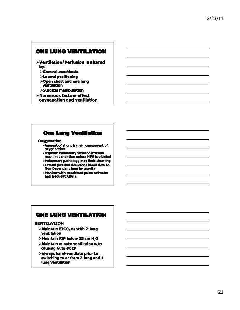
ONE LUNG VENTILATION
Ventilation/Perfusion is altered by: General anesthesia Lateral positioning Open chest and one lung
ventilation Surgical manipulation
Numerous factors affect oxygenation and ventilation
One Lung Ventilation
Oxygenation Amount of shunt is main component of
oxygenation Hypoxic Pulmonary Vasoconstriction
may limit shunting unless HPV is blunted Pulmonary pathology may limit shunting Lateral position decreases blood flow to
Non Dependent lung by gravity Monitor with consistant pulse oximeter
and frequent ABG’s
ONE LUNG VENTILATION
VENTILATION Maintain ETCO2 as with 2-lung
ventilation Maintain PIP below 35 cm H2O Maintain minute ventilation w/o
causing Auto-PEEP Always hand-ventilate prior to
switching to or from 2-lung and 1-lung ventilation

2/23/11
22
ONE LUNG VENTILATION Use large TV (10-12 ml/kg) Ventilation rate adjusted to avoid
hyperventilation Compliance is reduced and resistance is
increased (one lumen instead of two)
PIPs will be higher Some auto PEEP may be generated,
depend on size of DLT If pulse oximetry is <94% or PO2 <100,
recheck DLT or BB
O2 MANAGEMENT DURING ONE LUNG VENTILATION
Decrease shunt & minimize VL atelectasis D/C or avoid N2O prn to maintain PaO2 Check tube position and suction as needed PEEP to vented lung (may shunt blood to NVL) Apneic oxygenation to NVL q 10-20 minutes CPAP to non-ventilated lung (5-8 cmH2O) Reinflate NVL w/ 100% FiO2 prn, 2-lung vent Have surgeon clamp NVL PA or go to Bypass
EMERGENCE Prior to closing chest - Inflate lungs to 30 cm H2O to
reinflate atelectactic areas and to check for leaks Surgeon inserts chest tube to drain pleural cavity
and aid lung reexpansion Patient is extubated in OR, or exchange DL-ETT for
SL-ETT (HV-LP) if patient is to remain intubated Chest tubes to water seal and 20 cm H2O suction,
except in pneumonectomy => water seal only Patient transferred in head elevated position to ICU
on monitors and nonrebreathing mask O2

2/23/11
23
LUNG ISOLATION COMPLICATIONS
• Trauma – Dental and soft tissue injury – Large tube diameter causes laryngeal injury – TracheoBronchial wall ischemia/stenosis
• Malposition – Advancement of tube too far or too proximal
• Hypoxemia
• Aspiration
KEY CONCEPTS Spontaneous ventilation is sub-atmospheric pressure
process. Gas is “sucked” in Mechanical Ventilation is positive pressure, above
atmospheric pressure. Gas is “pushed” in Blood flow is primarily gravity dependant Negative pleural pressures coupled with the pale handle
effect pulls more gas to the dependant areas of lungs with spontaneous ventilation
Opening thorax alters negative intra-thoracic pressures altering lung dynamics ⇒ know details
Single lung ventilation gives 100% gas to one lung, Blood flow is split between both lungs= V/Q mismatch!
Questions