three arginine residues in apolipoprotein a-i are critical ... · critical for activation of...
TRANSCRIPT
Journal of Lipid Research
Volume 42, 2001
31
Three arginine residues in apolipoprotein A-I are critical for activation of lecithin:cholesterol acyltransferase
Stein Roosbeek,* Berlinda Vanloo,* Nicolas Duverger,
§
Hans Caster,* Joke Breyne,* Iris De Beun,* Hetal Patel,** Joël Vandekerckhove,
†
Carol Shoulders,** Maryvonne Rosseneu,
1,
* and Frank Peelman*
Laboratory for Lipoprotein Chemistry,* Department of Biochemistry, Ghent University, B-9000 Ghent, Belgium; Department of Medical Protein Research,
†
Flanders Interuniversity Institute for Biotechnology, B-9000 Ghent, Belgium; Aventis,
§
Vitry-sur-Seine 94403, France; MRC Molecular Medicine Group,**Clinical Sciences Centre, Hammersmith Hospital, London W12 ONN, UK
Abstract Previous studies have suggested that the helicalrepeat formed by residues 143–164 of apolipoprotein A-I(apoA-I) contributes to lecithin:cholesterol acyltransferase(LCAT) activation. To identify specific polar residues in-volved in this process, we examined residue conservationand topology of apoA-I from all known species. We ob-served that the hydrophobic/hydrophilic interface of helix143–164 contains a cluster of three strictly conserved argi-nine residues (R149, R153, and R160), and that these resi-dues create the only significant positive electrostatic poten-tial around apoA-I. To test the importance of R149, R153,and R160 in LCAT activation, we generated a series of mu-tant proteins. These had fluorescence emission, secondarystructure, and lipid-binding properties comparable to thoseof wild-type apoA-I. Mutation of conserved residues R149,R153, and R160 drastically decreased LCAT activity on lipid-protein complexes, whereas control mutations (E146Q,D150N, D157N, R171Q, and A175R) did not decrease LCATactivity by more than 55%. The markedly decreased activi-ties of mutants R149, R153, and R160 resulted from a de-crease in the maximal reaction velocity
V
max
because the ap-parent Michaelis-Menten constant
K
m
values were similarfor the mutant and wild-type apoA-I proteins. These datasuggest that R149, R153, and R160 participate in apoA-I-mediated activation of LCAT, and support the “belt” modelfor discoidal rHDL. In this model, residues R149, R153, andR160 do not form salt bridges with the antiparallel apoA-Imonomer, but instead are pointing toward the surface of thedisc, enabling interactions with LCAT.
—
Roosbeek, S., B.Vanloo, N. Duverger, H. Caster, J. Breyne, I. De Beun, H.Patel, J. Vandekerckhove, C. Shoulders, M. Rosseneu, and F.Peelman.
Three arginine residues in apolipoportein A-I arecritical for activation of lecithin:cholesterol acyltransferase
J. Lipid Res.
2001.
42:
31–40.
Supplementary key words
HDL
•
enzyme
•
lipoprotein • cholesterol
The role of apolipoprotein A-I (apoA-I) in the metabo-lism and maturation of different subclasses of high densitylipoproteins (HDL) has been well documented (1). ApoA-Iis synthesized as a prepropeptide, and is cleaved in plasma
to a 243-amino acid residues protein. ApoA-I associateswith phospholipids and cholesterol, and acts as acceptorof cellular lipids to promote “reverse cholesterol trans-port” (2). In this process, lipid-free apoA-I “solubilizes”cellular lipids through a direct interaction with the donorplasma membrane, or specific receptors, such as the scav-enger receptor class B type I (SR-BI) receptor (3) and theadenosine triphosphate (ATP)-binding cassette transporterABC-A1 (4–6). ApoA-I also mediates the maturation ofHDL through the activation of lecithin:cholesterol acyl-transferase (LCAT). LCAT converts cholesterol to choles-teryl esters (7), through the hydrolysis of the
sn
-2 fattyacid of lecithin and esterification of the hydroxyl group ofcholesterol. The cholesteryl esters are incorporated intothe core of HDL, and exchanged with either low densitylipoprotein (LDL) or very low density lipoprotein lipids,or taken up by the liver through the action of the SR-BI re-ceptor (3).
ApoA-I has been sequenced both at the protein andcDNA level (8, 9), and the occurrence of 11- and 22-resi-due helical repeats has been predicted by internal homol-ogy calculations (10). These bind lipids and have a uniqueamphipathic character, with separate hydrophilic and hy-drophobic faces (11). A crystal structure for lipid-freeapoA-I, apo
D
(1–43)A-I, has been determined at 4-Å reso-lution (12). ApoA-I forms a nearly continuous nonplanaramphipathic helix, with dimensions of 125
3
80
3
40 Å,in the shape of a horseshoe. The structure comprises 10
Abbreviations: apo, apolipoprotein; apo
D
(1–43)A-I, lipid-free apoA-I;DMPC, 1,2-dimyristoyl-
sn
-glycero-3-phosphatidylcholine; HDL, high den-sity lipoprotein; HPLC, high performance liquid chromatography; LCAT,lecithin:cholesterol acyltransferase; LDL, low density lipoprotein; PCR,polymerase chain reaction; PLPC, 1-palmitoyl-2-linoleoyl-
sn
-glycero-3-phosphatidylcholine; rHDL, reconstituted high density lipoprotein;SDS-PAGE, sodium dodecyl sulfate-polyacrylamide gel electrophoresis;SR-BI, scavenger receptor class B type I.
1
To whom correspondence should be addressed.
by guest, on August 28, 2018
ww
w.jlr.org
Dow
nloaded from
32 Journal of Lipid Research
Volume 42, 2001
helical segments, named A1–A10, which are punctuatedby small or pronounced kinks at regularly spaced prolineresidues. In the unit cell of the crystal, four apoA-I mono-mers form two antiparallel dimers that are associated viatheir hydrophobic faces. Such a structure is predicted tobe conserved in lipid-associated apoA-I, as the dimensionsof the dimer are compatible with those of discoidal andspherical HDL (12).
Two general models have been proposed for discoidalnascent HDL, the preferred substrate for LCAT. The “belt”model, proposed by Segrest et al. (13), consists of anapoA-I dimer made of two antiparallel monomers. Thesemonomers surround the edge of the disc and are orientedparallel to the face of the discoidal particle. This is remi-niscent of the “horseshoe” structure of crystalline apoA-I.In the alternative “picket-fence” model proposed by Bras-seur et al. (14) and by Phillips et al. (15), the helical re-peats are oriented perpendicular to the faces of the disc,and are parallel to the phospholipid acyl chains. Thismodel is supported by attenuated total reflection infraredmeasurements, performed on a Ge plate (14, 16), whereasthe belt model is favored by infrared measurements at anair-water interface (17).
Previous studies have suggested that helical repeats 6and 7 of apoA-I are critical for the activation of LCAT byapoA-I, but the specific residues that mediate this activa-tion have not been identified. Thus, it is well establishedthat monoclonal antibodies directed against residues143–164 of apoA-I suppress the activation of LCAT byapoA-I, while synthetic peptides that encompass helical re-peats 6 and 7 activate LCAT. Conversely, the activation ofLCAT by apoA-I is impaired by naturally occurring or ge-netically engineered apoA-I proteins that lack helical re-peat 6 and 7. Sorci-Thomas et al. (18) have further dem-onstrated that rotation of the hydrophobic face of helix143–164 of apoA-I by about 80
8
markedly reduces LCATactivation. Finally, this region of apoA-I harbors three nat-urally occurring missense mutations, R151C, R160L, andH162Q, associated with decreased LCAT activation.
In the present study, we have used the crystal structure ofapoA-I, comparative sequence analysis, and site-directedmutagenesis to address the issue of LCAT activation byapoA-I. We demonstrate that helical repeat 6 contains a clus-ter of three strictly conserved arginine residues (Arg-149,Arg-153, and Arg-160), and that these are critical for LCATactivation. Importantly, the data provide experimental sup-port for the belt model conformation of discoidal HDL.
MATERIALS AND METHODS
Sequence analysis and computer modeling
The electrostatic potential calculations were per formed onmonomer A in the crystalline structure of apo(
D
1–43)A-I (12),using the Delphi program (19), as implemented in the MSIInsight97 software package (Molecular Simulations, San Diego,CA). Calculations were performed with actual atomic charges, asdefined by the CHARMM atomic force field (20), a dielectricconstant of 8 for the solute, and 80 for the solvent. A focusingapproach was used to reduce grid boundary effects, where the
monomer filled 23% of a 63
3
63
3
63 points grid, during thefirst coarse calculation, and 80% of the 63
3
63
3
63 points gridin the focusing calculation.
Sequences of human (P02647), cynomolgus monkey (P15568),pig (P18648), cow (P15497), dog (P02648), rabbit (P09809), treeshrew (O18759), mouse (Q00623), rat (P04639), golden ham-ster (Q9Z2L4), hedgehog (Q9TS49), chicken (P08250), quail(P32918), and duck (O42296) apoA-I were aligned by CLUSTALW (21), available at http://www.ebi.ac.uk/clustalw/.
For sequence comparison between the helices of humanapoA-I, apoA-IV, apoA-II, apoE, apoC-I, apoC-II, and apoC-III,these sequences were divided in overlapping stretches of 36 resi-dues, which were displayed in a helical wheel projection.
Site-directed mutagenesis
Polymerase chain reaction (PCR) and the appropriate restric-tion sites were used to manipulate the apoA-I sequence. Mutagen-esis was performed by a two-step PCR-based strategy using
Pfu
DNA polymerase in accordance with the manufacturer’s instruc-tions (Stratagene, UK). The template was the plasmid pALTER/apoA-I, which encompasses nucleotides 1887 –2292 of the apoA-Isequence (GenBank accession number J0098). To facilitate clon-ing, the
Sst
I site at nucleotides 1923–1928 of apoA-I was mu-tated; this did not alter the encoded sequence. The first-roundPCRs were performed with a forward mutagenic oligonucleotide
.
35 bases in length. The sequence of the reverse oligonucle-otide was 5
9
-GACTATGGATCCTCACTGGGTGTTGAGCTTCTT-3
9
. The sequence of the forward primer for the second-roundPCR was 5
9
-AGATGGAGCTCTACCGCCAGAAGGTGGAGCCGCTGCGCGCAGAACTGCAAGAGGGCGCGCGCCAG-3
9
. The re-verse primer was the product of the first-round PCR. The mutantapoA-I cDNAs were introduced into the PXL2769.apoA-I vector(22). The sequences of all constructs were confirmed.
Expression and purification of the recombinant proteins
The expression system under the control of the T7 promotorhad been described previously for apoA-IV (22). The expressionvector pXL2769 for wild-type apoA-I, and the corresponding vec-tors for the apoA-I mutants, were introduced into
Escherichia coli
BL21(DE3) pLysS cells, for expression of histidine-tagged apoA-Imutants. Bacteria were grown overnight at 37
8
C in Terrific Brothmedium, supplemented with ampicillin (100
m
g/ml) andchloramphenicol (50
m
g/ml). Five milliliters of this culture wasused to inoculate 1 liter of the same medium. Cultures weregrown at 37
8
C until an absorbance at 610 nm of about 0.7 wasreached. Isopropyl-
b
-
d
-thiogalactoside was then added to a finalconcentration of 1 mM, followed by a 15-min incubation at 30
8
Cto induce apoA-I expression. Rifampicin was added to a concen-tration of 100
m
g/ml, and the cells were incubated for 60 min,harvested by centrifugation, and frozen at
2
20
8
C. ApoA-I ex-pression was verified by sodium dodecyl sulfate-polyacrylamidegel electrophoresis (SDS-PAGE).
For protein extraction, the pellets were resuspended by addi-tion of 5 ml of a 100 mM sodium phosphate lysis buffer, pH 7.4,containing 1 mM phenylmethylsulfonyl fluoride per gram ofbacteria. The cells were disrupted by five 5-min cycles of sonica-tion at 70 W, using a Vibracell
TM
sonifier (Sonics, Newtown, CT).Whole cells and cellular debris were removed by centrifugationat 2,000
g
for 15 min at 4
8
C, in an Eppendorf centrifuge. Nucleicacids were precipitated by addition of 10 ml of a 100 g/l solutionof streptomycin sulfate, per gram of bacterial protein in the su-pernatant fraction. The mixture was incubated overnight at 4
8
C,and centrifuged at 6,000
g
for 15 min at 4
8
C in an Eppendorfcentrifuge, to remove the precipitated material.
The apoA-I proteins were purified in the supernatant by affinitychromatography on a Ni-Probond column (InVitrogen, San Diego,
by guest, on August 28, 2018
ww
w.jlr.org
Dow
nloaded from
Roosbeek et al.
Arginine residues in apoA-I critical for LCAT activation 33
CA), equilibrated in a 100 mM phosphate buffer, pH 8.0, containing6 M guanidium hydrochloride. The wild-type and mutant apoA-Iproteins were eluted from the column by a stepwise decrease in theelution buffer pH, from 8.0 down to 6.0, 5.0, and 4.0. The elutedfractions were tested for the presence of apoA-I protein by SDS-PAGE. The fractions eluting at pH 4.0, containing pure apoA-I,were dialyzed against a 5 mM NH
4
HCO
3
buffer and lyophilized.
Preparation and isolation of theapolipoprotein-lipid complexes
Reconstituted HDL (rHDL) consisting of 1-palmitoyl-2-linoleoyl-phosphatidylcholine/cholesterol/apoA-I at a ratio of 2:0.1:1 (w/w/w) were prepared by the cholate dialysis method (23). Thehomogeneity and yield of the reconstituted HDL were tested byseparation on a Superose 6PG column equilibrated in a 10 mMTris-HCl buffer, pH 8.0, containing 150 mM NaCl in a fast pro-tein liquid chromatography system. Composition and size deter-mination were performed on the top fractions of the elutionpeak of the complex. The lipid and apolipoprotein composi-tions of the complexes were obtained by an enzymatic colorimet-ric assay for phospholipid and cholesterol (bioMérieux, Charou-nieres-les-Bains, France; Boehringer, Mannheim, Germany) andby protein quantification of apoA-I by phenylalanine assay byhigh performance liquid chromatography (HPLC) (24).
Physicochemical characterization of the reconstituted HDL
The intrinsic fluorescence of the tryptophan residues ofapoA-I wild-type and mutants, either lipid free or in rHDL com-plexes, was measured with an AMINCO-Bowman series 2 spec-trofluorimeter (Spectronic Unicam, Rochester, NY) at an excita-tion wavelength of 280 nm, with 4-nm excitation and emissionslit widths. Spectra were recorded between 310 and 450 nm. Themaximal wavelengths of tryptophan fluorescence emission werecalculated from the corrected spectra.
Circular dichroism spectra of the apoA-I mutants and theirrHDL complexes were measured on a JASCO (Tokyo, Japan)710 spectropolarimeter at wavelengths between 186 and 250nm. Measurements were carried out at a protein concentrationof 50
m
g/ml in a 0.01 M sodium phosphate buffer, pH 7.4. Ninespectra were collected and averaged for each sample. The sec-ondary structure was estimated by curve fitting with the CircularDichroism Deconvolution by back propagation Neural Net-works program (25).
Production of wild-type rLCAT in BHK cells
Recombinant LCAT was produced by a baby hamster kidney(BHK) cell line, stably transfected with the LCAT cDNA, undercontrol of the mouse metallothionein promotor (26). Stablytransfected BHK cells were incubated with Dulbecco’s modifiedEagle medium (DMEM; GIBCO-BRL, Gaithersburg, MD) con-taining 10% fetal bovine serum, streptomycin (100
m
g/ml), andpenicillin (100 units/ml). After 48 h, the confluent cells werewashed with Opti-MEM and the DMEM was replaced by serum-free Opti-MEM (GIBCO-BRL). After 48 h of incubation the me-dium was collected, filtered through a 0.22-
m
m pore size filter(Millipore, Bedford, MA), and dialyzed against 20 mM Tris-HClbuffer, pH 8.0. The rLCAT was purified on a Resource
TM
Q HRcolumn (Pharmacia, Uppsala, Sweden) with an NaCl gradientbetween 0 and 500 mM. rLCAT was collected in 1-ml fractionsand stored at
2
80
8
C, at a concentration of about 150
m
g/ml.
LCAT activation properties of the rHDL complexes
The activity of the LCAT enzyme with the various rHDL com-plexes as substrates was determined by measuring the amount ofcholesteryl esters, generated during the enzymatic reaction, byHPLC (27).
The initial velocities were determined in the linear portion ofthe curves, that is, between 0 and 25% cholesteryl ester forma-tion. The assay mixture consisted of variable amounts of rHDLcomplexes at cholesterol concentrations varying between 2 and20
m
M, defatted bovine serum albumin (4 mg/ml; Sigma, St.Louis, MO), and 6 mM 2-mercaptoethanol. Tris-HCl buffer (0.01M, pH 8.0), 0.15 M NaCl, 0.3 mM Na
1
-ethylenediaminetetraace-tic acid, and 1 mM NaN
3
were added to a final reaction volumeof 0.5–1.5 ml. Complexes were preincubated for 20 min at 37
8
C,before addition of 50–150
m
l of BHK rLCAT, to initiate the enzy-matic reaction. The reaction was performed at 37
8
C, and it wasstopped by extraction of the incubation mixture with hexane –isopropanol 3:2 (v/v), containing cholesteryl heptadecanoate(Sigma) as an internal standard, for cholesteryl ester quantification.
HPLC separation and quantification of the cholesteryl esterswere carried out as described previously (27). The apparent ki-netic parameters
V
max
and
K
m
, together with the estimated erroron the parameters, were determined for each apoA-I/rHDLcomplex. These values were calculated by fitting the initial veloc-ity
V
o
, as a function of the cholesterol concentration, to theMichaelis-Menten equation, using the SigmaPlot software (SPSS,Chicago, IL).
RESULTS
Selection of the mutants
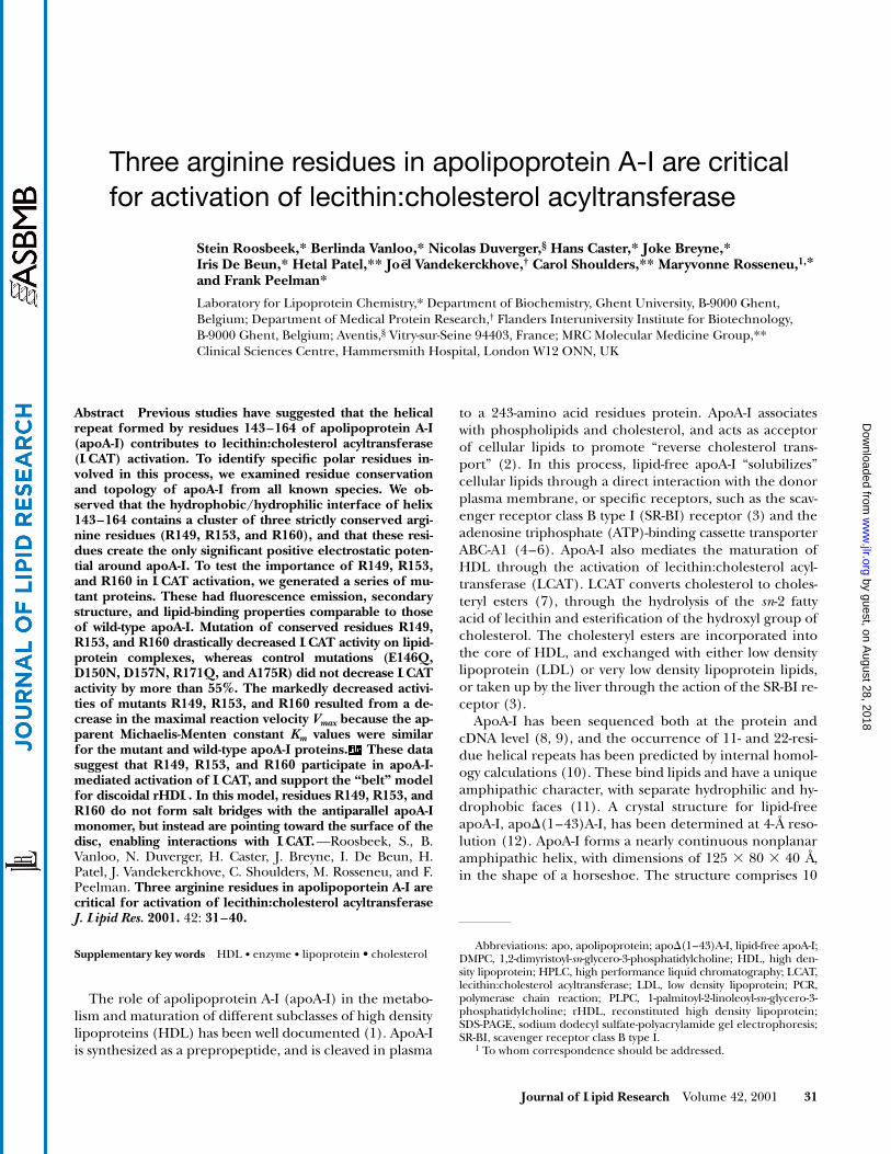
The electrostatic potential around an apoA-I monomeris predominantly negative (
Fig. 1
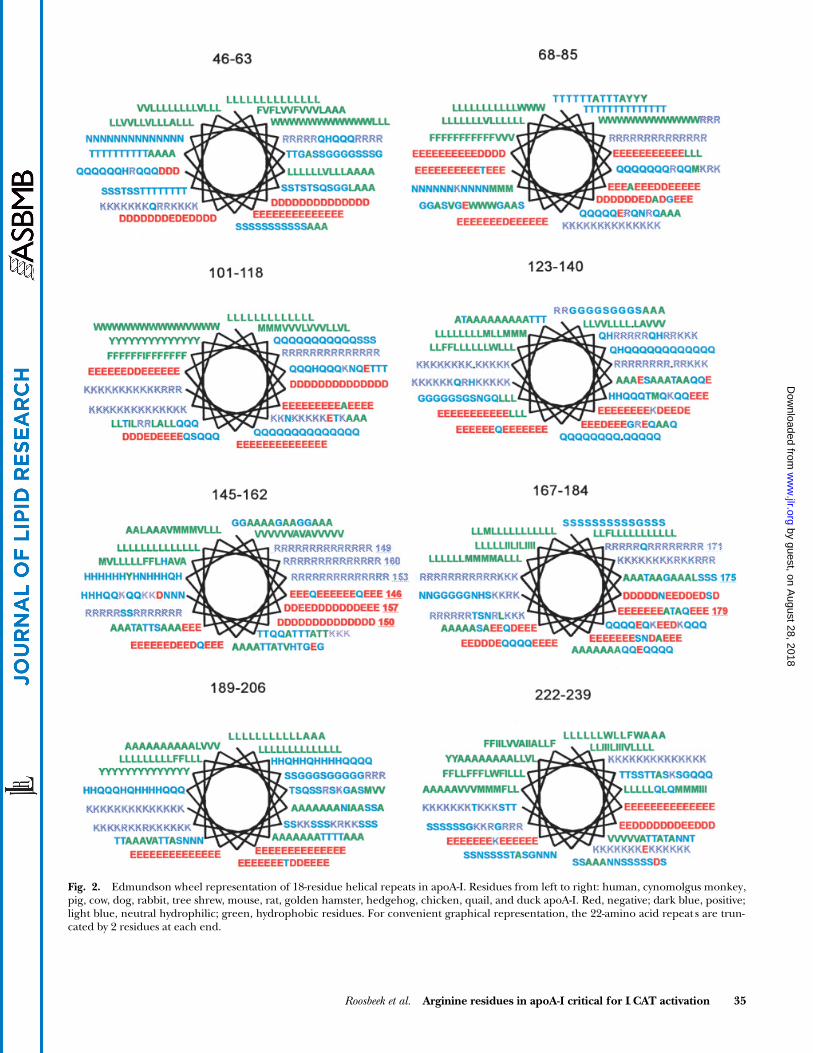
). The exception is a pos-itive electrostatic potential around the arginine clusterR149, R153, and R160 of helix 6, and around residuesR171 and K182 of helix 7. We examined the conservationand location of these arginine residues in all known apoA-Isequences of different species. The Edmundson wheelrepresentation of eight helical repeats is shown in
Fig. 2
,for human, cynomolgus monkey, pig, cow, dog, rabbit,tree shrew, mouse, rat, golden hamster, hedgehog,chicken, quail, and duck apoA-I (28). There is high se-quence conservation among apoA-I of the different spe-cies, and the amphipathic character of all helices is espe-cially well conserved. A specific feature of the repeat atresidues 145–162 is clearly demonstrated in Fig. 2, as thehydrophilic face of this amphipathic helix is characterizedby a cluster of three arginine residues at positions 149,153, and 160. These residues, which account for the posi-tive electrostatic potential described above, are totallyconserved among the different species. This cluster ofthree adjacent arginine residues on the Edmundsonwheel representation is unique to helix 145–162, as re-peats 101–118, 123–140, and 189–206 have only two adja-cent lysines, which are not perfectly conserved among thedifferent species (Fig. 2). A close inspection of the repeatsof apoA-IV, apoA-II, apoE, apoC-I, apoC-II, and apoC-IIIdid not reveal any similar cluster of three arginine resi-dues either (data not shown). In the crystal structure ofan apoA-I dimer, consisting of two antiparallel monomers,helix 143–164 interacts with repeat 99–120 of the othermonomer (
Fig. 3
). R149, R153, and R160 are not part ofthe dimer interface and cannot interact with the other re-peat. The same holds true for the belt model of an rHDLparticle proposed by Segrest et al. (13).
by guest, on August 28, 2018
ww
w.jlr.org
Dow
nloaded from
34 Journal of Lipid Research
Volume 42, 2001
To test the contribution of residues R149, R153, and R160,and of the electrostatic potential around the 22-residue re-peats 143–164 and 165–186 of apoA-I to LCAT activation, weconstructed the mutants listed in
Table 1
. R149, R153, andR160 were mutated to glutamine, either as single, double, ortriple mutants. In addition, acidic residues E146, D150, andD157, which might form ionic interactions with R149, R153,and R160 to stabilize the helical structure, were mutated to aglutamine or an asparagine (Table 1). The requirement foran arginine residue at position 153 was tested by the R153Ksubstitution. An R171Q mutation was performed to decreasethe positive electrostatic potential around repeat 167–184.In addition, A175 was mutated to an arginine, in order to in-crease the positive potential around this helix.
Expression and characterization of the apoA-I mutants
Wild-type and mutant apoA-I proteins were expressedin
E. coli
, and comparable yields were obtained (data notshown). The affinity-purified proteins migrated as a singleband on SDS-PAGE, with a molecular weight correspond-ing to that of apoA-I, carrying the N-terminal histidinetag. The secondary structure of the mutant apoA-I pro-teins was checked by circular dichroism (Table 1), and thepercentages of
a
helix,
b
sheet,
b
turns, and coil werefound to be similar to wild-type apoA-I. The secondarystructure is characterized by a high percentage of helicalstructure and low
b
-sheet content, in agreement with thecrystallized structure of apo
D
(1–43)A-I.
Lipid-binding properties of wild-typeand mutant apoA-I
The association of plasma, wild-type, and mutant apoA-Iwith 1,2-dimyristoyl-
sn
-glycero-3-phosphatidylcholine(DMPC) vesicles was followed by turbidity measure-ments at 325 nm, as a function of temperature. As shownin
Fig. 4
, wild-type recombinant apoA-I, the single mutantR153Q, the double mutant R153Q
1
R160Q, the triplemutant R149Q
1
R153Q
1
R160Q, and the D157N mu-tant, all reacted similarly with DMPC. Around the transi-tion temperature of DMPC, the large multilamellar vesi-cles were converted into smaller lipid-protein complexes.We also prepared rHDL particles consisting of 1-palmitoyl-2-linoleoyl-
sn
-glycero-3-phosphatidylcholine (PLPC), cho-lesterol, and recombinant apoA-I and examined their sizeand homogeneity (
Fig. 5
). The yield of rHDL formedfrom the mutant proteins was comparable to that of wild-type apoA-I, and each became fully associated with lipids.No peak of lipid-free apoA-I was detected by sensitive tryp-tophan fluorescence detection, at an elution volumelarger than that of the lipid-apolipoprotein complex. TherHDL particles were about 8 nm in diameter and con-tained 40–60 mol of phospholipid, 3–5 mol of cholesterolper mole of apoA-I, and two molecules of apoA-I. The sizeof the complexes was further estimated by native gradientgel electrophoresis, in a 4–20% polyacrylamide gradient.The sizes of complexes from the major protein fraction inthe gel were comparable for all apoA-I proteins (
Table 2
).
Fig. 1. Electrical potentials around a molecule of apoA-I(D1–43), calculated by the Delphi program on thebasis of the coordinates of Borhani and co-workers (12). Red, negative potential at 21 kt/e; blue, positivepotential at 10.5 kt/e.
by guest, on August 28, 2018
ww
w.jlr.org
Dow
nloaded from
Roosbeek et al.
Arginine residues in apoA-I critical for LCAT activation 35
Fig. 2. Edmundson wheel representation of 18-residue helical repeats in apoA-I. Residues from left to right: human, cynomolgus monkey,pig, cow, dog, rabbit, tree shrew, mouse, rat, golden hamster, hedgehog, chicken, quail, and duck apoA-I. Red, negative; dark blue, positive;light blue, neutral hydrophilic; green, hydrophobic residues. For convenient graphical representation, the 22-amino acid repeat s are trun-cated by 2 residues at each end.
by guest, on August 28, 2018
ww
w.jlr.org
Dow
nloaded from

36 Journal of Lipid Research
Volume 42, 2001
They were relatively small, as they were prepared at a 2:1(w/w) ratio of phospholipid to protein, to increase thehomogeneity of the preparation. Incubation of lipids withwild-type and mutant apoA-I at a 2.5:1 (w/w) ratio yieldeda more heterogeneous population of particles (data notshown).
To characterize further the lipid-binding properties ofthe mutant apoA-Is, we measured the maximal wavelengthfor the tryptophan fluorescence emission of lipid-freeapoA-I and rHDL. Consistent with the insertion of thetryptophan residues in apoA-I into a more hydrophobicenvironment, the tryptophan fluorescence of each apoA-Icomplex was characterized by a blue shift of 2–3 nm, com-
pared with the lipid-free protein (Table 2). The secondarystructure of lipid-bound apoA-I, determined by circulardichroism (Table 1), shows an increased helical content ofabout 15% for all complexes. This is due to the helix stabi-lization of the helical repeats by the lipids, accompaniedby a decrease of the percentage of coil structure, while
b
-sheet and
b
-turn content remain unchanged (Table 1).
LCAT activation properties of wild-typeand mutant apoA-I
Initial velocity measurements showed a linear increaseof up to 20–25% cholesterol esterification in the substrate.All subsequent measurements were carried out within that
Fig. 3. Representation of helix 143–164 of apoA-I in a dimer of apo(D1–43)A-I, together with the comple-mentary helix 99–120 in the antiparallel dimer. Representation: (A) parallel and (B) perpendicular to thelong axis of the helix.
by guest, on August 28, 2018
ww
w.jlr.org
Dow
nloaded from

Roosbeek et al.
Arginine residues in apoA-I critical for LCAT activation 37
range.
V
max
and
K
m
were determined for wild-type and mu-tant apoA-I, from
V
o
measurements at varying cholesterolconcentrations. The parameters were calculated by fittingthe experimental data to a Michaelis-Menten equation,using SigmaPlot software (SPSS).
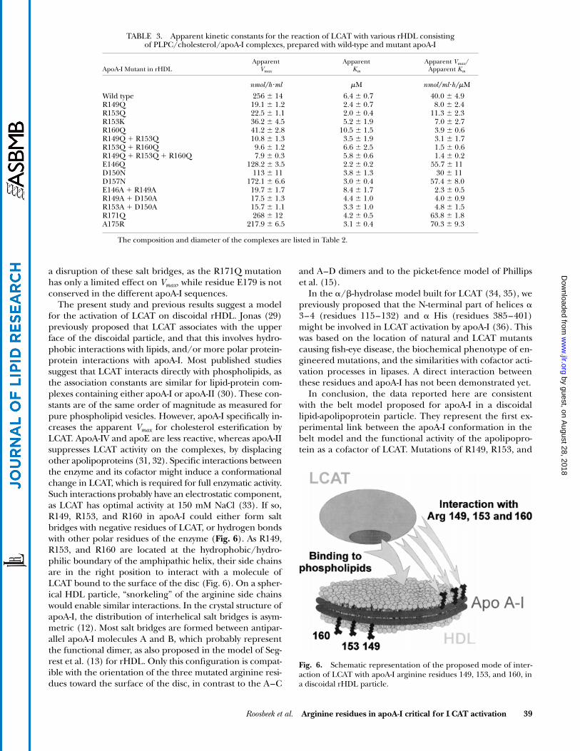
Table 3
summarizes thevalues obtained for
Vmax and Km and the enzymatic effi-ciency Vmax/Km, for wild-type and mutant apoA-I. Com-pared with wild-type apoA-I, we observed a decreased Vmaxfor all mutants in which one or more residues of the argi-nine cluster R149, R153, and R160 was mutated to aglutamine. This effect increased with the number of sub-stitutions in the mutant protein. Mutation of a single argi-nine, either R149, R153, or R160, resulted in a 6- to10-fold decrease in Vmax, compared with a 12- to 25-folddecrease for a double mutant, and a 30-fold decrease for a
triple mutant. Conversely, mutations of acidic residueE146, D150, or D157 had only limited effect, as Vmax valueswere about 50% of wild-type apoA-I. Mutation of both anarginine residue and an acidic residue decreased Vmax to avalue similar to that of a single arginine mutation, inagreement with the limited effect of mutating an acidicresidue of helix 143–160. Surprisingly, substitution ofR153 with a lysine residue was almost as deleterious as theR153Q substitution for LCAT activity. Mutation of R171 inthe next C-terminal helix had no effect on Vmax, while sub-stitution of A175 by an arginine did not affect this param-eter either.
The effect of the apoA-I mutations on the dissociationconstant Km was more limited, as Km values were of thesame order of magnitude, and varied at most 2- to 3-fold.Calculation of the catalytic efficiency Vmax/Km clearly showsthat mutation of one or more arginine residues of theR149, R153, R160 cluster drastically decreases LCAT enzy-matic activity on the mutant apoA-I/lipid complexes, to lessthan 20% of wild-type apoA-I. As the composition, size, andstructure of the complexes are comparable, this can be at-tributed to loss of activation of LCAT by mutation of thecofactor apoA-I.
DISCUSSION
We have used the crystal structure of apoD(1–43)A-I,sequence conservation calculations, and sequence align-ment programs to detect a unique feature in the hydro-philic face of helix 143–164. This helix had been previ-ously implicated in the activation of LCAT by apoA-I. Itcontains a cluster of three conserved arginine residues, atmatching positions on three turns of this helix. We there-fore generated mutations of polar residues in this helix,and measured the biochemical, biophysical, and LCAT ac-tivation properties of the mutants. We showed that thesecondary structure and lipid-binding properties of the
TABLE 1. Percentages of secondary structure of apoA-I mutants, lipid-free and inPLPC/cholesterol/apoA-I complexes, determined by circular dichroism
Lipid-Free ApoA-I PLPC/Cholesterol/ApoA-I Complexes
ApoA-I Mutant a Helix b Sheet b Turn Coil a Helix b Sheet b Turn Coil
% %
Wild type 58 7 14 21 76 3 10 11R149Q 65 5 13 17 70 4 11 15R153Q 43 12 16 29 74 3 10 13R153K 53 8 15 24 68 4 11 17R160Q 64 5 13 18 74 3 10 13R149Q 1 R153Q 61 8 13 18 71 4 11 14R153Q 1 R160Q 61 7 14 18 68 4 12 16R149Q 1 R153Q 1 R160Q 57 7 14 22 77 3 10 10E146Q 39 13 17 31 55 7 13 25D150N 48 11 15 26 64 5 12 19D157N 52 10 15 23 61 6 13 20E146A 1 R149A 55 6 18 19 66 4 20 11R149A 1 D150A 31 18 17 34 77 3 10 10R153A 1 D150A 58 7 14 21 79 3 9 9R171Q 44 11 16 29 65 5 12 18A175R 30 18 17 35 54 7 14 25
Fig. 4. Turbidity decrease of mixtures of apoA-I mutants withDMPC as a function of temperature, monitored by absorption mea-surements at 325 nm. Plasma apoA-I (open squares); wild-typeapoA-I (solid squares); R153Q mutant (open diamonds); R153Q 1R160Q mutant (solid trianges); R149Q 1 R153Q 1 R160Q mutant(solid circles); D157N mutant (3).
by guest, on August 28, 2018
ww
w.jlr.org
Dow
nloaded from

38 Journal of Lipid Research Volume 42, 2001
mutants were comparable to those of wild-type apoA-I.Mutations of conserved residues R149, R153, and R160 inhelix 6 drastically reduced LCAT activation by the mu-tants, due to a Vmax decrease. In contrast, mutations ofacidic residues in this helical repeat, or of R171 in thenext C-terminal repeat, did not have such an effect. Takentogether, these data suggest that residues R149, R153, andR160 mediate the activation of LCAT by apoA-I.
We have compared the orientation and hydrogen bond-ing of R149, R153, and R160 in the apoD(1–43)A-I crystalstructure, in the belt model proposed by Segrest et al.(13), and in the picket-fence model of Phillips et al. (15).In the belt model, as well as in the crystal structure ofapoD(1–43)A-I, R149, R153, and R160 do not form inter-helical salt bridges with the corresponding antiparallel re-peat 99–120. However, these arginines can form intraheli-cal salt bridges with E146, D150, and D157. Mutations ofthe negatively charged residues decreased Vmax only by
50%, whereas mutation of any of the conserved arginineresidues in helix 143–164 caused a 10-fold decrease of Vmax.According to these data, putative intrahelical salt bridges inhelix 143–164 would not be critical for the structure andcofactor activity of apoA-I in rHDL. This is suggested bythe similar helical content of all mutant proteins, and inaddition by the finding that the R153K mutation, whichwould preserve the potential salt bridge formation be-tween K153 and D150, impairs LCAT activation. In thepicket-fence model proposed by Phillips et al. (15), R149,R153, and R160 do not form a cluster of residues, as in thebelt model. They are located further apart, as R149 andR160 are at about 20 Å from each other. R153 forms an in-trahelical salt bridge with D150, while R149 can form asalt bridge with E179 in the antiparallel helical repeat atresidues 165–186. Similarly, D157 and R171 can form aninterhelical salt bridge with each other. The decrease inVmax observed with the arginine mutants cannot be due to
Fig. 5. Elution profiles of PLPC/cholesterol/apoA-Imutants separated on a Superose 6 PG column,monitored by tryptophan fluorescence measure-ment. Wild-type apoA-I (solid squares); R153Q mu-tant (open diamonds); R153Q 1 R160Q mutant(solid triangles); R149Q 1 R153Q 1 R160Q mutant(solid circles); D157N mutant (3).
TABLE 2. Maximal wavelength of tryptophan fluorescence of apoA-I lipid free and in rHDL; diameter and composition of rHDL particles consisting of PLPC/cholesterol/apoA-I
lmaxDiameter
ApoA-I MutantLipidFree rHDL
Gradient Gel
ElectrophoresisGel
Filtration
PLPC/Cholesterol
Apo/A-I
nm nm mol/mol/mol
Wild type 331 329 7.8 8.0 52/4/01R149Q 331 329 7.2 7.3 41/4/01R153Q 335 332 7.6 7.8 49/4/1R153K 336 333 8.3 8.4 67/5/1R160Q 335 333 7.4 7.6 46/4/1R149Q 1 R153Q 335 333 7.6 7.8 50/4/1R153Q 1 R160Q 335 333 8.1 8.2 59/5/1R149Q 1 R153Q 1 R160Q 331 329 8.1 8.2 60/5/1E146Q 336 333 7.6 7.8 50/5/1D150N 336 334 7.9 8.0 55/4/1D157N 335 332 7.7 8.0 53/4/1E146A 1 R149A 335 333 7.3 7.4 42/4/1R149A 1 D150A 331 329 8.2 8.2 64/6/1R153A 1 D150A 331 329 8.1 8.2 63/5/1R171Q 335 332 8.3 8.4 65/3/1A175R 336 334 7.6 7.8 49/3/1
Diameter was determined by native gradient gel electrophoresis (GGE), and by gel filtration on a Superose6PG column.
by guest, on August 28, 2018
ww
w.jlr.org
Dow
nloaded from
Roosbeek et al. Arginine residues in apoA-I critical for LCAT activation 39
a disruption of these salt bridges, as the R171Q mutationhas only a limited effect on Vmax, while residue E179 is notconserved in the different apoA-I sequences.
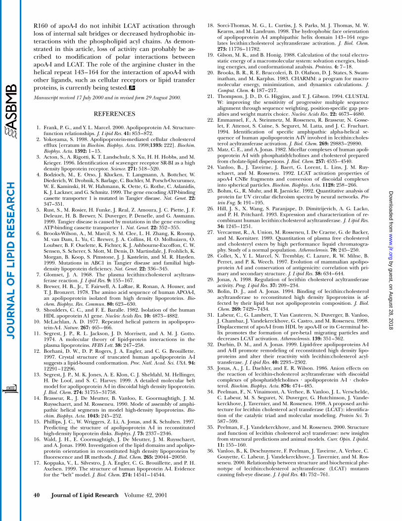
The present study and previous results suggest a modelfor the activation of LCAT on discoidal rHDL. Jonas (29)previously proposed that LCAT associates with the upperface of the discoidal particle, and that this involves hydro-phobic interactions with lipids, and/or more polar protein-protein interactions with apoA-I. Most published studiessuggest that LCAT interacts directly with phospholipids, asthe association constants are similar for lipid-protein com-plexes containing either apoA-I or apoA-II (30). These con-stants are of the same order of magnitude as measured forpure phospholipid vesicles. However, apoA-I specifically in-creases the apparent Vmax for cholesterol esterification byLCAT. ApoA-IV and apoE are less reactive, whereas apoA-IIsuppresses LCAT activity on the complexes, by displacingother apolipoproteins (31, 32). Specific interactions betweenthe enzyme and its cofactor might induce a conformationalchange in LCAT, which is required for full enzymatic activity.Such interactions probably have an electrostatic component,as LCAT has optimal activity at 150 mM NaCl (33). If so,R149, R153, and R160 in apoA-I could either form saltbridges with negative residues of LCAT, or hydrogen bondswith other polar residues of the enzyme (Fig. 6). As R149,R153, and R160 are located at the hydrophobic/hydro-philic boundary of the amphipathic helix, their side chainsare in the right position to interact with a molecule ofLCAT bound to the surface of the disc (Fig. 6). On a spher-ical HDL particle, “snorkeling” of the arginine side chainswould enable similar interactions. In the crystal structure ofapoA-I, the distribution of interhelical salt bridges is asym-metric (12). Most salt bridges are formed between antipar-allel apoA-I molecules A and B, which probably representthe functional dimer, as also proposed in the model of Seg-rest et al. (13) for rHDL. Only this configuration is compat-ible with the orientation of the three mutated arginine resi-dues toward the surface of the disc, in contrast to the A–C
and A–D dimers and to the picket-fence model of Phillipset al. (15).
In the a/b-hydrolase model built for LCAT (34, 35), wepreviously proposed that the N-terminal part of helices a3–4 (residues 115–132) and a His (residues 385–401)might be involved in LCAT activation by apoA-I (36). Thiswas based on the location of natural and LCAT mutantscausing fish-eye disease, the biochemical phenotype of en-gineered mutations, and the similarities with cofactor acti-vation processes in lipases. A direct interaction betweenthese residues and apoA-I has not been demonstrated yet.
In conclusion, the data reported here are consistentwith the belt model proposed for apoA-I in a discoidallipid-apolipoprotein particle. They represent the first ex-perimental link between the apoA-I conformation in thebelt model and the functional activity of the apolipopro-tein as a cofactor of LCAT. Mutations of R149, R153, and
TABLE 3. Apparent kinetic constants for the reaction of LCAT with various rHDL consisting
of PLPC/cholesterol/apoA-I complexes, prepared with wild-type and mutant apoA-I
ApoA-I Mutant in rHDLApparent
Vmax
ApparentKm
Apparent Vmax/Apparent Km
nmol/h?ml mM nmol/ml?h/mM
Wild type 256 6 14 6.4 6 0.7 40.0 6 4.9R149Q 19.1 6 1.2 2.4 6 0.7 8.0 6 2.4R153Q 22.5 6 1.1 2.0 6 0.4 11.3 6 2.3R153K 36.2 6 4.5 5.2 6 1.9 7.0 6 2.7R160Q 41.2 6 2.8 10.5 6 1.5 3.9 6 0.6R149Q 1 R153Q 10.8 6 1.3 3.5 6 1.9 3.1 6 1.7R153Q 1 R160Q 9.6 6 1.2 6.6 6 2.5 1.5 6 0.6R149Q 1 R153Q 1 R160Q 7.9 6 0.3 5.8 6 0.6 1.4 6 0.2E146Q 128.2 6 3.5 2.2 6 0.2 55.7 6 11D150N 113 6 11 3.8 6 1.3 30 6 11D157N 172.1 6 6.6 3.0 6 0.4 57.4 6 8.0E146A 1 R149A 19.7 6 1.7 8.4 6 1.7 2.3 6 0.5R149A 1 D150A 17.5 6 1.3 4.4 6 1.0 4.0 6 0.9R153A 1 D150A 15.7 6 1.1 3.3 6 1.0 4.8 6 1.5R171Q 268 6 12 4.2 6 0.5 63.8 6 1.8A175R 217.9 6 6.5 3.1 6 0.4 70.3 6 9.3
The composition and diameter of the complexes are listed in Table 2.
Fig. 6. Schematic representation of the proposed mode of inter-action of LCAT with apoA-I arginine residues 149, 153, and 160, ina discoidal rHDL particle.
by guest, on August 28, 2018
ww
w.jlr.org
Dow
nloaded from
40 Journal of Lipid Research Volume 42, 2001
R160 of apoA-I do not inhibit LCAT activation throughloss of internal salt bridges or decreased hydrophobic in-teractions with the phospholipid acyl chains. As demon-strated in this article, loss of activity can probably be as-cribed to modification of polar interactions betweenapoA-I and LCAT. The role of the arginine cluster in thehelical repeat 143–164 for the interaction of apoA-I withother ligands, such as cellular receptors or lipid transferproteins, is currently being tested.
Manuscript received 17 July 2000 and in revised form 29 August 2000.
REFERENCES
1. Frank, P. G., and Y. L. Marcel. 2000. Apolipoprotein A-I. Structure-function relationships. J. Lipid Res. 41: 853–872.
2. Yokoyama, S. 1998. Apolipoprotein-mediated cellular cholesterolefflux [erratum in Biochim. Biophys. Acta. 1998;1393: 222]. Biochim.Biophys. Acta. 1392: 1–15.
3. Acton, S., A. Rigotti, K. T. Landschulz, S. Xu, H. H. Hobbs, and M.Krieger. 1996. Identification of scavenger receptor SR-BI as a highdensity lipoprotein receptor. Science. 271: 518–520.
4. Bodzioch, M., E. Orso, J. Klucken, T. Langmann, A. Bottcher, W.Diederich, W. Drobnik, S. Barlage, C. Buchler, M. Porsch-Ozcurumez,W. E. Kaminski, H. W. Hahmann, K. Oette, G. Rothe, C. Aslanidis,K. J. Lackner, and G. Schmitz. 1999. The gene encoding ATP-bindingcassette transporter 1 is mutated in Tangier disease. Nat. Genet. 22:347–351.
5. Rust, S., M. Rosier, H. Funke, J. Real, Z. Amoura, J. C. Piette, J. F.Deleuze, H. B. Brewer, N. Duverger, P. Denefle, and G. Assmann.1999. Tangier disease is caused by mutations in the gene encodingATP-binding cassette transporter 1. Nat. Genet. 22: 352–355.
6. Brooks-Wilson, A., M. Marcil, S. M. Clee, L. H. Zhang, K. Roomp,M. van Dam, L. Yu, C. Brewer, J. A. Collins, H. O. Molhuizen, O.Loubser, B. F. Ouelette, K. Fichter, K. J. Ashbourne-Excoffon, C. W.Sensen, S. Scherer, S. Mott, M. Denis, D. Martindale, J. Frohlich, K.Morgan, B. Koop, S. Pimstone, J. J. Kastelein, and M. R. Hayden.1999. Mutations in ABC1 in Tangier disease and familial high-density lipoprotein deficiency. Nat. Genet. 22: 336–345.
7. Glomset, J. A. 1968. The plasma lecithin:cholesterol acyltrans-ferase reaction. J. Lipid Res. 9: 155–167.
8. Brewer, H. B., Jr., T. Fairwell, A. LaRue, R. Ronan, A. Houser, andT. J. Bronzert. 1978. The amino acid sequence of human APOA-I,an apolipoprotein isolated from high density lipoproteins. Bio-chem. Biophys. Res. Commun. 80: 623–630.
9. Shoulders, C. C., and F. E. Baralle. 1982. Isolation of the humanHDL apoprotein A1 gene. Nucleic Acids Res. 10: 4873–4882.
10. McLachlan, A. D. 1977. Repeated helical pattern in apolipopro-tein-A-I. Nature. 267: 465–466.
11. Segrest, J. P., R. L. Jackson, J. D. Morrisett, and A. M. J. Gotto.1974. A molecular theory of lipid-protein interactions in theplasma lipoproteins. FEBS Lett. 38: 247–258.
12. Borhani, D. W., D. P. Rogers, J. A. Engler, and C. G. Brouillette.1997. Crystal structure of truncated human apolipoprotein A-Isuggests a lipid-bound conformation. Proc. Natl. Acad. Sci. USA. 94:12291–12296.
13. Segrest, J. P., M. K. Jones, A. E. Klon, C. J. Sheldahl, M. Hellinger,H. De Loof, and S. C. Harvey. 1999. A detailed molecular beltmodel for apolipoprotein A-I in discoidal high density lipoprotein.J. Biol. Chem. 274: 31755–31758.
14. Brasseur, R., J. De Meutter, B. Vanloo, E. Goormaghtigh, J. M.Ruysschaert, and M. Rosseneu. 1990. Mode of assembly of amphi-pathic helical segments in model high-density lipoproteins. Bio-chim. Biophys. Acta. 1043: 245–252.
15. Phillips, J. C., W. Wriggers, Z. Li, A. Jonas, and K. Schulten. 1997.Predicting the structure of apolipoprotein A-I in reconstitutedhigh-density lipoprotein disks. Biophys. J. 73: 2337–2346.
16. Wald, J. H., E. Coormaghtigh, J. De Meutter, J. M. Ruysschaert,and A. Jonas. 1990. Investigation of the lipid domains and apolipo-protein orientation in reconstituted high density lipoproteins byfluorescence and IR methods. J. Biol. Chem. 265: 20044–20050.
17. Koppaka, V., L. Silvestro, J. A. Engler, C. G. Brouillette, and P. H.Axelsen. 1999. The structure of human lipoprotein A-I. Evidencefor the “belt” model. J. Biol. Chem. 274: 14541–14544.
18. Sorci-Thomas, M. G., L. Curtiss, J. S. Parks, M. J. Thomas, M. W.Kearns, and M. Landrum. 1998. The hydrophobic face orientationof apolipoprotein A-I amphipathic helix domain 143–164 regu-lates lecithin:cholesterol acyltransferase activation. J. Biol. Chem.273: 11776–11782.
19. Gilson, M. K., and B. Honig. 1988. Calculation of the total electro-static energy of a macromolecular system: solvation energies, bind-ing energies, and conformational analysis. Proteins. 4: 7–18.
20. Brooks, B. R., R. E. Bruccoleri, B. D. Olafson, D. J. States, S. Swam-inathan, and M. Karplus. 1983. CHARMM: a program for macro-molecular energy, minimization, and dynamics calculations. J.Comput. Chem. 4: 187–217.
21. Thompson, J. D., D. G. Higgins, and T. J. Gibson. 1994. CLUSTALW: improving the sensitivity of progressive multiple sequencealignment through sequence weighting, position-specific gap pen-alties and weight matrix choice. Nucleic Acids Res. 22: 4673–4680.
22. Emmanuel, F., A. Steinmetz, M. Rosseneu, R. Brasseur, N. Gosse-let, F. Attenot, S. Cuine, S. Seguret, M. Latta, and J. C. Fruchart.1994. Identification of specific amphipathic alpha-helical se-quence of human apolipoprotein A-IV involved in lecithin:choles-terol acyltransferase activation. J. Biol. Chem. 269: 29883–29890.
23. Matz, C. E., and A. Jonas. 1982. Micellar complexes of human apoli-poprotein A-I with phosphatidylcholines and cholesterol preparedfrom cholate-lipid dispersions. J. Biol. Chem. 257: 4535–4540.
24. Vanloo, B., J. Taveirne, J. Baert, G. Lorent, L. Lins, J. M. Ruy-schaert, and M. Rosseneu. 1992. LCAT activation properties ofapoA-I CNBr fragments and conversion of discoidal complexesinto spherical particles. Biochim. Biophys. Acta. 1128: 258–266.
25. Bohm, G., R. Muhr, and R. Jaenicke. 1992. Quantitative analysis ofprotein far UV circular dichroism spectra by neural networks. Pro-tein Eng. 5: 191–195.
26. Hill, J. S., X. Wang, S. Paranjape, D. Dimitrijevich, A. G. Lacko,and P. H. Pritchard. 1993. Expression and characterization of re-combinant human lecithin:cholesterol acyltransferase. J. Lipid Res.34: 1245–1251.
27. Vercaemst, R., A. Union, M. Rosseneu, I. De Craene, G. de Backer,and M. Kornitzer. 1989. Quantitation of plasma free cholesteroland cholesteryl esters by high performance liquid chromatogra-phy. Study of a normal population. Atherosclerosis. 78: 245–250.
28. Collet, X., Y. L. Marcel, N. Tremblay, C. Lazure, R. W. Milne, B.Perret, and P. K. Weech. 1997. Evolution of mammalian apolipo-protein A-I and conservation of antigenicity: correlation with pri-mary and secondary structure. J. Lipid Res. 38: 634–644.
29. Jonas, A. 1998. Regulation of lecithin cholesterol acyltransferaseactivity. Prog. Lipid Res. 37: 209–234.
30. Bolin, D. J., and A. Jonas. 1994. Binding of lecithin:cholesterolacyltransferase to reconstituted high density lipoproteins is af-fected by their lipid but not apolipoprotein composition. J. Biol.Chem. 269: 7429–7434.
31. Labeur, C., G. Lambert, T. Van Cauteren, N. Duverger, B. Vanloo,J. Chambaz, J. Vandekerckhove, G. Castro, and M. Rosseneu. 1998.Displacement of apoA-I from HDL by apoA-II or its C-terminal he-lix promotes the formation of pre-beta1 migrating particles anddecreases LCAT activation. Atherosclerosis. 139: 351–362.
32. Durbin, D. M., and A. Jonas. 1999. Lipid-free apolipoproteins A-Iand A-II promote remodeling of reconstituted high density lipo-proteins and alter their reactivity with lecithin:cholesterol acyl-transferase. J. Lipid Res. 40: 2293–2302.
33. Jonas, A., J. L. Daehler, and E. R. Wilson. 1986. Anion effects onthe reaction of lecithin-cholesterol acyltransferase with discoidalcomplexes of phosphatidylcholines ? apolipoprotein A-I ? choles-terol. Biochim. Biophys. Acta. 876: 474–485.
34. Peelman, F., N. Vinaimont, A. Verhee, B. Vanloo, J. L. Verschelde,C. Labeur, M. S. Seguret, N. Duverger, G. Hutchinson, J. Vande-kerckhove, J. Tavernier, and M. Rosseneu. 1998. A proposed archi-tecture for lecithin cholesterol acyl transferase (LCAT): identifica-tion of the catalytic triad and molecular modeling. Protein Sci. 7:587–599.
35. Peelman, F., J. Vandekerckhove, and M. Rosseneu. 2000. Structureand function of lecithin cholesterol acyl transferase: new insightsfrom structural predictions and animal models. Curr. Opin. Lipidol.11: 155–160.
36. Vanloo, B., K. Deschuymere, F. Peelman, J. Taveirne, A. Verhee, C.Gouyette, C. Labeur, J. Vandekerckhove, J. Tavernier, and M. Ros-seneu. 2000. Relationship between structure and biochemical phe-notype of lecithin:cholesterol acyltransferase (LCAT) mutantscausing fish-eye disease. J. Lipid Res. 41: 752–761.
by guest, on August 28, 2018
ww
w.jlr.org
Dow
nloaded from