three dimensional craniofacial morphometrics: …
TRANSCRIPT
USM SHORT TERM:
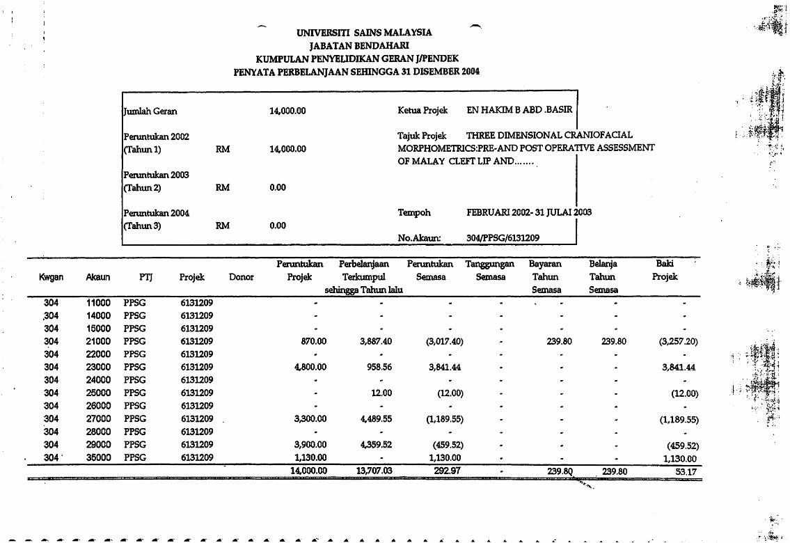
304/PPSG/6131209
THREE DIMENSIONAL
CRANIOFACIAL
MORPHOMETRICS: ANALYSIS
OF MALAY CLEFT LIP AND
PALATE INFANTS
ABDUL HAKIM ABDUL BASIR
D~. ZAINUL AHMAD RAJION / ASSOC. PROF. DR. AHMAD ~J. ZAKARIA
ASSOC. PROF. DR. IBRAHIM LUTFI SHUAIB
PROF. DR. AB. RANI SAMSUDIN
""·•·~\." ... ·z. , ... ·~·..,f.~A."· :.r,:. ~· ' ~ ,_._. :·-·. : .... ; ·-~; ,;·. . .. . . .. .-~:·:: -·. -~ ...
~·.;·~~-~-~--. ··-'- ·., . ·:·.:· .. ··~ ,·;.,·:u·s' 'M·':..7 • ·.J. ·1P· ···-.;o· 6. ~--·.··---·-·.-- ·;.
,.., : ~: "r" l\ l ~
. .
'1)
2)
3)
·:aAiiAGIAN PENYELIDIKAN & PEMBANGUNAN
CANSELORI
UNIVERSITI SAINS MALAYSIA
Laporan Akhir Projek PenyeHdikan Jangka Pendek
· Nama Penyelidik: •·••·······•·•••••••••············•···•·············••·•·····•··········•••·
..... ~ ....... ~ •.•..•.••..••...•.............•..•.••..••..•.•..•.......................•....•.....•......
Nanui. Pe~yelidik-Penyelldik Lain. (J.ikiz ·herladl!in) :
:~~t:::~~7.~:::?~:::~~~~::~::.~~~~:::~~~~::~ . ~ : .. ~.~~:~~. (. .. 1. 7.~.r:. ~:(.: ~Z/~~ .... ~ ~ .... : ........ . r /Of, flh. ·(( *1~; cfivn ct11clit'J . · .
.....••...•.. ~ ....••...........•..•......•....•........••...... ~
············•·············•···············•··•···········••····· . . .
·················~··········~····································
········~·······················································
····~~··,···········~·················· .. ·:· ... ····;·•.··············· ·•••·••••·····•••••·•••••··•···•···················•············
·····~··························································
.Pusat Peng!Yi~n/PusaUUnit · ... !.:. (.: .. ~~ ~ ~~. !::.1. ~!!.~~ ......... ., ............. ~. ·················································~--~~································~·················
Taj.uk P:rojek: ....................... r·........................................ • /~ Three /)i,'Y\ rAl d1 'oll!t f CYetVJ i o ~ G <i/ M cr pho ~ fl-1 ~ ~ · ·~ · · · · · · · • · •
· · · · · ·l;riq iy·~ i·~ · ·~r · 'iVi:rt~ · · · · · · r:te:r:;:· .. a;·· · ~ ~~j · ··A~~ ~e · · · Cfi::t-:vi·: · · · · · · ................................... 7. .................... 0 0.............. ... . . . . ·····•·•·•······••••···•·•·•• ·······················~············~~··········~·························································~
·············:·····························~~··~······················••t••··················· . . ........... .
USM liP~- 1 .
. . . . . . .: ·.· . ·-·· ..
• . . • ·_ .•. 4). . . < ~> · ·. · ·J>~ne~~aA ~:J~~:b:~· • :: .... i ::. , ·. •· . , - . ·:- : '.: -"1<:·; · -:·:''-' ·. : ;: --.· . ~->.· · : ·., _· ~:;·_:·:·· ;? ·,::-(' .·.: · · ·.. (PeriU.· dis_ediakarf 1J!il"lCZ:~ma~ di a_n~~r~) ~o. -:-. ·:2q~ per~~r.aan .-1i·_~dalam. BQJJ,~a : . .. . . . ..
I.·
I ...
'l
. J
~ I . 1
'. ..
•, :
..
• · Malqysia ."dan 'Bahasa ·f~ggeri.s Ini kemut/iann_ya ~~a~.· . f:li~ua~kpn. k~ .. da~am · Liipqtan · Tah.~na~ Bq~~cra~:-l'eeyelidiktJn. &. · P~bangiu_Ja_n · sebagni ·~a~· t!.art;~ ,; untuk
, ·.:··menyainpai~n .· f!~f~t~~ proiek_. ~il~n~~~q~: .ffP.Cfd~ p~h~·.tf~_iv~sitlJ. .. ,. .. :·.. ·
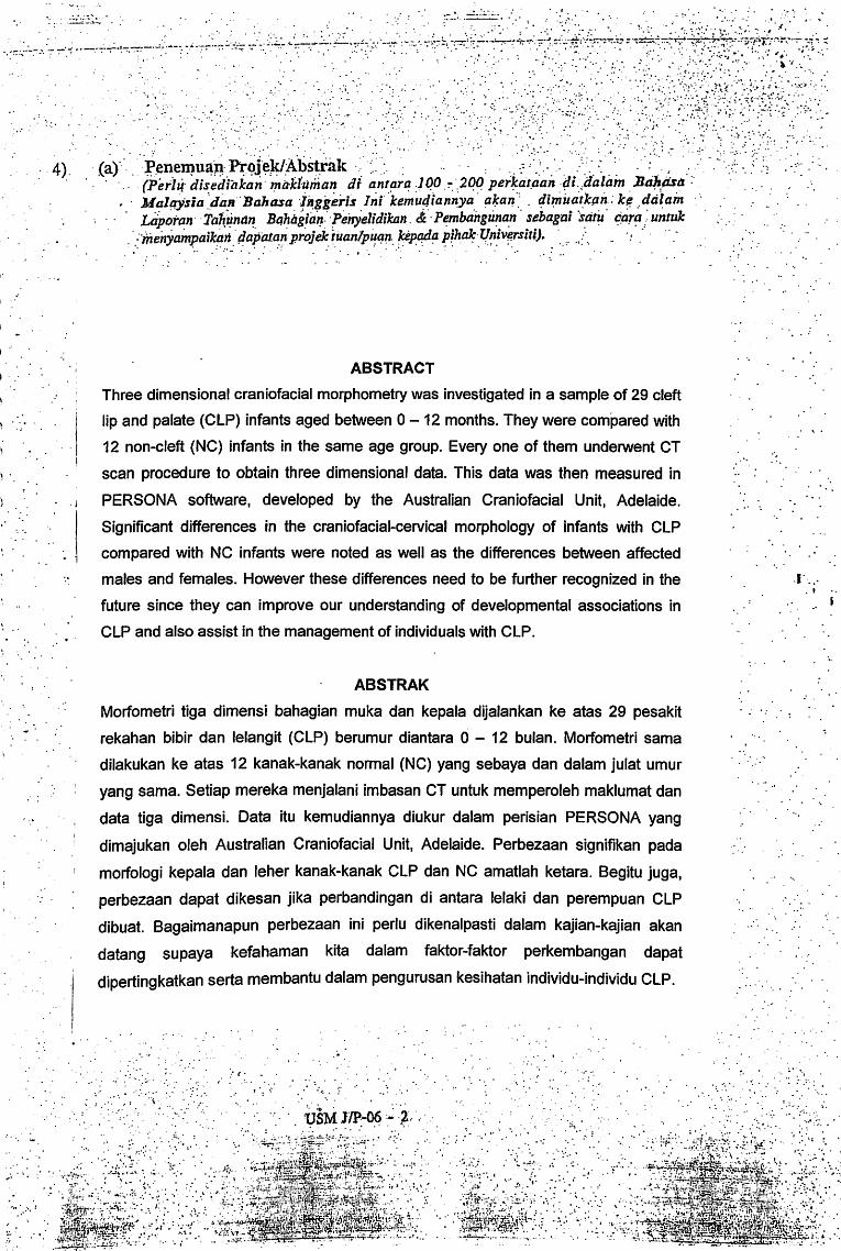
ABSTRACT Three dimensional craniofacial morphometry was investigated in a sample of 29 cleft
lip and palate (CLP) infants aged between 0-12 months. They were compared with
12 non-cleft (NC) infants in the same age group. Every one of them undeiWent CT
scan procedure to obtain three dimensional data. This data was then measured in
PERSONA software, developed by the Australian Craniofacial Unit, Adelaide.
Significant differences in the craniofacial-cervical morphology of infants with CLP
compared with NC infants were noted as well as the differences between affected
males and females. However these differences need to be further recognized in the
future since they can improve our understanding of developmental associations in
CLP and also assist in the management of individuals with CLP .
ABSTRAK Morfometri tiga dimensi bahagian muka dan kepala dijalankan ke atas 29 pesakit
rekahan bibir dan lelangit (CLP) berumur diantara 0 - 12 bulan. Morfometri sama
dilakukan ke atas 12 kanak-kanak normal (NC) yang sebaya dan dalam julat umur
yang sama. Setiap mereka menjalani imbasan CT untuk memperoleh maklumat dan
data tiga dimensi. Data itu kemudiannya diukur dalam perisian PERSONA yang
dimajukan oleh Australian Craniofacial Unit, Adelaide. Perbezaan signifikan pada
morfologi kepala dan leher kanak-kanak CLP dan NC amatlah ketara. Begitu juga,
perbezaan dapat dikesan jika perbandingan di antara lelaki dan perempuan CLP
dibuat. Bagaimanapun perbezaan ini perlu dikenalpasti dalam kajian-kajian akan
datang supaya kefahaman kita dalam faktor-faktor perkembangan dapat
dipertingkatkan serta membantu dalam pengurusan kesihatan individu-individu CLP.
1,·,.
:' ·.:. . ,·. ~ ... ~ .. ·.·: ~-. : .
.• (.
(.: .· ... · .. .· ,·: ·:
•••• 0
. . . ':.: '· . ~~ .· .. · .. .· ..
·.· ·.·:·:·:
. . .. .
. . .. ·•
. .·
... · .. · . t ..
•
.. ··.
. ... ',.
•. :.·.·., . . :·Y·:
. ··. ·. · ... · . : .· .. · ....
. ·· ... ::~ .. ;~:: ·: . .. . . ·.:: : . . ··.,·.· . · .. · ..
·: ~~¥N~ff' '<r; · <··;·!· .. •;:~.~:.,.~£ $/~ ~~~;~!~~:..~:~·. . · · ·,~~~t~~~l··: .. f. · :~·:· ·. ·. ·.~ '. ·:.:~.~~: ~-:~--i:;~'11 •1fh~;l:~-:?:: .. · .·, :• . . :~~-.;~~·:.;_/::·· ... ·.,: ... · .. ,._~-~-:.?4~. ·_.·_; ~--_~::>C:--..... ~·~··· ..... • ... • •. -~-... --.: .. ~· .• ·.· ..... ·~.:..~~·- •. ~_.····~ ~·--··· :~~-~~:·.~···.
--·- ·- •• ....:.·_·:~ 0
.:.:...
(b) Senaraikan Kata Kunci yang digunakan di dalant abstrak:
Bahasa Malaysia Bahasa Inegeris
· · · · O"ief.i -~~ · ;~~·j · ~~ i~ ~~ · · · · · · · · · c;i;io:p;~·~i· · · · · · ·w.;r, · · · ·
~ ............ " ... r ........ . . . . . C::.IY!f~ ~- .. 1:-?:r!~~l'.'f.' . . . . '!f:~(f./.or.!.~~?~ .. • ...... . .... ~-~~~-~f:c:: :( .. ~-7 ... .
. : .~~ .
'. ' ~ '
: :·.:·.~·
. . . . . . . . • ................... . ............................... .............................. . ... : .....
~·!'······~~········
. . .. . . . . ,. .......... . • , .• ! •••••• . ........................ • ...... .
. . . . . . . . . . . . . . . . . . . . . . . . . . . . . . ......................... .
5) Output pan Faedah Projek
(a) Penerbitan (tennasuk laporanlkertas seminar) (Sila nyatakan )enis. tajuk, pengarang, taiuln terbitan dan di mana telah diierbitldibentangkan). ·
~ ~ . ~ . . . . ~ A. :.:. . . . . . . .. . . . . . . . . . . . . . . . . . . . . . . . . . . 'f.~~.~ ..... •· ..... . ............ .
. . . . . . . . . . . . . . . . . . ' ...................... . . ...... . . .~ ....... .
. . . . . . . . . . . . . . . . . . . . . . . . . . . . . . . . . . . . . . . . . . . . . . . . . . . . . . . . . . . . . . . . . e e • e • t • t t e t t t • a t t e t t t t • • t t I t t a •• t .. ~ ...... . . ..... , .............................................. • ...... .
. . . . . . . . . . . . . . . . . . . . . . . . . . . . . . . . . . . . . . . . . . . . . . . . . . . . . . . . . . . . . . . . . . . . . . . . . . . . . . . . . . . ~ ........................................ .
. . . . . . . . . . . . . . . . . . . . . . . . . . . . . . . . . . . . . . . ~ ................ . . .. . ~ .·
USM J/P-06 - 3
6". Peralatan Yang Telah Dibeli:
••••• 41 ................................................... .
.. . . . . . . . . . . . . . . . . . . . . . . . . . . . . . . . . . . . . . . . . . . . . . . . . . . . . . .............................. "7. ................ · .......... .
. . . . . . . .... .. . . . . . . . . . . . . . . . '~\'~.: .......... .................. .
. . . · ................. : .. .. 7 .1' ............. ~ .................... .. . •. . . . . . . . •. . . . . . . . . . . . . . . . . . . . . . . . . . .
. . . . . . . . . . . . . . . . . . . . . . . . . . . . . . . . . . . . . . . . . . . . . . . . . . . .
............................... • .................................. . . . . . . . . . . . .. . . . . . . . . . . . ............. . . . . .. . . . . . . . ~ .....
. . . ... . . . . . . . . . . . . . . . . . . . . . . . . . . . . . . . . . . . . .. . . . . . . . ~ ........... . • • • • • • • • • • • • • • • • .• • • • • • • ! ••••••••••••••••
. . . . . . . . . . . .. . . . . . .. . . . . . . . . . . . ··-~ ......................... .
·.
UNTUK ~GUNAAN JAWATANKUASA PENYEL:(I)IkAN UNIVERSITI
. ~-...... " ..... . . :'\
··~ .......... -·· .......... . . f
~r ............ -• ............................. ' ....... . . . . . . . . . . . . . . . . . . . . . . . . . . . . . . . . . . . . . . . . . . . . . . . . . . . . . . . . ,. .
. . . . . . . . . . . . . . . . . . . . . ............................. . -------~· ............. ~ ............... : ............... .
TIT ANGAN PENGERUSI j/X PENYEIJOlKAN PUSA T PEN(;I\JIAN
USM J/P-06 - 5
. ....
. ....
.···. : . ~· ~· ~
' .
.i
., ·•
"· .. '·
I I
Kwgan Akaun
304 11000 i304 14000 304 15000 304 21000 304 22000 304 23000 304 24000 304 25000 304 26000 304 27000 304 28000 304 29000 304. 35000
Jumlah Geran
Peruntukan 2002 (Tahtm1)
Peruntukan 2003 (Tahun2)
Peruntukan 2004 (Tahun3)
PTJ Projek
PPSG 6131209 PPSG 6131209 PPSG 6131209 PPSG 6131209 PPSG 6131209 PPSG 6131209 PPSG 6131209 PPSG 6131209 PPSG 6131209 PPSG 6131209 PPSG 6131209 PPSG 6131209 PPSG 6131209
...-.. UNIVERSITI SAINS MALAYSIA
JABATAN BENDAHARI KUMPULAN PENYBUDIKAN GERAN JIPENDEK
PEN\'ATA PERBELANJAAN SEIDNGGA 31 DISEMBER 2004
14,000.00 Ketua Projek EN HAKIM B ABO .BASIR
Tajuk Projek THREE DIMENSIONAL CRANIOFACIAL
RM 14,000.00 MORPHOMETRICS:PRE·AND POST OPERATIVE ASSESSMENT
RM
RM
Donor
OF MALAY CLEFT LIP AND ...... .
0.00
Tempoh FEBRUAR12002· 31 ]ULAI2003 0.00
No.Akaun: 304/PPSG/6131209
Peruntukan Perbelanjaan Peruntukan Tanggungan Projek Terkumpul Semasa Semasa
sehingga Tahun lalu
Bayaran Tahun Semasa
Belanja Tahun
Semasa
870.00
4,800.00
3,300.00
3,900.00 1,130.00
14,000.00
3,887.40
958.56
12.00
4,489.55
4,359.52
13,707.03
(3,017.40) 239.80 239.80
3,841.44
(12.00)
(1,189.55)
(459.52) 1,130.00 292.97 239.8Q 239.80
Bald
Projek
3,841.44
(12.00)
(1,189.55)
(459.52) 1,130.00
53.17
tl •'4 :; .
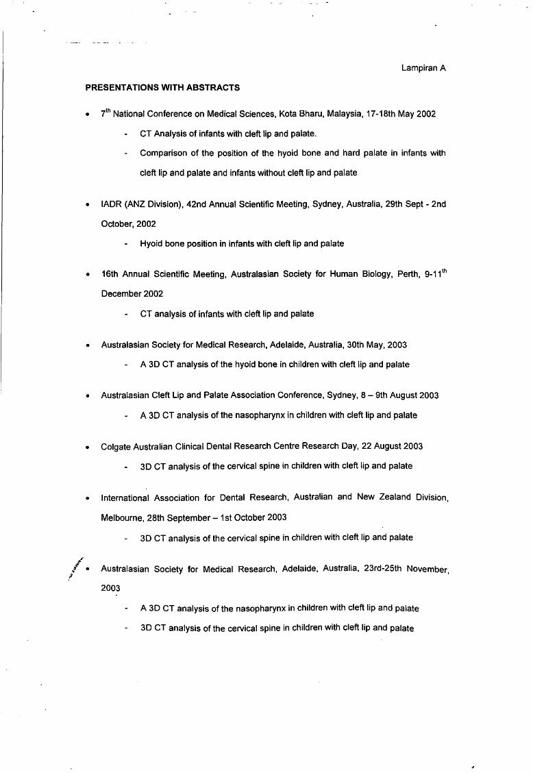
Lampiran A
PRESENTATIONS WITH ABSTRACTS
• ih National Conference on Medical Sciences, Kota Bharu, Malaysia, 17 -18th May 2002
CT Analysis of infants with cleft lip and palate.
Comparison of the position of the hyoid bone and hard palate in infants with
cleft lip and palate and infants without cleft lip and palate
• IADR (ANZ Division), 42nd Annual Scientific Meeting, Sydney, Australia, 29th Sept- 2nd
October, 2002
Hyoid bone position in infants with cleft lip and palate
• 16th Annual Scientific Meeting, Australasian Society for Human Biology, Perth, 9-11 1h
December 2002
CT analysis of infants with cleft lip and palate
• Australasian Society for Medical Research, Adelaide, Australia, 30th May, 2003
A 3D CT analysis of the hyoid bone in children with cleft lip and palate
• Australasian Cleft Lip and Palate Association Conference, Sydney, 8-9th August 2003
A 3D CT analysis of the nasopharynx in children with cleft lip and palate
• Colgate Australian Clinical Dental Research Centre Research Day, 22 August 2003
3D CT analysis of the cervical spine in children with cleft lip and palate
• International Association for Dental Research, Australian and New Zealand Division,
Melbourne, 28th September- 1st October 2003
3D CT analysis of the cervical spine in children with cleft lip and palate
.~ ·\; !! • Australasian Society for Medical Research, Adelaide, Australia, 23rd-25th November,
2003
A 3D CT analysis of the nasopharynx in children with cleft lip and palate
3D CT analysis of the cervical spine in children with cleft lip and palate
------~-- ----- ---~-
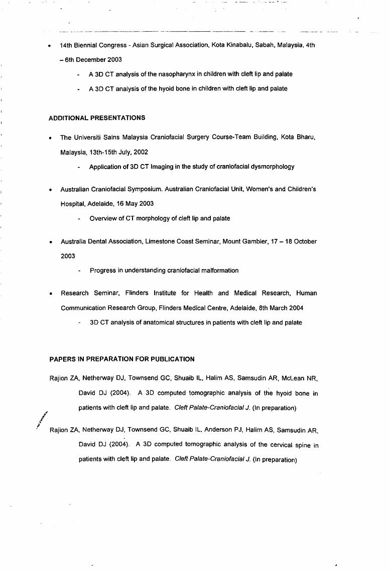
• 14th Biennial Congress- Asian Surgical Association, Kota Kinabalu, Sabah, Malaysia, 4th
- 6th December 2003
A 30 CT analysis of the nasopharynx in children with cleft lip and palate
A 30 CT analysis of the hyoid bone in children with cleft lip and palate
ADDITIONAL PRESENTATIONS
• The Universiti Sains Malaysia Craniofacial Surgery Course-Team Building, Kota Bharu,
Malaysia, 13th-15th July, 2002
Application of 3D CT Imaging in the study of craniofacial dysmorphology
• Australian Craniofacial Symposium. Australian Craniofacial Unit, Women's and Children's
Hospital, Adelaide, 16 May 2003
Overview of CT morphology of cleft lip and palate
• Australia Dental Association, Limestone Coast Seminar, Mount Gambier, 17-18 October
2003
Progress in understanding craniofacial malformation
• Research Seminar, Flinders Institute for Health and Medical Research, Human
Communication Research Group, Flinders Medical Centre, Adelaide, 8th March 2004
3D CT analysis of anatomical structures in patients with cleft lip and palate
PAPERS IN PREPARATION FOR PUBLICATION
Rajion ZA, Netherway OJ, Townsend GC, Shuaib IL, Halim AS, Samsudin AR, Mclean NR,
David OJ (2004). A 30 computed tomographic analysis of the hyoid bone in
patients with cleft lip and palate. Cleft Palate-Craniofacial J. (In preparation)
Rajion ZA, Netherway OJ, Townsend GC, Shuaib IL, Anderson PJ, Halim AS, Samsudin AR.
David OJ (2004). A 3D computed tomographic analysis of the cervical spine in
patients with cleft lip and palate. Cleft Palate-Craniofacial J. (In preparation)
Rajion ZA, Netherway DJ, Townsend GC, Shuaib IL, Halim AS, Samsudin AR, McLean NR,
David OJ (2004). A 30 computed tomographic analysis of the nasopharynx in
patients with cleft lip and palate. Cleft Palate-Craniofacial J. (In preparation)
Rajion ZA, Netherway OJ, Townsend GC, Shuaib IL, Halim AS, Samsudin AR, McLean NR,
David OJ (2004). A 3D computed tomographic analysis of the cranial base in
patients with cleft lip and palate. Cleft Palate-Craniofacial J. (In preparation)
Rajion ZA, Netherway OJ, Townsend GC, Shuaib IL, Halim AS, Samsudin AR, Mclean NR,
David OJ (2004). A 3D computed tomographic analysis of the spheno-occipital
synchondrosis in patients with cleft lip and palate. Cleft Palate-Craniofacial J. (In
preparation)
Three Dimensional Craniofacial Morphometries: Analysis of Malay Cleft Lip
and Palate Infants
1.0 Introduction
Cleft lip and palate (CLP) represents one of the most common forms of facial
deformity affecting one in every 500 to 1000 live births worldwide. It affects
individuals in all societies and has been the subject of considerable research. The
focus of previous studies has been on the aetiology, investigating the implications
and consequences for affected individuals, and surgical management.
The results of embryological studies have provided a clearer picture of what happens
during craniofacial development. This was highlighted by Diewert (1983) who
reported changes in craniofacial dimensions, proportions, and spatial relations during
the development of the secondary palate. Movements of the palatal shelves to the
horizontal position involve a complex interaction between the shelves and the tongue
that is influenced by developmental events in the shelves and the surrounding
craniofacial complex. Normal facial growth tends progressively to separate the
palatomaxillary processes from the tongue-mandibular complex as the nasa
maxillary complex lifts upward and the tongue shifts forward prior to shelf elevation.
This positional change may enhance palatal shelf elevation.
In addition to studies in humans, investigations using animal models show that,
during the period of shelf elevation, there is almost no growth in head width, but
constant growth in head height. This means that the position of least resistance for
the expanding palatal shelves is to occupy the space above the tongue (Ferguson,
1988).
Our understanding of the cellular and molecular events involved in craniofacial
development has improved greatly because of rapid advances in molecular biology.
During recent years, enormous progress has been made in our understanding of
normal and abnormal development of the head and neck. This progress has been
made possible through technical developments, particularly the application of
./· molecular techniques, and the development of animal models for studying the roles .,:t
! of genetic and environmental factors relevant to human CLP formation. The
application of precise cell marking procedures has led to a much better appreciation
of the cell movements and interactions involved in germ layer formation. The
techniques of scanning electron microscopy and in situ hybridisation methods for
studying gene expression have demonstrated the extensive contributions of neural
crest cells to craniofacial development.
·.·~
In CLP studies, anatomical differences have been observed. Excessive separation of
structures formed lateral to the tongue was observed by Maue-Dickson and Dickson
(1980) in a 15-week-old human foetus with cleft palate. Subtelny (1955) also found
that the nasopharynx was abnormally wide and the width between the maxillary
tuberosities was increased in unoperated CLP subjects.
Malformation resulting in CLP results from perturbations or insults during embryonic
development between the fourth and tenth weeks of gestation. Cleft lip and cleft of
the primary palate results from a failure of fusion of medial nasal, lateral nasal and
maxillary processes on either left, right or both sides of the forming craniofacial
complex. After primary palate fusion, secondary palate fusion takes place during the
ninth week to tenth week of gestation. Cleft palate may result from disturbances at
any stage of palate development: defective palatal shelf growth, delayed or failed
shelf elevation, defective shelf fusion, failure of medial edge cell death, post-fusion
rupture and failure of mesenchymal consolidation and differentiation (Ferguson,
1988).
CLP can occur in syndromic and non-syndromic forms. This study concentrated on
non-syndromic forms as they are less likely to have other pathological problems that
can affect the results. However, there may be some common mechanisms in both
types. Non-syndromic clefts of the oral cavity seem to be aetiologically distinctive;
however, clinically they make up the majority of cleft cases in the human population.
The non-syndromic forms of CLP have a multifactorial mode of inheritance with both
genetic and environmental factors operating. Currently, genes implicated in CLP
have been identified on different chromosomes, including chromosomes 6 and 11
(Juriloff and Mah, 1995; Eiberg eta/., 1987; Chenevix-Trench eta/., 1992). Genetic
analyses of non-syndromic oral clefts have produced significant results such as
association studies that point to polymorphisms at the TGF alpha locus playing a key
role in the aetiology of oral clefts. There is a suggestion that this locus may interact
with exposure to maternal smoking to influence the risk oral clefting (Shaw et a/.,
1996). The lack of consistent results from family studies highlights the fact that non-
~~ syndromic CLP is a heterogenous condition, undoubtedly caused by more than one :l
/ factor.
Many affected individuals appear as spontaneous events with no affected family
members. Multiple 'chance' combinations of genetic and environmental factors
(multifactorial aetiology) appear to be responsible for most of these CLP cases. The
2
most implicated environmental factors for human CLP have been cigarette smoking,
alcohol and nutritional factors such as folate deficiency (Wyszynski eta/., 1996).
This aetiology suggests that it is unlikely that the phenotypic effects will be limited
only to the cleft. It is likely that other structures will also be affected. It is also likely
that there will be a range of expressions of CLP, in other words phenotypic
heterogeneity.
The overall phenotypic pattern in CLP has not been well understood. The structures
affected in the crania-cervical region have not been well described previously. The
present study reports several anatomical anomalies not previously recognised. It is
not known whether these changes are a result of the CLP, a cause, or simply
pleiotropic effects associated with the clefting.
It has only been relatively recently that imaging techniques and 30 analytic
techniques have enabled a detailed assessment of the skeletal structures in CLP
patients. Most early knowledge has come from analyses of conventional radiographs
eg lateral head and AP views, which have several limitations such as superimposition
of structures, difficulty identifying landmarks and poor visualization of 30 structures.
The availability of 30 methods allows better opportunities to evaluate craniofacial
structures. There is now an opportunity of exploring the phenotypes of CLP
individuals in much more detail and to describe links, in terms of understanding the
mechanisms involved between wliat is happening at a molecular level and what
happens at the phenotypic level. There is a much better opportunity to link the
genotype, the molecular mechanisms and the phenotype.
By using 30 CT approaches, variables can now be defined that describe the size and
shape of bones and regions. Statistical analyses enable comparisons to be made
and help to clarify associations between structures. Multivariate analyses and
morphometric analyses are now possible with sophisticated computer software.
The particular advantage offered by this study is that CT data were obtained from
CLP individuals at infancy before they had been operated, and records were
available for unoperated non-cleft (NC) children, matched f<?r age, for comparison.
This study also used a sophisticated software package that enabled accurate and
reproducible location of landmarks from which variables could be derived thereby
offering advantages over conventional radiographs. This has allowed views that are
3 -
not possible with a conventional approach, including images of the hyoid bone,
cervical spine, nasopharynx, cranial base and spheno-occipital synchondrosis (SOS).
The description of the associations between the hyoid bone, cervical spine,
nasopharynx, cranial base, spheno-occipital synchondrosis (SOS) and CLP, which
have not been detailed in previous studies, is possibly the most important
contribution of this thesis. These areas were also selected because of their clinical
importance to swallowing, hearing, and speech in CLP. This study focussed on the
areas more distant from the cleft but within the craniofacial/cervical region. The
selection was also based on the hypothesis that CLP reflects part of a broader
problem, not just one in the region of the cleft. Previous studies have indicated that
CLP is associated with a variety of other anomalies (Maue-Dickson, 1979; Maue
Dickson and Dickson, 1980; Horowitz et a/., 1976; Krogman et a/., 1975; Molsted et
a/., 1993, 1995).
4
2.0 Methodology
2.1 Ethical Approval
Ethical approval was given by the Ethics and Research Committee USM dated
30/8/01, Number: USM/PPSG/Ethics Com/2001 [61.3(1 )] (Appendix 1). Data collection
took place in Malaysia from September 2001 to August 2002.
2.2 Data Collection
CT scans were obtained from 29 patients with unoperated non-syndromic cleft lip
and palate. Any syndromic patients were excluded. They were aged between 0-12
months and compared with 12 non-cleft patients (NC) in the same age group. The
NC patients had normal craniofacial morphology but had medical indications for
scanning including meningitis and hydrocephalus.
The distribution of clefts was cleft lip and/or alveolus (CL), n=7; unilateral cleft lip and
palate (UCLP), n=1 0; bilateral cleft lip and palate (BCLP), n=4; isolated cleft palate
(ICP), n=8; non-cleft patients (NC), n=12. Cephalometric analyses of cleft lip and/or
alveolus (cleft of the primary palate) have been shown to be different in craniofacial
morphology from other cleft types (Dahl, 1970; Smahel et al., 1991), for that reason
the CL was included in this study. Age and sex distribution of the cleft and NC groups
are shown in Table 2.1.
Table 2.1 Age and sex distribution of the cleft and NC groups
Group Sex No. Mean Age No. Range (Days) (min-max)
CLP F(12) M (17) 29 115 76 14-340
NC F(3) M (9) 12 145 86 19-297
Table 2.1 shows that the age range was greater in the CLP group and a few older
children were included. The reason for this is that in CLP group the primary operation
had been postponed because of other health problems such as upper respiratory
tract infection and aspiration pneumonia.
2.3 Imaging Procedure
Axial scans were obtained with a GE Lightspeed Plus CT Scanner System at the
Department of Radiology, Hospital USM. The protocol (Appendix II) used at the
Australian Craniofacial l:Jnit (ACFU), Women's and Children's Hospital, Adelaide
(Australia), was followed as the basis for the scanning procedure.
5
2.4 Image Measurement
The PERSONA software package developed by the Research Unit at the ACFU,
Women's and Children's Hospital, Adelaide (Abbott et al, 1990, 2000; Netherway et
al., 1997, 1999) was utilized for three-dimensional reconstruction of the images and
to determine the 30 coordinates of osseous landmarks on a Silicon Graphics
Computer workstation.
2.5 Statistical analysis
A linear model (PROC GLM, SAS 2001) incorporating the fixed effects 'sex' and 'cleft
group', and using •age' as a covariate, was fitted to all variables.
The model was as follows:
Variable= Age (14-340 days)
Sex (male, female)
Group (NC, UCLP, BCLP, ICP, CL)
Higher order interactions were not analysed for this small data set. Linear contrast
were arranged to compare the control group (NC) with all other groups, and to
compare the ICP group (a morphology distinct cleft-type) with other cleft groups. A
Chi-square test was used to test for any associations between anomalies of the
cervical spine and the incidence of cleft lip and palate. The level of significance was
set at 5o/o.
2.6 Errors of the method
Two determinations were performed to assess the reproducibility of landmark
determination and variables derived from these landmarks using Dahlberg's method
of double determination (1940). All measurements were repeated after a period of
one month. Student's t-tests were used to detect systematic errors (i.e. to ascertain
whether the mean difference between repeated measures deviated significantly from
zero).
6
3.0 Results
3.1 Hyoid bone
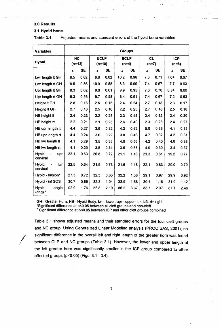
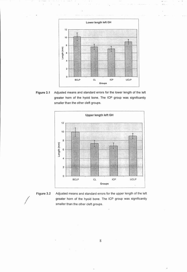
Table 3.1 Adjusted means and standard errors of the hyoid bone variables.
Variables Groups
NC UCLP BCLP CL Hyoid (n=12) (n=10) (n=4) (n=7)
X SE X SE x SE X SE
Lwr length It GH 8.5 0.62 8.8 0.62 10.2 0.96 7.6 0.71
Lwr length rt GH 8.5 0.56 10.0 0.58 8.3 0.90 7.4 0.67
Upr length It GH 8.3 0.62 9.0 0.61 9.9 0.95 7.3 0.70
Upr length rt GH 8.3 0.56 9.7 0.58 8.4 0.91 . 7.4 0.67
Height It GH 2.8 0.16 2.5 0.15 2.4 0.24 2.7 0.18
Height rt GH 2.7 0.16 2.5 0.16 2.2 0.25 2.7 0.18
HB height It 2.4 0.23 2.2 0.28 2.3 0.45 2.4 0.32
HB height rt 2.2 0.21 2.1 0.25 2.6 0.40 2.3 0.28
HB upr length It 4.4 0.27 3.9 0.32 4.3 0.52 5.0 0.36
HB upr length rt 4.4 0.24 3.6 0.29 3.8 0.46 4.7 0.32
HB lwr length It 4.1 0.29 3.5 0.35 4.0 0.56 4.2 0.40
HB lwr length rt 4.1 0.29 3.5 0.34 3.5 0.55 4.0 0.39
Hyoid - upr 22.1 0.63 20.8 0.72 21.1 1.16 21.3 0.81 cervical
Hyoid - lwr 22.5 0.64 21.9 0.73 21.6 1.18 22.1 0.83 cervical
Hyoid - basion* 27.5 0.72 32.3 0.86 32.2 1.38 29.1 0.97
Hyoid - inf SOS 30.7 0.86 32.3 1.04 33.9 1.68 30.4 1.18
Hyoid angle 92.9 1.76 85.8 2.10 86.2 3.37 88.7 2.37 (deg) *
GH= Greater Hom, HB= Hyoid Body, lwr= lower, upr= upper, It= left, rt= right *Significant difference at p<0.05 between all cleft groups and non-cleft • Significant difference at p<0.05 between ICP and other cleft groups combined
ICP (n=8)
x SE
7.0+ 0.67
7.7 0.63
6.8+ 0.66
7.2 0.63
2.3 0.17
2.5 0.18
2.4 0.30
2.4 0.27
4.1 0.35
4.2 0.31
4.0 0.38
3.4 0.37
19.2 0.77
20.0 0.79
29.9 0.92
31.9 1.12
87.1 2.46
Table 3.1 shows adjusted means and their standard errors for the four cleft groups
and NC group. Using Generalized Linear Modeling analysis (PROC SAS, 2001), no
... significant difference in the overall left and right length of the greater horn was found ~i
l~ between CLP and NC groups (Table 3.1 ). However, the lower and upper length of
the left greater horn was significantly smaller in the ICP group compared to other
affected groups (p<0.05) (Figs. 3.1 - 3.4).
7
-·' ,. /~
Low er length left GH
12 '
1 ~ ~
10
I;J ~
F-~ 8 r-- T
e & rr §.
I~~ .r: 6 r-- 1-c;,
lr• c "' ' .; -' lt d 4 - 1-
I• 1 ~
2 1-I• ••• --- --- --- 1-·.- .. I•
0 ~
BCLP CL ICP UCLP Groups
Figure 3.1 Adjusted means and standard errors for the lower length of the left
greater horn of the hyoid bone. The ICP group was significantly
smaller than the other cleft groups.
Figure 3.2
Upper length left GH
12 ..,. " , .
10
rl-1-
I
rl-1-
i+ I• 1- 1-
8 E .§. .s:: 6 0, c: Q)
-' 4 1- 1-
I•
2 1- .. 1-
I~
0 BCLP CL ICP UCLP
Groups
Adjusted means and standard errors for the upper length of the left
greater horn of the hyoid bone. The ICP group was significantly
smaller than the other cleft groups.
8
•
" !"
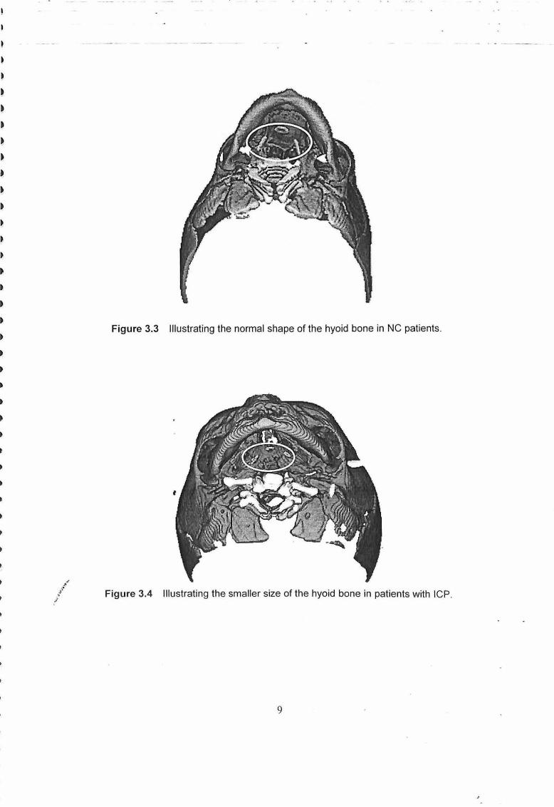
Figure 3.3 Illustrating the normal shape of the hyoid bone in NC patients .
,. / Figure 3.4 Illustrating the smaller size of the hyoid bone in patients with ICP.
/
9
/ l'
/
3.2 Cervical Spine
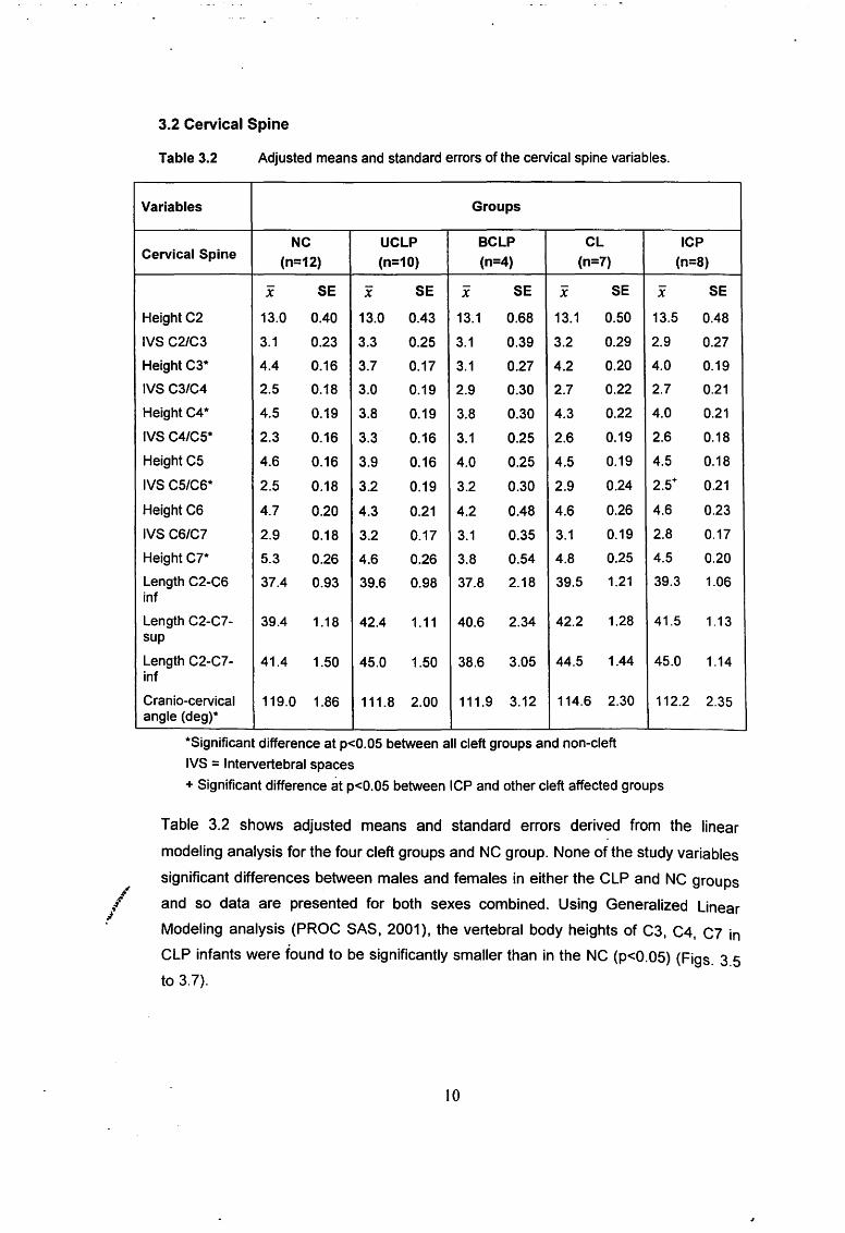
Table 3.2 Adjusted means and standard errors of the cervical spine variables.
Variables Groups
NC UCLP BCLP CL Cervical Spine
(n=12) (n=10) (n=4) (n=7)
X SE x SE X SE X SE
Height C2 13.0 0.40 13.0 0.43 13.1 0.68 13.1 0.50
IVS C2/C3 3.1 0.23 3.3 0.25 3.1 0.39 3.2 0.29
Height C3* 4.4 0.16 3.7 0.17 3.1 0.27 4.2 0.20
IVS C3/C4 2.5 0.18 3.0 0.19 2.9 0.30 2.7 0.22
Height C4* 4.5 0.19 3.8 0.19 3.8 0.30 4.3 0.22
IVS C4/C5* 2.3 0.16 3.3 0.16 3.1 0.25 2.6 0.19
Height C5 4.6 0.16 3.9 0.16 4.0 0.25 4.5 0.19
IVS C5/C6* 2.5 0.18 3.2 0.19 3.2 0.30 2.9 0.24
Height C6 4.7 0.20 4.3 0.21 4.2 0.48 4.6 0.26
IVS C6/C7 2.9 0.18 3.2 0.17 3.1 0.35 3.1 0.19
Height C7* 5.3 0.26 4.6 0.26 3.8 0.54 4.8 0.25
Length C2-C6 37.4 0.93 39.6 0.98 37.8 2.18 39.5 1.21 inf
Length C2-C7- 39.4 1.18 42.4 1.11 40.6 2.34 42.2 1.28 sup
Length C2-C7- 41.4 1.50 45.0 1.50 38.6 3.05 44.5 1.44 inf
Cranio-cervical 119.0 1.86 111.8 2.00 111.9 3.12 114.6 2.30 angle (deg)*
*Significant difference at p<0.05 between all cleft groups and non-cleft
IVS =Intervertebral spaces
ICP
(n=S)
X SE
13.5 0.48
2.9 0.27
4.0 0.19
2.7 0.21
4.0 0.21
2.6 0.18
4.5 0.18
2.5+ 0.21
4.6 0.23
2.8 0.17
4.5 0.20
39.3 1.06
41.5 1.13
45.0 1.14
112.2 2.35
+Significant difference at p<0.05 between ICP and other cleft affected groups
Table 3.2 shows adjusted means and standard errors derived from the linear
modeling analysis for the four cleft groups and NC group. None of the study variables
significant differences between males and females in either the CLP and NC groups
and so data are presented for both sexes combined. Using Generalized Linear
Modeling analysis (PROC SAS, 2001 ), the vertebral body heights of C3, C4, C7 in
CLP infants were found to be significantly smaller than in the NC (p<O.OS) (Figs. 3.S
to 3.7).
10
/ .,. .. /
5
4 E §. 3 -.r: -~ 2
C1.l :I:
0
~-~ i--1,1·
~· i--~~·. f • BCLP
·. rr-
'
CL
Height of C3
T r+-.... ph 1-
' a . ---
I" 1-
~ 1-
t-
II
ICP UCLP NC
Groups
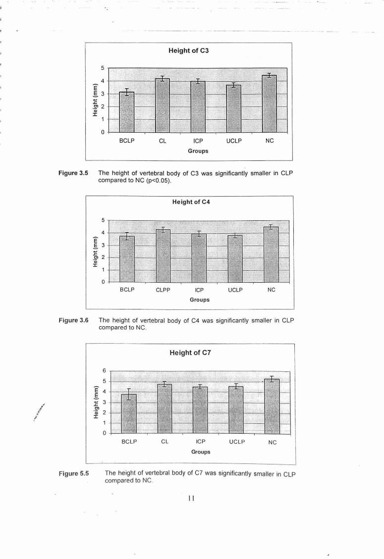
Figure 3.5 The height of vertebral body of C3 was significantly smaller in CLP compared to NC (p<O.OS).
5
4
E .§.. 3
:g, 2 'Qj :I:
0
r--t-r+-
t- 1•
t--
t-1-
BCLP CLPP
Height of C4
T rr-.... r- t-
t-I~
t--
I' t-
ICP UCLP NC
Groups
Figure 3.6 The height of vertebral body of C4 was significantly smaller in CLP compared to NC.
Figure 5.5
6
5
E4 .s :c 3 Cl
'Qj 2 :I:
0 .
T
r-~ r-
I--' -- ~ ...... --
BCLP
Height of C7
--'- *I-r±- --r- -+----1:'1 1-
--; --- :-
-- --- 1-I+ - i" - . - ...._ ....
CL ICP UCLP NC
Groups
The height of vertebral body of C7 was significantly smaller in CLP compared to NC .
II
..... . l
/
3.3 Nasopharynx
Table 3.3 Adjusted means and standard errors of the nasopharyngeal variables.
Variables Groups
NC UCLP BCLP CL ICP Nasopharynx
(n=12) (n=10) (n=4) (n=7) (n=B)
x SE x SE x SE x SE x SE
Inter hamular 25.6 0.77 33.5 0.83 34.3 1.30 29.6 0.96 29.3+ 0.91 notch*
Inter hamulus* 22.3 0.59 30.2 0.63 29.8 0.99 25.7 0.73 25.9+ 0.70
Inter-lateral 36.0 0.86 43.1 0.92 41.7 1.43 39.9 1.06 39.5 1.00 pterygoid*
Hamulus- 8.3 0.50 7.2 0.53 6.5 0.84 7.4 0.62 7.0 0.59 lateral Ptry.plate It*
Hamulus- 8.0 0.49 7.4 0.52 7.3 0.81 7.9 0.60 7.6 0.57 lateral Ptery. plate rt
Inter -maxillary 26.4 0.77 35.0 0.83 34.9 1.30 29.9 0.96 30.2+ 0.91 tuberosity*
Inter-zygomatic 62.3 1.23 70.0 1.32 68.5 2.07 67.4 1.53 66.2 1.44 distance*
Vomer- 18.2 0.47 20.1 0.50 21.5 0.78 20.1 0.58 18.7+ 0.55 hamulus It*
Vomer- 17.8 0.47 20.2 0.50 20.6 0.79 19.3 0.59 18.0+ 0.55 hamulus rt*
Vomer- basion 23.0 0.64 24.0 0.69 23.7 1.07 22.9 0.80 26.2+ 0.75
Basion- 26.8 0.65 28.6 0.69 27.7 1.08 28.0 0.80 27.2 0.76 hamulus-It
Basion- 26.5 0.63 28.4 0.66 27.9 1.04 27.6 0.77 27.1 0.73 hamulus rt
Hamulus angle 40.2 1.84 36.0 1.97 37.2 3.08 39.2 2.28 42.7 2.16 It
Hamulus angle 40.8 1.77 38.2 1.90 42.1 2.97 36.1 2.20 45.0+ 2.08 rt
Sphenopalatine 32.7 1.16 31.0 1.24 27.9 1.94 31.1 1.44 31.5 1.46 angle
Vomerine angle 21.2 1.19 19.4 1.28 17.2 2.00 17.0 1.48 21.4 1.51
* Significant difference at p<0.05 between all cleft groups and non-cleft + Significant difference at p<0.05 between ICP and combined cleft groups
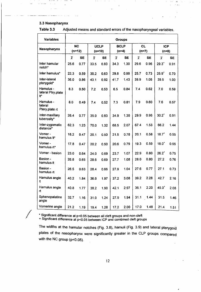
The widths at the hamular notches (Fig. 3.8), hamuli (Fig. 3.9) and lateral pterygoid
plates of the nasopharynx were significantly greater in the CLP groups compared
with the NC group (p<0.05).
12
.... ,.· .. .• ;1
40
35
~ 30
~ 25 :;; 20
:Q 15 ~ 10
Figure 3.8
5
0
Hamular notch width
J'
••. •:
-1~ "\. . ~ ., . -=- 7f " _ I; ...:;:. . ""'·
- r--- .. t-' ' -~ - !t t-.
;
1- " 'l t-. BCLP CL ICP UCLP NC
Groups
Adjusted mean values and standard errors for the hamular notch width
in CLP and NC groups. The CLP groups were significantly wider than
the NC group and the ICP group was significantly smaller when
compared to other CLP groups.
Hamulus width
35
30 -,.. .. -"
Figure 3.9
E 25
.s 20
£ 15 "0
~ 10
5
0
t-- ...:;:. r---:E-
I ~~-t-
1-- r-I< :II
t-,~·
1-. t- ' ·~ t-
BCLP CL ICP UCLP NC
Groups
Adjusted mean values and standard errors for the hamulus width in
CLP and NC groups. The CLP groups were significantly wider than
the NC group and the ICP group was significantly smaller when
compared to other CLP groups .
13
3.4 Cranial base
Table 3.4 Adjusted means and standard errors of the cranial base variables.
Variables Groups
Cranial Base NC UCLP BCLP, CL ICP
(n=12) (n=10) (n=4) (n=7) (n=B)
x SE x SE X SE x SE x SE Lt sphenoid
8.1 0.29 6.8 0.29 6.9 0.46 6.8 0.33 5.9+ 0.34 height* Rt sphenoid
7.9 0.26 6.7 0.27 6.8 0.42 7.0 0.31 5.7+ 0.32 height* Lt basioccipital
7.9 0.27 7.3 0.27 6.7 0.42 7.4 0.31 6.7 0.32 height* Rt
basioccipital 7.8 0.25 7.1 0.24 6.7 0.40 7.4 0.29 6.8 0.29 height* Basion-
64.1 1.31 62.2 1.41 61.8 2.19 62.0 1.63 nasion 60.5 1.66
Basion - sella 25.9 0.59 25.8 0.64 24.6 0.99 26.4 0.74 25.8 0.75
Sella - nasion* 44.9 0.92 42.0 0.98 42.5 1.53 41.3 1.14 40.7 1.16 Sella-sup.
9.2 0.34 9.9 0.34 9.5 0.54 10.2 0.40 9.5 0.41 sphenoid It Sella to sup.
9.2 0.36 10.0 0.36 9.5 0.57 10.3 0.42 9.5 0.43 sphenoid rt Basion- sup.
15.5 0.43 15.5 0.44 14.9 0.68 15.8 0.50 15.6 0.51 basioccipital It Basion-superior 15.7 0.47 15.3 0.47 14.4 0.73 15.8 0.54 15.7 0.55
basioccipital rt Basion - inf.
13.3 0.42 14.0 0.42 13.5 0.66 13.8 0.49 14.3 0.50 basioccipital It Basion - inf.
13.7 0.42 14.0 0.43 13.1 0.67 13.8 0.50 14.1 0.50 basioccipital rt Cranial base
131.4 2.00 131.9 2.13 134.1 3.34 132. 2.48 130.
2.51 an ole 1 7
*Significant difference at p<0.05 between all cleft groups and non-cleft
+ Significant difference at p<0.05 between ICP and other combined cleft groups
When the GLM model was applied to the height data for the basi-sphenoid and basi
occipital bones, statistically significant differences were found between the CLP and
NC groups (Table 3.4). The heights of the bones on both sides in CLP infants were
significantly smaller when compared to the NC (p<0.05). Furthermore, the heights of
the basi-sphenoid in the ICP group on both sides were significantly smaller when
~... compared with the other cleft groups (p<0.05) (Figs. 3.10 and 3.11 ). ·\" ,..,
!
14
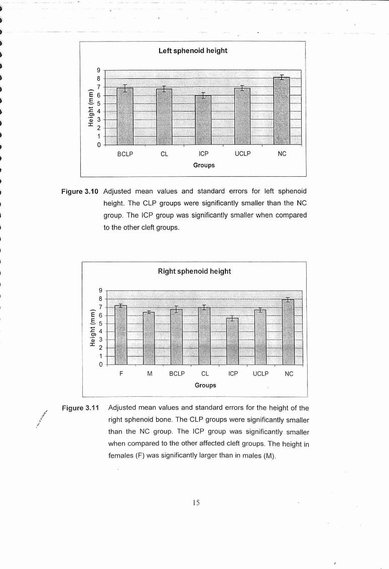
Left sphenoid height
9 .-------------------~~-----------------------. 8 +-------~------------~----------------~~~~ 7 +--=~=-------T-~~--~------~~~----~~
E 6 .Ss :c 4 Cl
'(ii 3 ::r: 2
1 0 +-~~-L~~~~~-r--~~~--~~~--r-~~~~
BCLP CL ICP UCLP NC
Groups
Figure 3.10 Adjusted mean values and standard errors for left sphenoid
height. The CLP groups were significantly smaller than the NC
group. The ICP group was significantly smaller when compared
to the other cleft groups.
Right sphenoid height
9.-----------------~---------------------------,
8 1-------~--~--------------~------------~~~ 7 +-~~-------~~~~--~~-------~--~----4
E 6 .s 5 -:c 4 Cl C1l 3
::r: 2
1 0 +-~-L-r-L~~-J--~~~-L-r-L~~~~~,_~-L~
F M BCLP CL ICP UCLP NC
Groups
Figure 3.11 Adjusted mean values and standard errors for the height of the
right sphenoid bone. The CLP groups were significantly smaller
than the NC group. The ICP group was significantly smaller
when compared to the other affected cleft groups. The height in
females (F) was significantly larger than in males (M).
15
•"' .l ... !
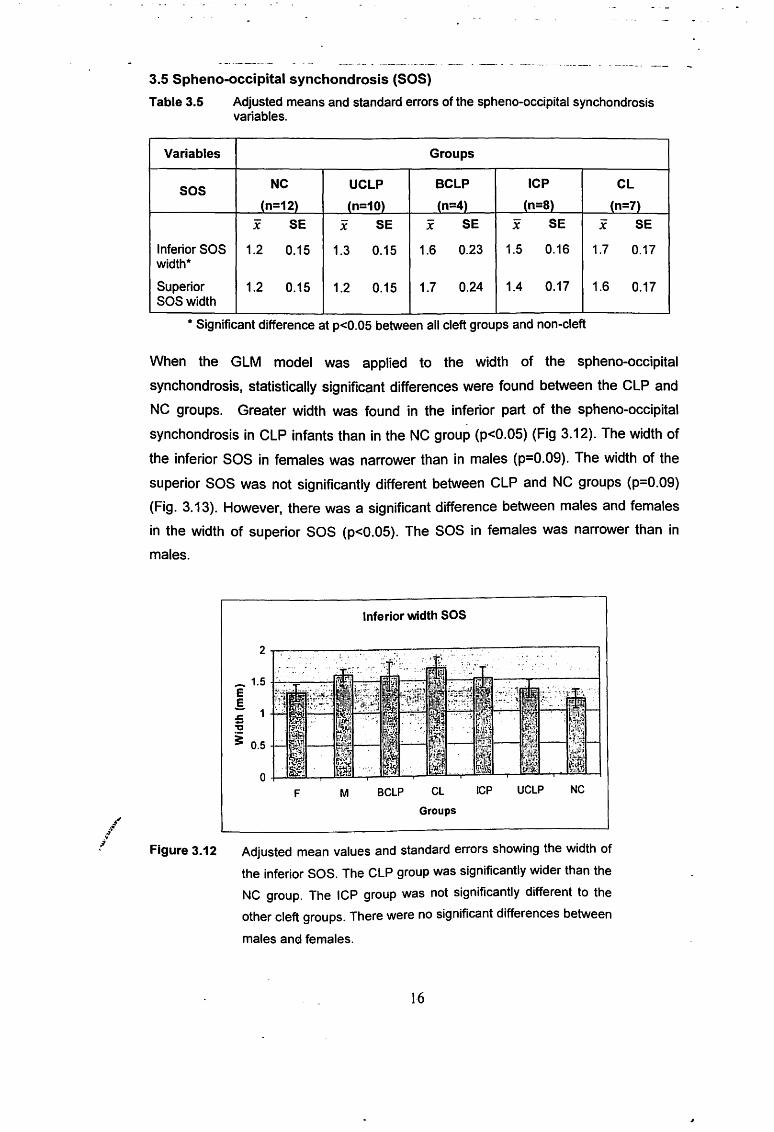
3.5 Spheno-occipital synchondrosis (SOS)
Table 3.5 Adjusted means and standard errors of the spheno-occipital synchondrosis variables.
Variables Groups
sos NC UCLP BCLP ICP CL
(n=12) (n=10) (n=4) (n=B) (n=7)
x SE X SE x SE x SE x SE
Inferior SOS 1.2 0.15 1.3 0.15 1.6 0.23 1.5 0.16 1.7 0.17 width*
Superior 1.2 0.15 1.2 0.15 1.7 0.24 1.4 0.17 1.6 0.17 SOSwidth
*Significant difference at p<0.05 between all cleft groups and non-cleft
When the GLM model was applied to the width of the spheno-occipital
synchondrosis, statistically significant differences were found between the CLP and
NC groups. Greater width was found in the inferior part of the spheno-occipital
synchondrosis in CLP infants than in the NC group (p<0.05) (Fig 3.12). The width of
the inferior SOS in females was narrower than in males (p=0.09). The width of the
superior SOS was not significantly different between CLP and NC groups (p=0.09)
(Fig. 3.13). However, there was a significant difference between males and females
in the width of superior SOS (p<0.05). The SOS in females was narrower than in
males.
Inferior width SOS
- 1.5 -I-'---=--~1':!!!-:'11----E E :;;- 1 -~ i 0.5 4-ll~lJJ11--H:fi~t--
F M BCLP CL
Groups
ICP UCLP NC
Figure 3.12 Adjusted mean values and standard errors showing the width of
the inferior SOS. The CLP group was significantly wider than the
NC group. The ICP group was not significantly different to the
other cleft groups. There were no significant differences between
males an<:.! females.
16
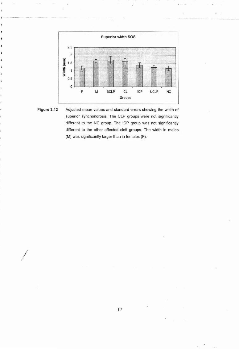
Figure 3.13
Superior width SOS
2.5
2
E ..§. 1.5 .s:;
-15 ~
0.5
0
F M BCLP CL ICP UCLP NC
Groups
Adjusted mean values and standard errors showing the width of
superior synchondrosis. The CLP groups were not significantly
different to the NC group. The ICP group was not significantly
different to the other affected cleft groups. The width in males
(M) was significantly larger than in females (F).
17
4.0 Discussion
After detailed analysis of the data collected for this study, several differences
between the CLP and NC groups became apparent. These differences pertained to
the five main areas of interest described below:
4.1 Hyoid Bone
This 3D CT study has shown, for the first time, details of the abnormalities of the
hyoid bone in CLP. The hyoid bone is smaller and in some cases there is no
ossification of the body of the hyoid bone. The hyoid is further from the cranial base.
There is smaller angulation and also it is at a low level in relation to the cervical
vertebrae.
These phenotypic changes in the hyoid bone relate to structures derived from the
first, second and third branchial arches. The hyoid bone is a composite endochondral
bone that develops from cartilage of the 2"d and 3rd branchial arches- lesser horn
from the 2"d branchial arches; greater horn from the 3rd branchial arches; body from
both 2"d and 3rd branchial arches (Koebke, 1978). In terms of embryology, this finding
indicates that the underlying factors associated with clefting anomalies not only affect
the labiomaxillary and palatine structures of the first arch, but also appear to
influence the development of structures derived from the 2"d and 3rd branchial arches.
Clinically there is an association between the low level of the epiglottis and the level
of the hyoid in relation to the cervical vertebrae with aspiration pneumonia. Alteration
in the position of the hyoid also presents significant potential problems in terms of
breathing, swallowing and head posture, because of alterations in attachments of the
muscles responsible for these functions.
In terms of clinical problems presented by the CLP groups 4/29 had aspiration
pneumonia and 6/29 had upper respiratory tract infections causing surgical
intervention to be deferred. When two or more anomalies present together, medical
complications can result and their coincidence carries implication·s for morbidity and
prognosis (Azmi et a/., 1983). Pandya and Boorman (2001) found failure to thrive
.:ti" (FTT) in babies with CLP, but with a feeding support nurse and airway management
./ it improved. It may also be that neonatal nurses may be able to provide more
effective care by understanding more of the nature of CLP and its effects on feeding.
The multidisciplinary nature of effective care of CLP infants also involves speech
pathology. A greater understanding of the differences in the morphology of the hyoid
· bone may improve the approaches to speech therapy in CLP infants. Therapy based
18
on current knowledge entirely overlooks the hyoid malformation. It is hoped that the
findings of this study may lead to new approaches to CLP speech therapy.
4.2 Cervical Spine
The cervical spine showed smaller vertebral body heights and greater intervertebral
spaces and smaller cervical angle. The presence of cervical spine anomalies was
noted and delayed ossification of the anterior arch of C 1. There was also an
association between the occurrence of CLP and the presence cervical spine
anomalies.
Endochondral ossification of the upper cervical vertebrae commences by the eight
week of foetal life and is completed by about three to six years of post-natal life
(Farman and Escobar, 1982; Sandham, 1986). Although no significant difference was
found in the overall length of the cervical spine, the smaller vertebral bodies and
greater intervertebral spaces suggest that there may be a difference in the pattern of
skeletal ossification or that maturation is delayed or altered in CLP compared with the
NC infants. This delay in maturation may influence the lifting of the head (during 6th -
1oth weeks in utero) and could also possibly be associated with the failure of the
elevation of the palatal shelves to meet leading to clefting of the palate. These
limitations of the extension of the head of the foetus could also interfere with the
descent of the glossa-mandibular complex. The wedging position of the tongue in
between the palatal shelves has been shown to be a major factor contributing to
failure of shelf elevation and clefting of the secondary palate (Diewert, 1983).
Abnormalities of the cervical spine in CLP, such as fusion of the posterior upper arch
and short posterior arch of C1, lipping of the atlas (C1) and anterior arch anomalies
of C1 which included two anterior arches instead of one and an asymmetric anterior
arch to the right, have not been demonstrated before the use of 3D CT.
The reduced cervical angle in CLP may be associated with postural changes to
facilitate airway maintenance. Anderson (1997), in his study on craniosynostosis
patients, reported that cervical spine fusion, particularly those affecting the higher
l" levels, may also have important consequences for head posture with resulting
.l influences on craniofacial growth and dental occlusion. Other researchers have also
proposed that cervical spine anomalies may alter head posture (Solow eta/., 1984;
Solow and Siersbaek-Nielsen, 1986; Hellsing et a/., 1987; Solow and Siersbaek
Nielsen, 1992; Nevard, 1994). These previous studies have also demonstrated
associations between head posture and craniofacial morphology. This study's
findings suggest that upper cervical spine anomalies may be more common in
19
Malaysian children with CLP (24°/o) than in American children (22o/o) (Horswell,
1991), Scottish children (13o/o) (Sandham, 1986), and Norwegian children (18o/o)
(Ugar and Semb, 2001 ). However, it must be stressed that the study groups referred
to include different proportions of cleft types so comparisons of incidence should be
undertaken with some caution. Furthermore, the present study was based upon 30
CT scans of subjects while earlier studies were based upon 20 cephalometric
radiographs. The enhanced clarity offered by CT images may well display anomalies
more clearly and thereby facilitate the diagnosis of CLP associated defects. Previous
studies have reported similar frequencies of fusion in NC groups or in the general
population, ranging from 0.5- 5o/o (Gray eta/., 1964; Brown eta/., 1964; Farman and
Escobar, 1982). In contrast, the author did not find any fusion anomalies, probably
due to the small sample size of the NC group. However, ethnicity cannot be ruled out
as an explanation.
Osborne et a/. (1971) suggested a smaller than normal anterior arch of the atlas
could have a direct effect on the anterior-posterior dimension of the pharynx. The
anterior arch of C1 is suggested to play a significant role in the establishment of
adequate veto-pharyngeal function and speech in children with CLP (Osborne et a/.,
1971; Sand ham, 1986). These findings suggest that the ossification of anterior arch
of C1 may be compromised in patients with CLP and this may later contribute to
problems in speech. The importance of the anterior arch of C1 and upper cervical
vertebrae was highlighted by Berkowitz (1996} in achieving adequate velopharyngeal
closure and speech.
The finding of short vertebral bodies in the cervical spines of infants with clefts is
consistent with a delay in growth in infancy. Previous studies have shown a delayed
growth in children with clefts of the lip and palate (Bowers eta/., 1987; Seth and
McWilliams, 1988; Harris and Hullings, 1990; Lilius and Nordstrom, 1992; Neiman
and Savage, 1997; Grippaudo and Kennedy, 1999; Spyropoulos and Burdi, 2001 ).
4.3 Nasopharynx
The findings in relation to the nasopharynx showed that there were increases in the
nasopharyngeal space, maxillary tuberosity, the zygoma, and a greater height of the
nasopharynx from the posterior part of the vomer (hormion) to hamulus left and right
in CLP.
The increased nasopharyngeal space may be associated with compression of
nasopharyngeal structures including the eustachian tube. The alteration of the ·
medial pterygoid plate and hamulus may alter the origin and orientation of the tendon
20
/ .l
/
of tensor veli palatini, affecting its function and pull, and lead to eustachian tube
dysfunction. These anatomical variations may compromise the dilatory mechanism
of the eustachian tube leading to clinical problems such as otitis media and hearing
loss. These anatomical variations could also play a possible role in the production of
velopharyngeal incompetence, hypernasality and upper respiratory tract infection.
4.4 Cranial Base
Midface hypoplasia is commonly associated with CLP. The cranial base in the CLP
group demonstrated smaller heights of the basisphenoid and basioccipital bones and
a smaller anterior cranial base distance from sella to nasion which may provide a
clue as to the origin of this facial feature. In a normal foetus the cranial base is a
border structure between the neurocranium and the facial skeleton. Thus, the
development and growth of the cranial base can interact both with the neurocrania!
and facial skeletal development. The cranial base is derived from the
chondrocranium (Sperber, 2001) and the formation of the chondrocranium starts
around the 5th foetal week. The elevation of the fusion of the palatal shelves takes
place around 7-1 0 weeks gestational age. At this time no ossification has occurred in
the occipital, sphenoid, ethmoid and frontal (Kjaer, 1990, 1992). Kjaer et a/. (1993)
have shown that the human basal cranium undergoes dimensional changes when
the palatal processes are elevated, and the primitive face, with its widely-spaced
eyes, changes to a face with the eyes closer together.
Since the cranial base develops from the chondrocranium, the possibility cannot be
excluded that an inborn alteration or a delayed maturation of the early development
of the cartilaginous cranial base affects not only the height of the basisphenoid and
basioccipital bones, but also the length of the cranial base, the width of the
nasopharynx, and the width of the cranial base and SOS, as all these structures
develop from the same basic structure.
The morphologic findings in the cranial base of CLP infants . could possibly be
ascribed to deficient development of the chondrocranium at the time of cleft
formation.
4.5 Spheno-occipital synchondrosis (SOS)
The main difference noted in the spheno-occipital synchondrosis in CLP was a
greater inferior width. The SOS is regarded as an important maturity and growth
centre of the facial skeleton (Ford, 1958; Stramrud, 1959; Thilander and lngervall,
1973; Melsen, 1974). Post-natal growth in the SOS is the major contributor to growth
in the cranial base, persisting into early adulthood. This prolonged growth period
21
allows for continued posterior expansion of the maxilla to accommodate future
erupting molars and provides space for the growing nasopharynx.
Previous studies have concentrated upon growth and closure of the SOS by
examining non-cleft human autopsy specimens (Ford, 1958; Thilander and lngervall,
1973; Melsen, 1974). The basicranium is also the first region of the skull to reach
adult size, and it is the structural foundation of many aspects of craniofacial
architecture. As the basicranium grows, it elongates and flexes in the spheno
ethmoid, mid-sphenoid, and spheno-occipital synchondroses (Lieberman et a/.,
2000).
The greater width found in the inferior part of the spheno-occipital synchondrosis
could be related to a defect in the chondrocranium of the cranial base. In the present
study, there was a significant difference between males and females in the width of
superior SOS. The SOS in females was narrower than in males. Previous studies
on autopsy specimens have shown that the SOS starts to fuse, beginning on its
cerebral surface, at 12 -13 years of age in girls and 14-15 of age in boys; with
ossification of the external aspect complete by around 20 years of age (Thilander and
lngervall, 1973; Melsen, 1974)). It is possible that a delay in skeletal maturation in
CLP contribute to its greater width in the inferior region where fusion normally occurs
at a later stage. The narrower SOS in CLP females compared with males might then
reflect a tendency to earlier skeletal maturation in females.
In this study infants, with CLP tended to have a wider SOS, in contrast to the
narrower SOS reported previously in Crouzon syndrome and Apert syndrome
Kreiborg et a/. (1993). A wider SOS could be associated with dysmorphic and
compensatory growth changes at a later age.
22
5.0 Summary of Findings
5.1 Overall Findings
The fact that several craniocervical structures are affected at the same time suggests
that clefting may be one aspect of a more general problem. While this study cannot
clarify whether the main aetiological factor is genetic or environmental, the reporting
of these common features should assist future researchers. This phenotypic study
should also assist molecular biologists searching for a molecular basis of CLP by
highlighting the fact that several regions of the developing craniofacial complex are
affected in CLP.
The phenotypic changes relate to structures derived from the first, second and third
arches and may reflect alterations in cartilage growth and/or ossification. The
findings of this study suggest there could be a common underlying defect or delay in
endochondral ossification. Development of the hyoid bone, cervical spine,
nasopharynx, cranial base and SOS all involve endochondral ossification.
This also could explain, in general, the reduced growth potential in CLP. Previous
studies have shown a delayed growth in children with clefts of the lip and palate
(Bowers et a/., 1987; Seth and McWilliams, 1988; Harris and Hullings, 1990; Lilius
and Nordstrom, 1992; Neiman and Savage, 1997; Grippaudo and Kennedy, 1999;
Spyropoulos and Burdi, 2001 ).
The principal feature of skeletal and connective tissue in the face is its dual origin
from neural crest cells and mesoderm. These cells establish the origins of the
skeletal and connective tissues. The cartilages are the first skeletal elements to
develop. The induction of cartilage from neural crest cells is often promoted by the
product of the epithelium. In the branchial region the morphology of the cartilages is
dependent on Homoebox gene activity. Most of these cartilages undergo
endochondral ossification (Johnston and Bronsky, 1995).
Two genes, core-binding factor alpha 1 (Cbfa-1) and Indian hedgehog (IHH), have
been shown to control osteoblast differentiation. Bone morphogenetic proteins
(BMP), members of the transforming growth factor-beta, and fibroblast growth factors
(FGR) and their receptors (FGFR) induce bone formation at genetically designated
sites (ossification centers) (Sperber, 2001 ). Delayed onset of osteogenesis will
reduce the final size of a bone, and premature onset of osteogenesis will increase its
final size. During the 7th week of intra-uterine life, mesenchymal cells condense as a
prelude to both intramembranous and endochondral bone formation. Although the
23
basic shape and size of bones may be genetically determined, extrinsic functional or
environmental factors become the predominant deterrninator of bone form.
Alterations in cartilage growth can lead to a reduced cranial base. Such a defect of
the chondrocranium will then have an inhibiting effect on the midface and maxilla
producing a dish-faced deformity of the middle third and dental malocclusion
(Sperber, 2001 ).
Overall, the 30 analysis has disclosed new information about phenotypic variation in
CLP and shown several significant differences from NC infants. It has also helped to
explain the possible reasons for the clinical problems faced by affected children such
as aspiration pneumonia, speech problems, otitis media and upper respiratory tract
infection.
The question of whether the phenotypic findings in the craniocervical structures are
the cause of the CLP, reflect a common underlying aetiological problem, or are an
effect cannot be answered definitively. However, the fact that there are several
structures affected together suggests that the clefting may be one aspect of a more
general problem.
5.21solated Cleft Palate (ICP)
In this study different CLP groups (CL, UCLP, BCLP and ICP) were compared with
an NC group. In addition the ICP group was also compared to the other three
affected groups. This was based on the fact that ICP seems to have a different
aetiology and the defect occurs at a later time during embryogenesis.
The findings for the ICP group compared with the other CLP groups included smaller
length of the left greater horn, smaller intervertebral spaces, smaller nasopharyngeal
width at various levels, a greater right hamulus angle, a larger vomer-basion distance
and a smaller basisphenoid height. These differences indicate that ICP is a related
but aetiologically different condition from CLP.
5.3 Comparison between Males and Females
In this study it was found that four variables were larger in the CLP and NC group
males than females; lateral pterygoid width, vome! to lateral pterygoid right,
interzygomatic distance and superior SOS width. These findings show that, even in
the infant stage, there is a tendency for some craniofacial structures in males to be
generally larger than females.
24