three-layered closure of persistent oroantral fistula...
TRANSCRIPT

Case ReportThree-Layered Closure of Persistent Oroantral Fistula UsingChin Graft, Buccal Fat Pad, and Buccal Advancement Flap: A CaseReport with Review of Literature
Shiv Prasad Sharma
Specialist Oral and Maxillofacial Surgeon, Zulfi General Hospital, Ministry of Health, King Abdulla Street, 11932, Saudi Arabia
Correspondence should be addressed to Shiv Prasad Sharma; [email protected]
Received 10 April 2019; Revised 20 July 2019; Accepted 30 July 2019; Published 14 August 2019
Academic Editor: Luis M. J. Gutierrez
Copyright © 2019 Shiv Prasad Sharma. This is an open access article distributed under the Creative Commons Attribution License,which permits unrestricted use, distribution, and reproduction in any medium, provided the original work is properly cited.
Various techniques have been used for the repair of oroantral fistula (OAF) but majority of them have focused on the soft tissueclosure alone, and most of the time, the osseous floor of the sinus was ignored. Existing literature supports that bone graftssupported by Buccal Fat Pad (BFP) heal well without undergoing significant resorption and necrosis. Through this case report,we wish to elaborate on the clinical success of using BFP and autogenous chin graft for simultaneous reconstruction of a largelong-standing oroantral fistula with underlying osseous defect. The combination technique can prove beneficial for osseousregeneration of sinus floor and improve chances for future implant prosthetic rehabilitation.
1. Introduction
An oroantral fistula can develop as a sequel of dental extrac-tions, infection, maxillary cyst/tumor excision, persistentinfection/abscess, or radiotherapy [1, 2]. A small oroantralfistula < 3mm can close spontaneously or by simple figureof eight suture, but large fistulae > 5mm need more compli-cated surgical management [3–5]. It is shown that about 50%of patients with unattended OAF will develop maxillary sinussymptoms in 48 hours, and within 2 weeks, 90% will havemaxillary sinusitis [6]. Early detection and management areadvised to avoid further complications.
Management of established OAF can be classified intononsurgical and surgical. Nonsurgical methods employ plac-ing materials into the defect to act as a mechanical barrierwithout attempting flap closure. Synthetic graft materials,fibrin glue, xenograft, absorbable implants, and acrylicsplints have all been used.
Surgical principle involves raising the adjacent or distantflap and advancing into the defect, for example, buccal flaps,palatal flaps, tongue flap, and nasolabial flaps [2]. All thesemethods have their own advantages and disadvantages.
The first clinical application of BFP was described byEgyedi in 1977 where he used it for reconstruction of palataldefect following tumor excision [7]. In recent years, BuccalFat Pad (BFP) has been used successfully for closing oraldefects due to its reliability and easy harvesting. With aproper technique of harvest, it can provide a 6 × 5 × 3 cmgraft which can cover an area of 10 cm2. The mean thicknessis about 6mm [8, 9]. Care should be taken while harvesting toavoid injury to the parotid duct and facial nerve branches.
Ignoring the underlying bony defect can have seriousconsequences with respect to prosthetic rehabilitation.Simultaneous use of autogenous bone graft for OAF closureis recommended to facilitate good prosthetic treatment. Bonegrafts from different sites have been used. In 1969, Proctorreported the use of iliac crest graft for large OAF closure[10]. This of course had the disadvantage of a separatesurgical procedure associated with unacceptable donor sitemorbidities sometimes. The chin area, retromolar area,tuberosity, and ramus of mandible and zygomatic bone arethe preferred alternative donor sites [11]. All the previousstudies have utilized the standard buccal flap (Rehrmann)to cover the graft. We would like to present the successful
HindawiCase Reports in DentistryVolume 2019, Article ID 8450749, 5 pageshttps://doi.org/10.1155/2019/8450749

use of autogenous chin graft covered with BFP and closurewith BAF in the reconstruction of OAF.
The use of chin grafts in OAF closure has been reportedwith successful outcomes. Haas et al. in their preliminarystudy showed successful results with chin grafts [12]. It isimportant to note that although chin grafts have been usedwith successful results in numerous applications, their usein OAF closure has been scarce or may be under reported.It provides high quality bone which is easy to harvest andcan be used without any significant donor site morbidity.The grafts from different origins show varied rates of miner-alization. In an interesting study by Schlegel et al., chin bonegrafts were found to be superior in terms of mineralizationover a 6-month period [13].
Several techniques have been utilized to reconstruct thebony defect. Most of the published articles are in the formof a case report or case series. There is a lack of large prospec-tive randomized studies comparing the reliability and out-comes of these existing methods. Kapustecki et al. in theirseries of 20 cases used autogenous grafts from symphysismandible (14 patients) and external oblique line (6 patients)covered with PRF (Platelet Rich Fibrin) membrane [14].They observed successful fistula closure along with restora-tion of adequate alveolar width and height in preparationfor future prosthetic solutions. In their series of 21 casesreconstructed with bone grafts from mandibular symphysisand retromolar areas, Watzak et al. realized a complicationrate of 14.3% in the form of wound dehiscence, which healedby secondary intention [15].
Er et al. performed two-layered closure with simulta-neous bone grafting using graft from different intraoral sites[16]. They noted wound dehiscence in 20% of their patients.It can be realized that wound breakdown can result frominadequate two-layered closure. Ahmed and Askar showedfavorable results with the coverage of bone graft with buccalmucoperiosteal flap [17]. In a 10-month follow-up case,Weinstock et al. demonstrated the additional benefit of thethree-layer closure with bone graft, buccal fat, and buccaladvancement flap [18]. The two layers over the graft not onlyprovide added support but also give a well vascularized bedfor the success of the graft.
Table 1 summarizes various studies demonstrating theuse of different modalities addressing the osseous defect asso-ciated with OAF.
2. Case Presentation
A 45-year-old male patient, chronic smoker, reported to uswith the chief complain of leakage of liquids through his nosewhile drinking. Otherwise, he was asymptomatic. He under-went extraction of his tooth number 16 about 6 months backelsewhere. On clinical examination, there were no signs andsymptoms suggestive of acute maxillary sinusitis. Intraorally,a fistulous opening round in shape with normal surroundingmucosa and an obvious bony defect was seen along the max-illary alveolus molar region (Figure 1). No active dischargewas present.
Radiographic examination was done to define the under-lying bony defect and also to rule out any foreign body
(fractured root tip). The sinuses showed normal appearance.The bony defect of size 2 × 1 8 cm along the sinus floor wasconfirmed (Figure 2).
After correlating the clinical and radiographic findings,surgical closure using buccal advancement flap (BAF)+BFP+autogenous bone graft from the chin was planned.Preoperatively, the patient was started on antral regime(Tab amoxicillin clavulanic 625mg+metronidazole 500mg+Tab ibuprofen+Tab chlorpheniramine 4mg+nasal decon-gestant) for 5 days.
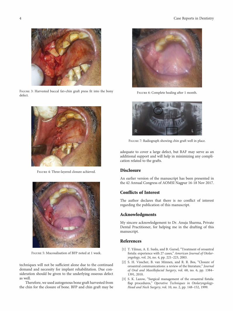
The entire procedure was performed under local anesthe-sia (2% lignocaine with 1 : 80000 adrenaline) using PosteriorSuperior Alveolar block supplemented by infiltration+greaterpalatine nerve block. The fistulous tract was excised in acircumscribed manner along the defect. A modified buccalmucoperiosteal flap was elevated in trapezoidal outline start-ing from the mesial and distal end of the fistulous opening.The periosteum was incised along the posterior aspect ofthe flap to identify BFP. Gentle blunt dissection was doneto harvest the BFP till we obtained the sufficient amount offat to cover the graft and the defect. Next, the cortical bonegraft was harvested from the chin of size matching the defect.The graft was press fit into the defect and did not requirescrew fixation. Then, the BFP was mobilized to cover thegraft, and finally, the mucoperiosteal flap was repositionedand sutured in place (Figures 3 and 4). This way, 3-layeredclosures were performed to reconstruct the bony as well assoft tissue defect. The patient was put on nasogastric tubefeed till the time of suture removal. Mucosalisation of thefat pad was noticed at follow-up visits after 1 week and 1month later (Figures 5 and 6).
The patient was observed for signs of infection, dehis-cence, and necrosis. Postoperative radiograph showed bonegraft well in place (Figure 7). No significant complicationwas noted in the follow-up appointments. Both the pri-mary site and the bone graft donor site healed withoutany dehiscence.
3. Discussion
The commonest cause for the development of OAF includesdental extractions of posterior teeth (molars, premolars) dueto close proximity of the root apices to the sinus floor. Fre-quencies of such occurrences have been reported to bebetween 0.31% and 4.7% [19]. Based on its location, the fistulacan be alveolosinusal, palatosinusal, or vestibulosinusal [20].
Ever since its first clinical use by Egyedi in 1977, BFP hasgained popularity and rightly so because of its advantages:rapid epithelialization, excellent reliable blood supply, ana-tomically favorable position, ease of harvest, low rate of com-plications, and minimal to no donor site morbidity.
Problems that we can come across mainly at the time ofharvesting BFP are perforation or shrinkage. Egyedi advo-cated covering the fat pad with split skin graft to overcomethese issues, but Tideman et al. have shown that BFP wascapable of self-epithelialization within 3-4 weeks of its inset[7, 21]. Nevertheless, covering the BFP might be essential incases of large defects and where the amount of BFP may beinadequate. In such cases, buccal advancement flap is the best
2 Case Reports in Dentistry

option. This combination technique provides more stabilityand provides additional tissue for cover. In our case, this lay-ered closure was needed to provide support to the bone graftand also to maintain a well vascularized environment for thegraft take up.
Our idea of three-layer closure is supported by the studiesof Er et al. and Weinstock et al. [16, 18]. Er et al. observedwound dehiscence in 20% of cases after two-layered closure.Weinstock et al. demonstrated additional benefit of thebuccal flap covering the BFP over the graft in a study witha 10-month follow-up.
There is so much heterogeneity in the methods for OAFclosure, and no particular technique is superior or inferiorto other. Each case is at the discretion of the treating surgeonbased on his experience and preference. The choice of thetechnique must be guided by 4 important assessment criteria:(a) size and type of defect, (b) presence or absence of sinusdisease, (c) minimal donor site morbidity to the patient,and (d) prosthetic considerations. The described techniquehas the added advantage for graft support and minimizingthe chance of graft resorption or wound dehiscence.
4. Conclusion
Repair of OAF is essential to prevent further complicationsrelated to the maxillary sinus. The routine flap based
Table 1: Summarizes various studies demonstrating the use of different modalities for OAF closure.
Author/year Number of cases Type of bone graft
(1) Waldrop TC et al. (1993) 1 Gelatin membrane+DFDBA+ePTFE
(2) Liversedge et al. (2002) 1 BFP+maxillary bone graft
(3) Haas et al. (2003) [12] 5 Chin graft+buccal flap
(4) Watzack G et al. (2005) 21 Retromolar/chin graft
(5) Delgado Galindez (2005) 22 Mandibular graft+mucoperiosteal flap
(6) Ogunsalu et al. (2005) 1 Biooss sandwiched between Biogide
(7) Scala M et al. (2007) bovine bone 3 Cryoplatelet gel+particulate bone maxilla+
(8) Penarrocha Diago M et al. (2007) 1 Zygomatic bone
(9) Lee BK (2008) 1 Ileac bone+palatal flap
(10) Doobrow JH et al. (2008) 1 Collagen matrix+FDB+CaSO4
(11) Scattarella et al. (2010) 1 Autologous bone+particulate xenograft+PTFE
(12) Ahmed MS et al. (2011) 8 Chin/ramus graft+BAF
(13) Er et al. (2013) [16] wall (3) 10 Chin (3), buccal (1), tuberosity (2), ramus (1), maxillary
(14) Cottam JR et al. (2013) 1 Recombinant rhBMP-2+collagen matrix
(15) Pourdanesh F et al. (2013) 1 BFP+pedicled coronoid process+mucosa
(16) Weinstock RJ et al. (2014) [18] 1 Maxilla+BFP flap+buccal advancement flap
(17) De Biasi M et al. (2014) 20 BFP+hydroxyapatite crystals+collagen sheath
(18) Choi N et al. (2015) 1 Scapular tip free flap
(19) Kapustecki M et al. (2016) [14] 20 Mental protuberance (14)+oblique line (6)
(20) S Amroun et al. (2018) 1 Maxillary tuberosity+mucosal closure
Abbreviations: DFDBA: Decalcified Freeze-Dried Bone Allograft; ePTFE: extended polytetrafluoroethylene; BFP: Buccal Fat Pad; Biooss: bone graft material;Biogide: a resorbable membrane; FDB: Freeze-Dried Bone; CaSO4: Calcium Sulphate; BAF: buccal advancement flap; rhBMP-2: recombinant human BoneMorphogenetic Protein-2.
Figure 1: Clinical picture showing fistulous opening withidentifiable bony defect.
Figure 2: Panoramic X-ray demonstrating the underlying osseousdefect.
3Case Reports in Dentistry
techniques will not be sufficient alone due to the continueddemand and necessity for implant rehabilitation. Due con-sideration should be given to the underlying osseous defectas well.
Therefore, we used autogenous bone graft harvested fromthe chin for the closure of bone. BFP and chin graft may be
adequate to cover a large defect, but BAF may serve as anadditional support and will help in minimizing any compli-cation related to the grafts.
Disclosure
An earlier version of the manuscript has been presented inthe 42 Annual Congress of AOMSI Nagpur 16-18 Nov 2017.
Conflicts of Interest
The author declares that there is no conflict of interestregarding the publication of this manuscript.
Acknowledgments
My sincere acknowledgement to Dr. Anuja Sharma, PrivateDental Practitioner, for helping me in the drafting of thismanuscript.
References
[1] T. Yilmaz, A. E. Suslu, and B. Gursel, “Treatment of oroantralfistula: experience with 27 cases,” American Journal of Otolar-yngology, vol. 24, no. 4, pp. 221–223, 2003.
[2] S. H. Visscher, B. van Minnen, and R. R. Bos, “Closure oforoantral communications: a review of the literature,” Journalof Oral and Maxillofacial Surgery, vol. 68, no. 6, pp. 1384–1391, 2010.
[3] S. K. Lazow, “Surgical management of the oroantral fistula:flap procedures,” Operative Techniques in Otolaryngology-Head and Neck Surgery, vol. 10, no. 2, pp. 148–152, 1999.
Figure 3: Harvested buccal fat+chin graft press fit into the bonydefect.
Figure 4: Three-layered closure achieved.
Figure 5: Mucosalisation of BFP noted at 1 week.
Figure 6: Complete healing after 1 month.
Figure 7: Radiograph showing chin graft well in place.
4 Case Reports in Dentistry

[4] R. Candamourty, M. K. Jain, K. Sankar, and M. R. RameshBabu, “Double-layered closure of oroantral fistula using buccalfat pad and buccal advancement flap,” Journal of Natural Sci-ence, Biology and Medicine, vol. 3, no. 2, pp. 203–205, 2012.
[5] E. T. Daif, “Long-term effectiveness of the pedicled buccal fatpad in the closure of a large oroantral fistula,” Journal of Oraland Maxillofacial Surgery, vol. 74, no. 9, pp. 1718–1722, 2016.
[6] H. Dym and J. C. Wolf, “Oroantral communication,” Oral andMaxillofacial Surgery Clinics of North America, vol. 24, no. 2,pp. 239–247, 2012.
[7] P. Egyedi, “Utilization of the buccal fat pad for closure of oro-antral and/or oro-nasal communications,” Journal of Maxillo-facial Surgery, vol. 5, pp. 241–244, 1977.
[8] P. M. J. Tostevin and H. Ellis, “The buccal pad of fat: a review,”Clinical Anatomy, vol. 8, no. 6, pp. 403–406, 1995.
[9] M.-K. Kim, W. Han, and S.-G. Kim, “The use of the buccal fatpad flap for oral reconstruction,” Maxillofacial Plastic andReconstructive Surgery, vol. 39, no. 1, article 5, 2017.
[10] B. Proctor, “Bone graft closure of large or persistent oro-maxillary fistula,” The Laryngoscope, vol. 79, no. 5, pp. 822–826, 1969.
[11] M. Peñarrocha-Diago, B. Garcia, D. Gomez, and J. Balaguer,“Zygomatic bone graft for oral-antral communication closureand implant placement,” The Journal of Oral Implantology,vol. 33, no. 5, pp. 305–309, 2007.
[12] R. Haas, G. Watzak, M. Baron, G. Tepper, G. Mailath, andG. Watzek, “A preliminary study of monocortical bone graftsfor oroantral fistula closure,”Oral Surgery, Oral Medicine, OralPathology, Oral Radiology, and Endodontology, vol. 96, no. 3,pp. 263–266, 2003.
[13] K. A. Schlegel, S. Schultze-Mosgau, J. Wiltfang, F. W. Neukam,S. Rupprecht, and M. Thorwarth, “Changes of mineralizationof free autogenous bone grafts used for sinus floor elevation,”Clinical Oral Implants Research, vol. 17, no. 6, pp. 673–678,2006.
[14] M. Kapustecki, I. Niedzielska, H. Borgiel-Marek, andB. Różanowski, “Alternative method to treat oroantral com-munication and fistula with autogenous bone graft and plateletrich firbin,” Medicina Oral Patología Oral y Cirugia Bucal,vol. 21, no. 5, pp. e608–e613, 2016.
[15] W. G, G. Tepper, W. Zechner, G. Monov, D. Busenlechner,and G. Watzek, “Bony press-fit closure of oro-antral fistulas:a technique for pre-sinus lift repair and secondary closure,”Journal of Oral and Maxillofacial Surgery, vol. 63, no. 9,pp. 1288–1294, 2005.
[16] N. Er, H. Y. Tuncer, Ç. Karaca, and S. Çopuroğlu, “Treatmentof oroantral fistulas using bony press-fit technique,” Journal ofOral and Maxillofacial Surgery, vol. 71, no. 4, pp. 659–666,2013.
[17] M. S. Ahmed and N. A. Askar, “Combined bony closure oforoantral fistula and sinus lift with mandibular bone graftsfor subsequent dental implant placement,” Oral Surgery, OralMedicine, Oral Pathology, Oral Radiology, and Endodontics,vol. 111, no. 4, pp. e8–14, 2011.
[18] R. J. Weinstock, L. Nikoyan, and H. Dym, “Composite three-layer closure of oral antral communication with 10 monthsfollow-up—a case study,” Journal of Oral and MaxillofacialSurgery, vol. 72, no. 2, pp. 266.e1–266.e7, 2014.
[19] B. Gacic, L. Todorovic, V. Kokovic et al., “The closure oforoantral communications with resorbable PLGA-coatedβ-TCP root analogs, hemostatic gauze, or buccal flaps: A
prospective study,” Oral Surgery, Oral Medicine, Oral Pathol-ogy, Oral Radiology, and Endodontology, vol. 108, no. 6,pp. 844–850, 2009.
[20] A. E. Borgonovo, F. V. Berardinelli, M. Favale, andC. Maiorana, “Surgical options in oroantral fistula treatment,”The Open Dentistry Journal, vol. 6, no. 1, pp. 94–98, 2012.
[21] H. Tideman, A. Bosanquet, and J. Scott, “Use of the buccal fatpad as a pedicled graft,” Journal of Oral and MaxillofacialSurgery, vol. 44, no. 6, pp. 435–440, 1986.
5Case Reports in Dentistry

DentistryInternational Journal of
Hindawiwww.hindawi.com Volume 2018
Environmental and Public Health
Journal of
Hindawiwww.hindawi.com Volume 2018
Hindawi Publishing Corporation http://www.hindawi.com Volume 2013Hindawiwww.hindawi.com
The Scientific World Journal
Volume 2018Hindawiwww.hindawi.com Volume 2018
Public Health Advances in
Hindawiwww.hindawi.com Volume 2018
Case Reports in Medicine
Hindawiwww.hindawi.com Volume 2018
International Journal of
Biomaterials
Scienti�caHindawiwww.hindawi.com Volume 2018
PainResearch and TreatmentHindawiwww.hindawi.com Volume 2018
Preventive MedicineAdvances in
Hindawiwww.hindawi.com Volume 2018
Hindawiwww.hindawi.com Volume 2018
Case Reports in Dentistry
Hindawiwww.hindawi.com Volume 2018
Surgery Research and Practice
Hindawiwww.hindawi.com Volume 2018
BioMed Research International Medicine
Advances in
Hindawiwww.hindawi.com Volume 2018
Hindawiwww.hindawi.com Volume 2018
Anesthesiology Research and Practice
Hindawiwww.hindawi.com Volume 2018
Radiology Research and Practice
Hindawiwww.hindawi.com Volume 2018
Computational and Mathematical Methods in Medicine
EndocrinologyInternational Journal of
Hindawiwww.hindawi.com Volume 2018
Hindawiwww.hindawi.com Volume 2018
OrthopedicsAdvances in
Drug DeliveryJournal of
Hindawiwww.hindawi.com Volume 2018
Submit your manuscripts atwww.hindawi.com