three protein cocktails mediate delayed-type ...three protein cocktails mediate delayed-type...
TRANSCRIPT
INFECTION AND IMMUNITY, Feb. 2011, p. 716–723 Vol. 79, No. 20019-9567/11/$12.00 doi:10.1128/IAI.00486-10Copyright © 2011, American Society for Microbiology. All Rights Reserved.
Three Protein Cocktails Mediate Delayed-Type Hypersensitivity ResponsesIndistinguishable from That Elicited by Purified Protein Derivative in the
Guinea Pig Model of Mycobacterium tuberculosis Infection�
Hongliang Yang,1 JoLynn Troudt,1 Ajay Grover,1 Kimberly Arnett,1 Megan Lucas,1 Yun Sang Cho,1,2
Helle Bielefeldt-Ohmann,3 Jennifer Taylor,1 Angelo Izzo,1 and Karen M. Dobos1*Colorado State University, Department of Microbiology, Immunology, and Pathology, Fort Collins, Colorado 805231; Bacteriology and
Parasitology Division, National Veterinary Research and Quarantine Service, Anyang 430-824, Republic of Korea2; andSchool of Veterinary Science, University of Queensland, Gatton Campus, Gatton Qld 4343, Australia3
Received 10 May 2010/Returned for modification 29 June 2010/Accepted 21 November 2010
Purified protein derivative (PPD) is a widely used reagent for the diagnosis of Mycobacterium tuberculosisinfection. Recently, the molecular composition of PPD was defined, with hundreds of mycobacterial proteinrepresentatives making up PPD. Which, if any, of these specific products drive the potency of PPD remains inquestion. In this study, two proteins (DnaK and GroEL2) previously identified as dominant proteins in PPDwere tested for the capacity to induce delayed-type hypersensitivity (DTH) responses in H37Rv-infected orBCG-vaccinated guinea pigs. These two proteins were used in pull-down assays to identify interacting PPDproducts. Six proteins were identified as interacting partners with DnaK and GroEL2, i.e., Rv0009, Rv0475,Rv0569, Rv0685, Rv2626c, and Rv2632c. These six proteins were tested alone and in combination with DnaKand GroEL2 for the capacity to induce a DTH response in the guinea pig model. From these studies, twococktails, DnaK/GroEL2/Rv0009 and DnaK/GroEL2/Rv0685, were found to induce DTH responses in H37Rv-infected or BCG-vaccinated guinea pigs that were indistinguishable from DTH responses driven by a PPDinjection. The mechanism by which DTH responses were induced was elucidated by histologic examination,analysis of activated CD4�/CD8� T cells, and cytokine mRNA expression at the site of the DTH response. PPDand the protein cocktails tested induced strong DTH responses in H37Rv-infected guinea pigs. Ex vivophenotyping of T cells at the DTH site indicated that this response is mediated by activated CD4� and CD8�
T cells, with increases in gamma interferon and tumor necrosis factor alpha, but not interleukin-10, at the siteof the DTH response. Our results demonstrate for the first time that the PPD response can be mimicked at themolecular level with defined protein cocktails. The use of this defined product will allow a more thoroughunderstanding of the DTH response and may provide a platform for more rapid and sensitive second-generation skin test reagents for the diagnosis of M. tuberculosis infection.
Tuberculosis remains one of the largest single causes ofdisease and death from an infectious agent, particularly indeveloping nations. With nearly 2 million deaths each year andan estimated 8 to 9 million new cases annually, tuberculosis isthe second leading cause of death worldwide among commu-nicable diseases (8). Further, it is estimated that one-third ofthe world’s population is latently infected with Mycobacteriumtuberculosis (9). The epidemic of human immunodeficiencyvirus infection (27) and the increase in the incidence of mul-tidrug-resistant M. tuberculosis (10) have further complicatedtuberculosis control efforts.
The most common way persons infected with M. tuberculosisare identified is through use of the tuberculin skin test (TST).Over the last century, the TST has been proven to be an impor-tant diagnostic tool for tuberculosis infection (25, 35). It is also animportant component of epidemiologic studies to evaluate theprevalence of latent tuberculosis in various populations.
The TST, which is also called the Mantoux test after theFrench physician Charles Mantoux (1877 to 1947), was devel-
oped by Florence B. Seibert (1897 to 1991) (33). The TST is amodification of Koch’s old tuberculin, resulting in purifiedprotein derivative (PPD). Several PPD standards exist, includ-ing PPD-S (1, 16), the first FDA standard; PPD-RT23 (7, 30),the standard produced by the Statens Serum Institut; andPPD-S2 (37), the new FDA standard of PPD. The basic meth-ods used to produce these PPD standards are similar. Proteinsare purified by repeated precipitation with trichloroacetic acid,ammonium sulfate, or both after cultures of M. tuberculosis aregrown to stationary phase and sterilized (34). The preparationsare dominated by M. tuberculosis proteins and peptides andcontain few polysaccharides, nucleic acids, and lipids. The elu-cidation of the molecular composition of PPD demonstratedthat PPD is a complex mixture of hundreds of denatured an-tigens (3, 19; Y. S. Cho et al., unpublished data). It is notsurprising, then, that little is known about the active compo-nents of PPD responsible for its potency. The presence of somany antigens likely contributes to the poor specificity ob-served with PPD testing; it cannot differentiate active tubercu-losis from latent infection or vaccination, complicating TSTuse as a tool for epidemiologic studies and diagnosis of tuber-culosis. PPD from different sources may vary in the delayed-type hypersensitivity (DTH) response because there is no wayto standardize the hundreds of antigenic components in PPD
* Corresponding author. Mailing address: 1682 Campus Delivery,Department of Microbiology, Immunology, and Pathology, ColoradoState University, Fort Collins, CO 80523-1682. Phone: (970) 491-6549.Fax: (970) 491-1815. E-mail: [email protected].
� Published ahead of print on 6 December 2010.
716
on March 1, 2020 by guest
http://iai.asm.org/
Dow
nloaded from
(23). In addition to issues arising from the number of proteinspresent in PPD, three of the most dominant proteins in PPD,Rv3418 (GroES), Rv0440 (GroEL2), and Rv0350 (DnaK) (3,19; Cho et al., unpublished), are highly conserved chaperones;reactivity to these proteins may also explain the diminishedspecificity of PPD. Lastly, the complexity of PPD limits ourcapacity to tease out the molecular interactions involved in theDTH response and, in particular, understand which of theseinteractions, if any, discern chronic infections from protectiveimmunity. Skin test reagents using defined antigens may beable to be used to address these questions.
Over the past 20 years, major efforts have focused on theidentification of highly purified M. tuberculosis recombinantproteins that may be used to develop a more specific diagnosticskin test (31). Some protein candidates, including ESAT-6(28), CFP10 (39), MPT64 (24), DPPD (6), and Rv0934 (17),were tested and found to induce strong DTH reactions inguinea pigs, but none of them were developed as second-generation skin test reagents. Several studies indicate that acombination of several purified antigens (antigen cocktails) ora combination of several DTH-inducing epitopes may be re-quired for second-generation skin test reagents to effectivelyreplace PPD (21, 24, 32).
The definition of the molecular composition of PPD allowsfor the development of a more refined product. Recently, fourheat shock proteins (GroES, GroEL2, HspX, and DnaK) wereidentified as dominant products in PPD-S2, the U.S. FDAstandard PPD (Cho et al., unpublished). In this study, DnaKand GroEL2, two of the major chaperones in PPD-S2 (Cho etal., unpublished) and bovine and avian PPD (3), were testedfor the capacity to induce a DTH response and were found toelicit DTH responses in H37Rv-infected and BCG-vaccinatedguinea pigs. As these proteins are conserved among othermycobacteria, their use in a second-generation TST may belimited. Thus, these proteins were used to pull out additionalprotein representatives from PPD, resulting in the identifica-tion of six highly interacting proteins. When these proteinswere tested in various formulations for the potential to elicit aDTH response, two cocktails, DnaK/GroEL2/Rv0685 andDnaK/GroEL2/Rv0009, were found to induce DTH responsesthat were indistinguishable from that induced by PPD in theguinea pig model of tuberculosis, as determined by skin testresponses and at the molecular level.
MATERIALS AND METHODS
Expression of recombinant proteins in Escherichia coli. The four chaperones,DnaK (Rv0350), GroEL2 (Rv0440), GroES (Rv3418c), and HspX (Rv2031c),and M. tuberculosis protein Rv2626c were expressed using plasmids availablefrom the Tuberculosis Vaccine Testing and Research Material contract(HHSN266200400091C; http://www.cvmbs.colostate.edu/mip/tb/recombinant.htm).
Gateway cloning-based (Invitrogen, Carlsbad, CA) entry plasmids containing theopen reading frames for M. tuberculosis proteins Rv0009, Rv0475, Rv0569, Rv0685,Rv2626c, and Rv2632c were obtained from the Pathogen Functional GenomicsResource Center (http://pfgrc.jcvi.org/fir_index.php/fir/available_libraries.html). LRClonase II Enzyme Mix (Invitrogen, Carlsbad, CA) was used to catalyze homologousrecombination between each entry plasmid and the pDEST17 destination vector(Invitrogen), thus creating an expression plasmid for the gene of interest. Theresulting expression clones were transformed to DH5� competent cells (Invitrogen).Plasmids were prepared using a QIAprep Spin Miniprep kit (Qiagen, Valencia, CA).The sequence fidelity of expression clones was confirmed by DNA sequencing(Proteomics and Metabolomics Facility, Colorado State University).
E. coli BL21(DE3)pLysS or BL21(DE3) host cells (Invitrogen) were trans-
formed with the expression clones. The pDEST17 destination vector contains anN-terminal His tag allowing for affinity purification of expressed proteins. Re-combinant His-tagged proteins were expressed following induction with IPTG(isopropyl-�-D-thiogalactopyranoside; Calbiochem, Darmstadt, Germany) andthen purified by affinity chromatography using nickel (Ni)-charged His-bind resin(Novagen, Darmstadt, Germany). Resin containing bound recombinant proteinwas washed with 10 mM Tris-HCl containing 0.5% ASB-14 (Calbiochem, Darm-stadt, Germany) to remove endotoxin prior to elution of proteins with imidazole.Purified proteins were dialyzed against 10 mM ammonium bicarbonate overnightat 4°C. The purity of recombinant proteins was assessed by sodium dodecylsulfate-polyacrylamide gel electrophoresis (SDS-PAGE), followed by silverstaining. The identification of His-tagged recombinant proteins was performedby Western blotting with mouse monoclonal IgG1 anti-DnaK and anti-GroEL2primary antibodies (IT41 and IT13, respectively, provided by the TuberculosisVaccine Testing and Research Materials contract) or mouse monoclonal IgG1Penta-His antibody (Qiagen, Valencia, CA). The reaction was detected with anti-mouse antibody conjugated to allophycocyanin, followed by development with 5-bromo-4-chloro-3-indolylphosphate (BCIP)–nitroblue tetrazolium (Sigma, St. Louis,MO). The concentration of recombinant proteins was determined with the bicin-choninic acid protein assay (Pierce, Rockford, IL). The endotoxin level for all theproteins was �10 ng�1 ml�1 mg�1 protein, as determined by the Limulus amebocytelysate QCL-1000 kit (Cambrex, Walkersville, MD), performed according to themanufacturer’s instructions.
Identification of GroEL2 and DnaK interacting proteins. Purified recombi-nant DnaK or GroEL2 or a mixture of the two proteins was used in pull-downassays to identify protein representatives in PPD which bound to DnaK andGroEL2. Briefly, 0.5 mg of recombinant DnaK, GroEL2, or a 1:1 mixture ofDnaK and GroEL2 was incubated with 5.0 mg of PPD overnight at 4°C. Theprotein-PPD mixture was then passed separately over Ni2� affinity resin (Hisband Quick 300 cartridges; Novagen, Darmstadt, Germany) to capture the re-combinant protein and any PPD products bound to it, the resin was washedextensively with 10 mM Tris-HCl to remove nonspecifically bound material, andthe bound material was eluted with imidazole as described above.
Mass spectrometry. The pull-down assay-eluted samples were reduced, alkyl-ated, trypsin digested, and dissolved in 5% acetonitrile–0.1% acetic acid forprotein identification by liquid chromatography-mass spectrometry (MS) as de-scribed previously (22). Tandem mass spectra were extracted, charge state de-convoluted, and deisotoped by BioWorks version 3.2. All tandem MS (MS/MS)samples were analyzed using Sequest (version 2.7; ThermoFinnigan, San Jose,CA) set up to search the M. tuberculosis database (GenBank accession no.AL123456, R9, 3913 entries) assuming the digestion enzyme trypsin and a max-imum of four missed cleavages. Sequest was searched with a fragment ion masstolerance of 1.00 Da and a parent ion tolerance of 2.5 Da. Scaffold (versionScaffold_2_05_02; Proteome Software Inc., Portland, OR) was used to validateMS/MS-based peptide and protein identifications. Peptide identifications wereaccepted if they could be established at greater than 95.0% probability as spec-ified by the Peptide Prophet algorithm. Protein identifications were accepted ifthey could be established at greater than 90.0% probability and contained at leasttwo identified peptides.
Guinea pig model. Specific-pathogen-free, outbred Hartley guinea pigs (450 to500 g at the beginning of the experiment) from Charles River Laboratories(Wilmington, MA) were maintained under biosafety level 3 conditions. Guineapigs were randomly assigned to one of three groups: BCG vaccinated, H37Rvinfected, or negative control. All animal experiment procedures were approvedby the Animal Care and Use Committee of Colorado State University.
BCG vaccination and aerosol challenge. Vaccinations were performed bysubcutaneous injection of 0.1 ml (103 CFU) of BCG Pasteur.
Guinea pigs were infected using a Madison chamber aerosol generation deviceto deliver M. tuberculosis H37Rv at a low aerosol dose of 20 bacilli. Subcutaneousinjection with 100 �l endotoxin-free phosphate-buffered saline (PBS) was used asa negative control.
Skin testing and biopsy. BCG-vaccinated or H37Rv-infected guinea pigs wereexamined for a DTH response induced by different formulations (PPD-S2, DnaK,GroEL2, GroES, HspX, DnaK/GroEL2, DnaK/GroEL2/Rv0009, DnaK/GroEL2/Rv0475, DnaK/GroEL2/Rv0569, DnaK/GroEL2/Rv0685, DnaK/GroEL2/Rv2626c,DnaK/GroEL2/Rv2632c, and PBS as a negative control). The skin test, according tothe Mantoux technique, was performed 10 weeks after aerosol infection or BCGvaccination. The hair on the ventral side was shaved off, and the guinea pigs wereinjected intradermally with 0.1 ml antigen solution diluted in endotoxin-free PBS.The PPD-S2 and formulations containing single proteins were used at a dose of 0.4�g. The DnaK/GroEL2 and three-protein formulations all used 0.13 �g of eachprotein.
Erythema and induration were measured with a micrometer after 24 and 48 h.
VOL. 79, 2011 NOVEL SECOND-GENERATION TUBERCULOSIS SKIN TEST REAGENT 717
on March 1, 2020 by guest
http://iai.asm.org/
Dow
nloaded from
The diameter (in millimeters) was defined as the mean of vertical and transversemeasurements.
Skin samples were obtained from infected group guinea pigs. The guinea pigswere euthanized by injection with sodium pentobarbital (Sleepaway; Fort DodgeLaboratories, Inc., Fort Dodge, IA) 48 h after antigen stimulation. The injectionsite skin was rinsed with 70% ethanol, removed, and then fixed with 10%formalin for histological examination. Skin samples were prepared for RNAextraction (30- to 50-mg skin sample in 0.6 ml RNAlater [Qiagen]) or cellisolation (the same area of skin sample in 1.5 ml of incomplete RPMI medium).The samples for RNA extraction were homogenized in RNAlater and immedi-ately stored at �20°C. The samples for cell isolation were kept on ice and usedimmediately after biopsy.
Histological examination. Following fixation in formalin, the skin tissues wereroutinely processed for paraffin embedding. Sections were cut at a 4- to 5-�mthickness, stained with hematoxylin and eosin (H&E), and examined on a NikonEclipse 50i microscope. Microphotographs were taken with a DS-Fi1 camera andassociated software (Nikon, Japan).
Flow cytometric analysis of cell surface markers. Flow cytometry was per-formed to investigate the subgroups of T cells in infected guinea pigs stimulatedwith different formulations. Briefly, after the removal of subcutaneous fat andmuscle tissues by scraping, the skin samples were minced into small pieces anddigested for 1 h at 37°C with 2 mg/ml collagenase type IV and 1.2 U/ml dispase(Worthington, Lakewood, NJ). The digested skin samples were further disruptedby gently pushing the tissue through a 70-�m cell strainer (BD Biosciences, SanJose, CA). Red blood cells were lysed with ACK buffer (0.14 M NH4Cl, 1.0 mMKHCO3, pH 7.2 to 7.4) and then washed and resuspended in complete RPMImedium. The viability of digested skin cells was determined by trypan bluestaining. Single-cell suspensions from each individual guinea pig were stainedwith fluorescently labeled monoclonal antibodies against CD4 (clone FITC CT7;Serotec Inc., Raleigh, NC) (36), CD8 (clone FITC CT6; Serotec Inc.) (36), pan-Tcells (clone APC CT5; Serotec Inc.) (36), and CD62L (clone PE lam1-116; SantaCruz Biotechnology, Santa Cruz, CA) (13) at 4°C for 30 min. Antibodies wereused at 0.2 �g/106 cells. Cells were gated on lymphocytes by forward and sidescatter according to their characteristic scatter profiles and further gated basedon pan-T and CD4 or CD8 expression.
All analyses were performed with an acquisition of at least 100,000 eventson a Becton Dickinson FACScalibur flow cytometer (BD Biosciences, SanJose, CA), and the data were analyzed using FlowJo software (Tree Star Inc.,Ashland, OR).
Total RNA isolation and real-time quantitative reverse transcription (RT)-PCR assay. To determine the quantities of cytokine mRNA in skin samples,real-time RT-PCR was performed for the H37Rv-infected guinea pig group.First, the skin samples were homogenized and released skin RNA was extractedusing the RNeasy mini kit with columns (Qiagen, Valencia, CA). ContaminatingDNA was removed by on-column treatment with RNase-free DNase (Qiagen,Valencia, CA) according to the manufacturer’s instructions. The yield and qual-ity of total RNA were examined using an ND1000 spectrophotometer (Nano-Drop Technologies, Wilmington, DE). The RNA was then stored at �80°C untilrequired for gene expression studies. RT was performed using an iScript cDNAsynthesis kit (Qiagen, Valencia, CA). Real-time RT-PCR, performed with iQCYBR green supermix (Bio-Rad, Hercules, CA), was carried out using primersfor guinea pig �-actin, gamma interferon (IFN-�), tumor necrosis factor alpha(TNF-�), and interleukin-10 (IL-10) as described previously (15). Results wererecorded as relative gene expression after normalization for �-actin thresholdcycle (CT) values by the 2���CT method (20). All real-time RT-PCR data arepresented such that the values for stimulated samples using different formula-tions are expressed relative to the value for the PBS-stimulated site on the sameanimal.
Statistical analysis. Descriptive analyses were carried out for histopathologi-cal features. All other data were expressed as the mean the SEM (standarderror of the mean). The DTH response data were analyzed by one-tailed Studentt test. The Mann-Whitney test was used to characterize changes in cytokinemRNA expression and cell surface markers between groups of guinea pigs. Allstatistical comparisons were carried out using GraphPad Prism version 4.00(GraphPad Software, San Diego, CA). P values of �0.05 were considered sta-tistically significant.
RESULTS
DTH responses of the dominant chaperones in PPD. Fourchaperones (DnaK, GroEL2, GroES, and HspX) were ex-pressed as recombinant proteins in E. coli BL21(DE3)pLysS
cells and purified using Ni-charged His-bind resin (data notshown). M. tuberculosis-infected, BCG-vaccinated, and naïveguinea pigs were used to evaluate DTH responses to thesechaperones, with PPD and saline as positive and negative con-trols, respectively. The results demonstrated that among thefour recombinant proteins used, only DnaK and GroEL2 elic-ited DTH responses ranging from weak to significant (onestudy demonstrated mean responses of 3.36 4.53 and10.04 2.29 mm for DnaK and GroEL2, respectively). Allfour chaperones were combined in various formulations andtested for the capacity to induce a DTH response. Only thecombination of DnaK and GroEL2 augmented the DTH re-sponse in infected and vaccinated animals (one study demon-strated mean responses of 10.68 2.32 mm for DnaK/GroEL2versus 9.46 1.83 mm for PPD). None of the naïve guinea pigsdeveloped a reaction to any of the antigens (data not shown).Dose-response assays were performed with injection dosesranging from 0.1 to 1.2 �g total protein/site. Positive DTHresponses were observed for all doses, with 0.25 to 0.4 �g totalprotein/site being the optimum dose (data not shown).
Pull-down assays. Ni chromatography was used to pull downprotein representatives and peptides in PPD that bound torecombinant DnaK and GroEL2. From this, DnaK andGroEL2 were found to interact with each other, and six addi-tional protein representatives were identified in the pull-downassay elution buffer, indicating an interaction with these twochaperones (Table 1). All six proteins are small-size proteinswith native molecular masses ranging from 9.5 to 22 kDa,except Rv0685, with a molecular mass of 44 kDa. As expectedfrom previous SDS-PAGE analyses of PPD (3; Cho et al.,unpublished), only recombinant DnaK and GroEL2 and noneof the six new products were observed as discrete proteinbands upon SDS-PAGE analysis of eluted samples (data notshown), due to the fact that PPD is composed mostly of proteinrepresentatives or aggregates of partially denatured proteinsand peptides rather than discrete protein molecules (3; Cho etal., unpublished).
DTH responses to protein cocktails. All of the proteinsidentified as interacting partners in the pull-down assay wereexpressed and purified as soluble recombinant products, ex-cept Rv0685, which was purified from inclusion bodies. Toestablish the diagnostic potential of these proteins, we evalu-ated DTH responses to these proteins, individually and com-
TABLE 1. Proteins identified in PPD-S2 through DnaK andGroEL2 pull-down assay
Accessionno. Product
Molecularmass
(kDa)
Binds DnaK,GroEL2, or
both
Rv0009 Probable iron-regulated peptidyl-prolyl cis-trans isomerase APpiA
19.2 Both
Rv0350 Probable chaperone protein DnaK 66.8 GroEL2Rv0440 60-kDa chaperone protein GroEL2 56.7 DnaKRv0475 Iron-regulated heparin-binding
hemagglutinin HbhA21.5 Both
Rv0569 Hypothetical protein 9.5 BothRv0685 Elongation factor Tu 43.6 BothRv2626c Hypothetical protein 15.5 BothRv2632c Hypothetical protein 10.6 Both
718 YANG ET AL. INFECT. IMMUN.
on March 1, 2020 by guest
http://iai.asm.org/
Dow
nloaded from
bined with DnaK and GroEL2, in M. tuberculosis H37Rv-in-fected, BCG-vaccinated, and naïve guinea pigs. None of thenaïve guinea pigs developed a reaction to any of the formula-tions (data not shown). The results showed that there was noDTH response when the six proteins were injected individually(data not shown); however, distinct DTH responses could bemeasured when proteins were combined with DnaK andGroEL2 (Fig. 1). As expected, PPD did not discriminate be-tween the infected and BCG-vaccinated guinea pigs, with DTHresponses of 8.68 0.34 mm (mean responses of erythema SEM) and 8.94 0.4 mm, respectively. Like PPD, all of theother formulations tested elicited almost identical DTH reac-tions in M. tuberculosis-infected and BCG-vaccinated guineapigs. However, both DnaK/GroEL2/Rv0009 and DnaK/GroEL2/Rv0685 induced stronger DTH responses than did the DnaK/GroEL2 formulation alone (P � 0.05); the DTH response toDnaK/GroEL2/Rv0569 was equivalent to that to DnaK/GroEL2alone. Interestingly, the DnaK/GroEL2/Rv0475, DnaK/GroEL2/Rv2626c, and DnaK/GroEL2/Rv2632c formulations weakenedthe DTH responses in both groups (P � 0.05) compared to thatelicited by DnaK/GroEL2 (data not shown for DnaK/GroEL2/Rv2626c).
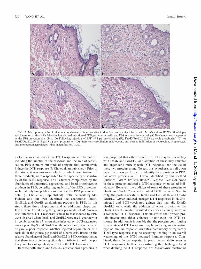
Histological examination of DTH sites from infected guineapigs. To assess if the same type of cellular and inflammatoryresponse was induced by using different formulations and PPD,microscopic examination of H&E-stained sections was per-formed on skin test sites from infected guinea pigs (Fig. 2).This revealed that the different protein formulations induced
pathological changes similar to that of PPD. In all cases, theinflammation in the dermis was characterized by vasodilation,edema, degranulation of mast cells, and infiltration of granu-locytes, mainly neutrophils, as well as lymphocytes and mono-cytes, at the site of antigen injection. These results suggest thatthe different formulations induced a DTH response thatclosely mimics that of PPD. No significant response was ob-served at the site of the saline injection in infected animals.
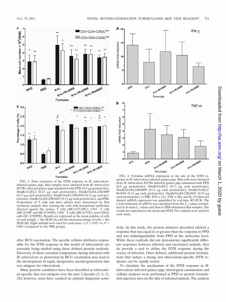
Flow cytometry. To determine the phenotype of the T cellsrecruited to the DTH reaction site, flow cytometry was per-formed on skin samples from infected guinea pigs stimulatedwith four different protein cocktails and PPD. The PBS injec-tion site was used to measure nonspecific T-cell accumulation.The absolute number and percentage of T subgroup cells pos-itive for each marker studied was determined by comparison toa sample stained with an isotype control antibody. We ob-served a 3- to 4-fold increase in both the absolute number andthe percentage of CD3� CD4� CD62Llow and CD3� CD8�
CD62Llow T cells in animals treated with the different formu-lations compared to those of animals treated with the PBScontrol (P � 0.05 or P � 0.001, Fig. 3A and B). An increasewas also observed in terms of the percentage of CD3� CD4�
CD62Llow T cells in the DnaK/GroEL2 and DnaK/GroEL2/Rv0685 groups compared to that in the PPD group (P � 0.05,Fig. 3B); however, there were no differences between differentformulations when the data on the absolute numbers of CD3�
CD4� CD62Llow and CD3� CD8� CD62Llow T cells in thesamples were analyzed (Fig. 3A).
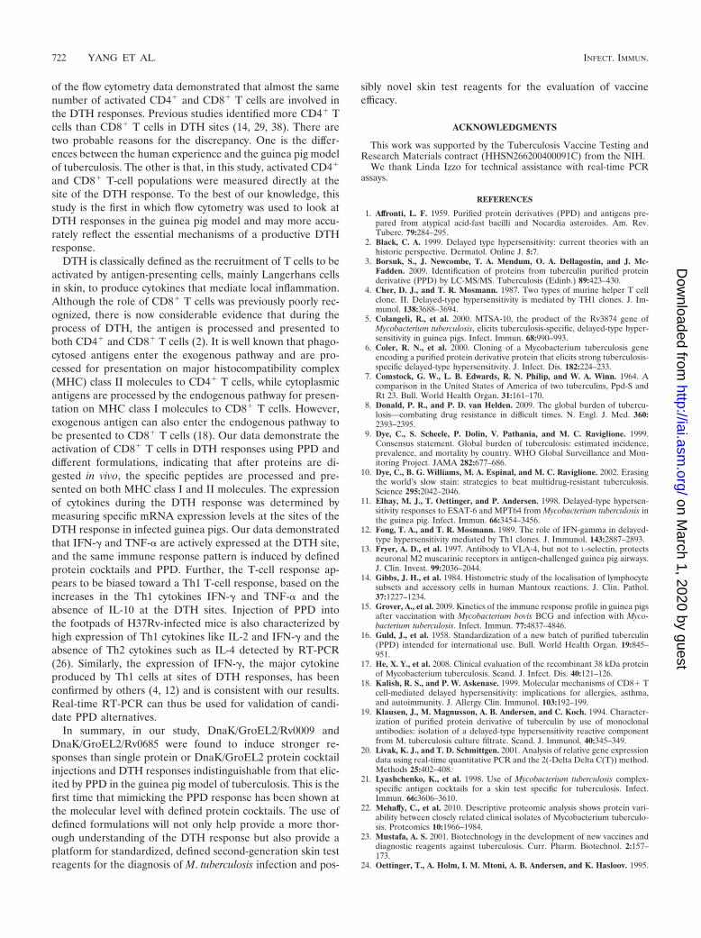
Cytokine expression. To investigate further the potentialdifferences in the cytokine secretion pattern of skin cells at theDTH sites, we analyzed cytokine expression by real-time RT-PCR in infected animals using four protein cocktails and PPD.The results demonstrate that IFN-� and TNF-� were signifi-cantly upregulated in response to all of the formulations overPBS (Fig. 4). While there were no significant differences incytokine induction between different formulations, there was atrend of higher TNF-� expression in three of the protein cock-tails and PPD versus the cocktail containing DnaK andGroEL2 only. The results also showed an absence of detect-able IL-10 mRNA in any of the samples.
DISCUSSION
In this study, we tested four dominant proteins found in PPDfor their potential to induce a DTH response in M. tuberculo-sis-infected or BCG-vaccinated guinea pigs. A baseline DTHresponse was established for two of these proteins, DnaK andGroEL2. Products in PPD that interacted with these two chap-erones were identified by pull-down assays and mass spectrom-etry, produced as recombinant antigens, and tested alone or incombination with DnaK and GroEL2 for the capacity to in-duce a DTH response. Three cocktails, each containing threeproteins, were as potent as PPD in the guinea pig model oftuberculosis. The primary molecular mechanisms responsiblefor inducing a DTH response were indistinguishable whendifferences between these cocktails and PPD were evaluated.Thus, the biological potency of PPD can be reproduced usinga simplified, three-protein cocktail mixture of DnaK/GroEL2/Rv0009, DnaK/GroEL2/Rv0569, or DnaK/GroEL2/Rv0685.These well-defined cocktails can be used to further define the
FIG. 1. Skin test responses in M. tuberculosis H37Rv-infected orBCG-vaccinated guinea pigs following intradermal injection with PPD(0.4 �g/site), DnaK/GroEL2 (0.13 �g of each protein/site), DnaK/GroEL2/Rv0009 (0.13 �g of each protein/site), DnaK/GroEL2/Rv0569(0.13 �g of each protein/site), DnaK/GroEL2/Rv0685 (0.13 �g of eachprotein/site), DnaK/GroEL2/Rv0475 (0.13 �g of each protein/site),DnaK/GroEL2/Rv2632c (0.13 �g of each protein/site), and PBS (datanot shown). The 48-h DTH (skin erythema measurement) response isshown. No DTH reactions were found in naïve guinea pigs stimulatedwith any of the antigens or in any animals stimulated with PBS alone.The results shown are from one representative experiment (eight an-imals per group; all experiments were duplicated) and are expressed asthe mean and SEM. *, P � 0.05 (compared to the DnaK/GroEL2group).
VOL. 79, 2011 NOVEL SECOND-GENERATION TUBERCULOSIS SKIN TEST REAGENT 719
on March 1, 2020 by guest
http://iai.asm.org/
Dow
nloaded from
molecular mechanisms of the DTH response in tuberculosis,including the kinetics of the response and the role of sensiti-zation. PPD contains hundreds of antigens that cumulativelyinduce the DTH response (3; Cho et al., unpublished). Prior tothis study, it was unknown which, or which combination, ofthese products, were responsible for the specificity or sensitiv-ity of the DTH response. This is further complicated by theabundance of denatured, aggregated, and lysed proteinaceousproducts in PPD, complicating analysis of the PPD proteome,such that only two publications describe the PPD proteome indetail (3; Cho et al., unpublished). Both the work by Mc-Fadden and our own identified the chaperones DnaK,GroEL2, and GroES as dominant products in PPD. In thisstudy, these three chaperones and an additional chaperone,HspX, were tested using the guinea pig model of M. tubercu-losis infection. DTH responses similar to that induced by PPDwere observed when DnaK and GroEL2 were used separately orin combination in M. tuberculosis-infected or BCG-vaccinatedguinea pigs. HspX and GroES, on the other hand, were negativeor gave a poor response, whether injected separately or as acocktail, in the guinea pig model of tuberculosis. Based on therelative abundance of DnaK and GroEL2 in PPD, we hypothesizethat these two proteins significantly contribute to both the po-tency and lack of specificity of PPD in the DTH response.
Because both DnaK and GroEL2 are chaperone proteins, it
was proposed that other proteins in PPD may be interactingwith DnaK and GroEL2, and addition of these may enhanceand engender a more specific DTH response than the use ofthese two proteins alone. To test this hypothesis, a pull-downexperiment was performed to identify these proteins in PPD.Six novel proteins in PPD were identified by this method(Rv0009, Rv0475, Rv0569, Rv0685, Rv2626c, Rv2632c). Noneof these proteins induced a DTH response when tested indi-vidually. However, the addition of some of these proteins toDnaK and GroEL2 elicited a potent DTH response. Specifi-cally, the protein cocktails DnaK/GroEL2/Rv0009 and DnaK/GroEL2/Rv0685 induced stronger DTH responses in H37Rv-infected and BCG-vaccinated guinea pigs than did DnaK/GroEL2 only, while the addition of other proteins to theDnaK/ GroEL2 mixture resulted in either an equally active ora weakened DTH response. This illustrates that protein-pro-tein interactions either enhance or abrogate the DTH re-sponse. In addition, it is possible that the proteins contributingto a weakened DTH response may be inducing an alternativetype of immune response. An anti-inflammatory or regulatoryT-cell-type response may be occurring, leading to an overallweakening of the DTH-mediated immune response. Com-bined, these factors explain, in part, the variability seen inDTH responses, further demonstrating the challenges facedwhen defining the DTH response in M. tuberculosis infection or
FIG. 2. Microphotographs of inflammatory changes at injection sites in skin from guinea pigs infected with M. tuberculosis H37Rv. Skin biopsyspecimens were taken 48 h following intradermal injection of PPD, protein cocktails, and PBS as a negative control. (A) No changes were apparentat the PBS injection site. (B to D) Following injection of PPD (0.4 �g protein/site) (B), DnaK/GroEL2 (0.13 �g each protein/site) (C), orDnaK/GroEL2/Rv0685 (0.13 �g each protein/site) (D), there was vasodilation, mild edema, and dermal infiltration of neutrophils, lymphocytes,and monocyte/macrophages. Final magnification, 200.
720 YANG ET AL. INFECT. IMMUN.
on March 1, 2020 by guest
http://iai.asm.org/
Dow
nloaded from
after BCG vaccination. The specific cellular attributes respon-sible for the DTH response in this model of tuberculosis arecurrently being studied using these defined protein cocktails.Discovery of these essential components during infection withM. tuberculosis or protection by BCG vaccination may lead tothe development of rapid, inexpensive second-generation skintest antigens for tuberculosis.
Many protein candidates have been described as tuberculo-sis-specific skin test antigens over the past 2 decades (5, 6, 11,24); however, none have reached an optimal diagnostic sensi-
tivity. In this study, the protein mixtures described elicited aresponse that was equal to or greater than the response to PPDand was indistinguishable from PPD at the molecular level.While these cocktails did not demonstrate significantly differ-ent responses between infected and vaccinated animals, theydo provide a tool to define the DTH response during thecourse of infection. Once defined, additional protein formula-tions that induce a strong and tuberculosis-specific DTH re-sponse can be rapidly tested.
To elucidate the mechanism of the DTH response in M.tuberculosis-infected guinea pigs, histological examination andcellular analyses were performed at PPD or protein formula-tion injection sites on the skin of infected animals. The analysis
FIG. 3. Flow cytometry of the DTH response in M. tuberculosis-infected guinea pigs. Skin samples were obtained from M. tuberculosisH37Rv-infected guinea pigs stimulated with PPD (0.4 �g protein/site),DnaK/GroEL2 (0.13 �g each protein/site), DnaK/GroEL2/Rv0009(0.13 �g each protein/site), DnaK/GroEL2/Rv0569 (0.13 �g each pro-tein/site), DnaK/GroEL2/Rv0685 (0.13 �g each protein/site), and PBS.Proportions of T cells and their subsets were determined by flowcytometry analysis after staining the cells with monoclonal antibodiesdirected against the surface T cells (MCA751APC), CD4� T cells(MCA749F and MCA749PE), CD8� T cells (MCA752F), and CD62Lcells (SC-13505PE). Results are expressed as the mean number of cellsof each sample the SEM (A) and the mean percentage of cells theSEM (B). Eight animals were used for each assay. *, P � 0.05; **, P �0.001 (compared to the PBS group).
FIG. 4. Cytokine mRNA expression at the site of the DTH re-sponse in M. tuberculosis-infected guinea pigs. Skin cells were obtainedfrom M. tuberculosis H37Rv-infected guinea pigs stimulated with PPD(0.4 �g protein/site), DnaK/GroEL2 (0.13 �g each protein/site),DnaK/GroEL2/Rv0009 (0.13 �g each protein/site), DnaK/GroEL2/Rv0569 (0.13 �g each protein/site), DnaK/GroEL2/Rv0685 (0.13 �geach protein/site), or PBS. IFN-� (A), TNF-� (B), and IL-10 (data notshown) mRNA expression was quantified by real-time RT-PCR. Then-fold induction of mRNA was calculated from the CT values normal-ized to �-actin CT values and then to PBS-stimulated skin samples. Theresults are expressed as the mean and SEM. Five animals were used foreach assay.
VOL. 79, 2011 NOVEL SECOND-GENERATION TUBERCULOSIS SKIN TEST REAGENT 721
on March 1, 2020 by guest
http://iai.asm.org/
Dow
nloaded from
of the flow cytometry data demonstrated that almost the samenumber of activated CD4� and CD8� T cells are involved inthe DTH responses. Previous studies identified more CD4� Tcells than CD8� T cells in DTH sites (14, 29, 38). There aretwo probable reasons for the discrepancy. One is the differ-ences between the human experience and the guinea pig modelof tuberculosis. The other is that, in this study, activated CD4�
and CD8� T-cell populations were measured directly at thesite of the DTH response. To the best of our knowledge, thisstudy is the first in which flow cytometry was used to look atDTH responses in the guinea pig model and may more accu-rately reflect the essential mechanisms of a productive DTHresponse.
DTH is classically defined as the recruitment of T cells to beactivated by antigen-presenting cells, mainly Langerhans cellsin skin, to produce cytokines that mediate local inflammation.Although the role of CD8� T cells was previously poorly rec-ognized, there is now considerable evidence that during theprocess of DTH, the antigen is processed and presented toboth CD4� and CD8� T cells (2). It is well known that phago-cytosed antigens enter the exogenous pathway and are pro-cessed for presentation on major histocompatibility complex(MHC) class II molecules to CD4� T cells, while cytoplasmicantigens are processed by the endogenous pathway for presen-tation on MHC class I molecules to CD8� T cells. However,exogenous antigen can also enter the endogenous pathway tobe presented to CD8� T cells (18). Our data demonstrate theactivation of CD8� T cells in DTH responses using PPD anddifferent formulations, indicating that after proteins are di-gested in vivo, the specific peptides are processed and pre-sented on both MHC class I and II molecules. The expressionof cytokines during the DTH response was determined bymeasuring specific mRNA expression levels at the sites of theDTH response in infected guinea pigs. Our data demonstratedthat IFN-� and TNF-� are actively expressed at the DTH site,and the same immune response pattern is induced by definedprotein cocktails and PPD. Further, the T-cell response ap-pears to be biased toward a Th1 T-cell response, based on theincreases in the Th1 cytokines IFN-� and TNF-� and theabsence of IL-10 at the DTH sites. Injection of PPD intothe footpads of H37Rv-infected mice is also characterized byhigh expression of Th1 cytokines like IL-2 and IFN-� and theabsence of Th2 cytokines such as IL-4 detected by RT-PCR(26). Similarly, the expression of IFN-�, the major cytokineproduced by Th1 cells at sites of DTH responses, has beenconfirmed by others (4, 12) and is consistent with our results.Real-time RT-PCR can thus be used for validation of candi-date PPD alternatives.
In summary, in our study, DnaK/GroEL2/Rv0009 andDnaK/GroEL2/Rv0685 were found to induce stronger re-sponses than single protein or DnaK/GroEL2 protein cocktailinjections and DTH responses indistinguishable from that elic-ited by PPD in the guinea pig model of tuberculosis. This is thefirst time that mimicking the PPD response has been shown atthe molecular level with defined protein cocktails. The use ofdefined formulations will not only help provide a more thor-ough understanding of the DTH response but also provide aplatform for standardized, defined second-generation skin testreagents for the diagnosis of M. tuberculosis infection and pos-
sibly novel skin test reagents for the evaluation of vaccineefficacy.
ACKNOWLEDGMENTS
This work was supported by the Tuberculosis Vaccine Testing andResearch Materials contract (HHSN266200400091C) from the NIH.
We thank Linda Izzo for technical assistance with real-time PCRassays.
REFERENCES
1. Affronti, L. F. 1959. Purified protein derivatives (PPD) and antigens pre-pared from atypical acid-fast bacilli and Nocardia asteroides. Am. Rev.Tuberc. 79:284–295.
2. Black, C. A. 1999. Delayed type hypersensitivity: current theories with anhistoric perspective. Dermatol. Online J. 5:7.
3. Borsuk, S., J. Newcombe, T. A. Mendum, O. A. Dellagostin, and J. Mc-Fadden. 2009. Identification of proteins from tuberculin purified proteinderivative (PPD) by LC-MS/MS. Tuberculosis (Edinb.) 89:423–430.
4. Cher, D. J., and T. R. Mosmann. 1987. Two types of murine helper T cellclone. II. Delayed-type hypersensitivity is mediated by TH1 clones. J. Im-munol. 138:3688–3694.
5. Colangeli, R., et al. 2000. MTSA-10, the product of the Rv3874 gene ofMycobacterium tuberculosis, elicits tuberculosis-specific, delayed-type hyper-sensitivity in guinea pigs. Infect. Immun. 68:990–993.
6. Coler, R. N., et al. 2000. Cloning of a Mycobacterium tuberculosis geneencoding a purified protein derivative protein that elicits strong tuberculosis-specific delayed-type hypersensitivity. J. Infect. Dis. 182:224–233.
7. Comstock, G. W., L. B. Edwards, R. N. Philip, and W. A. Winn. 1964. Acomparison in the United States of America of two tuberculins, Ppd-S andRt 23. Bull. World Health Organ. 31:161–170.
8. Donald, P. R., and P. D. van Helden. 2009. The global burden of tubercu-losis—combating drug resistance in difficult times. N. Engl. J. Med. 360:2393–2395.
9. Dye, C., S. Scheele, P. Dolin, V. Pathania, and M. C. Raviglione. 1999.Consensus statement. Global burden of tuberculosis: estimated incidence,prevalence, and mortality by country. WHO Global Surveillance and Mon-itoring Project. JAMA 282:677–686.
10. Dye, C., B. G. Williams, M. A. Espinal, and M. C. Raviglione. 2002. Erasingthe world’s slow stain: strategies to beat multidrug-resistant tuberculosis.Science 295:2042–2046.
11. Elhay, M. J., T. Oettinger, and P. Andersen. 1998. Delayed-type hypersen-sitivity responses to ESAT-6 and MPT64 from Mycobacterium tuberculosis inthe guinea pig. Infect. Immun. 66:3454–3456.
12. Fong, T. A., and T. R. Mosmann. 1989. The role of IFN-gamma in delayed-type hypersensitivity mediated by Th1 clones. J. Immunol. 143:2887–2893.
13. Fryer, A. D., et al. 1997. Antibody to VLA-4, but not to L-selectin, protectsneuronal M2 muscarinic receptors in antigen-challenged guinea pig airways.J. Clin. Invest. 99:2036–2044.
14. Gibbs, J. H., et al. 1984. Histometric study of the localisation of lymphocytesubsets and accessory cells in human Mantoux reactions. J. Clin. Pathol.37:1227–1234.
15. Grover, A., et al. 2009. Kinetics of the immune response profile in guinea pigsafter vaccination with Mycobacterium bovis BCG and infection with Myco-bacterium tuberculosis. Infect. Immun. 77:4837–4846.
16. Guld, J., et al. 1958. Standardization of a new batch of purified tuberculin(PPD) intended for international use. Bull. World Health Organ. 19:845–951.
17. He, X. Y., et al. 2008. Clinical evaluation of the recombinant 38 kDa proteinof Mycobacterium tuberculosis. Scand. J. Infect. Dis. 40:121–126.
18. Kalish, R. S., and P. W. Askenase. 1999. Molecular mechanisms of CD8� Tcell-mediated delayed hypersensitivity: implications for allergies, asthma,and autoimmunity. J. Allergy Clin. Immunol. 103:192–199.
19. Klausen, J., M. Magnusson, A. B. Andersen, and C. Koch. 1994. Character-ization of purified protein derivative of tuberculin by use of monoclonalantibodies: isolation of a delayed-type hypersensitivity reactive componentfrom M. tuberculosis culture filtrate. Scand. J. Immunol. 40:345–349.
20. Livak, K. J., and T. D. Schmittgen. 2001. Analysis of relative gene expressiondata using real-time quantitative PCR and the 2(-Delta Delta C(T)) method.Methods 25:402–408.
21. Lyashchenko, K., et al. 1998. Use of Mycobacterium tuberculosis complex-specific antigen cocktails for a skin test specific for tuberculosis. Infect.Immun. 66:3606–3610.
22. Mehaffy, C., et al. 2010. Descriptive proteomic analysis shows protein vari-ability between closely related clinical isolates of Mycobacterium tuberculo-sis. Proteomics 10:1966–1984.
23. Mustafa, A. S. 2001. Biotechnology in the development of new vaccines anddiagnostic reagents against tuberculosis. Curr. Pharm. Biotechnol. 2:157–173.
24. Oettinger, T., A. Holm, I. M. Mtoni, A. B. Andersen, and K. Hasloov. 1995.
722 YANG ET AL. INFECT. IMMUN.
on March 1, 2020 by guest
http://iai.asm.org/
Dow
nloaded from
Mapping of the delayed-type hypersensitivity-inducing epitope of secretedprotein MPT64 from Mycobacterium tuberculosis. Infect. Immun. 63:4613–4618.
25. Pai, M., K. Dheda, J. Cunningham, F. Scano, and R. O’Brien. 2007. T-cellassays for the diagnosis of latent tuberculosis infection: moving the researchagenda forward. Lancet Infect. Dis. 7:428–438.
26. Pais, T. F., R. A. Silva, B. Smedegaard, R. Appelberg, and P. Andersen. 1998.Analysis of T cells recruited during delayed-type hypersensitivity to purifiedprotein derivative (PPD) versus challenge with tuberculosis infection. Im-munology 95:69–75.
27. Pillay, T., M. Khan, J. Moodley, M. Adhikari, and H. Coovadia. 2004.Perinatal tuberculosis and HIV-1: considerations for resource-limited set-tings. Lancet Infect. Dis. 4:155–165.
28. Pollock, J. M., et al. 2003. Specific delayed-type hypersensitivity responses toESAT-6 identify tuberculosis-infected cattle. J. Clin. Microbiol. 41:1856–1860.
29. Poulter, L. W., G. J. Seymour, O. Duke, G. Janossy, and G. Panayi. 1982.Immunohistological analysis of delayed-type hypersensitivity in man. Cell.Immunol. 74:358–369.
30. Rangel-Frausto, M. S., S. Ponce-De-Leon-Rosales, C. Martinez-Abaroa, andK. Haslov. 2001. Tuberculosis and tuberculin quality: best intentions, mis-leading results. Infect. Control Hosp. Epidemiol. 22:481–484.
31. Reed, S. G., and A. Campos-Neto. 2003. Vaccines for parasitic and bacterialdiseases. Curr. Opin. Immunol. 15:456–460.
32. Rhodes, S. G., et al. 2000. Antigen specificity in experimental bovine tuber-culosis. Infect. Immun. 68:2573–2578.
33. Seibert, F. B. 1934. The isolation and properties of the purified proteinderivative of tuberculin. Am. Rev. Tuberc. 30:713–720.
34. Seibert, F. B., and J. T. Glen. 1941. Tuberculin purified protein derivative:preparation and analyses of a large quantity of standard. Am. Rev. Tuberc.44:9–24.
35. Shingadia, D., and V. Novelli. 2008. The tuberculin skin test: a hundred, notout? Arch. Dis. Child. 93:189–190.
36. Tan, B. T., et al. 1985. Production of monoclonal antibodies defining guineapig T-cell surface markers and a strain 13 Ia-like antigen: the value ofimmunohistological screening. Hybridoma 4:115–124.
37. Villarino, M. E., et al. 2000. Comparison testing of current (PPD-S1) andproposed (PPD-S2) reference tuberculin standards. Am. J. Respir. Crit. CareMed. 161:1167–1171.
38. Vukmanovic-Stejic, M., J. R. Reed, K. E. Lacy, M. H. Rustin, and A. N.Akbar. 2006. Mantoux test as a model for a secondary immune response inhumans. Immunol. Lett. 107:93–101.
39. Weldingh, K., and P. Andersen. 2008. ESAT-6/CFP10 skin test predictsdisease in M. tuberculosis-infected guinea pigs. PLoS One 3:e1978.
Editor: J. L. Flynn
VOL. 79, 2011 NOVEL SECOND-GENERATION TUBERCULOSIS SKIN TEST REAGENT 723
on March 1, 2020 by guest
http://iai.asm.org/
Dow
nloaded from