thymidine kinase-negative herpes establish trigeminal
TRANSCRIPT

Proc. Natl. Acad. Sci. USAVol. 86, pp. 4736-4740, June 1989Microbiology
Thymidine kinase-negative herpes simplex virus mutants establishlatency in mouse trigeminal ganglia but do not reactivate
(stages of viral pathogenesis/acyclovir-resistance/latency-associated transcript/superinfection rescue/neuronal vectors)
DONALD M. COEN*t, MAGDALENA KOSZ-VNENCHAKt§, JENNIE G. JACOBSON¶, DAVID A. LEIBt II,CONNIE L. BOGARDt II, PRISCILLA A. SCHAFFERt II, KENNETH L. TYLERt**, AND DAVID M. KNIPEtt
*Department of Biological Chemistry and Molecular Pharmacology, tDepartment of Microbiology and Molecular Genetics, lCommittee on Virology,IILaboratory of Tumor Virus Genetics, Dana-Farber Cancer Institute, and **Department of Neurology, Massachusetts General Hospital, HarvardMedical School, Boston, MA 02115
Communicated by Bernard N. Fields, March 20, 1989 (received for review January 19, 1989)
ABSTRACT Herpes simplex virus infection ofmammalianhosts involves lytic replication at a primary site, such as thecornea, translocation by axonal transport to sensory gangliaand replication, and latent infection at a secondary site,ganglionic neurons. The virus-encoded thymidine kinase,which is a target for antiviral drugs such as acyclovir, is notessential for lytic replication yet evidently is required at thesecondary site for replication and some phase of latent infec-tion. To determine the specific stage in viral pathogenesis atwhich this enzyme is required, we constructed virus deletionmutants that were acyclovir resistant and exhibited no detect-able thymidine kinase activity. After corneal inoculation ofmice, the mutants replicated to high titers in the eye but wereseverely impaired for acute replication in trigeminal gangliaand failed to reactivate from ganglia upon cocultivation withpermissive cells. Nevertheless, latency-associated transcriptswere expressed in neuronal nuclei of ganglia from mutant-infected mice and superinfection of the ganglia with a secondvirus rescued the latent mutant virus. Thus, contrary to awidely accepted hypothesis, the thymidine kinase-negativemutants established latent infections, implying that neitherthymidine kinase activity nor ganglionic replication is neces-sary for establishment of latency. Rather, thymidine kinaseappears to be necessary for reactivation from latency. Theseresults suggest that acyclovir-resistant viruses could establishlatent infections in clinical settings and have implications forthe use of genetically engineered herpesviruses to deliverforeign genes to neurons.
Infection of mammalian hosts by herpes simplex virus (HSV)involves several stages. Initially, the virus replicates lyticallyat a primary site in the skin, cornea, or mucosa. The virusthen gains access to nerve terminals and is transported byaxonal transport to the secondary site of replication, sensoryganglia. Subsequently, HSV can establish and maintain alatent infection in ganglionic neurons during which infectiousvirus is not detectable and viral gene expression and DNAreplication are severely restricted (1-6). In humans, reacti-vation of virus from latent infections is responsible forrecurrent episodes of disease. Latent infections cannot becured by currently available antiviral drugs including acy-clovir (2-8).HSV encodes at least 70 proteins (9), of which 30-40 are
required for lytic replication in growing mammalian cells (10).The HSV-encoded thymidine kinase (tk), which phosphoryl-ates deoxypyrimidine nucleosides and nucleoside analogssuch as acyclovir, is among those viral gene products that arenot required for lytic replication (11-13). Furthermore, stud-ies in mice have shown that tk is not required for replication
at the primary site of infection, at least when that site is thecornea. These studies have suggested instead that tk isrequired both for acute ganglionic replication and reactivat-able latent ganglionic infections (for review, see refs. 3 and 6).Thus, tk appears to have evolved, at least in part, to functionin the interaction of HSV with the ganglionic neuron, whichis the site for three stages in HSV pathogenesis-secondaryreplication, latency, and reactivation.A widely accepted hypothesis (14-16) for the role of tk in
these stages ofHSV pathogenesis is that tk is required for theestablishment of infection of ganglionic neurons. However, ithas not been technically possible until recently to distinguishbetween a requirement for tk during establishment of infec-tion and a requirement at later stages such as reactivationfrom latency or a requirement during earlier events such astransport of viralDNA to neuronal nuclei (for review, see ref.6). Moreover, certain mutant viruses have been describedthat express little or no tk, yet establish reactivatable latentinfections in mice (17-21). Thus, a definitive role for tk inlatent infection has not been determined.The role of tk in viral pathogenesis is of special clinical
interest because tk-deficient mutants are resistant to thewidely used anti-HSV drug, acyclovir. This resistance stemsfrom the requirement for viral tk to activate acyclovir effi-ciently (12, 13, 22-25). Such tk-deficient, acyclovir-resistantmutants can arise during treatment of human herpesvirusinfections with acyclovir, especially in immunocompromisedpatients, and increasingly have been associated with poorresponse to acyclovir therapy (26-35). Iftk is not required forthe establishment of latent infection, acyclovir-resistant mu-tants may be able to persist in an infected patient. Moreover,there are anti-HSV drugs under development that are de-signed to block viral replication in mammalian hosts byinhibition oftk (36, 37) and thus depend upon an essential rolefor tk during human infections.To define more precisely the role of tk in HSV pathogen-
esis, we constructed tk-negative mutants containing definedlesions in the tk gene and tested these viruses in a mouse eyemodel for acute replication in the eye and trigeminal gangliaand for reactivatable latent infections. The finding that spe-cific RNA species (latency-associated transcripts, LAT) areprominent in latently infected neurons (38-42) and the de-velopment ofa superinfection-rescue assay (43) allowed us todetermine whether the genomes of these mutant viruses canreach trigeminal ganglia and whether they exhibit biologicalactivity characteristic of the latent state. This work has beenpresented in part.tt
Abbreviations: HSV, herpes simplex virus; tk, thymidine kinase;LAT, latency-associated transcript; PFU, plaque-forming unit(s).tTo whom reprint requests should be addressed.§Permanent address: Jagiellonian University, Institute of MolecularBiology, Cracow, Poland.
4736
The publication costs of this article were defrayed in part by page chargepayment. This article must therefore be hereby marked "advertisement"in accordance with 18 U.S.C. §1734 solely to indicate this fact.

Proc. Natl. Acad. Sci. USA 86 (1989) 4737
MATERIALS AND METHODS
Cells and Viruses. African green monkey kidney (Vero)cells, 143 cells, and the KOS strain ofHSV were propagatedas described (44). The replication-incompetent ICP27 dele-tion mutant 5dll.2 was propagated and assayed on 3.3 cellsas described by McCarthy et al. (45).To construct dlsptk, a plasmid containing HSV strain KOS
sequences from the Bgl II site (Fig. 1) to the Pvu II sitedownstream of the tk gene was digested to completion withPst I and Sph I and treated with T4 DNA polymerase toremove 3' overhanging sequences. The larger fragment waspurified from an agarose gel, recircularized with T4 DNAligase, and used to transform Escherichia coli. Bacterialcolonies containing a plasmid with the appropriate deletionwere identified by restriction enzyme mapping. One suchplasmid was transfected into Vero cells with infectious KOSDNA as described (49). Progeny virus were plated at 370C in100 uM acyclovir; plaques resistant to acyclovir were pickedand screened for the deletion mutation as described (50) afterBamHI digestion. Two independently derived isolates wereobtained.To construct dlsactk, pKOS17B2 (44) was digested with
Sst I (an isoschizomer of Sac I), the 4-base 3' overhangingsequences were removed with T4 DNA polymerase, and theDNA was recircularized with T4 DNA ligase. The ligationmixture was digested again with Sst I and used to transformE. coli. Bacterial colonies containing an appropriately de-leted plasmid were screened by resistance ofplasmid DNA toSst I and by digestion with Alu I, which recognizes the fourcentral bases recognized by Sac I. Transfection, acyclovirselection, and screening were performed as above, exceptthat virion DNAs were screened for the loss of the Sac I site,both by digestion with Alu I and with BamHI plus Sst I. Bothdeletion plasmids transferred acyclovir resistance to recipi-ent infectious DNAs at frequencies severalfold above thoseobtained with infectious DNA alone.Plaque reduction assays to measure susceptibilities to
acyclovir were performed as described (51). tk assays wereperformed as described (22, 23) using 143 cells.
Assays of Acute and Latent Infections in Mice. Acute virustiters in the eye and trigeminal ganglia and reactivatablelatent infections in trigeminal ganglia were assayed aftercorneal inoculation of CD-1 mice (Charles River BreedingLaboratories) as described (21, 43).In Situ Hybridization. The methods used for in situ hybrid-
ization have been described (52, 53). The labeled double-stranded DNA probe used in Fig. 3 was plasmid pIPHcontaining a 1.4-kilobase-pair Pst I-Hpa I fragment from theLAT coding sequences. Similar results were obtained withlabeled purified insert DNA sequences from pIPH.
Superinfection-Rescue Assays. Rescue of virus from la-tently infected ganglia by superinfection with HSV mutant5dll.2 was performed as described by Leib et al. (43). Briefly,ganglia were removed from mice infected 30 days previously,dissociated, and the dissociated cells infected at high multi-plicity with 5dll.2. Vero cells in suspension were then addedand allowed to settle. A methylcellulose overlay was thenadded. After plaques formed, they were visualized withneutral red, counted, and picked. Viral DNA was obtainedfrom plaque isolates and analyzed by Southern blot hybrid-ization as described (50).
RESULTSConstruction of Acyclovir-Resistant HSV Mutants with De-
rmed Deletions in the tk Gene. Previous studies of the role oftk in HSV pathogenesis mainly have used spontaneouslyarising tk-deficient mutants (for review, see ref. 6). A capac-ity to revert and/or the lack of penetrance of tk mutations incertain of these mutants may have contributed to their abilityto establish reactivatable latent infections in some cases (e.g.,refs. 17, 18, 20, and 21). We, therefore, constructed two HSVmutants with specific deletions in the tk gene, reasoning thatdeletion mutations would be more penetrant and less likely torevert. One mutant, dlsptk, contains a 360-base-pair deletionremoving roughly the middle one third of tk coding se-quences; the other, dlsactk, contains a small deletion thatremoves a Sac I restriction site and should shift the tktranslational reading frame (Fig. 1). Neither mutation lieswithin the open reading frame of the UL24 gene, whichoverlaps the tk gene; mutations in UL24 can decrease HSVgrowth in cell culture (9, 54). The mutants were selected forresistance to acyclovir after transfection of Vero cells withwild-type HSV strain KOS DNA and plasmids containing thedeletion mutations. The presence of the deletions in theviruses was confirmed by restriction enzyme digestion,Southern blotting, and hybridization with radiolabeled tksequences. Isolates containing the deletions were plaquepurified three times, after which no undeleted tk genes weredetectable by Southern blot hybridization.Both mutants were tested for resistance to acyclovir in
plaque reduction assays (51) and proved highly resistant.Doses of about 200 uM acyclovir were required to reduceplaque formation 50-i.e., about 50-fold higher than that oftheir wild-type parent. The mutants failed to induce detect-able tk activity in tk-deficient human 143 cells (Table 1).Similar results were obtained by measuring thymidine anab-olism in infected 143 cells (D.M.C., unpublished results).
Replication and Latency Competence of the tk Mutants in aMouse Eye Model. We tested the replication and latencycompetence of these two mutants after corneal inoculation ofCD-1 mice. By this route HSV establishes reactivatablelatent infections in trigeminal ganglia (14-16). Inoculation ofmice with either of the two deletion mutants or wild-typeKOS at a dose of 2 x 10⁶ plaque-forming units (PFU) pereye led to comparable titers of infectious virus in the eye 24hr after infection (Fig. 2; Table 1). These titers representedviral growth rather than residual inocula, because infectiousvirus was only just at or below the limit of detection 3 hr afterinfection (Fig. 2). Titers of mutant viruses in the eye declinedto lower levels more quickly during the next 3 days than didthe titer of wild-type virus (Fig. 2).
In contrast to their replication competence at the primarysite of infection, infectious tk mutant viruses were nearlyundetectable in trigeminal ganglia during the first severaldays after infection. Only 1 of 22 ganglia from mice infectedwith the mutants yielded any infectious virus and in thatinstance only just at the limit of detection (5 PFU perganglion). Thus, titers of infectious mutant virus in ganglia 3days after infection were at least four orders of magnitudebelow those of wild-type KOS (Table 1). The behaviors of thetwo deletion mutants during the acute phase of infectiongenerally agree with those reported for spontaneously arisingtk-negative viruses (14-16), although the more rapid declinein mutant virus titers in the eye has not been reportedpreviously.To examine the ability of the mutants to reactivate from
latent infections, we cultured explanted ganglia with Verocells. None of 14 ganglia from mice infected 30 days previ-ously with mutant dlsptk yielded infectious virus upon co-
cultivation with Vero cells, although 10/10 ganglia from miceinfected with KOS did (Table 1). We attempted to stimulate
ttJacobson, J., Goldstein, D., Weller, S., Leib, D., Bogard, C.,Kosz-Vnenchak, M., Schaffer, P., Knipe, D. & Coen, D., 13thInternational Herpesvirus Workshop, Aug. 7-13, 1988, Universityof California, Irvine, CA.
Microbiology: Coen et A

Proc. Natl. Acad. Sci. USA 86 (1989)
Barn EcoRi BglII I Sphl Sacl PSU
i dlsptkdt sacnt
FIG. 1. Locations of deletion mutations in the HSV tk gene. (Upper) HSV genome in the prototype arrangement with the major repeatsequences shown as solid boxes. The location of the tk gene is indicated at map coordinate 0.3. (Lower) Diagonal lines connect to an expandedview of this region with selected restriction endonuclease restriction sites indicated. The ATG and TGA marked above the line indicate thebeginning and end of the tk coding region (46-48); the ATG and TGA marked below the line indicate the beginning and end of the UL24 openreading frame (9). Locations of the deletion mutations in mutants dlsptk and dlsactk are shown.
reactivation by co-cultivation in the presence of 200 mMdimethyl sulfoxide, a treatment that greatly enhances thereactivation frequency of certain wild-type strains and mu-tants from latently infected ganglia (43, 55). However, novirus was recovered from ganglia from mice infected witheither of the tk mutants reactivated under these conditions(Table 1). Thus, the two deletion mutants were unable toreactivate from latent infections in agreement with most paststudies of mutants that do not express detectable levels of tk(for review, see ref. 6).The tk Mutant Viruses Express LAT. The results just
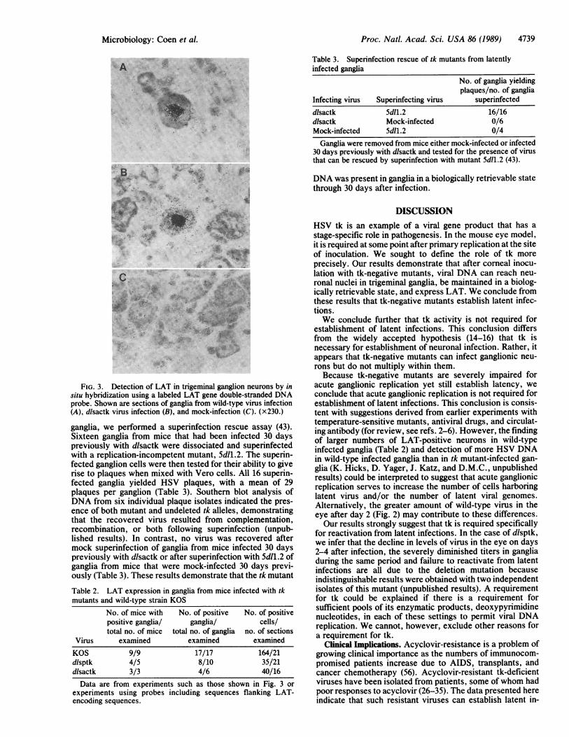
described left unanswered the question of whether the tkmutants were defective in their ability to reactivate fromlatent infections or at earlier stages, such as their ability toestablish latency. To address this question, we first examinedmutant-infected ganglia for LAT expression. Mice weremock-infected or infected with KOS or with either of the tkdeletion mutants. Thirty days later, ganglia were sectionedand prepared for in situ hybridization with a radiolabeledplasmid containing sequences encoding LAT. As shown inFig. 3, no signal was detected in ganglionic sections frommock-infected mice, whereas silver grains were concentratedover neuronal nuclei in ganglionic sections from mice in-fected with either KOS or mutant dlsactk. Similar hybrid-ization was observed in ganglionic sections from mice in-fected with mutant dlsptk (data not shown). The numbers ofgrains per nucleus were similar in sections from mice infectedwith wild-type virus or tk deletion mutants. No specific signal
Table 1. tk activities and acute and latent infections by tkmutants and wild-type strain KOS
No. of gangliareactivating/
% tk Acute virus titert total no. of gangliaVirus activity* Eye Ganglia - DMSO + DMSOKOS 100 5.4 ± 0.3 4.8 ± 0.2 10/10 2/2dlsptk 0.3 5.2 ± 0.3 <0.7 0/14 0/5dlsactk 0.2 5.6 ± 0.1 <0.7 ND 0/5DMSO, dimethyl sulfoxide at 200 mM during co-cultivation; ND,
not done.*Activities were normalized to those of KOS-infected cell extracts.Mock-infected activity was 0.4% that of KOS.tAcute virus titers are presented as log titer (mean ± SD). Peak titersin the eye occurred 1 day after infection. Peak titers in ganglia (meanof four samples) occurred 3 days after infection.
was obtained when sections were probed with plasmidscontaining HSV sequences encoding the major DNA-bindingprotein, ICP8, or with vector sequences alone as controls fornonspecific hybridization (data not shown).Data from several in situ hybridization experiments are
compiled in Table 2. LAT-positive sections were detectedfrequently in ganglia from mice infected with the tk deletionmutants (12/16). Although the frequencies of positive sec-tions (17/17) and positive cells per section were somewhat (2-to 5-fold) higher in ganglia from wild-type infected mice, thefact that LAT expression was readily detected in most gangliafrom mice infected with the tk mutants demonstrated thatmutant virus DNA reached the neuronal nuclei and wasbiologically active.The tk Mutants Can Be Rescued by a Superinfecting Virus.
As a second test for biologically active mutant DNA in
7
6
<0.
W 4
A.
the 3ie inicte afenetoacutekelcto i h y a
'Ui
dilsptk0
0 12 3 4DAY
FIG. 2. Growth of tk deletion mutants and KOS in the eye. Atthe times indicated after infection, acute replication in the eye wasmeasured by swabbing with cotton moistened with medium, dippingthe swabs in 1 ml ofmedium, freezing, thawing, and assaying directlyfor infectious virus on Vero cells. Each point represents the loga-rithmic mean of infectious virus from at least four eye swabs. Thezero time point shown indicates the size of the virus inocula, 2 X 10⁶PFU. The break in the ordinate indicates the limits of detection, 5PFU per eyeswab [<0.7 log (PFU)]. Points below this break yieldedno detectable infectious virus.
4738 Microbiology: Coen et al.
Proc. Natl. Acad. Sci. USA 86 (1989) 4739
Table 3. Superinfection rescue of tk mutants from latentlyinfected ganglia
No. of ganglia yieldingplaques/no. of ganglia
Infecting virus Superinfecting virus superinfecteddlsactk 5d11.2 16/16dlsactk Mock-infected 0/6Mock-infected 5d11.2 0/4
Ganglia were removed from mice either mock-infected or infected30 days previously with dlsactk and tested for the presence of virusthat can be rescued by superinfection with mutant 5d11.2 (43).
8.. ..eqALI *' *~jMI I
p1
*r gr.
j'm'
Jo- |si
U" . .
FIG. 3. Detection of LAT in trigeminal ganglion neurons by insitu hybridization using a labeled LAT gene double-stranded DNAprobe. Shown are sections of ganglia from wild-type virus infection(A), dlsactk virus infection (B), and mock-infection (C). (x230.)
ganglia, we performed a superinfection rescue assay (43).Sixteen ganglia from mice that had been infected 30 dayspreviously with dlsactk were dissociated and superinfectedwith a replication-incompetent mutant, 5d11.2. The superin-fected ganglion cells were then tested for their ability to giverise to plaques when mixed with Vero cells. All 16 superin-fected ganglia yielded HSV plaques, with a mean of 29plaques per ganglion (Table 3). Southern blot analysis ofDNA from six individual plaque isolates indicated the pres-ence of both mutant and undeleted tk alleles, demonstratingthat the recovered virus resulted from complementation,recombination, or both following superinfection (unpub-lished results). In contrast, no virus was recovered aftermock superinfection of ganglia from mice infected 30 dayspreviously with dlsactk or after superinfection with 5d11.2 ofganglia from mice that were mock-infected 30 days previ-ously (Table 3). These results demonstrate that the tk mutant
Table 2. LAT expression in ganglia from mice infected with tkmutants and wild-type strain KOS
No. of mice with No. of positive No. of positivepositive ganglia/ ganglia/ cells/total no. of mice total no. of ganglia no. of sections
Virus examined examined examinedKOS 9/9 17/17 164/21dlsptk 4/5 8/10 35/21dlsactk 3/3 4/6 40/16Data are from experiments such as those shown in Fig. 3 or
experiments using probes including sequences flanking LAT-encoding sequences.
DNA was present in ganglia in a biologically retrievable statethrough 30 days after infection.
DISCUSSIONHSV tk is an example of a viral gene product that has a
stage-specific role in pathogenesis. In the mouse eye model,it is required at some point after primary replication at the siteof inoculation. We sought to define the role of tk more
precisely. Our results demonstrate that after corneal inocu-lation with tk-negative mutants, viral DNA can reach neu-ronal nuclei in trigeminal ganglia, be maintained in a biolog-ically retrievable state, and express LAT. We conclude fromthese results that tk-negative mutants establish latent infec-tions.We conclude further that tk activity is not required for
establishment of latent infections. This conclusion differsfrom the widely accepted hypothesis (14-16) that tk isnecessary for establishment of neuronal infection. Rather, itappears that tk-negative mutants can infect ganglionic neu-rons but do not multiply within them.Because tk-negative mutants are severely impaired for
acute ganglionic replication yet still establish latency, weconclude that acute ganglionic replication is not required forestablishment of latent infections. This conclusion is consis-tent with suggestions derived from earlier experiments withtemperature-sensitive mutants, antiviral drugs, and circulat-ing antibody (for review, see refs. 2-6). However, the findingof larger numbers of LAT-positive neurons in wild-typeinfected ganglia (Table 2) and detection of more HSV DNAin wild-type infected ganglia than in tk mutant-infected gan-glia (K. Hicks, D. Yager, J. Katz, and D.M.C., unpublishedresults) could be interpreted to suggest that acute ganglionicreplication serves to increase the number of cells harboringlatent virus and/or the number of latent viral genomes.Alternatively, the greater amount of wild-type virus in theeye after day 2 (Fig. 2) may contribute to these differences.Our results strongly suggest that tk is required specifically
for reactivation from latent infections. In the case of dlsptk,we infer that the decline in levels of virus in the eye on days2-4 after infection, the severely diminished titers in gangliaduring the same period and failure to reactivate from latentinfections are all due to the deletion mutation becauseindistinguishable results were obtained with two independentisolates of this mutant (unpublished results). A requirementfor tk could be explained if there is a requirement forsufficient pools of its enzymatic products, deoxypyrimidinenucleotides, in each of these settings to permit viral DNAreplication. We cannot, however, exclude other reasons fora requirement for tk.
Clinical Implications. Acyclovir-resistance is a problem ofgrowing clinical importance as the numbers of immunocom-promised patients increase due to AIDS, transplants, andcancer chemotherapy (56). Acyclovir-resistant tk-deficientviruses have been isolated from patients, some of whom hadpoor responses to acyclovir (26-35). The data presented hereindicate that such resistant viruses can establish latent in-
A r-
Microbiology: Coen et al.
&
P,
Its?4I .,:,
f.., A.-I. -

Proc. Natl. Acad. Sci. USA 86 (1989)
fections. If one can extrapolate from the mouse model to thehuman setting, it is conceivable that latent acyclovir-resistantvirus might be reactivated by superinfection with tk-positiveviruses, leading to mixed populations of virus that couldretain both pathogenicity and resistance (57, 58) and greaterpersistence in the affected patient populations.
In addition, efforts have been made to develop drugs thatcould act against HSV infections in humans by specificinhibition of viral tk activity (36, 37). Our results suggest thatsuch drugs could inhibit HSV reactivation from latency suchthat they might be useful prophylactically during immuno-suppressive treatments. However, based on our results, ifsuch drugs were given during the acute phase of infectionthey may not prevent the establishment of latency, whileremoval of drug inhibition might lead to reactivation. Wehave previously argued that such drugs may also have to begiven at relatively high doses, because levels of tk activityless than 10% those of wild-type are sufficient for acutereplication and reactivatable latent infections in the mouseeye model (21).A Potential Vector for Neurons. Based on studies using
neuronal cell cultures in vitro, HSV has been proposed as avector to deliver genes to mammalian neurons in vivo (59, 60).For this purpose, it would be advantageous for the virus toinfect neurons specifically and nondestructively and to ex-press the gene product of interest efficiently and stably. Thetk deletion mutants described here do not replicate in ganglia,yet express LAT efficiently and stably in mouse neurons invivo. Modified to place the gene of interest under the controlof LAT or neuron-specific gene expression signals, perhapsby replacement of tk sequences, these mutants may offer allof these advantages.
We thank J. Stevens for providing his in situ hybridization protocoland for pointing out the sensitivity ofLAT hybridization as an assayfor latent infections, B. Fields for critically reading the manuscript,D. Yager for providing the starting plasmid for construction ofdlsptk, K. Ruffner for technical assistance, and J. Boni for help inisolation of the tk mutants. This work was supported by Grants P01A124010 and R01 A126126 from the National Institutes of Health andResearch Grant MV 242 from the American Cancer Society. J.G.J.was a predoctoral trainee supported by Public Health ServiceTraining Grants 5T32 GM07306 and A107245, D.A.L. was supportedby a National Multiple Sclerosis Society postdoctoral fellowship(FG766-A-1), K.L.T. was supported by a National Institutes ofHealth physician-scientist grant and an Alfred P. Sloan researchfellowship, and D.M.K. was supported by an American CancerSociety faculty research award.
1. Stevens, J. G. & Cook, M. L. (1971) Science 173, 843-845.2. Stevens, J. G. (1977) Adv. Cancer Res. 26, 227-256.3. Hill, T. J. (1982) in The Herpesviruses, ed. Roizman, B. (Plenum, New
York), Vol. 3, pp. 175-240.4. Wildy, P., Field, H. J. & Nash, A. A. (1982) in Virus Persistence, eds.
Mahy, B. W. J., Minson, A. C. & Darby, G. K. (Cambridge Univ. Press,Cambridge), pp. 134-167.
5. Price, R. W. (1985) Cancer Invest. 3, 285-292.6. Price, R. W. (1985) Cancer Invest. 3, 389-403.7. Corey, L. & Spear, P. G. (1986) N. Engl. J. Med. 314, 686-691.8. Corey, L. & Spear, P. G. (1986) N. Engl. J. Med. 314, 749-756.9. McGeoch, D. J., Dalrymple, M. A., Davison, A. J., Dolan, A., Frame,
M. C., McNab, D., Perry, L. J., Scott, J. E. & Taylor, P. (1988) J. Gen.Virol. 69, 1531-1574.
10. Schaffer, P. A., Wagner, E. K., Devi-Rao, G. B. & Preston, V. G. (1987)in Genetic Maps, ed. O'Brien, S. J. (Cold Spring Harbor Lab., ColdSpring Harbor, NY), pp. 93-98.
11. Dubbs, D. R. & Kit, D. (1964) Virology 22, 493-502.12. Elion, G. B., Furman, P. A., Fyfe, J. A., deMiranda, P., Beauchamp, L.
& Schaeffer, H. J. (1977) Proc. Natl. Acad. Sci. USA 74, 5716-5720.13. Fyfe, J. A., Keller, P. M., Furman, P. A., Miller, R. L. & Elion, G. B.
(1978) J. Biol. Chem. 253, 8721-8727.14. Tenser, R. B., Miller, R. L. & Rapp, F. (1979) Science 205, 915-917.
15. Tenser, R. B. & Dunstan, M. E. (1979) Virology 99, 417-422.16. Tenser, R. B., Ressel, S. & Dunstan, M. E. (1981) Virology 112,328-341.17. Field, H. J. & Wildy, P. (1978) J. Hyg. 81, 267-277.18. Gordon, Y. J., Gilden, D. M., Shtram, Y., Asher, Y., Tabor, E., Wellish,
M., Devlin, M., Snipper, D., Hadar, J. & Becker, Y. (1983) Arch. Virol.76, 39-49.
19. Sears, A. E., Meignier, B. & Roizman, B. (1985) J. Virol. 55, 410-416.20. Tenser, R. B. & Edris, W. A. (1987) J. Virol. 61, 2171-2174.21. Coen, D. M., Irmiere, A. F., Jacobson, J. G. & Kerns, K. M. (1989)
Virology 168, 221-231.22. Coen, D. M. & Schaffer, P. A. (1980) Proc. Natl. Acad. Sci. USA 77,
2265-2269.23. Coen, D. M., Dixon, R. A. F., Ruby, S. W. & Schaffer, P. A. (1980) in
Animal Virus Genetics, eds. Fields, B. N., Jaenisch, R. F. & Fox, C. F.(Academic, New York), pp. 581-590.
24. Field, H. J., Darby, G. & Wildy, P. (1980) J. Gen. Virol. 49, 115-124.25. Schnipper, L. E. & Crumpacker, C. S. (1980) Proc. Natl. Acad. Sci.
USA 77, 2270-2273.26. Crumpacker, C. S., Schnipper, L. E., Marlowe, S. I., Kowalsky, P. N.,
Hershey, B. J. & Levin, M. J. (1982) N. Engl. J. Med. 306, 343-346.27. Bums, W. H., Saral, R., Santos, G. W., Laskin, 0. L., Lietman, P. S.,
McLaren, C. & Barry, D. W. (1982) Lancet 1, 421-423.28. Sibrack, C. D., Gutman, L. T., Wilfert, C. M., McLaren, C., St. Clair,
M. H., Keller, P. M. & Barry, D. W. (1982) J. Infect. Dis. 146, 673-682.29. Wade, J. C., McLaren, C. & Meyers, J. D. (1983) J. Infect. Dis. 148,
1077-1082.30. Svennerholm, B., Vahlne, A., Lowhagen, G. B., Widell, A. & Lycke, E.
(1985) Scand. J. Infect. Dis. 47, 149-154.31. Collins, P. & Oliver, N. M. (1986) J. Antimicrob. Chemother. 18, Suppl.
B, 103-112.32. Christophers, J., Sutton, R. N. P., Noble, R. V. & Anderson, H. (1986)
J. Antimicrob. Chemother. 18, Suppl. B, 121-125.33. Schinazi, R. F., del Bene, V., Scott, R. T. & Dudley-Thorpe, J. B. (1986)
J. Antimicrob. Chemother. 18, Suppl. B, 127-134.34. Erlich, K. S., Mills, J., Chatis, P., Mertz, G. J., Busch, D. F., Follans-
bee, S. E., Grant, R. M. & Crumpacker, C. S. (1989) N. Engl. J. Med.320, 293-296.
35. Chatis, P. A., Miller, C. H., Schrager, L. E. & Crumpacker, C. S. (1989)N. Engl. J. Med. 320, 297-300.
36. Nutter, L. M., Grill, S. P., Dutschman, G. E., Sharma, R. A., Bobek,M. & Cheng, Y.-C. (1987) Antimicrob. Agents Chemother. 31, 368-374.
37. Focher, F., Hildebrand, C., Freese, S., Ciarrocchi, G., Noonan, T.,Sangalli, S., Brown, N., Spadari, S. & Wright, G. (1988) J. Med. Chem.31, 1496-1500.
38. Stevens, J. G., Wagner, E. K., Devi-Rao, G. B., Cook, M. L. & Feld-man, L. T. (1987) Science 235, 1056-1059.
39. Deatly, A. M., Spivack, J. G., Lavi, E. & Fraser, N. W. (1987) Proc.Natl. Acad. Sci. USA 84, 3204-3208.
40. Rock, D. L., Nesburn, A. B., Ghiasi, H., Ong, J., Lewis, T. L., Lo-kensgard, J. R. & Wechsler, S. L. (1987) J. Virol. 61, 3820-3826.
41. Croen, K. D., Ostrove, J. M., Dragovic, L. J., Smialek, J. E. & Straus,S. E. (1987) N. Engl. J. Med. 317, 1427-1432.
42. Gordon, Y. J., Johnson, B., Romanowski, E. & Araullo-Cruz, T. (1988)J. Virol. 62, 1832-1835.
43. Leib, D. A., Coen, D. M., Bogard, C. L., Hicks, K. A., Yager, D. R.,Knipe, D. M., Tyler, K. L. & Schaffer, P. A. (1989) J. Virol. 63, 759-768.
44. Weller, S. K., Aschman, D. P., Sacks, W. R., Coen, D. M. & Schaffer,P. A. (1983) Virology 130, 290-305.
45. McCarthy, A., McMahan, L. & Schaffer, P. A. (1989) J. Virol. 63, 18-27.
46. McKnight, S. L. (1980) Nucleic Acids Res. 8, 5949-5964.47. Wagner, M. J., Sharp, J. A. & Summers, W. C. (1981) Proc. Natl. Acad.
Sci. USA 78, 1441-1445.48. Irmiere, A. F., Manos, M. M., Jacobson, J. G., Gibbs, J. S. & Coen,
D. M. (1989) Virology 168, 210-220.49. Chiou, H. C., Weller, S. K. & Coen, D. M. (1985) Virology 145,213-226.50. Coen, D. M., Weinheimer, S. P. & McKnight, S. L. (1986) Science 234,
53-59.51. Coen, D. M., Fleming, H. E., Jr., Leslie, L. K. & Retondo, M. J. (1985)
J. Virol. 53, 477-488.52. Haase, A., Brahic, M., Stowring, L. & Blum, H. (1984) Methods Virol.
7, 189-226.53. Stroop, W. G., Rock, D. L. & Fraser, N. W. (1984) Lab. Invest. 51, 27-
38.54. Jacobson, J. G., Martin, S. L. & Coen, D. M. (1989) J. Virol. 63,
1839-1843.55. Whitby, A. J., Blyth, W. A. & Hill, T. J. (1987) Arch. Virol. 97, 137-144.56. Hirsch, M. S. & Schooley, R. T. (1989) N. Engl. J. Med. 320, 313-314.57. Field, H. J. (1982) Antimicrob. Agents Chemother. 21, 744-752.58. Field, H. J. & Lay, E. (1984) Antiviral Res. 4, 43-52.59. Palella, T. D., Silverman, L., Schroll, C. T., Homa, F. L., Levine, M.
& Kelley, W. N. (1988) Mol. Cell Biol. 8, 457-460.60. Geller, A. I. & Breakefield, W. X. (1988) Science 241, 1667-1669.
4740 Microbiology: Coen et al.