thyroid nodules: pathophysiological insight on oncogenesis and
TRANSCRIPT

PHYSIOLOGICAL RESEARCH bull ISSN 0862-8408 (print) bull ISSN 1802-9973 (online) copy 2014 Institute of Physiology vvi Academy of Sciences of the Czech Republic Prague Czech Republic Fax +420 241 062 164 e-mail physresbiomedcascz wwwbiomedcasczphysiolres
Physiol Res 63 (Suppl 2) S263-S275 2014
REVIEW
Thyroid Nodules Pathophysiological Insight on Oncogenesis and Novel Diagnostic Techniques J KRAacuteTKYacute1 H VIacuteTKOVAacute1 J BARTAacuteKOVAacute1 Z TELIČKA1 M ANTOŠOVAacute1 Z LIacuteMANOVAacute1 J JISKRA1 1Third Department of Medicine First Faculty of Medicine Charles University General University Hospital Prague Czech Republic
Received March 20 2014 Accepted April 30 2014 Summary
Thyroid nodules are a very frequent pathology among common
population Despite the vast majority of them are of benign
origin the incidence of thyroid cancer is currently rather rising
Although there are several risk factors of thyroid cancer and
several clinical ultrasound biochemical and molecular diagnostic
markers the exact mechanisms of thyroid oncogenesis and the
linkage between thyroid nodule ultrasound appearance and its
biological character remain unclear While ionizing radiation is the
only one well-known risk factor for thyroid cancer the
significance of some others remains unclear The aim of our
review was to discuss some not completely known
pathophysiological mechanisms involved in thyroid oncogenesis
as hypothyroidism mutations of genes regulating cell
proliferation thyroid autoimmunity and pregnancy and to
describe pathophysiological background of some ultrasound
markers of thyroid cancer (size echogenicity vascularization
calcifications and stiffness) Better knowledge in this field is
crucial for development of novel diagnostic techniques and
therapeutic approaches For example the analysis of BRAF RAS
and other mutations in cytological samples may help to
distinction between follicular thyroid carcinoma and follicular
thyroid adenoma and may significantly decrease the number of
unnecessary surgery among patients with thyroid nodules
Alternatively the different malign cells growth angiogenesis
destructions of thyroid follicles reparative changes growth
retardation fibrosis and increased interstitial fluid pressure
implicate the typical ultrasound appearance of papillary
thyroid cancer (hypoechogenicity irregular vascularization
microcalcifications stiffness) which is essential to catch the
suspicious nodules on the basis of their ultrasound appearance
among large amount of benign nodules
Key words
Thyroid nodule bull Thyroid cancer bull Hypothyroidism bull Thyroid
autoimmunity bull BRAF mutation bull RAS mutation bull Thyroid
ultrasound bull Microcalcifications bull Thyroid elastography bull
Pregnancy
Corresponding author
J Kraacutetkyacute Third Department of Medicine First Faculty of
Medicine Charles University and General University Hospital
U Nemocnice 1 128 08 Prague 2 Czech Republic Fax
+420224919780 E-mail jankratkyvfncz
With increasing resolution of modern ultrasound
equipment the prevalence of thyroid nodules rises up to 76 (Ferraz et al 2011) while risk of malignancy is distinctly lower (5-15 ) (Cooper et al 2009) The most common is papillary thyroid carcinoma (PTC) which represents 87 of thyroid malignancies (Kamran et al 2013) Over the last 40 years the incidence of PTC has almost tripled in US population predominantly due to microcarcinomas (le1 cm) (Davies et al 2002) Although there are several risk factors of thyroid cancer and several clinical ultrasound biochemical and molecular diagnostic markers (Table 1) the exact mechanisms of thyroid oncogenesis and the linkage between thyroid nodule ultrasound appearance and its biological character remain unclear While ionizing radiation is the only one good proved risk factor for thyroid cancer the
S264 Kraacutetkyacute et al Vol 63 significance of some others remains unclear In the first part of the review some not completely known pathophysiological mechanisms involved in thyroid oncogenesis as hypothyroidism mutations of genes regulating cell proliferation thyroid autoimmunity and
pregnancy are discussed and the second part is aimed to describe pathophysiological background of some ultrasound markers of thyroid cancer (size echogenicity vascularization calcifications and stiffness)
Table 1 Clinical ultrasound biochemical and molecular markers of thyroid cancer
Clinical Ultrasound Biochemical and molecular
History of head or neck irradiation Hoarseness or dysphonia dysphagia and dyspnea Presence of cervical lymphadenopathy Present of thyroid cancer in first degree relative Multiple endocrine neoplasia syndrome 2 Age under 20 or over 70 years Pregnancy Accumulation of FDG on PET scanning
Diameter gt1-2 cm Hypoechogenicity Increased intra-nodular vascularization Irregular margins Microcalcifications Absence of hypoechoic hallo Higher stiffness measured by elastography
TSHR-mRNA in peripheral blood Increased calcitonin serum level BRAF RAS RETPTC mutations in FNAC samples
PET positron emission tomography FDG Fluordeoxyglucose TSHR-mRNA messenger RNA of thyrotropin receptor FNAC fine needle aspiration cytology
Hypothyroidism Hypothyroidism is well-known stimulator of
thyroid growth In peripheral hypothyroidism thyroid hormone synthesis and production and thyroid follicular cells proliferation are mediated by secretion of thyroid stimulating hormone (TSH) in pituitary gland Physiologically activation of the TSH receptor by binding of TSH induces intracellular production of cyclic AMP (cAMP) by adenylyl cyclase The higher level of cAMP activates c-AMP-dependent protein kinase A which subsequently phosphorylates its substrates One of them is nuclear factor CREB which activates the transcription of genes which are among other responsible for thyroid cells proliferation (Kondo et al 2006) This signalizing cascade is analogically responsible for the thyroid growth in hypothyroidism and also well-differentiated thyroid cancer usually preserves this dependence The levothyroxine suppression treatment in patient after thyroidectomy for differentiated thyroid carcinoma is based on the tumor cells growing inhibition by suppressed TSH level (Biondi et al 2005 Fiore et al
2009) Moreover a raising risk of malign thyroid nodule with increasing TSH level and higher TSH level among patients with disease recurrence or progression have been reported (Boelaert et al 2006) Consistently thyroid nodules with autonomous thyroid hormone secretion (ldquotoxic nodulesrdquo) and TSH serum levels lt04 mIUl were associated with significantly lower probability of thyroid cancer (Fiore et al 2009) and this mild hyperthyroidism patient may be a protective factor against the development of PTC It seems to be reasonable to maintain TSH level rather in the lower part of normal range in the patients with thyroid nodules presenting other risk factors of PTC eg autoimmune thyroiditis previous neck irradiation pregnancy etc
Molecular changes
Mutations of genes regulating cell cycle cell
proliferation and apoptosis certainly play a crucial role in thyroid oncogenesis Main molecular changes involved in thyroid oncogenesis are BRAF and RAS point mutations and RET-PTC and PAX8PPAR-gamma rearrangements
2014 Oncogenesis of Thyroid Nodules S265
Fig 1 RASRAFMAP signalization pathway GF growth factor RET RET receptor with tyrosine kinase activity RAS GTP binding RAS protein with kinase activity BRAF BRAF protein with serine-threonine kinase activity MEK MEK protein with kinase activity ERK ERK proteins with kinase activity activates the nuclear transcription by phosphorylation
BRAF is a cytoplasmic serine-threonine kinase
involved in RASRAFMEK signaling pathway (Fig 1) Normally the BRAF kinase is activated by linking of RAS protein and it triggers of the whole signaling cascade which leads to increased growth and cell proliferation Most proven BRAF V600E mutation leads to permanent phosphorylation activity of the BRAF protein independent on the binding of RAS protein Schulten et al (2012) found activated mutation of BRAF gen in 72 of 115 (63 ) PTCs Cantara et al (2010) analyzed 235 FNAC samples and proved 33 cases of BRAF mutations All the BRAF mutations were associated with the diagnosis of PTC Similarly RETPTC rearrangement (RETPTC) was found only in patients with PTCs in this study (Cantara et al 2010)
RET is the proto-oncogene located at chromosome 10 coding cell membrane receptor with thyrosine kinase activity which after binding of its ligand activates among others the RASBRAFMEK pathway In some PTCs the RET gen is fused with different gen At least 11 types of RETPTC variants have been already isolated (Santoro et al 2006 Marotta et al 2011) The resulting chimeric protein lacks its extracellular and transmembrane domain but has a permanent phosphorylation activity (Marotta et al 2011)
Another common mutation in the aforementioned signaling pathway is the activating mutation of RAS gene It was found not only in thyroid malignancies but also in benign follicular adenomas
(FTA) (Cantara et al 2010) However the presence of RAS mutation in FTA may increase the risk of later progression to follicular thyroid carcinoma (FTC)
It seems that the molecular steps in the carcinogenesis are different in FTC and PTC Direct inception as microcarcinoma is supposed in PTC whereas FTC probably develops from FTA (Fig 2)
Fig 2 Hypothetical carcinogenesis of different types of thyroid cancer (adapted Fuchshuber et al 1998) RAS mutation + presence of mutation of RAS oncogene RAS mutation ndash absence of mutation of RAS oncogene RETPTC presence of RETPTC rearrangement BRAF presence of BRAF oncogene mutation p53 presence of tumor suppressor protein p53 mutation
That advance in thyroid nodules and cancer
research allowed introducing of further diagnostic tools as molecular analysis of fine needle aspiration cytology (FNAC) samples Since 2009 the revised guidelines of American Thyroid Association (ATA) recommended to consider the molecular analysis of FNAC samples if the cytological result is indeterminate Although cytology and histology remain the gold standard for diagnosis of thyroid malignancies molecular analysis of FNAC samples may help in the management of patients with indeterminate cytology in which based on cytology cannot be differentiated between FTC and FTA The surgery should be chosen in patient with RAS or any other risk mutation in cytology sample
Thyroid autoimmunity
The association between Hashimoto thyroiditis
(HT) and PTC is a long-debated question Some experts even consider HT to be a risk factor for the development of PTC Indeed PTC and HT share some epidemiological (exposure to ionizing radiation higher incidence in
S266 Kraacutetkyacute et al Vol 63 women and areas with high dietary iodine intake) and molecular features (Weetman et al 2004 Cunha et al 2011) The results of a recently published meta-analysis (Lee et al 2013) involving a total of 10648 PTCs demonstrated a significant association between histologically proved HT and PTC The HT was found in 2471 (232 ) with PTC Interestingly PTCs with coexisting HT were associated with longer recurrence-free survival absence of extrathyroidal invasion and absence of lymph node metastasis An explanation might be an enhanced immune antitumor activity in patients with HT This idea is also supported by the frequent finding of noninvasive capsulated microPTCs in thyroid autopsies with HT (Ferraz et al 2011) The HT includes the formation of proliferating nodules as well as cytological alterations and nuclear modification similar to PTC (Cunha et al 2011) On the contrary it must be noted that an inflammatory changes occur in the majority of tumors in response to the presence of pathological tissue and there is no relevant evidence of causal link between HT and PTC so far One hypothesis is that development of inflammatory process leads to depletion of follicular thyroid cells and their replacement by mononuclear cells producing chemokines cytokines and growth factors Most of them are under transcriptional control of the Nuclear factor-kappa B (NF-κb) The activity of NF-κb could spread to the remaining follicular cells and may lead to the tumor inception in susceptible follicular cells (Pacifico et al 2010) Above mentioned RASBRAFMEK signalization pathway plays a key role in thyroid carcinogenesis The mutations of BRAF gene are mostly associated with PTC however is not linked with higher frequency of HT (Kim et al 2009) Conversely RETPTC whose product is the ligand-independent kinase at the beginning of the whole pathway is more often present at PTCs associated with HT and is even found in benign lesions such as HT (Muzza et al 2010) There may be three hypothetic mechanisms of the linkage between RETPTC and inflammation 1) chronic inflammation acts as a source of mutagenic agents such as reactive oxygen radicals that damage DNA and increase the likelihood of random creation of RETPTC rearrangement 2) the presence of RETPTC rearrangement may be the source of other pro-inflammatory transcription factors 3) molecules released by inflammation could sustain the survival of thyroid cells in which RETPTC rearrangement randomly occurs and thereby enabling acquire additional mutation and thus become resistant to apoptosis (Cunha et al 2011)
Pregnancy Prevalence of thyroid nodules among pregnant
women is 9-34 About 12-15 of thyroid nodules discovered in pregnancy can be malignant predominantly owing to papillary (micro)carcinomas It is either similar to or possibly greater than seen in general population (De Groot et al 2012 Jiskra et al 2014) About 10 of thyroid cancers occurring during the reproductive years are diagnosed during pregnancy or early after delivery (Gibelli et al 2011) A history of one or more pregnancies is associated with a small increase in the risk of thyroid cancer (relative risk 18) (McTiernan et al 1984) Moreover thyroid cancer diagnosed during pregnancy may be associated with a poor prognosis as compared with tumors that developed in non-gravid periods (Alves et al 2011 Mazzaferri et al 2011)
Pathophysiological background of increased risk of thyroid cancer development and progression in pregnancy is unknown The mechanisms most frequently discussed are human chorionic gonadotropin (hCG) and estrogen stimulation increased vascularity and lack of immune surveillance of cancer (Alves et al 2011) (Fig 3)
Fig 3 Main factors influencing oncogenesis and tumor progression in thyroid gland during pregnancy
Human chorionic gonadothropin Human chorionic gonadothropin (hCG) belongs
to the family of glycoprotein hormones with the structural homology not only in the hCG and TSH molecules (identical α-subunit) but also in their receptors This homology suggests the basis for the reactivity of hCG with the TSH receptor (Yoshimura et al 1995 Voigt et al 2007) At the time of the peak hCG levels in physiological pregnancy serum TSH levels fall and bear a mirror image to the hCG peak Maternal thyroid gland
2014 Oncogenesis of Thyroid Nodules S267
secretes more thyroid hormone during early pregnancy in response to the thyrotropic activity of hCG that overrides the operation of hypothalamic-pituitary-thyroid feedback system (Hershman et al 2004 Alves et al 2011) Analogically hCG is the most potent stimulator of thyroid growth in the first trimester of pregnancy Hence in susceptible thyroid follicular cells (eg in cells with BRAF and RAS mutations or RETPTC and PAX8-PPAR gamma rearrangements) excessive hCG stimulation may lead to inception of rapid progression of cancer
Estrogens
Estrogens with their receptors may play role in the thyroid oncogenesis reprogramming and progression (Tafani et al 2014) Indirect estrogen effect is increasing the thyroxine binding globulin (TBG) direct effect on cellular signaling is mediated classically after the binding estradiol (E2) on two soluble intracellular estrogen receptors ERα (ESR1) and ERβ (ESR2) when ERs form stable dimers that interact with specific sequences called estrogen response elements (EREs) and initiate the transcription of target genes Additionally a rapid signaling such as activation of kinases (eg phosphatidylinositol 3-kinase) and phosphatases an increase in ion fluxes across membranes and a transmembrane intracellular non-classical ER mediating with a G protein-coupled receptor (GPCR) has been described in human follicular thyroid carcinoma cell lines in vitro (Santin et al 2011)
The important role play estrogen receptors ERα and ERβ in thyroid stem and progenitor cells with eight times higher expression levels of ERα mRNA compared with the differentiated thyrocytes The levels are higher in younger women than in older women or men (Horn-Ross et al 2011 Santin et al 2011) The proliferation and growth of thyroid cancer cells are promote by an ERα agonist in contrast toERβ that would promote apoptotic actions and other suppressive functions Therefore ERαERβ ratio could have an important role in the pathophysiology of thyroid cancer similar to that postulated for breast cancer (Santin et al 2011) Furthermore 17-beta-estradiol significantly increased the expression of Cyclin D1 in a human thyroid carcinoma cell line lacking endogenous TSH receptor (Santin et al 2011) Estrogen-responsive regulatory region of Cyclin D1 regulates the cell progression mediated by the facilitating G1 to S phase transition Its expression has been associated with an aggressive behavior of papillary
micro-carcinoma (more than 90 of the metastasizing micro-carcinomas expressed Cyclin D1 (Khoo et al 2002) In contrast TSH-induced differentiation of progenitor cells was significantly inhibited by E2 Also estrogens stimulated the growth with a lower extent than other growth factors and simultaneously inhibited the differentiation of thyroid nodule-derived stemprogenitor cells (Xu 2013) Despite of the experimental evidence the vast majority of clinical studies has not proved the use of oral contraceptives or other exogenous estrogens to be associated with higher risk of thyroid cancer or its unfavourable prognosis (Mazzaferri et al 2011) Only in one study an increased risk of thyroid cancer with the number of pregnancies was observed but only in women who prematurely stopped lactation (Mack et al 1999)
Estrogen aids in angiogenesis of various tumors during hypoxic conditions however the role of estrogen within the hypoxic environment of thyroid cancer is not known Hypoxia with estrogens modulate the hypoxia inducible factor-1 (HIF-1) signaling which is abrogated by the anti-estrogen fulvestrant and the HIF-1 inhibitor YC-1 (3-(5-hydroxymethyl-2-furyl)-1-benzylindazole) (Rajoria et al 2013) HIF-1 inhibitor can abolish migration and tubulogenesis of human umbilical vein endothelial cells (HUVECs) in thyroid cancer cells HIF-1 and NF-κB are two transcription factors very frequently activated in tumors and involved in tumor growth progression inflammatory-reparative response and resistance to chemotherapy (Tafani et al 2014)
Increased vascularization
Increased vascularization of thyroid parenchyma which can be visualized by colorpower doppler ultrasound is typical finding in pregnancy It is mediated predominantly by hCG and VEGF stimulation High tumorigenic potential is associated with an elevated vascular endothelial growth factor (VEGF) expression in human thyroid tumor cell lines In contrast placental growth factor (PIGF) expression is severely down regulated in the majority of thyroid tumor cell lines and in tumors (Viglietto et al 1995) Both the VEGF receptors (FLT-1 and flkKDR) are expressed in endothelial cells that line tumor-embedded micro-vascular vessels suggesting that VEGF but not PIGF contributes to thyroid tumor development (Viglietto et al 1995) VEGF and hCG mediated vascularization of thyroid parenchyma may facilitate cancer cell angioinvasion and contribute to the worse prognosis of thyroid cancer diagnosed in pregnancy
S268 Kraacutetkyacute et al Vol 63 Changes of immune system in pregnancy
Several changes in immune system which can support development and progression of malignity were described in pregnancy They include immune tolerance to fetus as the semi-allograft shift T1 helper lymphocytes (Th1) to T2 helper lymphocytes (Th2) immunosuppressive effect of progesteron and tolerogenic effect of regulatory T cells The important fact is that 40 of decidual cells are cells of the innate immune system (NK cells macrophages dendritic cells and T reg cells) Regulatory T cells and Th2 cells are important mediators of tolerance in both pregnancy and cancer Expression of human leucocyte antigen G (HLAG) on trophoblast cells and cancer cells has important immunomodulatory effects Its expression correlated amount of invasive interstitial and endovascular extravillous trophoblast cells The role of HLAG is to suppress cytolytic killing by both NK and cytotoxic T cells to induce apoptosis of immune cells to regulate cytokine production in blood mononuclear cells and to reduce stimulatory capacity and impair maturation of dendritic cells (Holtan et al 2009)
Pathophysiological background of ultrasound markers of thyroid cancer
Increasing quality and resolution of modern
ultrasound equipment make thyroid ultrasound the key non-invasive technique to predict biological nature of the thyroid nodule
Size
The size of nodules itself as a risk factor of malignancy is questionable Whereas benign adenomas may reach large proportion PTC is often found as microcarcinoma (in up to one third of adult thyroid autopsy) (Vanderpump et al 2011) So far nodules gt1 cm in diameter were considered to have malign potential and should be referred to US-FNAC Recently in a large study including 7348 thyroid nodules gt1 cm in diameter Kamran et al (2013) referred a higher risk of thyroid carcinoma among nodules gt2 cm in comparison to nodules 1-2 cm in diameter Unlike PTC the frequency of the FTC was rising with increasing size of the nodules It implies that whereas PTC is largely predetermined at its inception and does not transform with growth the FTC develops from FTA only if expands to certain size (Kamran et al 2013) One explanation is that nodule growth increases risk of secondary genomic mutations
that transform FTA to FTC The second one is that the histological hallmarks of FTC (angioinvasion andor capsular invasion) may occur only when FTA reaches certain cellular number The idea of transforming FTA to FTC is also supported by findings of known risk oncogenic mutation (RAS PAX 8PPARgamma) in FTA (Suarez et al 1990 Cantara et al 2010) The FTA with RAS mutation could be the precursors of FTC and may be best characterized as ldquocarcinoma in siturdquo (Nikiforof et al 2011) Another attractive hypothesis of FTA malignant transformation is the increased activity of metaloproteinase-9 the enzyme which degrades collagen-4 Collagen-4 is an essential compound of basal membrane and its degradation may allow the cell infiltration Friguglietti et al (2000) found significantly higher immunohistochemical positivity of metaloproteinase-9 in the FTC cells compared to the FTA Apparently the nodules with cytological finding follicular lesion of indeterminate significance should be carefully monitored for the growth progression The presence of progression may indicate risk of developing FTC and the patient should be refer to surgery In fact the size of thyroid nodule as a predictor of thyroid malignancy may be relevant in follicular lesions whereas the risk of PTC is probably equal in small and large lesions
Vascularization
It is well known fact that increased intra-nodular vascularity proven on Doppler ultrasound is a risk factor for thyroid carcinoma especially for PTC Angiogenesis plays an important role in tumor growth and proliferation Akslen et al (2000) analyzed histological samples of 128 PTC and they found 314 times higher microvessel density in the carcinomas compared to normal surrounding thyroid tissue however increased tumor vascularization was not associated with a poorer prognosis They suggest that this may be due to the fact that PTC unlike the FTC locally spread primarily through lymphatic veins However there are also papers demonstrating increasing microvessel density with rising invasiveness of PTC According the results of Chinese study by Liang et al (2006) the density of lymphatic vessels and also microvessels was the lowest in the group of microPTC (le1 cm) raised in the tumors gt1 cm and was the highest in the group of metastatic PTC Consistently the expression of vascular endothelial growth factor C (VGEF-C) was increasing in these groups in the same way
2014 Oncogenesis of Thyroid Nodules S269
Increased neovascularization in PTC seems to be caused by an imbalance between pro-angiogenic and anti-angiogenic factors Tanaka et al (2002) analyzed the expression patterns and levels of several anti- and pro-angiogenic factors and its receptors in 75 PTCs and found that VEGF expression strongly positively correlated with the other pro-angiogenic factors (VEGF-C Angiopoietin-2 and Tie-2) and their higher expression was associated with increased microvascular density Conversely Thrombospondin-1 (antiangiogenic factor) expression correlated with the degree of invasion negatively They counted VEGFThrombospondin-1 and Angiopoietin-2Thrombospondin-1 ratios that significantly correlated with the degree of tumor infiltration (Tanaka et al 2002) Presence of Doppler signal in the central part of the nodule surly increases the risk of malignancy However current high-sensitive ultrasound devices can receive Doppler signals also of regular intra-nodular vascularization of benign nodule and to distinguish it from the irregular chaotic vascularization in malign nodules may not be always easily possible
Echogenicity and nodule margin
PTC ultrasound image typically appears as an irregular hypoechoic nodule The irregular shape is due to the uneven tumor cells proliferation The cause why PTC is hypoechoic is still not clear We suggest it could be caused by the lack of follicular tissue arrangement that is specific for thyroid gland and that implies the typical hyperechoic picture of the healthy thyroid parenchyma in comparison to neck muscles and other surrounding tissues The solid PTC cell mass unlike the normal follicles does not contain as many interface with different acoustic impedance This may reduce the ultrasound reflection in the PTC Nevertheless majority of the thyroid nodules are hypoechoic in comparison with the normal thyroid tissue but only marked hypoechogenicity seems to be suspect The evaluation of the nodules echogenicity is often affected by the physicianrsquos experience and his subjective view Few years ago the automatic texture analyzer was successfully used to distinguish between the texture of normal thyroid gland and thyroiditis in Czech Republic (Smutek et al 2003) Similarly automatic analyzer evaluating thyroid nodules echogenicity could be developed and it could eliminate the influence of the examiner
Calcifications
The presence of calcifications in thyroid
nodules regardless of their appearance may be associated with higher risk of malignancy (Taki et al 2004 Reading et al 2005) Macrocalcifications are large (gt2 mm) hyperechoic spots that cause acoustic shadow and may be present both in benign and malign nodules They originate in areas of tiny hemorrhages and destructions in thyroid nodules as a result of reparative changes with precipitations of calcium salts (dystrophic calcifications) Microcalcifications are visualized as small (le2 mm) hyperechoic points without acoustic shadow They are associated with the papillary (nipple-like) histomorphology and occur very common in malign nodules with frequency 26-70 (Frates et al 2006 Kwak et al 2011) but are relatively rare in benign lesions Histopathologically microcalcifications are bdquopsammoma bodiesldquo (PBs) The term is derived from the Greek word ldquopsammosrdquo meaning ldquosandrdquo Despite numerous ancillary studies over a span of three and half decades formation of PBs bodies remains a poorly understood mechanism Ultrastructural study of PTC has shown that thickening of the base lamina in vascular stalk of neoplastic papillae followed by thrombosis calcification tumor cell necrosis and collagen production by neoplastic cells to formation of PBs (Johannessen et al 1980 Das et al 2009) It is suggested that rather than being the outcome of dystrophic calcification of dead or dying tissue PBs may indeed represent an active biologic process ultimately leading to degenerationdeath of tumor cells and retardation of growth of the neoplasm It may also serve as a barrier against the spread of neoplasm (Das et al 2009) This is in concordance with the common clinical finding that microcalcifications are most frequently found in small (lt2 cm) papillary thyroid cancers (Shi et al 2012) with slow non-invasive tumor growth and good clinical prognosis Fortunately microcalcifications can be easily confused with dense colloid aggregations (in benign ldquocolloidrdquo nodules) or with the fibrosis of the tissue (in Hashimoto`s thyroiditis) In contrast to dense colloid aggregations which vibrate due to ultrasonic wave and cause an artifact called ldquocomet tailrdquo the microcalcifications stay motionless and in contrast to fibrosis they are separate structures and do not form irregular stripes connecting with each other (Lewinski and Adamczewski 2013) A novel ultrasound diagnostic technique ldquoMicroPurerdquo was recently developed by Toshiba Medical System Corporation (httpwwwtoshiba-medicaleuenOur-Product-Range UltrasoundTechnologiesMicroPure2) aimed to distinguish microcalcifications from the artifacts and
S270 Kraacutetkyacute et al Vol 63 fibrosis Usefulness of MicroPure imaging in measuring of microcalcifications was demonstrated both in breast and thyroid lesions (Sankaye et al 2010 Ciledag et al 2012)
Nodule stiffness
Uncontrolled proliferation and irregular growth of malign cells may cause a higher stiffness of tumor mass compared to normal thyroid tissue On the higher stiffness of the carcinomas may contribute some of the following factors Increased interstitial fluid pressure (IFP) which has been observed in many of human carcinomas and reach up to 60 mm Hg The mechanism of elevated IFP is not exactly known It involves an increased permeability of the defect tumor blood vessels Leak of the plasma proteins which are not effectively drained through the pathological lymphatic vessels increased interstitial oncotic pressure The leakiness of tumor vessels might be due to the overproduction of VEGF by the tumor cells which has been shown to increase permeability and due to the tumor stromal
infiltration by the activated macrophages and other immune cells and by their cytokines as well Moreover the extracellular matrix of the tumor contains much denser collagen fiber network and increased number of the fibroblast Therefore the tumor is more rigid in comparison with normal tissue (Heldin et al 2004) The presence of hard calcification fairly typical for thyroid carcinoma makes the tumor stiffer as well
Tissue elasticity can be determined by elastography the relatively new ultrasound modality It was first introduced in 1991 by Orphir (Orphir et al 1991) It is assumed that higher stiffness may predict a higher risk of thyroid cancer Actually there are two different types of elastography ndash Real time (strain) elastograpy (RTE) and Shear wave elastography (SWE) Both methods do not differ only by their principle but also by their output While SWE can directly quantified tissue elasticity using the Youngs modulus of elasticity RTE shows only the relative distribution of elasticity displayed by map of relative elasticity distribution called elastogram (Fig 4)
Fig 4 Elastogram and strain-ratio (21) of papillary thyroid carcinoma
According to a meta-analysis published in 2010 which involved a total of 639 nodes from eight studies RTE reached the sensitivity 92 and specificity of 90 in detecting thyroid malignancies (Bojunga et al 2010) Recently high negative predictive value of quantitative RTE for thyroid malignancy (991 and 972 respectively) was found (Azizi et al 2013 Mehrota et al 2013) Higher reproducibility of RTE can be achieved by semi-quantitative analysis using strain ratio (the ratio of elasticity of the nodule as compared to surrounding normal thyroid tissue) (Cantisani et al 2014) Although
there is lack of information about usefulness of semi-quantitative RTE in differential diagnosis of thyroid nodules our own experiences showed promising results To date we determined the strain-ratio of 35 thyroid nodules (10 malign and 25 benign) in comparison to the surrounding thyroid tissue to the carotid artery and to the neck muscles In all three categories we found significant differences of strain-ration between benign and malignant lesions (unpublished results)
For all that there are several limitations of elastography cystic nodules appear to be falsely stiff due
2014 Oncogenesis of Thyroid Nodules S271
to the non-compressible fluid content (Bathia et al 2011) the fibroticatrophic involution in the benign lesions may increases the stiffness of the benign nodules (Cantisani et al 2014) etc Bojunga et al (2010) reported that most of the false negative results of RTE were FTC because FTC might be soft and may be considered as a benign nodule Therefore elastography seems to be useful predominantly for diagnosis of PTC
Conclusion
It seems that persistently elevated serum levels
of TSH in patients with thyroid nodules may participate in cancer genesis However the stimulation of the growth of already present latent microcarcinomas then real cancer inception is the pathophysiological mechanism On the other hand hyperthyroidism linked with TSH suppression may act as a protection factor for thyroid cancer development
The causal relationship between thyroid cancer and thyroid autoimmunity remains still unclear Epidemiological association of papillary thyroid cancer and thyroid autoimmunity was reported however the causal relationship is not adequately proved so far Moreover even better prognosis of patients with coexisting HT and PTC was reported Hypothetical mechanisms of causal relationship between thyroid cancer and thyroid autoimmunity may be the replacement of thyroid follicular cells by mononuclear cells producing several growth factors production of reactive oxygen radicals that damage DNA and better survival of thyroid cells with RETPTC rearrangement in chronic autoimmune inflammation
Human chorionic gonadotropin and estrogen stimulation increased vascularization and lack of immune surveillance of cancer are the most important mechanisms that cause the increased risk of thyroid cancer development and progression in pregnancy
Mutations of genes regulating cell cycle cell proliferation and apoptosis certainly play a crucial role in thyroid oncogenesis However it seems that the molecular steps in the carcinogenesis are different in PTC (direct inception as microcarcinoma) and FTC (probably develops from FTA) The different origin and development contribute to different diagnostic possibilities of both carcinomas as well Except for the size the most of the above-described clinical ultrasound and molecular characteristics are especially associated with PTC and its diagnostic options are quite sufficient
now PTC is assumed to arise de novo and not by the oncogenesis in already pre-existing benign nodule Thanks to that the PTC has a relatively typical ultrasound appearance (hypoechogenicity irregular vascularization irregular nodule margin microcalcifications and stiffness) Probably the different character of malignant cells growth angiogenesis destruction of thyroid follicles reparative changes growth retardation fibrosis and increased interstitial fluid pressure implicate the typical ultrasound appearance of PTC On the contrary the size of nodule itself as a risk factor of PTC is problematic As above mentioned PTC is largely predetermined at its inception and does not transform with growth whereas the FTC develops from follicular thyroid adenoma (FTA) only if expands to certain size Therefore the size as a risk factor for malignancy may be used perhaps in follicular lesions but not in PTC
In the future especially the pathogenesis of FTC which could help in the development of innovative methods to distinguish between FTC and FTA should be focused on Based on the ultrasound appearance and cytological examination is not possible to distinguish between both pathologies Currently only the histological examination after diagnostic surgery with the proved angioinvasion or invasion through the nodule capsule may distinguish between the both Currently the study of certain tumor-specific mutations is the most promising way It seems that RAS mutation could be the risk factor of the FTA transformation into FTC in the future
Conflict of Interest There is no conflict of interest Acknowledgement Supported by Research Project of Charles University Prvouk P25 Abbreviations ATA American Thyroid Association BRAF BRAF protein (oncogene) cAMP cyclic adenosine monophosphate E2 estradiol EREs estrogen response elements FNAC fine needle aspiration cytology FTA follicular adenoma FTC follicular thyroid carcinoma GPCR G protein-coupled receptor hCG human chorionic gonadothropin HIF-1 hypoxia inducible factor-1 HLAG human leukocyte antigen HT Hashimoto thyroiditis IFP increased interstitial fluid pressure NF-κb nuclear factor-kappa B PBs
S272 Kraacutetkyacute et al Vol 63 psammoma bodies PIGF placental growth factor PTC papillary thyroid carcinoma RAS RAS protein (oncogene) RETPTC RETPTC rearrangement RTE real time elastography SWE shear wave elastography TBG thyroxin binding globulin Th1 T1 helper
lymphocytes Th2 T2 helper lymphocytes TSH thyroid stimulating hormone US-FNAC ultrasound guided fine needle aspiration cytology VEGF vascular endothelial growth factor VGEF-C vascular endothelial growth factor C
References AKSLEN LA LIVOLSI VA Increased angiogenesis in papillary thyroid carcinoma but lack of prognostic importance
Hum Pathol 31 439-442 2000 ALVES VG SANTIN PA FURLANETTO WT Prognosis of thyroid cancer related to pregnancy a systematic review
J Thyroid Res 10 1-5 2011 AZIZI G KELLER J LEWIS M PUETT D RIVENBARK K MALCHOFF C Performance of elastography for the
evaluation of thyroid nodules a prospective study Thyroid 23 734-740 2013 BIONDI B FILETTI S SCHLUMBERGER M Thyroid-hormone therapy and thyroid cancer a reassessment Nat Clin
Pract Endocrinol Metab 1 32-34 2005 BHATIA KS RASALKAR DP LEE YP WONG KT KING AD YUEN HY AHUJA AT Cystic change in thyroid
nodules a confounding factor for real-time qualitative thyroid ultrasound elastography Clin Radiol 66 799-807 2011
BOELAERT K HORACEK J HOLDER RL WATKINSON JC SHEPPARD MC FRANKLYN JA Serum thyrotropin concentration as a novel predictor of malignancy in thyroid nodules investigated by fine-needle aspiration J Clin Endocrinol Metab 91 4295-4301 2006
BOJUNGA J HERRMANN E MEYER G WEBER S ZEUZEM S FRIEDRICH-RUST M Real-time elastography for the differentiation of benign and malignant thyroid nodules a meta-analysis Thyroid 20 1145-1150 2010
CANTARA S CAPEZZONE M MARCHISOTTA S CAPUANO S BUSONERO G TOTI P DI SANTO A CARUSO G CARLI AF BRILLI L MONTANARO A PACINI F Impact of proto oncogene mutation detection in cytological specimens from thyroid nodules improves the diagnostic accuracy of cytology J Clin Endocrinol Metab 95 1365-1369 2010
CANTISANI V LODISE P GRAZHDANI H MANCUSO E MAGGINI E DI ROCCO G DAMBROSIO F CALLIADA F REDLER A RICCI P CATALANO C Ultrasound elastography in the evaluation of thyroid pathology Current status Eur J Radiol 83 420-428 2014
CILEDAG N ARDA K ARIBAS BK AKTAS E KOumlSE SK The utility of ultrasound elastography and MicroPure imaging in the differentiation of benign and malignant thyroid nodules AJR Am J Roentgenol 198 244-249 2012
COOPER DS DOHERTY GM HAUGEN BR KLOOS RT LEE SL MANDEL SJ MAZZAFERRI EL MCIVER B PACINI F SCHLUMBERGER M SHERMAN SI STEWARD DL TUTTLE RM Revised American Thyroid Association management guidelines for patients with thyroid nodules and differentiated thyroid cancer Thyroid 19 1167-1214 2009
CUNHA LL FERREIRA RC MARCELLO MA VASSALLO J WARD LS Clinical and pathological implications of concurrent autoimmune thyroid disorders and papillary thyroid cancer J Thyroid Res 2011 1-13 2011
DAS DK Psammoma body a product of dystrophic calcification or of a biologically active process that aims at limiting the growth and spread of tumor Diagn Cytopathol 37 534-541 2009
DAVIES L WELCH HG Increasing incidence of thyroid cancer in the United States 1973-2002 JAMA 295 2164-2167 2006
DE GROOT L ABALOVICH M ALEXANDER E AMINO N BARBOUR L COBIN R EASTMAN C LAZARUS J LUTON D MANDEL S MESTMAN J ROVET J SULLIVAN S Management of thyroid dysfunction during pregnancy and postpartum an Endocrine Society clinical practice guideline J Clin Endocrinol Metab 97 2543-2565 2012
2014 Oncogenesis of Thyroid Nodules S273
FERRAZ C ESZLINGER M PASCHKE R Current state and future perspective of molecular diagnosis of fine-needle aspiration biopsy of thyroid nodules J Clin Endocrinol Metab 96 2016-2026 2011
FIORE E RAGO T PROVENZALE M SCUTARI M UGOLINI C BASOLO F DI COSCIO G BERTI P GRASSO L ELISEI R PINCHERA A VITTI P Lower levels of TSH are associated with a lower risk of papillary thyroid cancer in patients with thyroid nodular disease thyroid autonomy may play a protective role Endocr Relat Cancer 16 1251-1260 2009
FRATES MC BENSON CB CHARBONEAU JW CIBAS ES CLARK OH COLEMAN BG CRONAN JJ DOUBILET PM EVANS DB GOELLNER JR HAY ID HERTZBERG BS INTENZO CM JEFFREY RB LANGER JE LARSEN PR MANDEL SJ IDDLETON WD READING CC SHERMAN SI TESSLER FN Management of thyroid nodules detected at US Society of Radiologists in Ultrasound consensus conference statement Ultrasound Q 22 231-238 2006
FRIGUGLIETTI CU MELLO ES CASTRO IV FILHO GB ALVES VA Metalloproteinase-9 immunoexpression and angiogenesis in thyroid follicular neoplasms relation to clinical and histopathologic features Head Neck 22 373-379 2000
FUCHSHUBER P LOREE TR DELACURE MD HICKS WL JR Differentiated thyroid carcinoma risk group assignment and management controversies Oncology (Williston Park) 1 99-106 1998
GIBELLI B ZAMPERINI P PROH M GIUGLIANO G Management and follow-up of thyroid cancer in pregnant women Acta Otorhinolaryngol Ital 31 358-365 2011
HELDIN CH RUBIN K PIETRAS K OSTMAN A High interstitial fluid pressure ndash an obstacle in cancer therapy Nat Rev Cancer 4 806-813 2004
HERSHMAN MJ Physiological and pathological aspects of the effect of human chorionic gonadotropin on the thyroid Best Pract Res Clin Endocrinol Metab 18 249-265 2004
HOLTAN GS CREEDON JD HALUSKA P MARKOVIC NS Cancer and pregnancy parallels in growth invasion and immune modulation and implications for cancer therapeutic agents Mayo Clin Proc 84 985-1000 2009
HORN-ROSS LP CANCHOLA JA MA H REYNOLDS P BERNSTEIN L Hormonal factors and the risk of papillary thyroid cancer in the California Teachers Study cohort Cancer Epidemiol Biomarkers Prev 20 1751-1759 2011
JISKRA J KRAacuteTKYacute J LIacuteMANOVAacute Z Thyroid carcinoma in gravidity case reports (in Czech) Prakt Gyn 1 47-53 2014
JOHANNESSEN JV SOBRINHO-SIMOtildeES M The origin and significance of thyroid psammoma bodies Lab Invest 43 287-296 1980
KAMRAN SC MARQUSEE E KIM MI FRATES MC RITNER J PETERS H BENSON CB DOUBILET PM CIBAS ES BARLETTA J CHO N GAWANDE A RUAN D MOORE FD JR POU K LARSEN PR ALEXANDER EK Thyroid nodule size and prediction of cancer J Clin Endocrinol Metab 98 564-570 2013
KHOO CLM EZZAT S FREEMAN LJ ASA LS Cyclin D1 protein expression predicts metastatic behavior in thyroid papillary microcarcinomas but is not associated with gene amplification J Clin Endocrinol Metab 87 1810-1813 2002
KIM SK SONG KH LIM SO Clinical and pathological features and the BRAF mutation in patients with papillary thyroid carcinoma with and without Hashimoto thyroiditis Thyroid 19 137-141 2009
KONDO T EZZAT S ASA SL Pathogenetic mechanisms in thyroid follicular-cell neoplasia Nat Rev Cancer 6 292-306 2006
KWAK JY HAN KH YOON JH MOON HJ SON EJ PARK SH JUNG HK CHOI JS KIM BM KIM EK Thyroid imaging reporting and data system for US features of nodules a step in establishing better stratification of cancer risk Radiology 260 892-899 2011
LEE JH KIM Y CHOI JW KIM YS The association between papillary thyroid carcinoma and histologically proven Hashimotos thyroiditis a meta-analysis Eur J Endocrinol 168 343-349 2013
LEWINSKI A ADAMCZEWSKI Z Decision making for surgery in the suspect thyroid nodule Thyroid Int 1 3-18 2013
S274 Kraacutetkyacute et al Vol 63 LIANG QC WEI QY FAN SQ Expression of VEGF-C and angiogenesis and lymphangiogenesis in papillary thyroid
carcinoma Zhong Nan Da Xue Xue Bao Yi Xue Ban 31 414-416 2006 MACK WJ PRESTON-MARTIN S BERNSTEIN L QIAN D XIANG M Reproductive and hormonal risk factors for
thyroid cancer in Los Angeles County females Cancer Epidemiol Biomarkers Prev 8 991-997 1999 MAROTTA V GUERRA A SAPIO MR VITALE M RETPTC rearrangement in benign and malignant thyroid
diseases a clinical standpoint Eur J Endocrinol 165 499-507 2011 MAZZAFERRI LE Approach to the pregnant patient with thyroid cancer J Clin Endocrinol Metab 96 265-272 2011 MCTIERNAN MA WEISS SN DALING RJ Incidence of thyroid cancer in women in relation to reproductive and
hormonal factors Am J Epidemiol 120 423-435 1984 MEHROTRA P MCQUEEN A KOLLA S JOHNSON SJ RICHARDSON DL Does elastography reduce the need for
thyroid FNAs Clin Endocrinol (Oxf) 78 942-949 2013 MUZZA M DEGLINNOCENTI D COLOMBO C PERRINO M RAVASI E ROSSI S CIRELLO V BECK-
PECCOZ P BORRELLO MG FUGAZZOLA L The tight relationship between papillary thyroid cancer autoimmunity and inflammation clinical and molecular studies Clin Endocrinol (Oxf) 72 702-708 2010
NIKIFOROV YE OHORI NP HODAK SP CARTY SE LEBEAU SO FERRIS RL YIP L SEETHALA RR TUBLIN ME STANG MT COYNE C JOHNSON JT STEWART AF NIKIFOROVA MN Impact of mutational testing on the diagnosis and management of patients with cytologically indeterminate thyroid nodules a prospective analysis of 1056 FNA samples J Clin Endocrinol Metab 96 3390-3397 2011
OPHIR J CEacuteSPEDES I PONNEKANTI H YAZDI Y LI X Elastography a quantitative method for imaging the elasticity of biological tissues Ultrason Imaging 13 111-1134 1991
PACIFICO F LEONARDI A Role of NF-kB in the thyroid cancer Mol Cell Endocrinol 321 29-35 2010 RAJORIA S NICOLINI A HANLY E GEORGE LA GELIEBTER J SHIN JE SURIANO R CARPI A TIWARI
KR Interlinking of hypoxia and estrogen in thyroid cancer progression Curr Med Chem 10 1-10 2013 READING CC CHARBONEAU JW HAY ID SEBO TJ Sonography of thyroid nodules a ldquoclassic patternrdquo
diagnostic approach Ultrasound Q 21 157-165 2005 SANKAYE P PORTER G DOYLE S Initial experience with MicroPure a new ultrasound image processing function
to improve calcification visualization Breast Cancer Res 12 39 2010 SANTIN PA FURLANETTO WT Role of estrogen in thyroid function and growth regulation J Thyroid Res 10 1-5
2011 SANTORO M MELILLO RM FUSCO A RETPTC activation in papillary thyroid carcinoma European Journal of
Endocrinology Prize Lecture Eur J Endocrinol 155 645-653 2006 SCHULTEN HJ SALAMA S AL-MANSOURI Z ALOTIBI R AL-GHAMDI K AL-HAMOUR OA SAYADI H
AL-ARADATI H AL-JOHARI A HUWAIT E GARI M AL-QAHTANI MH AL-MAGHRABI J BRAF mutations in thyroid tumors from an ethnically diverse group Hered Cancer Clin Pract 10 1186-1197 2012
SHI C LI S SHI T LIU B DING C QIN H Correlation between thyroid nodule calcification morphology on ultrasound and thyroid carcinoma J Int Med Res 40 350-357 2012
SMUTEK D SAacuteRA R SUCHARDA P TJAHJADI T SVEC M Image texture analysis of sonograms in chronic inflammations of thyroid gland Ultrasound Med Biol 29 1531-1543 2003
SUAREZ HG DU VILLARD JA SEVERINO M CAILLOU B SCHLUMBERGER M TUBIANA M PARMENTIER C MONIER R Presence of mutations in all three ras genes in human thyroid tumors Oncogene 5 565-570 1990
TAFANI M DE SANTIS E COPPOLA L PERRONE AG CARNEVALE I RUSSO A PUCCI B CARPI A BIZZARRI M RUSSO AM Bridging hypoxia inflammation and estrogen receptors in thyroid cancer progression Biomed Pharmacother 68 1-5 2014
TAKI S TERAHATA S YAMASHITA R KINUYAD K NOBARAE K KAKUDAE K KODAMAA Y YAMAMOTOA I Thyroid calcifications sonographic patterns and incidence of cancer Clin Imaging 28 368-371 2004
TANAKA K SONOO H KUREBAYASHI J NOMURA T OHKUBO S YAMAMOTO Y YAMAMOTO S Inhibition of infiltration and angiogenesis by thrombospondin-1 in papillary thyroid carcinoma Clin Cancer Res 8 1125-1131 2002
2014 Oncogenesis of Thyroid Nodules S275
VANDERPUMP MP The epidemiology of thyroid disease Br Med Bull 99 39-51 2011 VIGLIETTO G MAGLIONE D RAMBALDI M CERUTTI J ROMANO A TRAPASSO F FEDELE M IPPOLITO
P CHIAPPETTA G BOTTI G ET AL Upregulation of vascular endothelial growth factor (VEGF) and downregulation of placenta growth factor (PlGF) associated with malignancy in human thyroid tumors and cell lines Oncogene 11 1569-1579 1995
VOIGT V MAHER G WOLF HH SCHMOLL JH Human chorionic gonadotropin-induced hyperthyroidism in germ cell cancer ndash a case presentation and review of the literature Onkologie 30 330-334 2007
WEETMAN AP Cellular immune response in autoimmune thyroid disease Clin Endocrinol 61 405-413 2004 XU S CHEN G PENG W RENKO K DERWAHL M Oestrogen action on thyroid progenitor cells relevant for the
pathogenesis of thyroid nodules J Endocrinol 218 125-133 2013 YOSHIMURA M HERSHMAN MJ Thyrotropic action of human chorionic gonadotropin Thyroid 5 425-434 1995
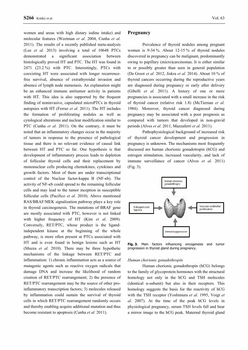
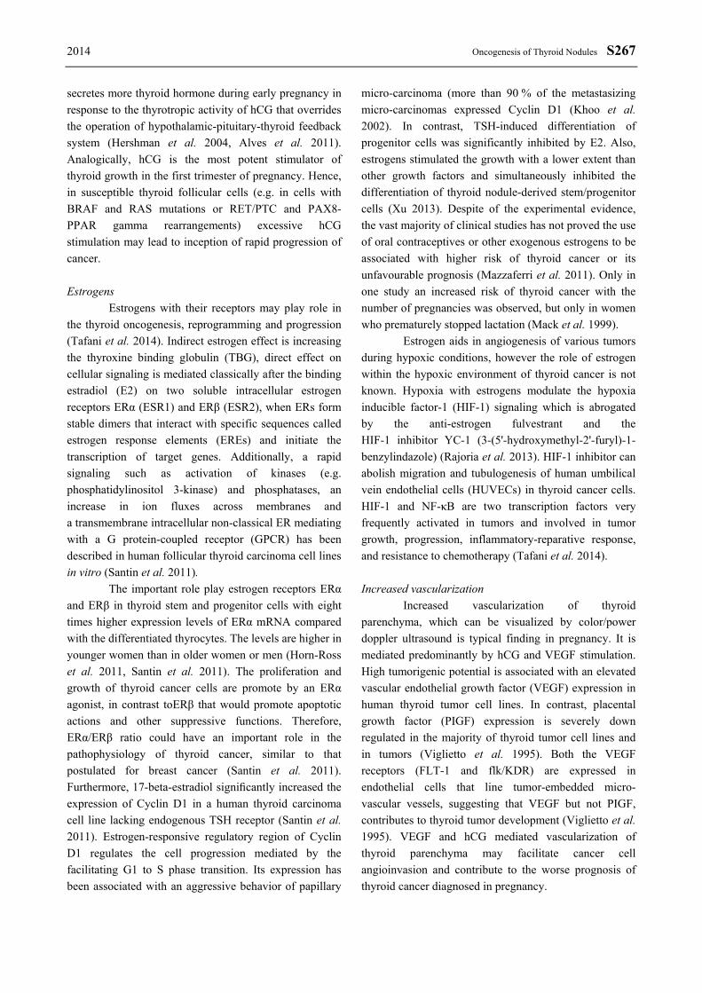
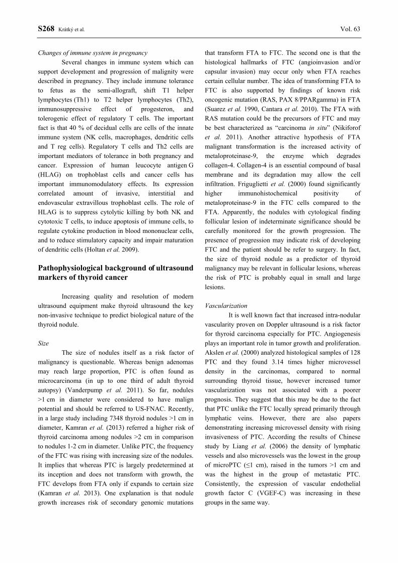