thyrotropin and thyroglobulin...
TRANSCRIPT
Thyrotropin and thyroglobulin testing
Stefan K. G. Grebe, MD, PhDProfessor of Laboratory Medicine and Pathology
Mayo Clinic, Rochester, MN
Conflicts of interest
• None pertaining to this talk
Off label use of FDA regulated in vitro diagnostic devices
• None
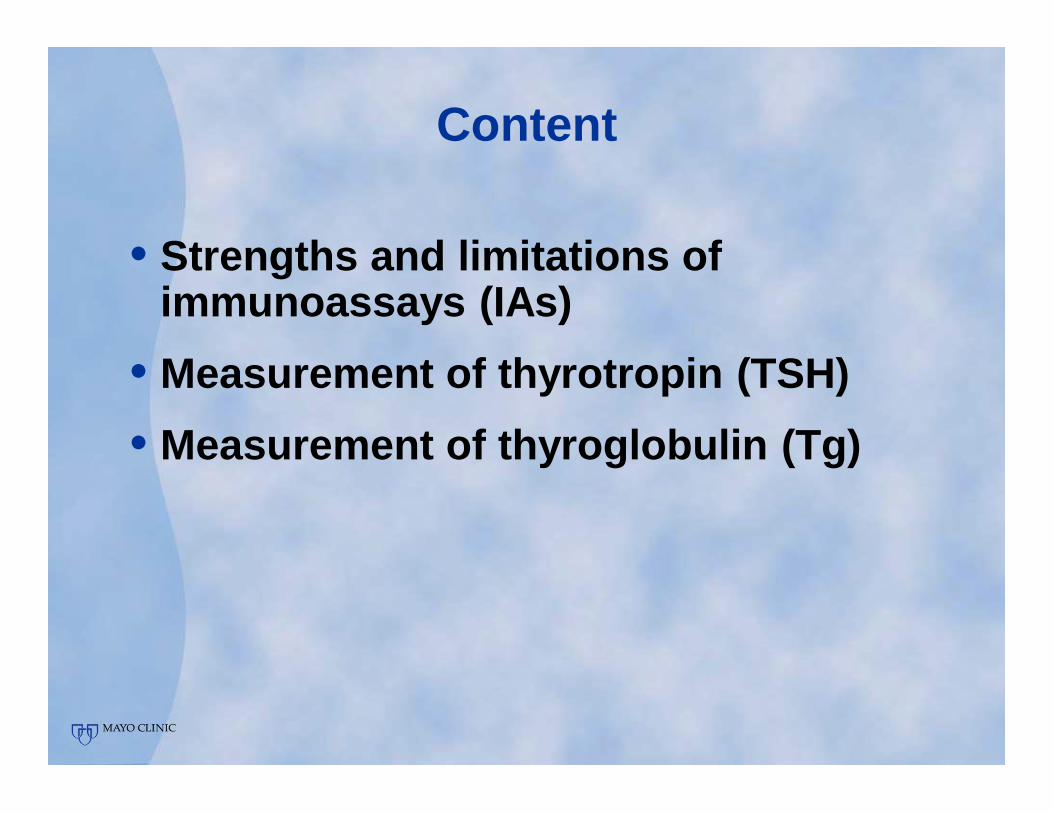
Content
• Strengths and limitations of immunoassays (IAs)
• Measurement of thyrotropin (TSH)• Measurement of thyroglobulin (Tg)
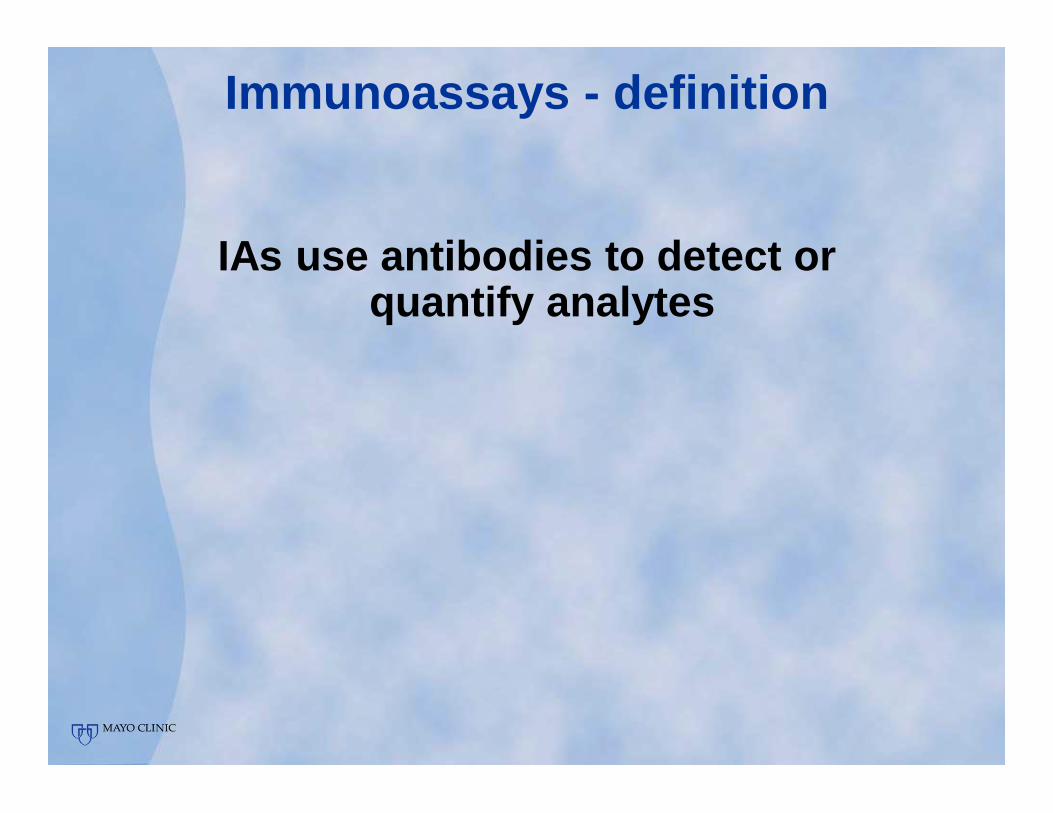
Immunoassays - definition
IAs use antibodies to detect or quantify analytes
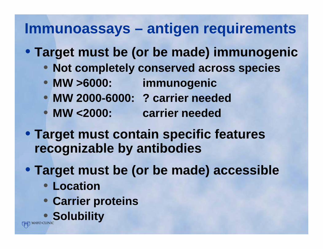
• Target must be (or be made) immunogenic• Not completely conserved across species• MW >6000: immunogenic• MW 2000-6000: ? carrier needed• MW <2000: carrier needed
• Target must contain specific features recognizable by antibodies
• Target must be (or be made) accessible• Location• Carrier proteins• Solubility
Immunoassays – antigen requirements
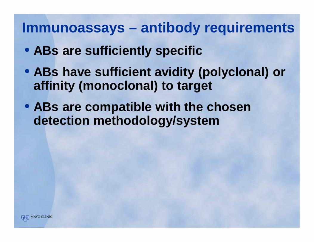
Immunoassays – antibody requirements• ABs are sufficiently specific• ABs have sufficient avidity (polyclonal) or
affinity (monoclonal) to target• ABs are compatible with the chosen
detection methodology/system
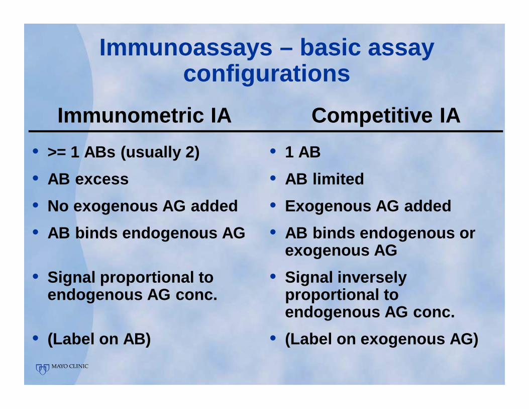
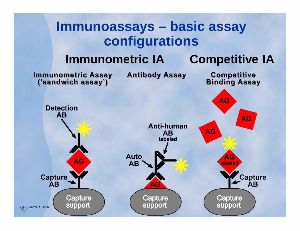
Immunoassays – basic assay configurations
• >= 1 ABs (usually 2)• AB excess• No exogenous AG added• AB binds endogenous AG
• Signal proportional to endogenous AG conc.
• (Label on AB)
• 1 AB• AB limited• Exogenous AG added• AB binds endogenous or
exogenous AG• Signal inversely
proportional to endogenous AG conc.
• (Label on exogenous AG)
Competitive IAImmunometric IA
Immunoassays – basic assay configurations
Competitive IAImmunometric IA
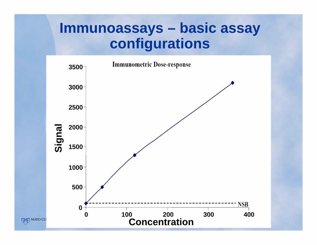
Immunoassays – basic assay configurations
0
2500
1000
500
0
2000
1500
3500
200100
3000
300 400Concentration
Sign
al
Immunoassays – basic assay configurations
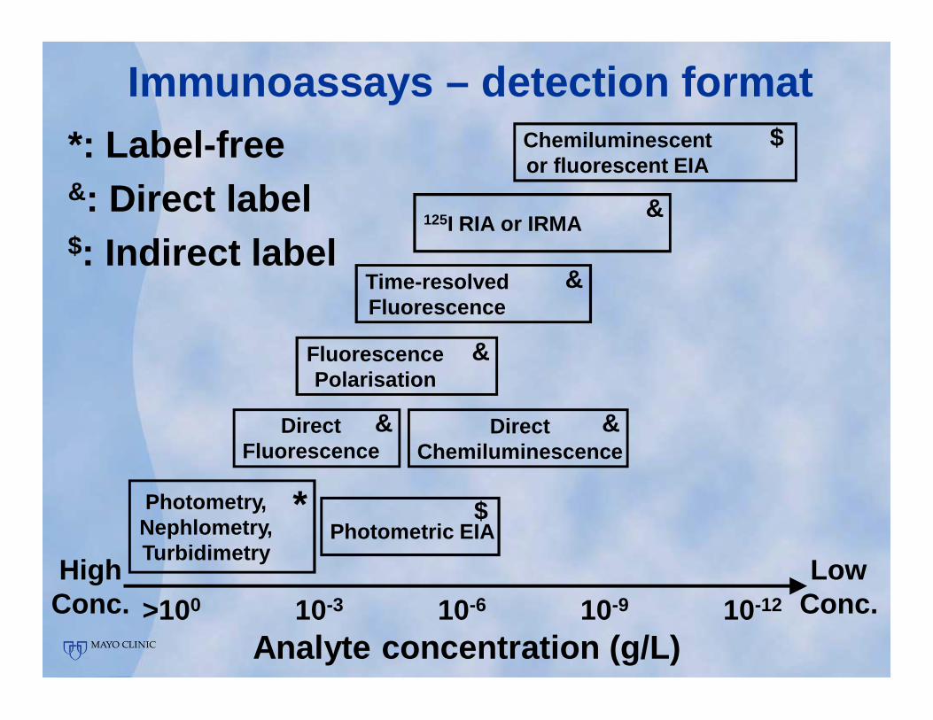
Immunoassays – detection format
>100 10-3 10-9 10-1210-6
Analyte concentration (g/L)
HighConc.
LowConc.
Photometry,Nephlometry,Turbidimetry
*
DirectFluorescence
DirectChemiluminescence
& &
FluorescencePolarisation
&
Time-resolvedFluorescence
&
125I RIA or IRMA &
Photometric EIA$
Chemiluminescentor fluorescent EIA
$*: Label-free&: Direct label$: Indirect label
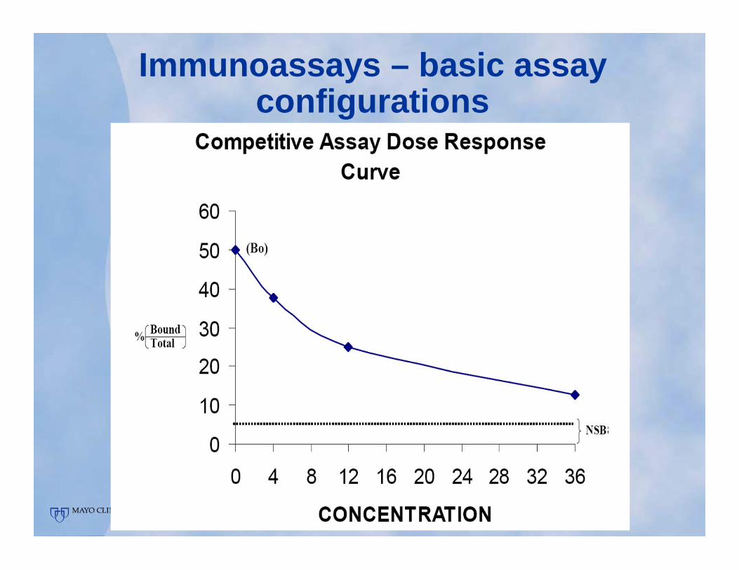
Immunoassays – problems & interferences
• Competitive assays - limited dynamic range
• Immunometric assays - hook
Assay format specific problems
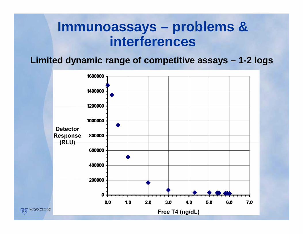
Immunoassays – problems & interferences
Limited dynamic range of competitive assays – 1-2 logs
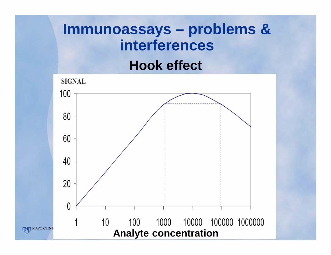
Immunoassays – problems & interferences
Hook effect
Analyte concentration
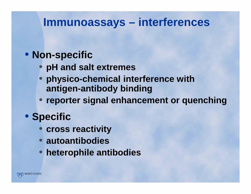
Immunoassays – interferences
• Non-specific• pH and salt extremes• physico-chemical interference with
antigen-antibody binding • reporter signal enhancement or quenching
• Specific• cross reactivity• autoantibodies• heterophile antibodies
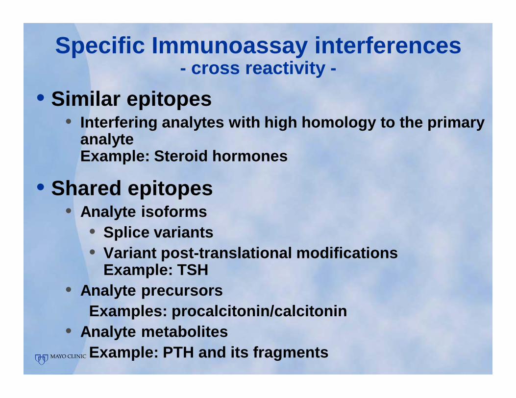
Specific Immunoassay interferences- cross reactivity -
• Similar epitopes• Interfering analytes with high homology to the primary
analyteExample: Steroid hormones
• Shared epitopes• Analyte isoforms
• Splice variants• Variant post-translational modifications
Example: TSH• Analyte precursors
Examples: procalcitonin/calcitonin• Analyte metabolites
Example: PTH and its fragments
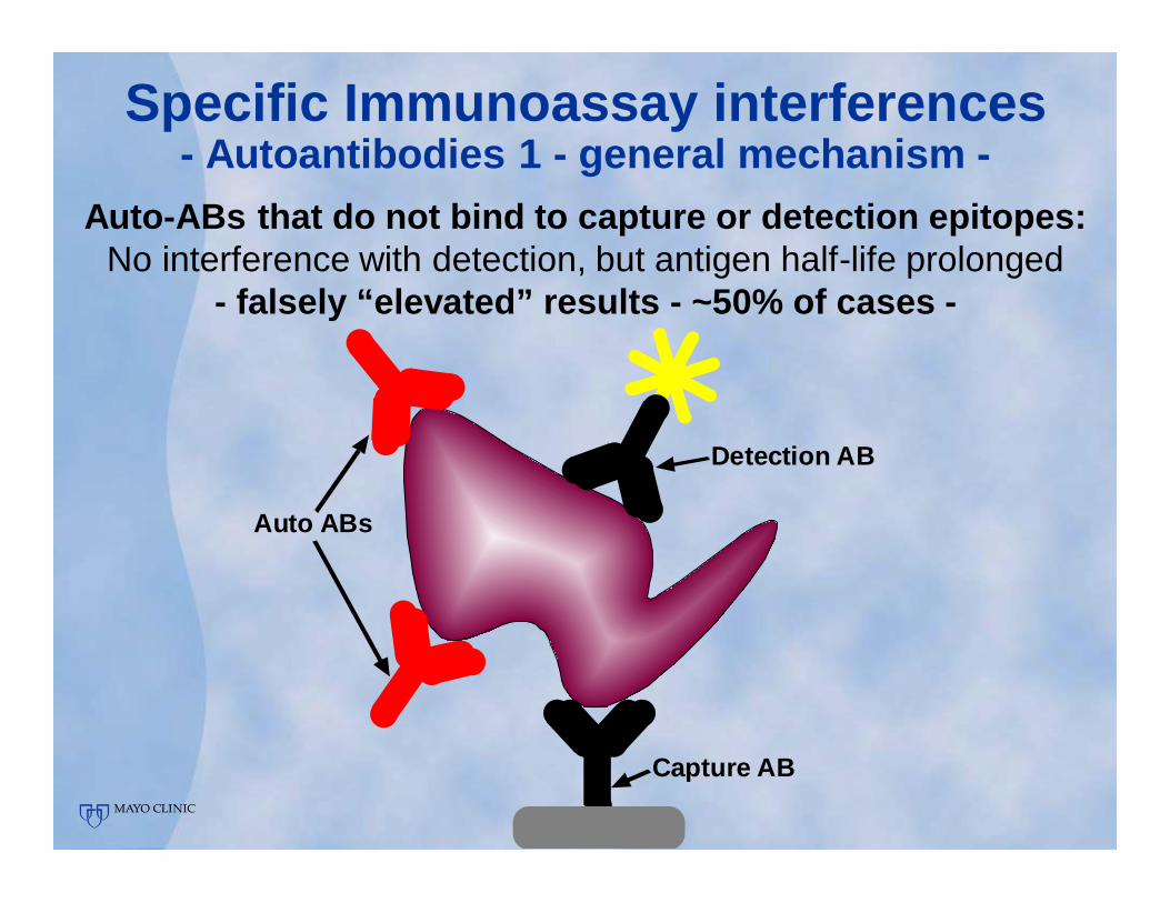
Auto ABs
Capture AB
Detection AB
Auto-ABs that do not bind to capture or detection epitopes:No interference with detection, but antigen half-life prolonged
- falsely “elevated” results - ~50% of cases -
Specific Immunoassay interferences- Autoantibodies 1 - general mechanism -
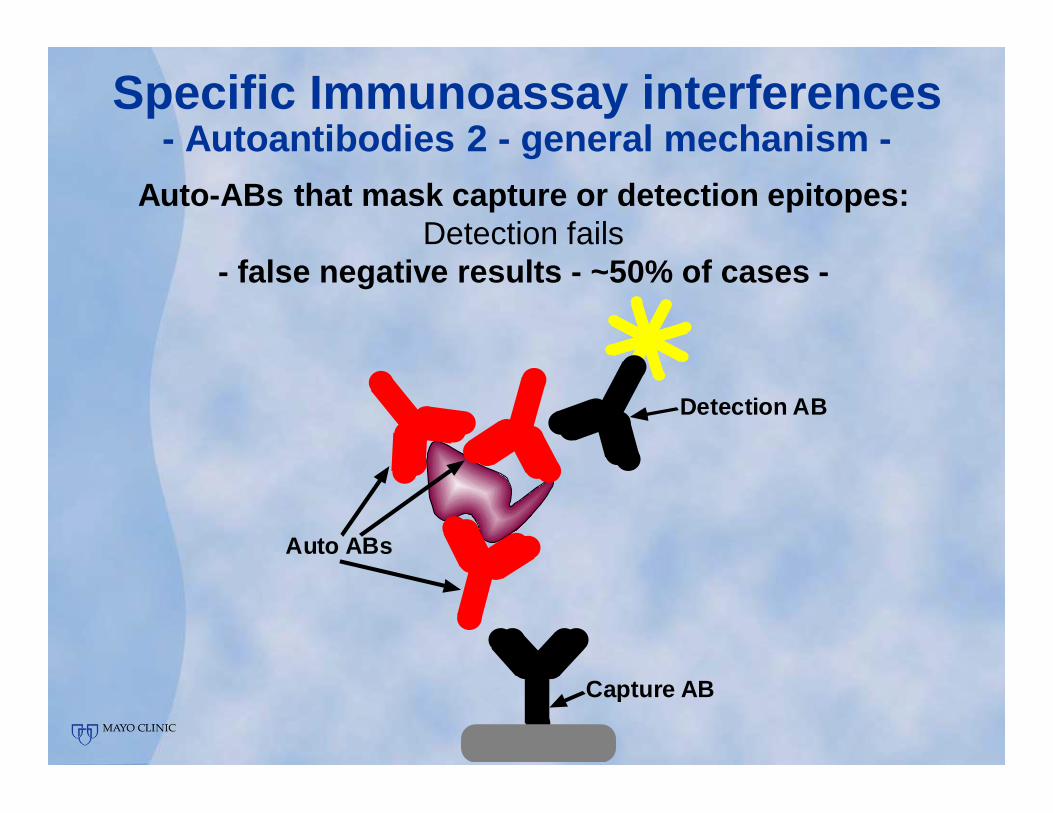
Capture AB
Detection AB
Auto ABs
Auto-ABs that mask capture or detection epitopes:Detection fails
- false negative results - ~50% of cases -
Specific Immunoassay interferences- Autoantibodies 2 - general mechanism -
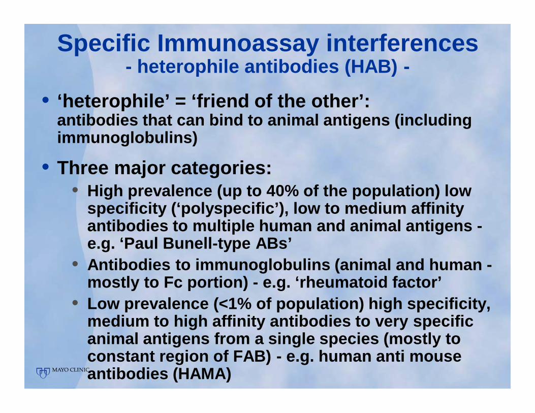
Specific Immunoassay interferences- heterophile antibodies (HAB) -
• ‘heterophile’ = ‘friend of the other’:antibodies that can bind to animal antigens (including immunoglobulins)
• Three major categories:• High prevalence (up to 40% of the population) low
specificity (‘polyspecific’), low to medium affinity antibodies to multiple human and animal antigens -e.g. ‘Paul Bunell-type ABs’
• Antibodies to immunoglobulins (animal and human -mostly to Fc portion) - e.g. ‘rheumatoid factor’
• Low prevalence (<1% of population) high specificity, medium to high affinity antibodies to very specific animal antigens from a single species (mostly to constant region of FAB) - e.g. human anti mouse antibodies (HAMA)
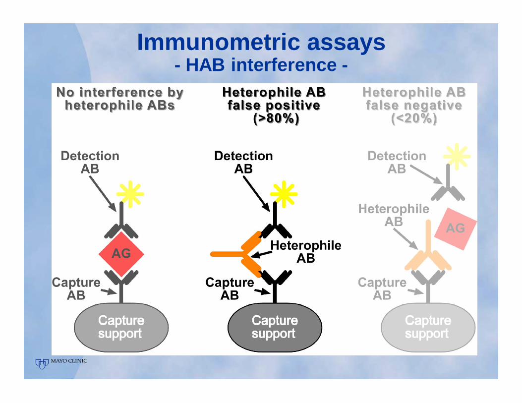
Immunometric assays- HAB interference -
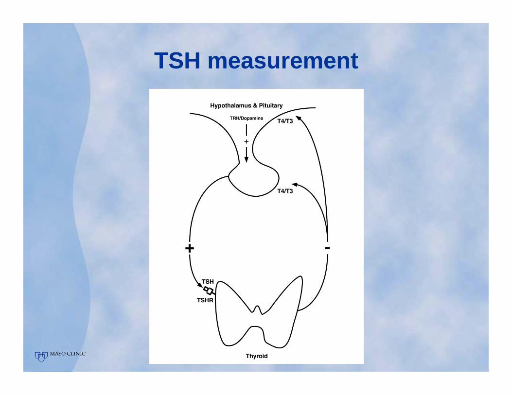
TSH measurement
TSH measurement
• The best single marker to assess thyroid function status
• The best single marker to assess thyroid function status
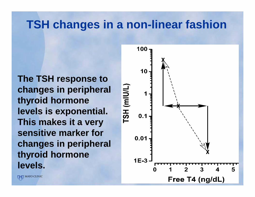
The TSH response to changes in peripheral thyroid hormone levels is exponential.This makes it a very sensitive marker for changes in peripheral thyroid hormone levels.
TSH changes in a non-linear fashion
TSH measurement
• The best single marker to assess thyroid function status
• A TSH within the ‘normal’ range should correlate with euthyroidism
• The best single marker to assess thyroid function status
• A TSH within the ‘normal’ range should correlate with euthyroidism
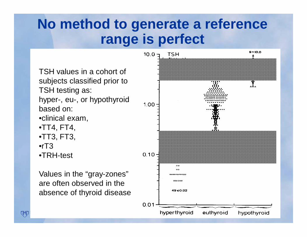
No method to generate a reference range is perfect
TSH values in a cohort of subjects classified prior to TSH testing as:hyper-, eu-, or hypothyroid based on:•clinical exam,•TT4, FT4, •TT3, FT3,•rT3•TRH-test
Values in the “gray-zones” are often observed in the absence of thyroid disease
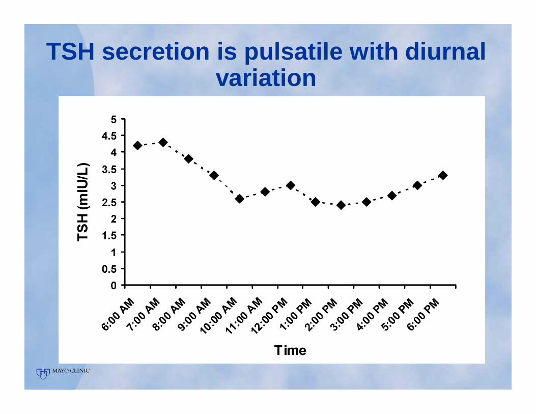
TSH secretion is pulsatile with diurnal variation
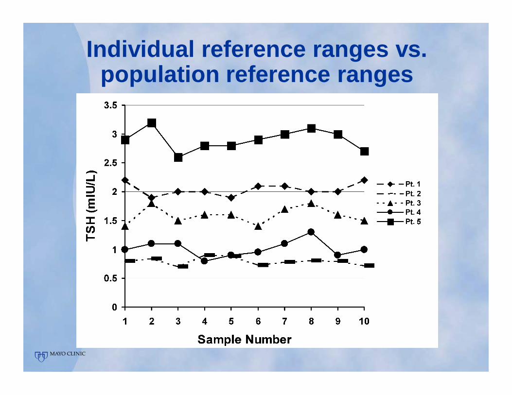
Individual reference ranges vs. population reference ranges

TSH assays differ from each otherand also over time
• 10-20% average difference between different assays
• Much larger difference may be observed
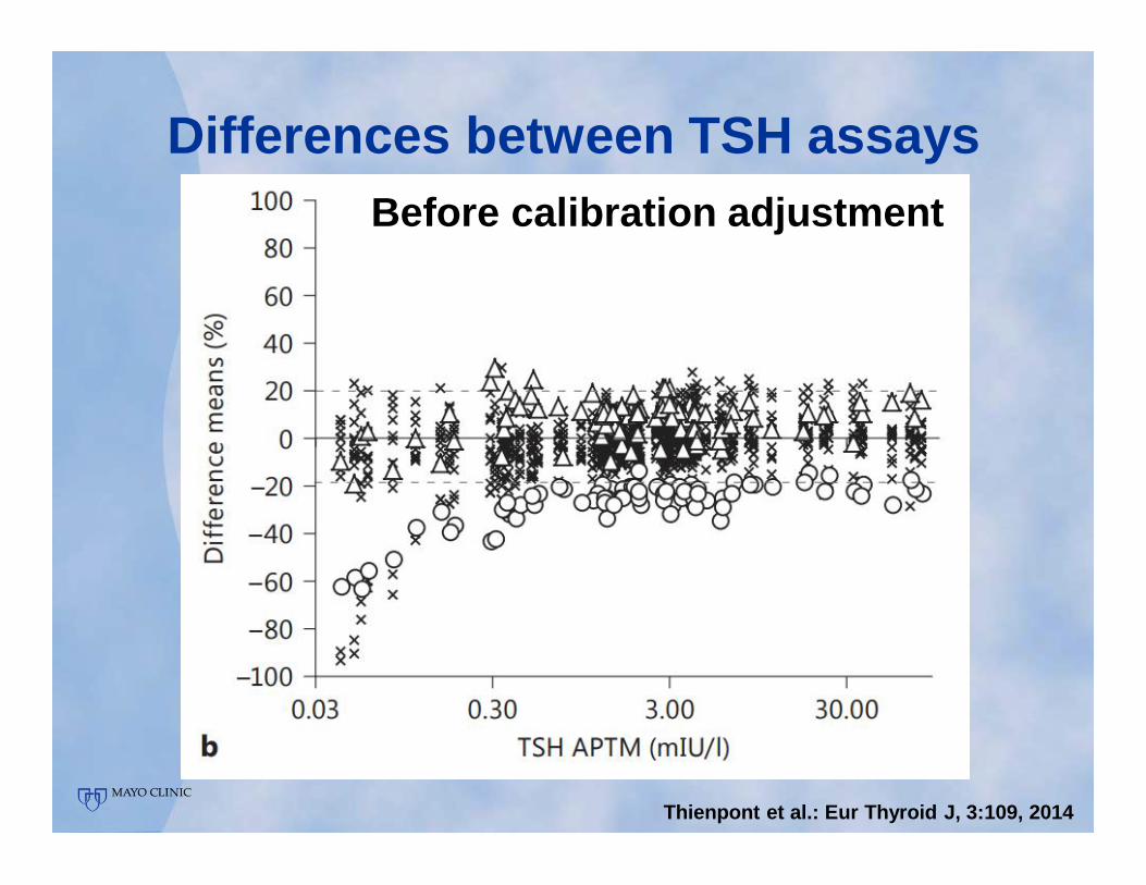
Differences between TSH assays
Thienpont et al.: Eur Thyroid J, 3:109, 2014
Before calibration adjustment
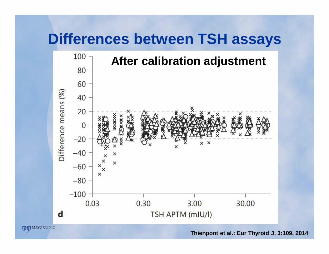
Differences between TSH assays
Thienpont et al.: Eur Thyroid J, 3:109, 2014
After calibration adjustment
TSH assays differ from each otherand also over time
• 10-20% average difference between different assays
• Much larger difference may be observed• Lot-to-lot variability of +/- 5-10% is common,
more might be observed occasionally
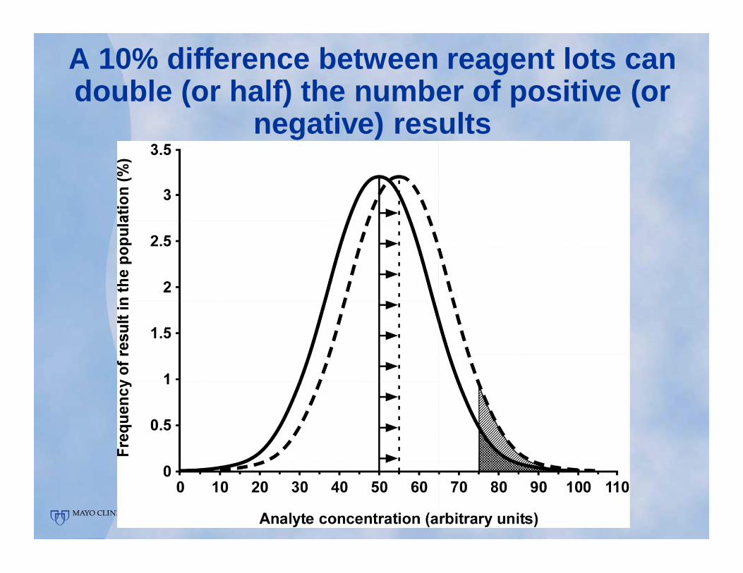
A 10% difference between reagent lots can double (or half) the number of positive (or
negative) results
Recommendations on interpreting TSH results and management - 1
• Re-measure borderline elevated or low TSH levels at least twice, regardless of reference range used
• A narrow TSH reference range is unsuitable in tertiary (and likely in secondary) care patient populations - about 20% of individuals would be classified as ‘biochemically hypothyroid’
• In treated hypothyroid patients, aim for a TSH between 0.3-3 mIU/L, if the patient is young/middle aged; higher levels in the elderly
• Avoid excessive micro-titration of TSH levels
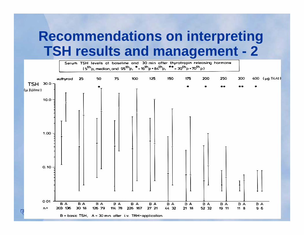
Recommendations on interpreting TSH results and management - 2
• Overtreatment is likely with T4 doses of >150 mcg/day
Recommendations on interpreting TSH results and management - 2
Recommendations on interpreting TSH results and management - 2
• Overtreatment is likely with T4 doses of >150 mcg/day
• In most thyroid cancer patients do not aim for complete TSH suppression
• Suppression to <0.1 mIU/L is only indicated for high risk patients and those with persistent disease
Measurement of thyroglobulin (Tg)
The role of Tg measurements
• Thyroid tumor marker• Detection of tumor recurrence
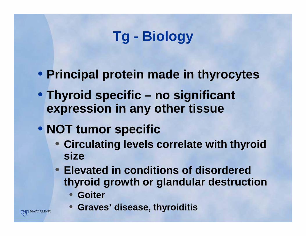
Tg - Biology
• Principal protein made in thyrocytes
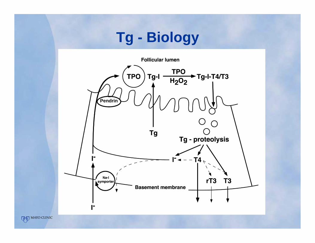
Tg - Biology
Pendrin

Tg - Biology
• Principal protein made in thyrocytes• Thyroid specific – no significant
expression in any other tissue
Tg - Biology
• Principal protein made in thyrocytes• Thyroid specific – no significant
expression in any other tissue• NOT tumor specific
• Circulating levels correlate with thyroid size
• Elevated in conditions of disordered thyroid growth or glandular destruction• Goiter• Graves’ disease, thyroiditis
Tg – Clinical role
• Follow-up of treated thyroid cancer patients
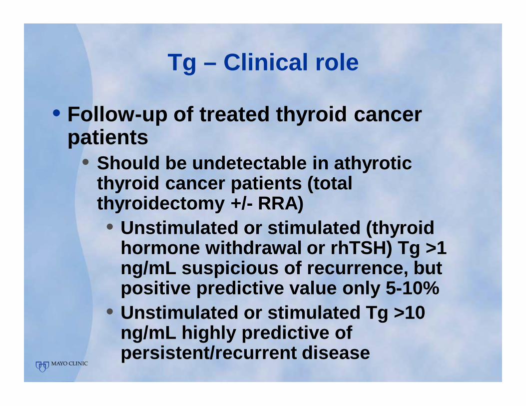
Tg – Clinical role
• Follow-up of treated thyroid cancer patients
• Should be undetectable in athyrotic thyroid cancer patients (total thyroidectomy +/- RRA)• Unstimulated or stimulated (thyroid
hormone withdrawal or rhTSH) Tg >1 ng/mL suspicious of recurrence, but positive predictive value only 5-10%
• Unstimulated or stimulated Tg >10 ng/mL highly predictive of persistent/recurrent disease
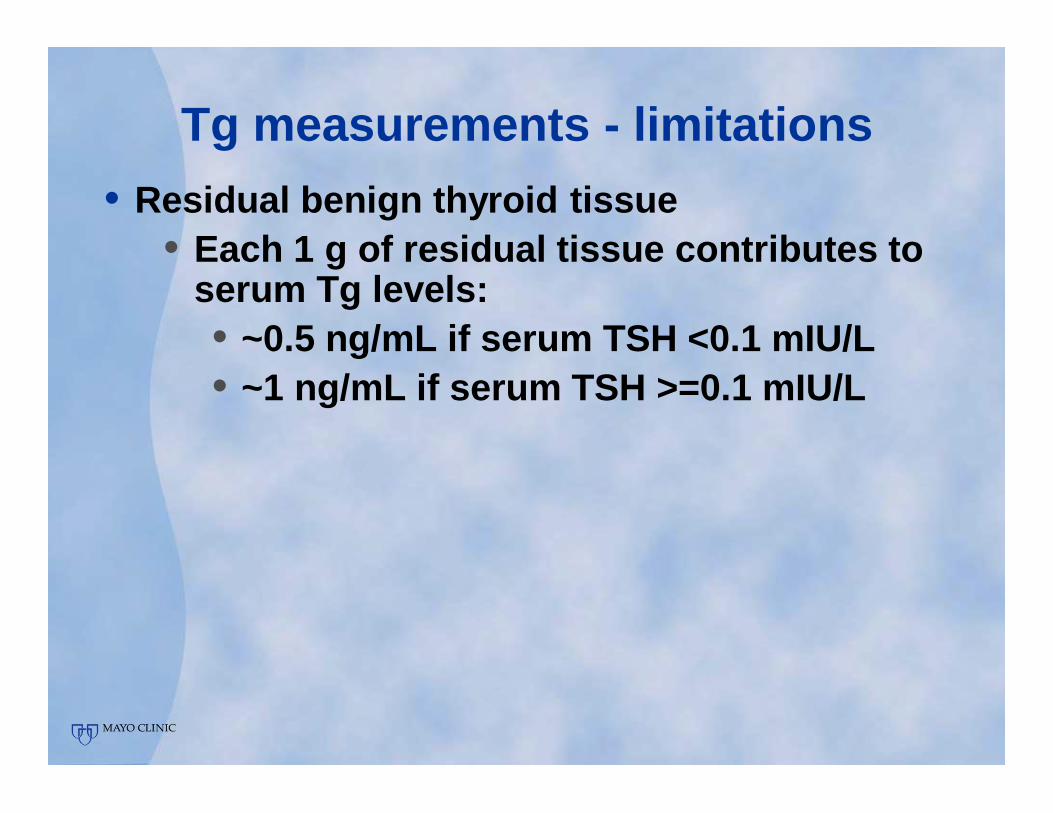
Tg measurements - limitations• Residual benign thyroid tissue
• Each 1 g of residual tissue contributes to serum Tg levels:• ~0.5 ng/mL if serum TSH <0.1 mIU/L• ~1 ng/mL if serum TSH >=0.1 mIU/L
Tg measurements - limitations• Residual benign thyroid tissue• Autoantibody interferences – pot. false low Tg
• ~25% of thyroid cancer patients have detectable anti thyroglobulin auto-antibodies (TgAb)
• Always measure both Tg & TgAb• Use LOQ of TgAb assay as cut-off for
TgAb positivity, rather than manufacturer recommended cut-offs
• “True” Tg conc. might be impossible to determine in TgAb+ patients
Tg measurements - limitations• Residual benign thyroid tissue• Autoantibody interferences – pot. false low Tg• Other limitations/interferences
• As discussed in the first section:• Non-specific interferences• Hook• Heterophile antibody interferences
Tg measurements - limitations• Residual benign thyroid tissue• Autoantibody interferences – pot. false low Tg• Other limitations/interferences• Poor low-end sensitivity necessitates
stimulated Tg measurements for some assays
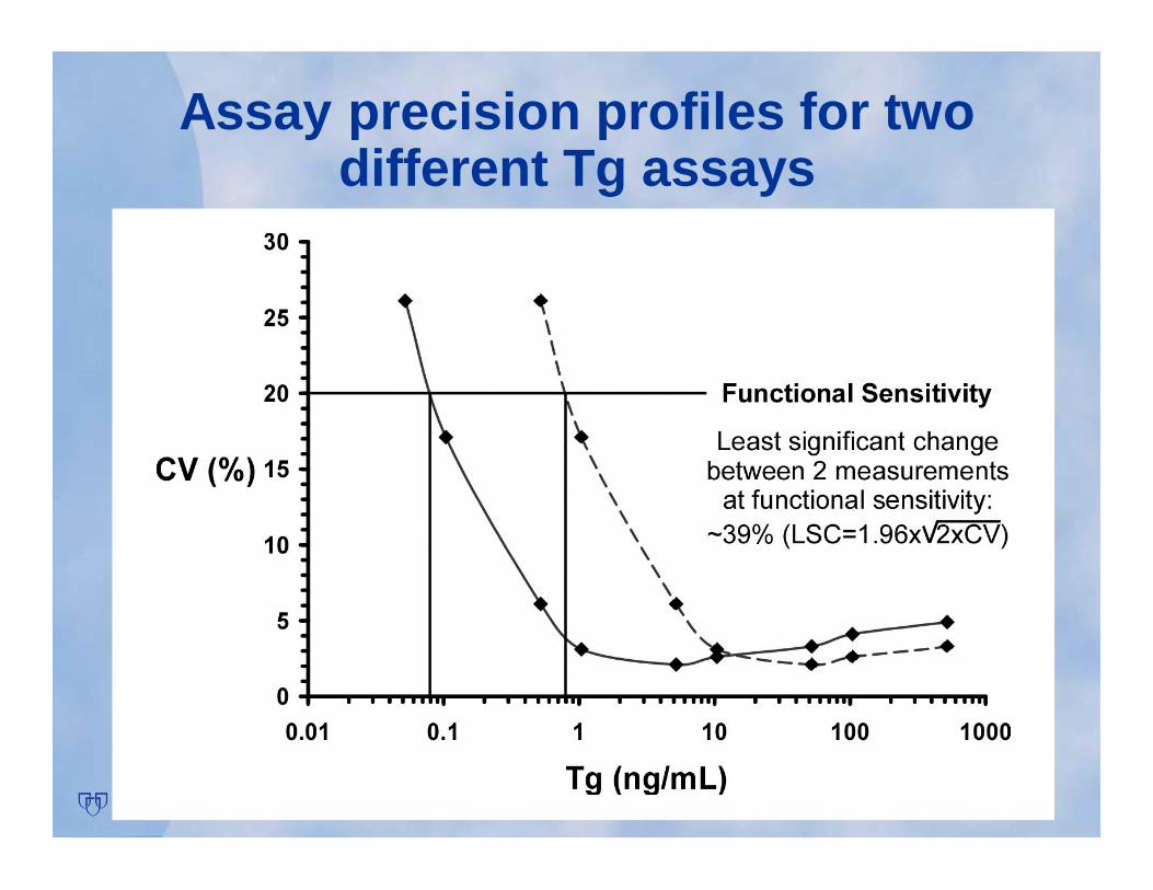
Assay precision profiles for two different Tg assays
Tg measurements - limitations• Residual benign thyroid tissue• Autoantibody interferences – pot. false low Tg• Other limitations/interferences• Poor low-end sensitivity necessitates
stimulated Tg measurements in some assays• Calibration differences between different Tg
assays, or reagent lot-to-lot variability can lead to false positive and false negative test results
Tg measurements – current ATA Guidelines (2015) and other sources
• Use assay calibrated against CRM457• All current assays fulfil this criterion
Tg measurements – current ATA Guidelines (2015) and other sources
• Use assay calibrated against CRM457• Always measure TgAb alongside Tg
• This ensures that potentially falsely low biased Tg measurements are identified
Tg measurements – current ATA Guidelines (2015) and other sources
• Use assay calibrated against CRM457• Always measure TgAb alongside Tg• Tg assay with a LOQ of ≤0.2 ng/mL preferred
• Assays with higher LOQ often will force use of stimulated Tg measurements

Tg measurements – current ATA Guidelines (2015) and other sources
• Use assay calibrated against CRM457• Always measure TgAb alongside Tg• Tg assay with a LOQ of ≤0.2 ng/mL preferred• Perform postoperative Tg measurement
• ≥3-4 weeks post-op• On T4 or stimulated• Unstimulated Tg <0.2 ng/mL or stimulated Tg <1
ng/mL denote persistent disease risk <4%• Higher values do not distinguish between remnant
and persistent disease

Tg measurements – current ATA Guidelines (2015) and other sources
• Use assay calibrated against CRM457• Always measure TgAb alongside Tg• Tg assay with a LOQ of ≤0.2 ng/mL preferred• Perform postoperative Tg measurement• Regular follow-up Tg measurements
• Use the same assay in a given individual• Re-baseline if assay is changed• Usually unstimulated measurements
• Low/intermediate risk patients 6-12 monthly, later 12-24 monthly
• Incomplete thyroidectomy: use patients previous values and doubling time, rather than fixed cut-offs
• High risk patients at least 6-12 monthly

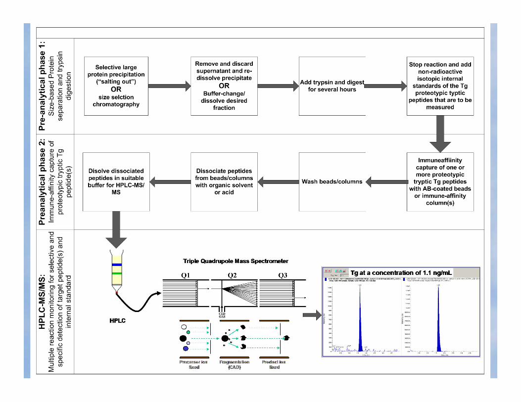
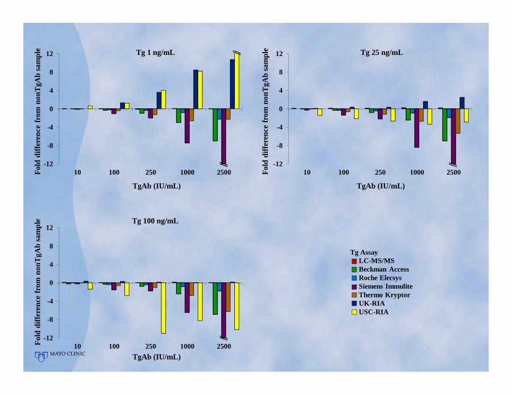
(Partially) overcoming TgAb interference :Tg measurement by mass spectrometry (MS)
• MS can overcome the TgAb and heterophile AB (HAB) interferences that plague Tg IAs

• MS can overcome the TgAb and heterophile AB (HAB) interferences that plague Tg immunoassays
• Trypsin digestion of sample before MS cleaves Tg, TgAb and HAB (along with all other serum proteins)
• MS detects Tg-proteotypic peptides after digest with high specificity
(Partially) overcoming TgAb interference :Tg measurement by mass spectrometry (MS)
-12
-8
-4
0
4
8
12
10 100 250 1000 2500-12
-8
-4
0
4
8
12
10 100 250 1000 2500Fold
diff
eren
ce fr
om n
onTg
Ab
sam
ple
TgAb (IU/mL)
Fold
diff
eren
ce fr
om n
onTg
Ab
sam
ple
Fold
diff
eren
ce fr
om n
onTg
Ab
sam
ple
TgAb (IU/mL) TgAb (IU/mL)
Tg AssayLC-MS/MSBeckman AccessRoche ElecsysSiemens ImmuliteThermo KryptorUK-RIAUSC-RIA
Tg 1 ng/mL
Tg 100 ng/mL
Tg 25 ng/mL
-12
-8
-4
0
4
8
12
10 100 250 1000 2500
Roche Tg II – Tg LC-MS/MS)/Tg LC-MS/MS
FoldedProbability
0
0.1
0.2
0.3
0.4
0.5
-100% -50% 0% 50% 100% 150% 200%
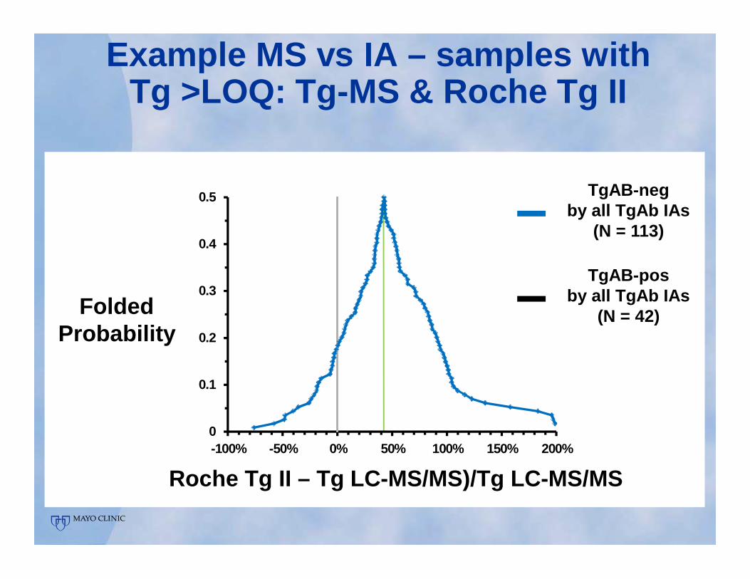
Example MS vs IA – samples with Tg >LOQ: Tg-MS & Roche Tg II
TgAB-negby all TgAb IAs
(N = 113)
TgAB-posby all TgAb IAs
(N = 42)
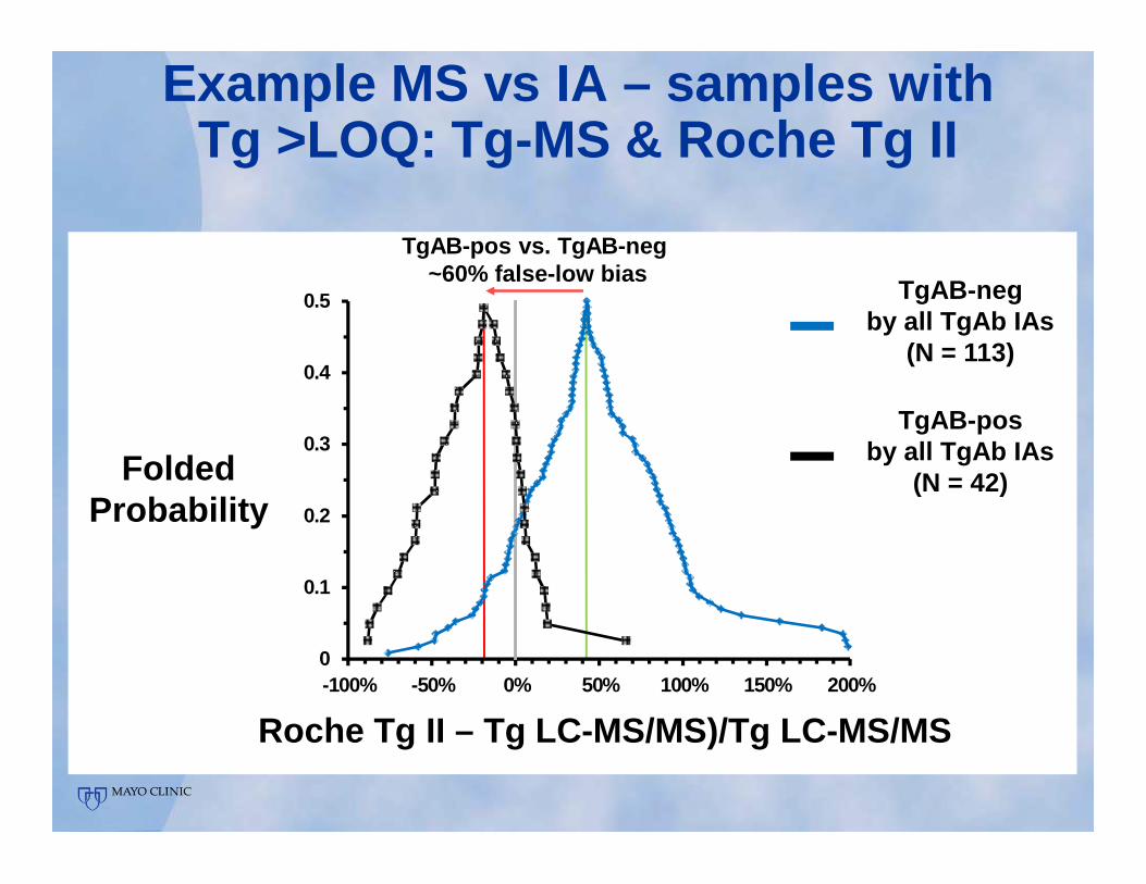
Roche Tg II – Tg LC-MS/MS)/Tg LC-MS/MS
FoldedProbability
0
0.1
0.2
0.3
0.4
0.5
-100% -50% 0% 50% 100% 150% 200%
TgAB-pos vs. TgAB-neg~60% false-low bias
Example MS vs IA – samples with Tg >LOQ: Tg-MS & Roche Tg II
TgAB-negby all TgAb IAs
(N = 113)
TgAB-posby all TgAb IAs
(N = 42)
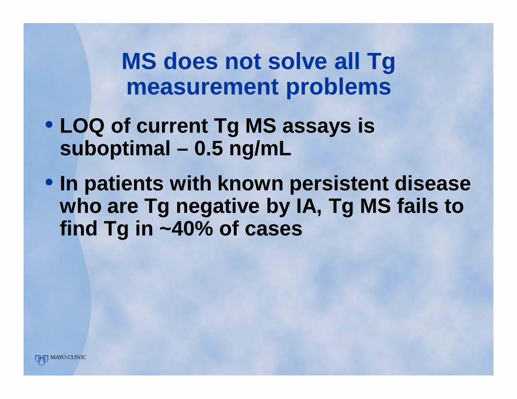
MS does not solve all Tg measurement problems
• LOQ of current Tg MS assays is suboptimal – 0.5 ng/mL
• In patients with known persistent disease who are Tg negative by IA, Tg MS fails to find Tg in ~40% of cases

Most patients with undetectable Tg by IA have low Tg values by Tg MS
N=105, 20 with detectable Tg by LC-MS/MS
Individual specimens, all with Tg <0.1 ng/mL by Beckman IA
Tg b
y LC
-MS
/MS
, ng/
mL
Recommendation for use of Tg MS assays
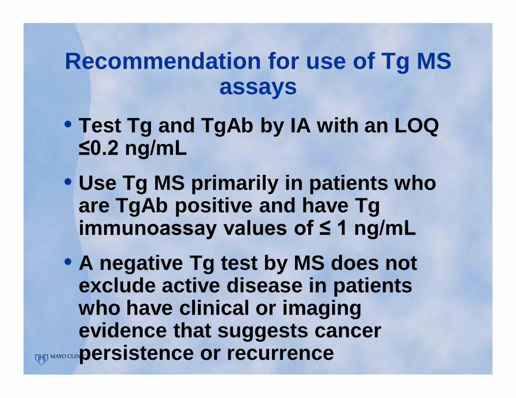
• Test Tg and TgAb by IA with an LOQ ≤0.2 ng/mL
• Use Tg MS primarily in patients who are TgAb positive and have Tg immunoassay values of ≤ 1 ng/mL
• A negative Tg test by MS does not exclude active disease in patients who have clinical or imaging evidence that suggests cancer persistence or recurrence

Thank You
Questions?