tia in the swedish stroke register (riksstroke). aspects...
TRANSCRIPT
LUND UNIVERSITY
PO Box 117221 00 Lund+46 46-222 00 00
TIA in the Swedish Stroke Register (Riksstroke). Aspects on diagnostic validation, riskfactors, investigations, and therapies
Buchwald, Fredrik
2018
Document Version:Publisher's PDF, also known as Version of record
Link to publication
Citation for published version (APA):Buchwald, F. (2018). TIA in the Swedish Stroke Register (Riksstroke). Aspects on diagnostic validation, riskfactors, investigations, and therapies. Lund: Lund University, Faculty of Medicine.
General rightsCopyright and moral rights for the publications made accessible in the public portal are retained by the authorsand/or other copyright owners and it is a condition of accessing publications that users recognise and abide by thelegal requirements associated with these rights.
• Users may download and print one copy of any publication from the public portal for the purpose of private studyor research. • You may not further distribute the material or use it for any profit-making activity or commercial gain • You may freely distribute the URL identifying the publication in the public portalTake down policyIf you believe that this document breaches copyright please contact us providing details, and we will removeaccess to the work immediately and investigate your claim.

1
TIA in the Swedish Stroke Register (Riksstroke)

2

3
TIA in the Swedish Stroke Register (Riksstroke)
Aspects on diagnostic validation, risk factors, investigations, and therapies
Fredrik Buchwald
DOCTORAL DISSERTATION
by due permission of the Faculty of Medicine at Lund University, Sweden.
To be defended at Kvinnoklinikens aula, Skåne University Hospital in Malmö on 16 March 2018, at 9.00 a.m.
Faculty opponent Christina Sjöstrand, Karolinska Institutet

4
Organization LUND UNIVERSITY
Document name DOCTORAL DISSERTATION
Department of Clinical Sciences, Neurology Date of issue
Author Fredrik Buchwald Sponsoring organization
Title and subtitle: TIA in the Swedish Stroke Register (Riksstroke). Aspects on diagnostic validation, risk factors, investigations, and therapies.
Abstract Background: Transient ischemic attacks (TIA) indicate an increased risk of stroke, one of the leading causes of death and disability worldwide. In order to prevent stroke, our knowledge on diagnosis, demographics, risk factors, investigations, and treatment of patients with TIA needs to be improved. Aims: The aims of this thesis were to validate data and diagnoses in the Riksstroke TIA module (Riksstroke-TIA), to clarify the role of atrial fibrillation (AF) in TIA and the extent of oral anticoagulant (OAC) treatment in patients with AF, to assess characteristics, risk factors, and secondary preventive treatment in TIA patients with a history of stroke in comparison to those without, and evaluate the degree of carotid imaging and determinants for its non-use in patients with TIA. Methods: Paper I was based on a study sample of 180 patients from 6 different hospitals, extracted from the cohort of patients registered in Riksstroke-TIA between 1/7/2011 to 30/6/2012 (n=7825). Medical files were retrieved from each hospital. Paper II – IV were based on data from patients registered in Riksstroke-TIA between 1/7/2011 to 30/6/2013 (n=15064). For comparison, data on patients with ischemic stroke (IS) registered in Riksstroke during the corresponding period of time were included in paper II – IV (n=44416). Results Paper I: Two independent assessors agreed on a likely or possible diagnosis of TIA in 77% (137/180), in 3% (5/180) on a diagnosis of IS, and in 2% (3/180) that a diagnosis of TIA was unlikely. The quality of documentation was fair. Paper II: AF was present in 19% (2779/14980) of patients with TIA compared to 30% (13258/44173) in those with IS. Proportions of AF increased markedly with age. At discharge, 64% (1778/2771) of patients with TIA and AF and 50% (5502/10899) of patients with IS and AF were treated with OACs. Paper III: Patients with TIA and a history of stroke were older, more likely to be male, and they had higher proportions of AF, hypertension, and diabetes mellitus than those without a history of stroke. In TIA patients with prior stroke aged ≥85 years, AF was present in 41% (300/724) compared to 30% (604/2028) in those without prior stroke. At discharge, levels of OAC treatment in TIA patients with AF and prior stroke were lower than in those without prior stroke. Paper IV: Carotid imaging was performed in 70% (10545/15023) of patients with TIA. Determinants for its non-use were age ≥85 years, age 74-84 years, female sex, AF, a history of stroke, and care at a non-university hospital. There were substantial regional variations regarding proportions of carotid imaging, especially in the very elderly. Conclusions: There was interobserver agreement on TIA diagnoses in a majority of cases. More systematic documentation aided by a guide or checklist might improve diagnostic certainty. Data registered in Riksstroke-TIA was valid and suited for scientific evaluation. AF was a common but insufficiently treated risk factor in TIA. Certain patient groups appeared neglected with regard to carotid imaging and secondary preventive treatment, namely the very elderly, women, those with AF, and a history of stroke. Opportunities of secondary prevention were likely missed in a substantial number of patients.
Key words: transient ischemic attack, diagnostic validation, ischemic stroke, risk factors, secondary prevention
Classification system and/or index terms (if any)
Supplementary bibliographical information Language: English
ISSN and key title 1652-8220 ISBN 978-91-7619-591-8
Recipient’s notes Number of pages Price
Security classification
I, the undersigned, being the copyright owner of the abstract of the above-mentioned dissertation, hereby grant to all reference sources permission to publish and disseminate the abstract of the above-mentioned dissertation.
Signature Date 2018-02-07

5
TIA in the Swedish Stroke Register (Riksstroke)
Aspects on diagnostic validation, risk factors, investigations, and therapies
Fredrik Buchwald
6
Cover art by Petrea Frid
Copyright Fredrik Buchwald
Faculty of Medicine, Department of Clinical Sciences, Lund University ISBN 978-91-7619-591-8 ISSN 1652-8220 Printed in Sweden by Media-Tryck, Lund University Lund 2018

7
...für Papa. If you can meet with triumph and disaster and treat those two impostors just the same...
Rudyard Kipling
8
Content
Content ...................................................................................................................... 8Original papers ............................................................................................. 10Abbreviations ............................................................................................... 11
Introduction ............................................................................................................. 13Definition ...................................................................................................... 13Diagnosis ...................................................................................................... 14Epidemiology ............................................................................................... 16Risk of stroke ................................................................................................ 16Etiology ........................................................................................................ 17Investigation ................................................................................................. 19
Aim of the thesis ..................................................................................................... 21Subjects and methods .............................................................................................. 23
Study materials ............................................................................................. 23The Swedish Stroke Register (Riksstroke) .......................................... 23The Swedish Stroke Register TIA module (Riksstroke-TIA) ............. 24
Study subjects and procedure ....................................................................... 26Paper I .................................................................................................. 26Papers II - IV ....................................................................................... 27
Statistical methods ........................................................................................ 27Ethical considerations ................................................................................... 28
Results ..................................................................................................................... 29Validation of diagnoses of TIA in Riksstroke-TIA (Paper I) ....................... 31
Documentation of time aspects and neurological examination ........... 31TIA diagnosis and interobserver agreement ........................................ 32
Atrial fibrillation in TIA versus ischemic stroke (Paper II) ........................ 33Proportions of atrial fibrillation ........................................................... 33Patient characteristics .......................................................................... 33Oral anticoagulant treatment at discharge ........................................... 34
TIA and ischemic stroke patients with or without prior stroke (Paper III) .. 35
9
Patient characteristics .......................................................................... 36Atrial fibrillation .................................................................................. 37Medication at discharge ....................................................................... 37
Carotid imaging in patients with TIA or ischemic stroke (Paper IV) .......... 39Patient characteristics .......................................................................... 39Hospital type and regional differences ................................................ 40Independent determinants for not undergoing carotid imaging ........... 42
Discussion ............................................................................................................... 45Methodological considerations ..................................................................... 45
Coverage and selection bias ................................................................. 45Quality and completeness of registered data ....................................... 46Confounding ........................................................................................ 47
General discussion ........................................................................................ 48Validation of Riksstroke-TIA .............................................................. 48Atrial fibrillation in TIA versus ischemic stroke ................................. 49TIA patients with or without prior stroke ............................................ 51Carotid imaging ................................................................................... 52General considerations ......................................................................... 53
Conclusions ............................................................................................................. 55Future perspectives ................................................................................................. 57Swedish summary ................................................................................................... 59
Svensk sammanfattning ................................................................................ 59Acknowledgements ................................................................................................. 61References ............................................................................................................... 63

10
Original papers
This thesis is based on the following four papers referred to in the text by their Roman numerals. The papers are appended in the end of this thesis with due permission from the publishers.
I. Buchwald F, Ström JO, Norrving B, Petersson J. Validation of diagnoses of transient ischemic attack in the Swedish Stroke Register (Riksstroke) TIA-module. Neuroepidemiology. 2015;45:40-43
II. Buchwald F, Norrving B, Petersson J. Atrial fibrillation in transient ischemic attack versus ischemic stroke. A Swedish Stroke Register (Riksstroke) study. Stroke. 2016;47:2456-2461
III. Buchwald F, Norrving B, Petersson J. Transient ischemic attack and ischemic stroke with or without prior stroke. Acta Neurologica Scandinavica. 2017;136:654-659
IV. Buchwald F, Norrving B, Petersson J. Is carotid imaging underused in patients with transient ischemic attack and ischemic stroke? A Swedish Stroke Register (Riksstroke) study. Acta Neurologica Scandinavica. 2017 Dec 18. doi: 10.111/ane.12886. [Epub ahead of print]

11
Abbreviations
AF Atrial fibrillation
CI Confidence interval
CT Computed tomography
CTA Computed tomography angiography
DWI Diffusion weighted imaging
ICD International Classification of Diseases
IS Ischemic stroke
MRA Magnetic resonance angiography
MRI Magnetic resonance imaging
NIHSS National Institutes of Health Stroke Scale
NINDS National Institute of Neurological Disorders and Stroke
NOAC Novel oral anticoagulant
OAC Oral anticoagulant
OR Odds ratio
PD Prevalence difference
RLS Reaction Level Scale
SD Standard deviation
TIA Transient ischemic attack

12

13
Introduction
Transient ischemic attacks (TIAs) are episodes of transitory neurological symptoms that should leave the affected person without any residual deficits. So what is the importance of a TIA? It is not the TIA in itself that carries a potential harm to the affected individual but the increased risk of stroke associated with this condition. Stroke is one of the leading causes of death and disability worldwide,1, 2 and a TIA represents a challenge and a chance to prevent a potentially devastating stroke.
Definition
In 1975, the National Institute of Neurological Disorders and Stroke (NINDS) launched a “Classification and Outline of Cerebrovascular Diseases”3 stating that there are
[...] episodes of temporary and focal cerebral dysfunction of vascular origin, rapid in onset [...], which are variable in duration, commonly lasting 2 to 15 minutes but occasionally lasting as long as a day (24 hours). [...] Each attack leaves no persistent neurological deficit.
Over the past decades, a TIA has been defined as an episode of neurological deficit with sudden onset and rapid resolution, caused by focal cerebral or retinal ischemia lasting less than 24 hours. With the advent of magnetic resonance imaging (MRI) neuroradiological evidence accumulated that a considerable number of patients with a TIA according to this “time-based” definition actually had suffered a persistent brain tissue lesion.4 Hence, a “tissue-based” definition has been proposed, namely a transient episode of neurological dysfunction caused by focal brain or retinal ischemia, without evidence of acute infarction.5 The application of the tissue-based definition requires an MRI with diffusion weighted imaging (DWI) as this modality is significantly more sensitive than computed tomography (CT) in detecting brain infarctions in the setting of short-lasting neurological symptoms.6 Today, both definitions are used, at least in part based on the available neuroradiological modality during assessment of a patient with a TIA. In the 11th revision of the International Classification of Diseases (ICD-11),

14
which is due to be published in 2018, the tissue-based definition will be incorporated.7
Diagnosis
Today, the gold standard test of diagnosis of TIA is expert opinion only. In most cases, diagnosis is based on a description of symptoms presented to the physician by the patient or a bystander after their resolution. In a minority of patients, symptoms and clinical findings are still present on examination and give supportive information to the physician. Radiological or laboratory findings are used to exclude structural lesions that may cause transient neurological symptoms of non-vascular origin. In 1975, NINDS published TIA criteria (Table 1),3 that acknowledged that there are episodes of transient neurological symptoms whose nature will remain uncertain. This statement also included an attempt to correlate symptoms to vascular territories. However, the application of these criteria in real life can be challenging as patients and bystanders often describe transient neurological episodes in words that do not match typical medical phrasings such as those used by NINDS.

15
Table 1. TIA criteria according to NINDS3
Carotid system Vertebrobasilar system Either carotid or vertebrobasilar system
Uncertain TIA Symptoms not to be included as TIA
Unilateral motor deficit
Motor deficit of any combination of extremities up to quadriplegia, sometimes changing from side to side
Isolated dysarthria Isolated vertigo
Unconsciousness including syncope
Unilateral sensory deficit
Sensory deficit in any combination of extremities including all four or involving both sides of the face or mouth
Isolated homonymous hemianopia
Isolated dysarthria
Tonic and/or clonic activity
Aphasia Loss of vision, complete or partial in both homonymous fields (bilateral homonymous hemianopia)
Isolated dysphagia
March of a sensory deficit
Loss of vision in one eye or in part of one eye
Homonymous hemianopia
Isolated diplopia
Incontinence of bowel or bladder
Homonymous hemianopia
Ataxia, imbalance, unsteadiness, or dysequilibrium not associated with vertigo
Dizziness or wooziness alone
Combination of the above
Either vertigo (with or without nausea and vomiting), diplopia, dysphagia, or dysarthria in combination with one another or with any of the above
Loss of vision associated with alteration of consciousness
Combinations of the above
Focal symptoms associated with migraine
Scintillating scotomata
It is up to the treating physician to decide whether the presented symptoms are actually caused by a cerebral or retinal ischemia. Both knowledge of the temporal development of focal ischemia and neurovascular anatomy are necessary requisites. But, as reflected in the NINDS criteria,3 there will remain uncertainty about the true cause of at least some episodes that might be suggestive of a TIA, e.g. in the case of isolated dysarthria or isolated vertigo. On the other hand, there are rare types of TIAs that contradict the proposed criteria, such as limb-shaking TIAs in the presence of high-grade carotid stenosis.8 Several conditions causing transient neurological symptoms can mimic TIA. These include focal seizures, migraine, syncope, metabolic derangements such as hypoglycemia or septicemia, peripheral vestibulopathies, amyloid angiopathy, and subdural hematoma, among others.

16
With regard to these diagnostic challenges it is hardly surprising that agreement on a diagnosis of TIA between health care professionals may be far from perfect. Of all patients referred to a TIA clinic, less than 50% might get a final diagnosis of TIA.9 Interobserver agreement on TIA diagnosis assessed in diagnostic validation studies is not more than fair, both between general physicians and neurologists,10 emergency department physicians and neurologists,11 among neurologists,12 and also between fellowship-trained stroke physicians.13
Epidemiology
Recent epidemiological studies from New Zealand, Italy, Spain, Brazil, France, and Denmark report incidence rates of first-ever TIA (standardized to the European population) ranging from 25 to 73 per 100 000 inhabitants per year.14-19 Reported incidence rates including recurrent TIAs, so called attack rates, ranged from 63 to 81 per 100 000 inhabitants.14, 18, 19 In these studies, the traditional time-based definition was applied. To date, there are no substantial epidemiological studies on tissue-based TIA incidence.
Risk of stroke
Up to a quarter of ischemic stroke (IS) events are preceded by a TIA, most of them occurring during hours or days before the stroke.20 In the beginning of the 21st century the early risk of stroke after TIA was highlighted.21-23According to two separate meta-analyses on the risk of stroke early after TIA published in 2007, pooled absolute stroke risk was at least 3%, 5% and 8% at 2, 7 and 30 days after TIA, respectively.24, 25 Historically, patients with TIA were only rarely admitted to hospital and investigation and initiation of treatment would often be performed with a delay of weeks or sometimes months. In the wake of the aforementioned studies the effect of urgent assessment and treatment on the early risk of stroke after TIA was tested. Two landmark trials, the EXPRESS study in Oxford and the Parisian SOS-TIA study reported that the stroke rate after TIA could be dramatically diminished. In the EXPRESS trial, the 90-day stroke rate was reduced from 10% to 2% and in the SOS-TIA trial from a predicted rate of 6% to 1%.26, 27 In spite of the dramatic effect of early treatment described in these studies, there remains a substantial risk associated with TIA. A meta-analysis published in 2017 based on studies performed from 2007 to 2015 reported a pooled stroke risk of 1.4%, 2.1% and 2.8% at 2, 7, and 30 days after TIA, respectively. A multicentre study on cardiovascular events after TIA or minor

17
stroke performed from 2009 through 2011 showed a risk of fatal or nonfatal cardiovascular events including stroke of 6% (Figure 1) and a risk of stroke of 5% within the first year.28 In comparison, a 70-year-old man in the general population has an annual probability of suffering any kind of stroke of about 1%.29
Figure 1. Cumulative incidence of fatal and nonfatal cardiovascular events in patients with TIA or minor stroke (Amarenco et al. NEJM 2016)28
Etiology
In their monograph “Transient ischaemic attacks of the brain and eye”,30 Hankey & Warlow acknowledged that
TIA and ischaemic stroke are qualitatively part of the same spectrum and anything, which causes a TIA, may, if more severe and/or prolonged, cause an ischaemic stroke while anything, which causes ischaemic stroke, may, if less severe and/or prolonged, cause a TIA.
Unfortunately, it is often not possible to determine what caused the TIA. Generally, the attempt to visualize the symptomatic clot, stenosis or occlusion that resulted in transient ischemia will be in vain. On radiologic investigation, the patient has usually recovered or is in the process of recovering, and the vascular compromise has resolved.
18
Classifications used to categorize potential causes of an IS are TOAST (Trial of Org 10172 in Acute Stroke Treatment),31 CCS (Causative Classification of Stroke System)32 based on the SSS-TOAST algorithm,33 and ASCO (Atherosclerosis Small Vessel Disease Cardiac Source Other Cause).34 Primarily developed for use in patients with IS, they have been found to be useful in TIA, as well.35, 36
As in IS, categories for potential etiologies are large artery disease, a cardioembolic source including atrial fibrillation (AF), small vessel disease, other determined etiology, and undetermined etiology. The ASCO classification has recently been updated to include dissection as a specified cause (ASCOD).37
Historically, it has been assumed that causes of TIA are the same as those in IS.30,
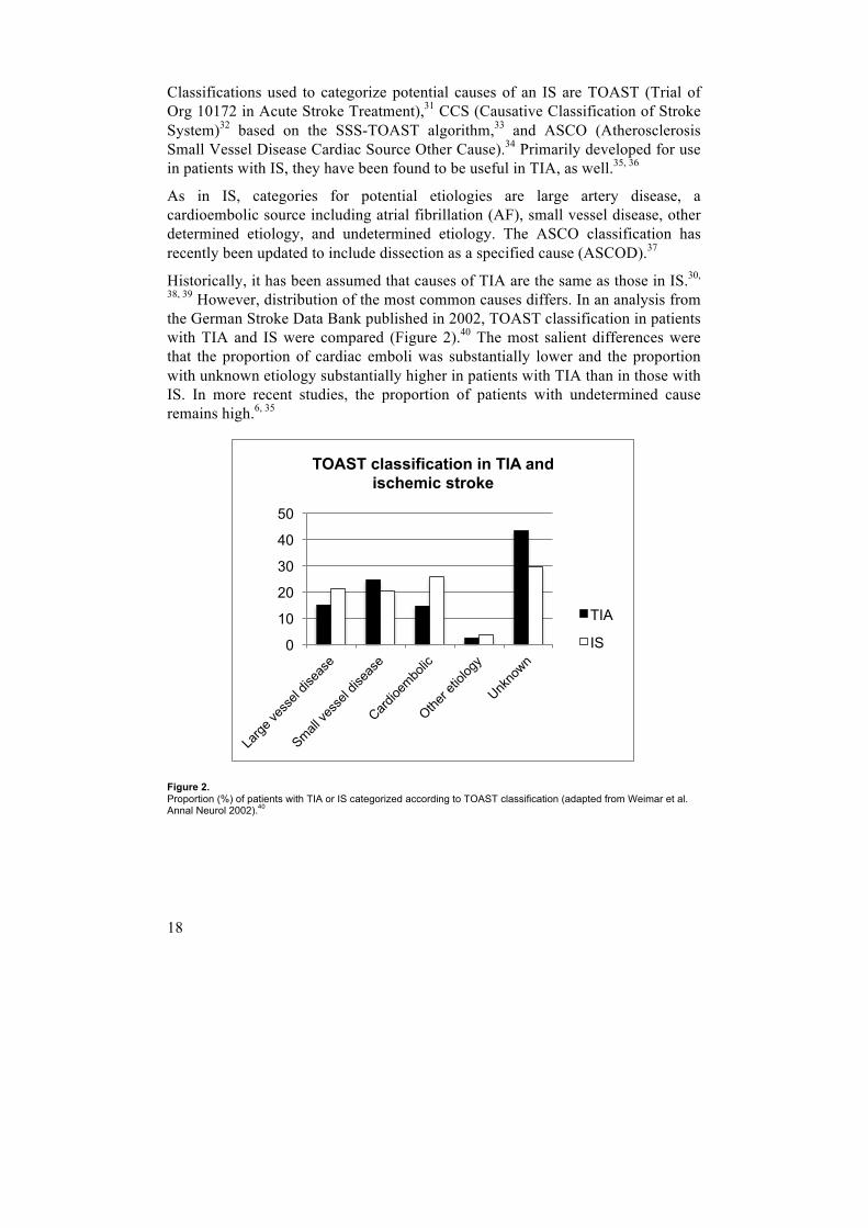
38, 39 However, distribution of the most common causes differs. In an analysis from the German Stroke Data Bank published in 2002, TOAST classification in patients with TIA and IS were compared (Figure 2).40 The most salient differences were that the proportion of cardiac emboli was substantially lower and the proportion with unknown etiology substantially higher in patients with TIA than in those with IS. In more recent studies, the proportion of patients with undetermined cause remains high.6, 35
Figure 2. Proportion (%) of patients with TIA or IS categorized according to TOAST classification (adapted from Weimar et al. Annal Neurol 2002).40
0
10
20
30
40
50
TOAST classification in TIA and ischemic stroke
TIA
IS
19
Investigation
The purpose of investigating patients with TIA is
1. to verify the diagnosis and rule out potential TIA mimics,
2. to identify the cause,
3. to identify risk factors for cerebrovascular disease which may be treatable, and
4. to offer a guide to prognosis and treatment.
Taking a thorough history and performing a physical examination are keystones in the assessment of TIA patients. In order to rule out TIA mimics such as transient neurological symptoms of non-ischemic cause, neuroimaging is recommended. A CT scan will help to rule out an intracranial mass lesion or hemorrhage whereas MRI scanning offers additional information in excluding inflammatory changes e.g. seen in multiple sclerosis that may not be detected by CT. MRI DWI might show signs of recent ischemia (IS according to the tissue-based definition) but the value of its use as a prognostic tool in patients with TIA is still debated.41, 42
According to the Swedish guideline on TIA and stroke provided by the National Board of Health and Welfare (Socialstyrelsen), the etiological workup should include urgent vessel imaging and continuous cardiac rhythm monitoring in-hospital and/or Holter ECG. Immediate instigation of antithrombotic medication, including antiplatelet or oral anticoagulants (OACs) dependent on the presence of AF, statins, and blood pressure lowering drugs is recommended.43

20

21
Aim of the thesis
The overall aim of this thesis was to assess the quality of TIA diagnosis and extent of evaluation and treatment in patients with TIA in Sweden. We intended to further explore potential causes of TIA, its demographic features, and risk factors.
The specific aims were:
I. Paper I: To validate data and diagnoses in the Riksstroke TIA module (Riksstroke-TIA) by assessing patient characteristics, quality of documentation and interobserver agreement regarding diagnosis.
II. Paper II: To assess the proportion of atrial fibrillation in patients with TIA in comparison to patients with IS, and clarify patient characteristics and secondary preventive measures.
III. Paper III: To compare TIA and IS patients with versus without a prior stroke with respect to patient characteristics, risk factors, and secondary preventive medical treatment at discharge.
IV. Paper IV: To assess the proportions of carotid imaging in patients with TIA and IS and determinants for its non-use with respect to baseline demographics, risk factors, hospital characteristics, and geographical region.

22

23
Subjects and methods
Study materials
All studies included in this thesis were observational studies. In Paper I, TIA cases were retrieved from Riksstroke-TIA including a representative study sample of 180 patients. Research material was derived from medical records that were provided by the respective hospitals and from the register. The other three studies (Paper II – IV) were based on registered data from the TIA and acute stroke modules of Riksstroke.
The Swedish Stroke Register (Riksstroke)
Riksstroke is the Swedish quality register of stroke care, and was founded in 1994,44 the Worlds’ first ever quality registry for stroke covering an entire country. Since 1998, all Swedish hospitals treating acute stroke contribute with data. Every year, 22-25000 new episodes of stroke are reported and registered online, including first time and recurring events.
Riksstroke is used for continuous quality improvement in the management of stroke patients. It also aims at a high and equally distributed quality of stroke care throughout the country. Another important purpose is scientific evaluation of data registered in Riksstroke’s database on patient characteristics, stroke management, and outcome in routine clinical settings. The results are reported yearly towards the public, patient organizations, health professionals, and politicians within health care and social services. Since 2015, Riksstroke provides hospital specific data to all contributing hospitals via online dashboards.
In Riksstroke, data on demography, vascular risk factors, functional ability and symptoms preadmission, level of medical care, acute stroke treatment, diagnostic measures, medical treatment at admission and discharge, stroke-related complications, rehabilitation, mortality, and follow-up are registered. As a measure of stroke severity at admission the Reaction Level Scale RLS8545 is used, with categories of fully awake (RLS 1), stuporous (RLS 2-3), and comatose (RLS 4-8), in addition to the NIHSS (National Institutes of Stroke Scale). It is registered

24
in which hospital each patient is taken care of and regional location of each hospital.
The Swedish Stroke Register TIA module (Riksstroke-TIA)
In 2010, a registry module for TIA was added to Riksstroke, and all Swedish hospitals treating TIA are encouraged to contribute. During the first year, 59 out of 74 hospitals in Sweden registered patients. The number of participating hospitals has increased and in 2016, 68 out of 72 hospitals registered TIA events.
In concordance with the acute stroke module, the TIA registry aims to register as many TIA patients as possible and to support a high and equally distributed quality of TIA care. It also provides important and useful data for research on demography, risk factors, logistic details, diagnostic evaluation, and treatment.
As in Riksstroke, results are reported yearly towards the public, patient organizations, health professionals, and politicians within health care and social services. Hospitals are provided with online dashboards.
The first TIA report included the period January to June 2011; from then on the reports were published every year. The first two full year reports covered July 2011 to June 2012 and July 2012 to June 2013. Since then, TIA reports have been published as a joint report together with data on acute stroke and 3 months follow-up after stroke covering a calendar year.
Riksstroke-TIA is a hospital-based register, and patient data are collected at discharge from hospital. Hospital personnel records items into the web-based register. Registration routines and registration rates vary between hospitals.
Items collected in Riksstroke-TIA cover patient baseline characteristics, process indicators, investigations, and medical treatment at admission and discharge (Table 2).
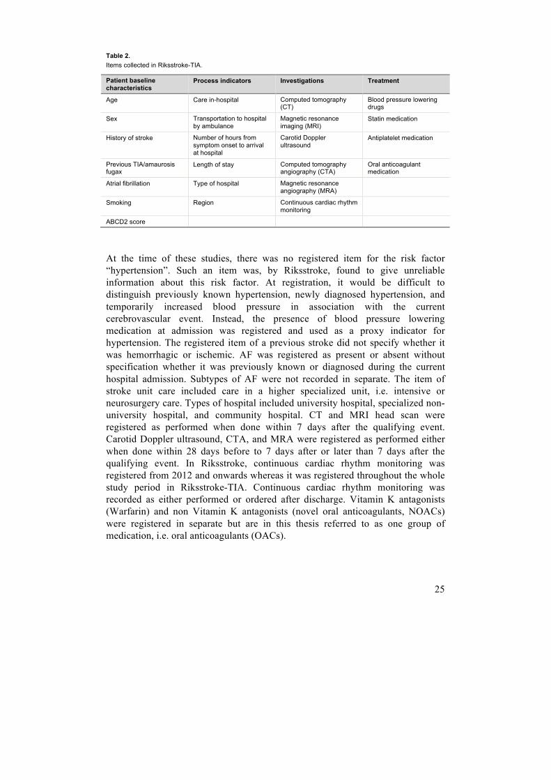
25
Table 2. Items collected in Riksstroke-TIA.
Patient baseline characteristics
Process indicators Investigations Treatment
Age Care in-hospital Computed tomography (CT)
Blood pressure lowering drugs
Sex Transportation to hospital by ambulance
Magnetic resonance imaging (MRI)
Statin medication
History of stroke Number of hours from symptom onset to arrival at hospital
Carotid Doppler ultrasound
Antiplatelet medication
Previous TIA/amaurosis fugax
Length of stay Computed tomography angiography (CTA)
Oral anticoagulant medication
Atrial fibrillation Type of hospital Magnetic resonance angiography (MRA)
Smoking Region Continuous cardiac rhythm monitoring
ABCD2 score
At the time of these studies, there was no registered item for the risk factor “hypertension”. Such an item was, by Riksstroke, found to give unreliable information about this risk factor. At registration, it would be difficult to distinguish previously known hypertension, newly diagnosed hypertension, and temporarily increased blood pressure in association with the current cerebrovascular event. Instead, the presence of blood pressure lowering medication at admission was registered and used as a proxy indicator for hypertension. The registered item of a previous stroke did not specify whether it was hemorrhagic or ischemic. AF was registered as present or absent without specification whether it was previously known or diagnosed during the current hospital admission. Subtypes of AF were not recorded in separate. The item of stroke unit care included care in a higher specialized unit, i.e. intensive or neurosurgery care. Types of hospital included university hospital, specialized non-university hospital, and community hospital. CT and MRI head scan were registered as performed when done within 7 days after the qualifying event. Carotid Doppler ultrasound, CTA, and MRA were registered as performed either when done within 28 days before to 7 days after or later than 7 days after the qualifying event. In Riksstroke, continuous cardiac rhythm monitoring was registered from 2012 and onwards whereas it was registered throughout the whole study period in Riksstroke-TIA. Continuous cardiac rhythm monitoring was recorded as either performed or ordered after discharge. Vitamin K antagonists (Warfarin) and non Vitamin K antagonists (novel oral anticoagulants, NOACs) were registered in separate but are in this thesis referred to as one group of medication, i.e. oral anticoagulants (OACs).
26
Study subjects and procedure
Study subjects in all papers were patients aged ≥18 years registered in Riksstroke-TIA. Riksstroke recommended application of a “time-based” definition, i.e. a focal neurological deficit with acute onset caused by cerebral or retinal ischemia with symptom duration less than 24 hours, irrespective of neuroradiological findings. TIA diagnoses eligible for inclusion were classified according to ICD-10: vertebro-basilar artery syndrome (G45.0), carotid artery syndrome (G45.1), multiple and bilateral precerebral artery syndromes (G45.2), amaurosis fugax (G45.3), other transient cerebral ischemic attacks and related syndromes (G45.8), and unspecified transient cerebral ischemic attack (G45.9). Transient global amnesia (G45.4) is not included.
Paper I
Study subjects in paper I were patients aged ≥18 years registered in Riksstroke-TIA from 1 July 2011 to 30 June 2012 with a diagnosis of TIA which included both patients treated in-hospital and patients not admitted to hospital. In this period of time, 7825 events were registered at 59 of 74 Swedish hospitals (1-377 patients per hospital). Six hospitals registered more than 250 events, 14 hospitals 150 to 250, and 17 hospitals 75 to 149. Two hospitals from each of these groups (total 6) were selected on the basis of varying geographical location and size of catchment population. A list of 30 registered patients per hospital (total 180) was prepared at the Riksstroke secretariat, creating a simple random sample that was matched by age and sex to the overall population in Riksstroke-TIA.
Each participating hospital provided anonymized copies of medical records covering the acute in-hospital stay. A secretary at the study center created a first set of medical records containing only details on symptoms and signs including clinical examinations, with all other parts censored, and a second set including the complete medical record. Two physicians at two separate Swedish hospitals performed the assessment independently – one house officer with special interest in neurology (Jakob Ström, Örebro, assessor A) and one stroke neurologist (Fredrik Buchwald, Malmö, assessor B). For evaluation, a pre-specified protocol with a two step approach was used 1 : In step 1 (using first record set) documentation was evaluated regarding time (duration, onset, resolution), reported symptoms, and neurological examinations at arrival and later during in-hospital stay. Assesment was based on NINDS criteria3 and a time-based TIA definition. The clinical events were categorized into one of the following groups: likely TIA, 1 For details, see paper I, supplementary material. 2 For details, see paper IV, supplementary tables 1 and 2.

27
possible TIA, unlikely TIA, or IS/retinal infarction. In step 2 (using second record set) data on age, sex, vascular risk factors, history of cerebrovascular events, ABCD2 score,46 neuroimaging, and neurovascular investigations were collected. Missing ABCD2 scores were retrospectively acquired from Riksstroke. Presented results on patient characteristics, diagnostic procedures, time aspects, and five key features of neurological examination - consciousness, speech/language, vision, motor function and sensation - are based on both assessors findings with post-assessment consensus. In each patient, assessor B determined a principal symptom or symptom complex. In cases with multiple symptoms the leading one was identified.
Papers II - IV
Papers II - IV were based on patients aged ≥18 years registered in Riksstroke-TIA from 1 July 2011 to 30 June 2013. Only patients treated in-hospital were included. For comparison, patients with IS registered in Riksstroke during the same period of time were assessed. Stroke diagnoses were set according to the World Health Organization (WHO) definition of stroke;47 ICD-10 diagnosis was I63 (IS). For patients with more than one stroke during the course of 28 days, only the first event was included.
In paper II, patients without registered information on the presence or absence of atrial fibrillation (<1%) were excluded.
In paper III, patients in whom information on the presence or absence of a history of prior stroke was missing (<1%) were excluded.
In paper IV, patients who were evaluated with either carotid Doppler ultrasound or CTA or both in association with the current TIA or IS had per definition undergone carotid imaging. Those patients with no registered information on the performance of both carotid Doppler ultrasound and CTA (<1%) were excluded. Data on MRA were excluded from further analyses due to uncertainty about the included vascular territory. The proportion of patients who were tested with MRA but no other vascular modality was low, i.e. 0.7% (102/14597) of patients with TIA and 0.9% (392/43847) of patients with IS.
Statistical methods
In paper I – IV, SPSS 22.0 was used for all statistical analyses. Categorical variables were summarized as proportions and quantitative variables as means or medians. Proportions were derived from the total of patients in whom the

28
respective item was registered. Potential differences were tested by chi2 testing and Student’s T-test, as appropriate. A difference with a p value <0.05 was considered statistically significant. Age groups were reported in the intervals <45, 45-54, 55-64, 65-74, 75-84, and ≥85 years.
In paper I, interobserver agreement was presented in percentage of agreement and by Cohen Kappa statistics (κ) with regard to expected uneven distribution of items. Calculations were performed for the four diagnostic categories (likely, possible, unlikely TIA, IS/retinal infarction) and for likely and possible TIA merged into one group. The strength of agreement for kappa values were “poor” (0-0.2), “fair” (0.21-0.4), “moderate” (0.41-0.6), “good” (0.61-0.8), and “very good” (0.81-1).
In paper II, baseline data were compared with prevalence differences (PD) and 95% confidence intervals (CI).
In paper IV, variables associated with not undergoing carotid imaging in univariate analyses were included in multivariate logistic regression models with stepwise elimination in order to assess independent association. Both in TIA and IS, references were defined as age <65 years, male sex, no AF, no hypertension, no diabetes, no smoking, no history of stroke, care at a university hospital, and care in the region with highest proportion of carotid imaging. In TIA, this was the southeastern region and in IS, the Stockholm region. References in variables only registered in patients with IS were defined as care at a stroke unit and being alert at admission. The NIHSS score was not included in the multivariate logistic regression model as this item was registered in less than 50% of patients.
Ethical considerations
The Regional Ethical Review Board in Lund approved the studies of this thesis (Dnr 2013/719).
All patients or their next of kin were informed about the registration in the quality register Riksstroke, and that data may be used for compiling statistics, for continuous quality assessment of stroke care, and for research purposes. Patients had the choice of not participating (opt-out consent). Consent was not collected for specific research studies. Use of data from Riksstroke for research purposes required an application approved by the Riksstroke secretariat. Data delivered by Riksstroke to the researcher was anonymized.
By registration in Riksstroke, personal and potentially sensitive information was stored, but direct risk for patients is not expected.
29
Results
Paper I is based on 7825 TIA events registered in Riksstroke-TIA from 01/07/2011 to 30/06/2012. The study sample of 180 patients was extracted from this cohort. Papers II – IV were based on 15064 TIA events registered from 01/07/2011 to 30/06/2013 in Riksstroke-TIA.
For comparison, IS events registered during the same 2-year period (n=44416) were analysed in papers II – IV.
Demographic and baseline characteristics of patients included in the four papers are summarized in Table 3.

30
Table 3. Patient characteristics
Study Paper I Papers II - IV Diagnosis TIA TIA TIA Ischemic
stroke
Cohort Study sample (n=180)
Riksstroke-TIA (n=7825)
Riksstroke-TIA (n=15064)
Riksstroke (n=44416)
Period of registration 01/07/11 – 30/06/12
01/07/11 – 30/06/12
01/07/11 – 30/06/13
01/07/11 – 30/06/13
Female n (%) 78 (43.3) 3746 (47.9) 7270 (48.3) 21400 (48.2)
Age mean (range) 76.4 (45-97) 72.9 (18-101) 73.2 (20-102) 75.7 (18-104)
Hypertension* n (%) 109 (60.6) 4588 (58.6) 9075 (60.5) 27723 (62.8)
Atrial fibrillation n (%) 43 (23.9) 1391 (17.8) 2779 (18.6) 13258 (30.0)
Diabetes mellitus n (%) 22 (12.2) 1211 (15.5) 2362 (15.8) 9388 (21.2)
Smoking n (%) 19 (10.6) 902 (11.5) 1663 (11.9) 5875 (14.3)
History of stroke n (%) 43 (23.9) 1505 (19.2) 2892 (19.3) 10853 (24.6)
History of TIA n (%) 22 (12.2) 1386 (17.7) 2724 (18.3) 3991 (9.1)
CT n (%) 175 (97.2) 7542 (96.4) 14661 (97.4) 43763 (98.6)
MRI n (%) 4 (2.2) 627 (8.0) 1257 (8.4) 6967 (15.8)
Neither CT nor MRI n (%)
3 (1.7) 225 (2.9) 310 (2.1) 301 (0.7)
Carotid Doppler ultrasound n (%)
99 (55.0) 4695 (60.0) 8970 (59.7) 18212 (41.3)
CTA n (%) 23 (12.8) 1098 (14.0) 2293 (15.3) 8072 (18.3)
MRA n (%) 0 (0) 208 (2.7) 357 (2.4) 1469 (3.3)
No vascular imaging n (%)
58 (32.0) 2323 (29.7) 4358 (29.0) 19852 (45.1)
Cardiac arrhythmia detection† n (%)
172 (96.0) 3579 (45.7) 7097 (47.7) 9500 (43.2)
ABCD2 score (mean) 4.2 4.2 4.2 not registered
NIHSS score‡ Mean (SD) Median Interquartile range
not registered not registered not registered 5.8 (6.6) 3 1-8
Level of consciousness§
Alert Stuporous Comatose
not registered not registered not registered 37629 (85.8) 4792 (10.9) 1412 (3.2)
* Hypertension was defined as treatment with blood pressure lowering medication at admission † In the study sample of paper I, a default 12-channel ECG in-hospital was accepted as cardiac arrhythmia detection; in Riksstroke-TIA and Riksstroke, only continuous cardiac rhythm monitoring in-hospital was registered. ‡ NIHSS score was registered in 21720/44416 patients § based on RLS8545, i.e. alert = RLS 1, stuporous = RLS 2-3, comatose = RLS 4-8

31
Validation of diagnoses of TIA in Riksstroke-TIA (Paper I)
In paper I, 7825 TIA events registered in Riksstroke-TIA from 01/07/2011 to 30/06/2012 were included. 97% (7600/7825) of patients were treated in-hospital. From the total cohort, a study sample of 180 patients registered at 6 different hospitals was extracted. All 180 patients had been admitted to hospital. Demographic and other patient characteristics in the study sample were consistent with the total cohort, except for AF being more common and the use of MRI and MRA less frequent in the study sample compared to the total cohort (Table 3). Registered symptoms are shown in Table 4. Non-localizing symptoms were reported in 13% (24/180), i.e. loss of consciousness, amnesia, confusion, and other non-focal or not clearly focal symptoms.
Table 4. Distribution of principal symptom or combination of symptoms in the study sample
Symptoms % (n)
Motor 25 (45)
Sensorimotor 13.9 (25)
Speech and/or language 12.2 (22)
Speech and/or language and hemisymptoms* 10.6 (19)
Amaurosis fugax 9.4 (17)
Non-focal or not clearly focal symptoms† 8.3 (15)
Isolated diplopia, vertigo or dysarthria 6.7 (12)
Confusion, amnesia, and/or loss of consciousness 5.0 (9)
Isolated sensory 3.9 (7)
Isolated homonymous hemianopia 2.8 (5)
Positive visual symptoms 1.1 (2)
Complete blindness 1.1 (2)
Total 100 (180) * Homonymous hemianopia, unilateral motor or sensory deficit. † Multiple concomitant symptoms that could not clearly be assigned to a focal vascular territory.
Documentation of time aspects and neurological examination
In 23% (42/180) of patients in the study sample, neither exact nor estimated duration of symptoms was documented. The mode of onset was documented in 71% (127/180) and the mode of resolution in 37% (67/180). The most frequent documented features of neurological examination were motor function (92%; 166/180) and level of consciousness (90%; 163/180), followed by sensory

32
function (66%, 119/180), vision (55%; 99/180) and speech/language (44%; 79/180). All 5 key features of neurological examination were documented in 26% (46/180). The ABCD2 score was not recorded in any case.
TIA diagnosis and interobserver agreement
In 93% (167/180), at least one assessor found the clinical event to be a likely or possible TIA. The two independent observers agreed in 77% (138/180) that the documented event was a likely or possible TIA, in 3% (5/180) that the event was an IS or retinal infarction, and in 2% (3/180) that TIA diagnosis was unlikely. The observers disagreed in 8% (15/180) on a likely or possible diagnosis of TIA versus IS, and in 11% (19/180) on a vascular versus non-vascular cause (Table 5).
For the categories likely TIA, possible TIA, unlikely TIA and IS/retinal infarction, the κ value of interobserver agreement was 0.326 (standard error 0.055). Merging the categories of likely and possible TIA resulted in a κ value of 0.378 (standard error 0.093).
Table 5. Ratings and agreement on TIA diagnosis in the study sample
Investigator B
Likely TIA Possible TIA Unlikely TIA IS/RI Total
Likely TIA Possible TIA Unlikely TIA IS/RI Total
Investigator A
Likely TIA 36.6 (66) 10 (18) 2.8 (5) 0.6 (1) 150 (90)
Possible TIA 12.8 (23) 17.2 (31) 3.3 (6) 2.2 (4) 135.5 (64)
Unlikely TIA 0.6 (1) 1.1 (2) 1.7 (3) 0 33.3 (6)
IS/RI 1.1 (2) 4.4 (8) 2.8 (5) 2.8 (5) 11.1 (20)
Total 51.1 (92) 32.8 (59) 10.6 (19) 5.6 (10) 100 (180)
Ratings are expressed as percentages with numbers in parentheses. Bold signals agreement; italics ”likely TIA” vs. ”possible TIA”; and normal font disagreement. RI indicates retinal infarction.

33
Atrial fibrillation in TIA versus ischemic stroke (Paper II)
Paper II was based on data from 15064 TIA events and 44416 IS events, registered in Riksstroke-TIA and Riksstroke from 01/07/2011 to 30/06/2013. The presence or absence of AF was documented in >99% of cases, i.e. 14980/15064 of TIA and 44173/44416 of IS patients.
Proportions of atrial fibrillation
Previously known or newly diagnosed AF was registered in 19% (2779/14980) of patients with TIA and 30% (13258/44173) of patients with IS (PD, -11.5; 95% CI, -12.2 to -10.7). The proportion of AF increased with age, both in patients with TIA and IS. In TIA, proportions of AF ranged from 2% in patients <45 years to 33% in those ≥85 years, and in IS, from 2% to 47% (Figure 3).
Figure 3. Proportion (%) of AF in patients with TIA versus IS dependent on age. Above columns p qui2 values for TIA versus IS are noted. ns indicates non-significant. ** = p<0.0001; * = p<0.05.
Patient characteristics
Baseline characteristics of all patients with TIA and IS included in this study are shown in Table 3. In patients with TIA, mean age and proportions of AF, hypertension, diabetes mellitus, smoking, and a history of stroke were lower than
ns * *
**
**
**
**
0
10
20
30
40
50
<45 45-54 55-64 65-74 75-84 ≥85 Total
Atrial fibrillation in TIA versus ischemic stroke
TIA
IS

34
in patients with IS whereas the proportion of a history of TIA was higher in patients with TIA compared to those with IS.
In both TIA and IS patients, the parameters age, hypertension, being a non-smoker, and a history of stroke and TIA were associated with the presence of AF. Among patients with TIA, AF was less common in women than in men, whereas in IS, women had a higher proportion of AF than men. In TIA, diabetes mellitus was associated with AF. In IS, no such association was observed (Table 6).
Table 6. Proportions of atrial fibrillation according to patient characteristics in patients with TIA compared with patients with ischemic stroke
TIA Ischemic stroke Difference TIA - IS
AF present AF present
N % (95% CI) N % (95% CI) PD % (95% CI) P value
Sex F 1283/7229 17.7 (16.9, 18.6) 7089/21 297 33.3 (32.7, 33.9) -15.5 (-16.6, -13.2) **
M 1496/7751 19.3 (18.4, 20.2) 6169/22 876 27.0 (26.4, 27.5) -7.7 (-8.7, -5.4) **
Hypertension Yes 2261/9033 25.0 (24.1, 26.0) 9639/27 629 34.9 (34.3, 35.5) -9.9 (-10.9, -7.8) **
No 509/5893 8.6 (7.9, 9.4) 3544/16 386 21.6 (21.0, 22.3) -13.0 (-14.0, -12.0) **
Diabetes mellitus
Yes 497/2345 21.2 (19.5, 22.9) 2760/9342 29.5 (28.6, 30.5) -8.4 (-10.3, -4.4) **
No 2265/12558 18.0 (17.4, 18.7) 10471/34788 30.1 (29.6, 30.6) -12.1 (-12.9, -10.3) **
Smoking Yes 162/1653 9.8 (8.4, 11.2) 944/5848 16.1 (15.2, 17.1) -6.3 (-8.1, -1.2) **
No 2362/12246 19.3 (18.6, 20.0) 11157/35255 31.7 (31.2, 32.1) -12.4 (-13.2, -10.6) **
History of stroke
Yes 785/2868 27.4 (25.7, 29.0) 3896/10806 36.1 (35.2, 37.0) -8.7 (-10.6, -5.2) **
No 1983/12077 16.4 (15.8, 17.1) 9311/33225 28.0 (27.5, 28.5) -11.6 (-12.4, -9.7) **
History of TIA
Yes 551/2707 20.4 (18.8, 21.9) 1281/3968 32.3 (30.8, 33.7) -11.9 (-14.0, -7.7) **
No 2191/12143 18.0 (17.4, 18.7) 11764/39685 29.6 (29.2, 30.1) -11.6 (-12.4, -9.8) **
Age <65 222/3306 6.7 (5.9, 7.6) 711/7634 9.3 (8.7, 10.0) -2.6 (-3.7, -1,5) **
65-74 554/4204 13.2 (12.2, 14.2) 2206/10504 21.0 (20.2, 21.8) -7.8 (-9.1, -4.5) **
75-84 1094/4707 23.2 (22.0, 24.5) 4824/14197 34.0 (33.2, 34.8) -10.7 (-12.2, -7.9) **
≥85 909/2763 32.9 (31.2, 34.7) 5517/11838 46.6 (45.7, 47.5) -13.7 (-15.7, -10.4) **
CI indicates confidence interval; PD, prevalence difference. ** = p<0.0001.
Oral anticoagulant treatment at discharge
Antithrombotic treatment at discharge was registered in >99% of TIA and surviving IS patients. Patients with TIA and AF were treated with OACs in 64% (1778/2771) compared to 50% (5502/10899) in those with IS and AF. The proportion of OAC treatment in patients with AF decreased with increasing age, and ranged from 81% (177/220) in TIA patients aged <65 years to 46% (412/905) in those aged ≥85 years. In patients with IS and AF, 71% (472/664) of patients <65 years and 33% (1396/4218) of those aged ≥85 years were treated with OAC. 5% (126/2769) of patients with TIA and AF and 9% (1011/10899) of patients with IS and AF were discharged without any antithrombotic medication. Proportions of treatment, separately for female and male patients are detailed in Table 7.

35
Table 7. Oral anticoagulant treatment in female and male patients with atrial fibrillation at discharge, separately for TIA and ischemic stroke.
Female Male Difference Female - Male
Age N % (95% CI) N % (95% CI) PD % (95% CI)
TIA <65 51/61 83.6 (74.3, 92.9) 126/159 79.2 (72.9, 85.6) 4.4 (-6.9, 15.6)
65-74 162/208 77.9 (72.2, 83.5) 253/346 73.1 (68.5, 77.8) 4.8 (-2.6, 12.1)
75-84 329/471 69.9 (65.7, 74.0) 445/621 71.7 (68.1, 75.2) -1.8 (-7.3, 3.7)
≥85 243/539 45.1 (40.1, 49.3) 169/366 46.2 (41.1, 51.3) -1.1 (-7.7, 5.5)
Total 785/1279 61.4 (58.7, 64.0) 993/1492 66.6 (64.2, 69.0) -5.2 (-8.8, -1.6)
IS <65 123/172 71.5 (64.8, 78.3) 349/492 70.9 (66.9, 75.0) 0.6 (-7.3, 8.4)
65-74 470/717 65.6 (62.1, 69.0) 847/1232 68.8 (66.2, 71.3) -3.2 (-7.5, 1.1)
75-84 1072/1984 54.0 (51.8, 56.2) 1245/2084 59.7 (57.6, 61.9) -5.7 (-8.8, -2.7)
≥85 854/2769 30.8 (29.1, 32.6) 542/1449 37.4 (34.9, 39.9) -6.6 (-9.6, -3.5)
Total 2519/5642 44.7 (43.4, 45.9) 2983/5257 56.7 (55.4, 58.1) -12.1 (-14.0, -10.2)
CI indicates confidence interval; PD, prevalence difference.
TIA and ischemic stroke patients with or without prior stroke (Paper III)
Included in paper III were patients with TIA or IS in whom the presence or absence of a history of stroke was registered. This was the case in >99% of patients registered during the 2-year study period (TIA: 15012/15064; IS: 44169/44416).
A prior history of stroke was present in 19% (2892/15012) of patients with TIA and 25% (10853/44169) of patients with IS. The proportion of patients with a history of stroke increased with age in both patients with TIA and IS (Figure 4).

36
Figure 4. Proportion of a prior stroke (%) in patients with TIA and IS dependent on age.
Patient characteristics
Patients with a history of stroke, both in those with TIA and IS, were older and more likely to be male compared to patients without a prior stroke. They had higher proportions of AF, hypertension, and diabetes mellitus whereas they were less likely to be smokers than those without a prior stroke (Table 8).
Table 8. Characteristics and risk factors of patients admitted for TIA or ischemic stroke with or without a previous stroke
TIA Ischemic stroke
Prior stroke n=2892
No prior stroke n=12120 P value Prior stroke
n=10853 No prior stroke n=33316 P value
Female n (%) 1301/2892 (45.0) 5946/12120 (49.1) <0.0001 5013/10853 (46.2) 16279/33316 (48.9) <0.0001
Age mean (SD) 77.0 (10.4) 72.2 (12.5) <0.0001 78.1 (10.5) 74.9 (12.8) <0.0001
AF n (%) 785/2868 (27.4) 1983/12077 (16.4) <0.0001 3896/10806 (36.1) 9311/33225 (28.0) <0.0001
Hypertension n (%) 2236/2884 (77.5) 6809/12073 (56.4) <0.0001 8023/10806 (74.2) 19590/33197 (59.0) <0.0001
Diabetes mellitus n (%) 639/2878 (22.2) 1717/12057 (14.2) <0.0001 2845/10835 (26.3) 6498/33283 (19.5) <0.0001
Smoking n (%) 274/2630 (10.4) 1382/11296 (12.2) 0.010 1173/10074 (11.6) 4681/31036 (15.1) <0.0001
0 5
10 15 20 25 30 35
<45 45-54 55-64 65-74 75-84 ≥85 total
A history of prior stroke in patients with TIA or ischemic stroke
TIA
IS

37
Atrial fibrillation
AF was present in 27% (785/2868) of TIA patients with a history of stroke compared to 16% (1983/12077) in those without (p<0.0001). The proportion of AF increased with age. In TIA patients ≥85 years, 41% (300/724) of those with a prior stroke had AF compared to 30% (604/2028) in those without a prior stroke (p<0.0001).
In patients with IS and a history of stroke, AF was registered in 36% (3896/10806) compared to 28% (9311/33225) in patients without a history of stroke (p<0.0001). As in TIA, the proportion of AF in IS patients increased with age and reached 51% (1698/3319) in those aged ≥85 years with a prior history of stroke and 45% (4676/8466) in those without (p<0.0001).
Medication at discharge
Figures 4A–C show treatment patterns at discharge of (A) OAC in the presence of AF, (B) blood pressure lowering drugs, and (C) statins. Proportions of OAC medication in the presence of AF decreased with increasing age both in patients with TIA and IS. Patients with a prior stroke were less likely to receive OACs than patients without a prior stroke. Proportions of blood pressure lowering drugs use increased with age and were higher in those with a prior stroke compared to those without. Regarding statins, there were no significant differences between patients with or without a history of stroke but proportions of treatment dropped in patients aged 75–84 years and in those ≥85 years.

38
(A) Oral anticoagulants in the presence of AF at discharge
(B) Blood pressure lowering medication at discharge
(C) Statins at discharge
Figure 4A–C. Proportion of treatment (%) at discharge: (A) oral anticoagulants in the presence of AF (B) blood pressure lowering medication, and (C) statin treatment in relation to a history of stroke, separately for patients with TIA and IS.
0 20 40 60 80
100
TIA IS
prior stroke
no prior stroke
0 20 40 60 80
100 TIA IS
prior stroke
no prior stroke
0 20 40 60 80
100 TIA IS
prior stroke
no prior stroke

39
Carotid imaging in patients with TIA or ischemic stroke (Paper IV)
In more than 99% of patients with TIA (15023/15064) and IS (44075/44416) it was registered whether or not patients had undergone carotid Doppler ultrasound or CTA. Any carotid imaging procedure, i.e. either carotid Doppler ultrasound or CTA or both, was performed in 70% (10545/15023) of patients with TIA and in 54% (23772/44075) of patients with IS.
Patient characteristics
Patients with TIA who did not undergo carotid imaging had a mean age of 78.7 years (SD 12.1) compared to 70.8 years (SD 11.6) in those who were examined. In patients with IS, mean age in those who were not examined with a carotid imaging procedure was 80.9 years (SD 10.8) compared to 71.2 years (SD 11.8) in patients who underwent carotid imaging. Proportions of carotid imaging decreased with increasing age (Figures 5A and B).
According to univariate analyses in Table 9, female sex, hypertension, AF, diabetes mellitus, non-smoking, and a history of stroke were factors associated with not being investigated in both TIA and IS patients. Patients with IS in whom carotid imaging was not performed had higher proportions of decreased level of consciousness at admission and lower proportions of stroke unit care than those who were investigated.

40
Table 9. Carotid imaging in patients with TIA or ischemic stroke dependent on patient characteristics, risk factors, stroke unit care, hospital type, and region
TIA n=15023
Ischemic stroke n=44075
Carotid imaging n (%)
P value Carotid imaging n (%)
P value
Sex Female 4871/7252 (67) <0.0001 10034/21230 (47) <0.0001
Male 5674/7771 (73) 13738/22845 (60)
Hypertension Yes 6002/9050 (66) <0.0001 13897/27531 (51) <0.0001
No 4503/5919 (76) 9785/16324 (60)
Atrial fibrillation Yes 1509/2773 (54) <0.0001 4824/13133 (37) <0.0001
No 8984/12171 (74) 18843/30745 (61)
Diabetes mellitus
Yes 1605/2357 (68) 0.015 4871/9319 (52) <0.0001
No 8890/12591 (71) 18855/34646 (54)
Smoking Yes 1326/1661 (80) <0.0001 4198/5842 (72) <0.0001
No 8521/12265 (70) 18264/35119 (52)
History of stroke Yes 1530/2880 (53) <0.0001 4298/10769 (40) <0.0001 No 8982/12094 (74) 19375/33099 (59)
Level of consciousness*
Alert not registered 22051/37413 (59)
Stuporous 1316/4737 (28) <0.0001 Comatose 219/1399 (16)
Care at stroke unit
Yes not registered 19675/34524 (57) <0.0001 No 4017/9432 (43)
Hospital type University 2079/2654 (78) <0.0001 5704/9240 (62) <0.0001
Specialized non-university
4815/7098 (68) 10430/19428 (53)
Community 3651/5271 (69) 7638/14977 (51)
Region Northern 614/1066 (58) <0.0001 1878/4609 (41) <0.0001
Uppsala-Örebro 2305/3622 (64) 4566/10036 (46) Stockholm 914/1173 (78) 5590/8645 (65)
Southeastern 1384/1769 (78) 2734/4567 (60) Western 2454/3753 (68) 3738/7874 (48)
Southern 2783/3640 (77) 5266/8344 (63) * based on RLS8545, i.e. alert = RLS 1, stuporous = RLS 2-3, comatose = RLS 4-8
Hospital type and regional differences
In university hospitals, proportions of carotid imaging were higher than in specialized non-university hospitals and community hospitals; in the two latter, proportions were on similar levels. This applied to both patients with TIA and IS (Table 9).
Proportions of carotid imaging also differed dependent on geographical region. In patients with TIA, proportions of carotid imaging varied from 58% (614/1066) in

41
the northern to 78% (1384/1769) in the southeastern region of Sweden. In IS, proportions ranged from 41% (1878/4609) in the northern region to 65% (5590/8645) in the Stockholm region (Table 9).
As seen in Figures 5A and B, proportions of carotid imaging dropped with increasing age in all regions, both in patients with TIA and IS. In patients aged ≥85 years, carotid imaging in TIA patients ranged from 4% (5/139) in the northern region to 55% (196/354) in the southeastern region and in IS patients, from 8% (77/989) in the northern region to 34% (818/2408) in Stockholm.
(A)
(B)
Figures 5A and B. Proportion of carotid imaging (%) in patients with (A) TIA and (B) ischemic stroke divided in age groups <65, 65-74, 75-84, and ≥85 years, separately for each region
0 20 40 60 80
100
<65
65-7
4 75
-84
≥85
<65
65-7
4 75
-84
≥85
<65
65-7
4 75
-84
≥85
<65
65-7
4 75
-84
≥85
<65
65-7
4 75
-84
≥85
<65
65-7
4 75
-84
≥85
Northern n=1066
Uppsala-Örebro n=3622
Stockholm n=1173
South-eastern n=1769
Western n=3753
Southern n=3640
TIA
0 20 40 60 80
100
<65
65-7
4 75
-84
≥85
<65
65-7
4 75
-84
≥85
<65
65-7
4 75
-84
≥85
<65
65-7
4 75
-84
≥85
<65
65-7
4 75
-84
≥85
<65
65-7
4 75
-84
≥85
Northern n=4609
Uppsala-Örebro
n=10036
Stockholm n=8645
South-eastern n=4567
Western n=7874
Southern n=8344
Ischemic stroke
42
Independent determinants for not undergoing carotid imaging
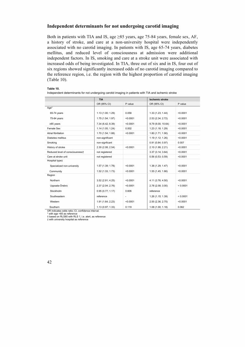
Both in patients with TIA and IS, age ≥85 years, age 75-84 years, female sex, AF, a history of stroke, and care at a non-university hospital were independently associated with no carotid imaging. In patients with IS, age 65-74 years, diabetes mellitus, and reduced level of consciousness at admission were additional independent factors. In IS, smoking and care at a stroke unit were associated with increased odds of being investigated. In TIA, three out of six and in IS, four out of six regions showed significantly increased odds of no carotid imaging compared to the reference region, i.e. the region with the highest proportion of carotid imaging (Table 10).
Table 10. Independent determinants for not undergoing carotid imaging in patients with TIA and ischemic stroke
TIA Ischemic stroke
OR (95% CI) P value OR (95% CI) P value Age*
65-74 years
75-84 years
≥85 years
1.13 (1.00; 1.29)
1.75 (1.54; 1.97)
7.34 (6.42; 8.39)
0.058
<0.0001
<0.0001
1.33 (1.23; 1.44)
2.53 (2.34; 2.72)
9.79 (9.00; 10.64)
<0.0001
<0.0001
<0.0001
Female Sex 1.14 (1.05; 1.24) 0.002 1.23 (1.18; 1.29) <0.0001
Atrial fibrillation 1.70 (1.54; 1.88) <0.0001 1.80 (1.71; 1.90) <0.0001
Diabetes mellitus non-significant 1.19 (1.12; 1.26) <0.0001
Smoking non-signficant 0.91 (0.84; 0.97) 0.007
History of stroke 2.30 (2.08; 2.54) <0.0001 2.10 (1.99; 2.21) <0.0001
Reduced level of consciousness† not registered 3.37 (3.14; 3.64) <0.0001
Care at stroke unit not registered 0.56 (0.53; 0.59) <0.0001 Hospital type‡
Specialized non-university
Community
1.57 (1.39; 1.78)
1.52 (1.33; 1.73)
<0.0001
<0.0001
1.38 (1.29; 1.47)
1.55 (1.45; 1.66)
<0.0001
<0.0001 Region
Northern
Uppsala-Örebro
Stockholm
Southeastern
Western
Southern
3.52 (2.91; 4.25)
2.37 (2.04; 2.76)
0.95 (0.77; 1.17)
reference
1.91 (1.64; 2.23)
1.13 (0.97; 1.33)
<0.0001
<0.0001
0.606
-
<0.0001
0.119
4.11 (3.76; 4.50)
2.78 (2.58; 3.00)
reference
1.26 (1.15; 1.39)
2.55 (2.36; 2.75)
1.08 (1.00; 1.18)
<0.0001
< 0.0001
-
< 0.0001
<0.0001
0.062 OR indicates odds ratio; CI, confidence interval. * with age <65 as reference † based on RLS85 with RLS 1, i.e. alert, as reference ‡ with university hospital as reference

43
Proportions of investigation might potentially be lower in patients with signs of severe stroke. Therefore we reassessed the cohort of patients with IS after exclusion of those with decreased level of consciousness at admission (i.e. patients with signs of severe stroke). In this subgroup of IS patients, proportions of carotid imaging and determinants for not undergoing this investigation were similar compared to the total cohort of patients with IS.2
It cannot be excluded that patient cohorts treated at respective hospital type and respective region might differ with respect to age, patient characteristics, and risk factors. Therefore corresponding multivariate logistic regression models were performed in patients with TIA aged <75 years without AF and a history of stroke and in patients with IS aged <75 years without AF and a history of stroke, who were independent pre-admission and alert at admission. Both in TIA and IS, odds ratios (ORs) for no carotid imaging were on similar levels in these selected subgroups as in the total cohorts, i.e. hospital-dependent and regional differences remained unchanged (data not shown).
2 For details, see paper IV, supplementary tables 1 and 2.

44
45
Discussion
Methodological considerations
The presented papers were observational cross-sectional descriptive studies based on evaluation of medical files (Paper I), registered data from Riksstroke-TIA (Paper I – IV) and Riksstroke (Paper II – IV) registered during a defined period of time. The strength of this quality register-based research is the large number of included patients in an unselected cohort, as inclusion is not restricted by age, functional status or co-morbidity. This leads to generalizable results. However, data may be less detailed compared to e.g. randomized controlled trials.
Methodological issues that might influence the reliability of the study results include coverage, selection bias, imprecision of registered data, and confounding.
Coverage and selection bias
In papers I – IV, patients registered in Riksstroke-TIA were included. In papers II – IV, patients with IS registered in Riksstroke during the same period of time were included for comparison.
Riksstroke is a hospital-based register, and patients with TIA not taken care of at a hospital (but e.g. at a primary care unit) were not registered. Patients who were registered at a hospital but taken care of in an outpatient setting (e.g. at an emergency department or a TIA clinic) and not admitted to hospital were excluded from papers II - IV due to expected insufficient data quality. Obviously, patients experiencing a TIA who did not seek any medical attention could not be included.
In the period 01/07/2011 to 30/06/2012, 59 out of 74 Swedish hospitals contributed with registration of TIA events, and during the following year, 59 out of 72 hospitals. Registration rates per capita of catchment population varied substantially between hospitals,48, 49 and it is therefore likely that a number of patients taken care of in-hospital were not registered. Hospitals that did not register any patients in Riksstroke-TIA included university hospitals, specialized non-university hospitals, and community hospitals. According to the annual Riksstroke report on TIA for the period July 2012 to June 2013, the national mean
46
registration rate of TIA events was 79 per 100 000 inhabitants which included both first-time and recurrent TIA.49 For the year 2012, it has been estimated that 66% of the Swedish population were included in the catchment area of participating hospitals.50
Epidemiological studies on TIA are difficult to perform, in part due to the lack of a gold standard investigation. Diagnosis is based on expert opinion but interobserver agreement is not more than fair.13 Estimates from the Oxford Vascular Study (OXVASC)51 indicate that the standardized incidence of definite or probable TIA is approximately 108 per 100 000 population52. Incidence rates of first-ever TIA ranged from 25 to 73 per 100 000 inhabitants according to recent epidemiological studies from New Zealand, Brazil, Spain, Italy, France, and Denmark,14-19 and in Sweden, incidence rate of first-ever TIA was 73 per 100 000 inhabitants for the years 2011 and 2012 based on data from Riksstroke-TIA.50
Patients with TIA not registered in Riksstroke-TIA included those who were taken care of in-hospital but not registered, those seen at medical units such as primary care facilities that would not participate in registration, and those individuals who did not seek any medical attention. It is obviously difficult to assess patients not registered in Riksstroke-TIA and compare them to those registered. Potentially, factors associated with a delay in seeking medical attention might be the same as those associated with not seeking at all. In patients with TIA, motor deficits lasting one hour or longer53 and presence of a witness54 might be associated with reduced delay in seeking medical attention, whereas time factors such as the occurrence of symptoms during the weekend might cause increased delay.53 Recognition of symptoms as TIA or stroke appeared not to be associated with a more urgent visit to a medical care facility.54
Hence, coverage is not complete. However, patient characteristics in our cohort were comparable to TIA studies with a high degree of case ascertainment, namely the OXVASC register,27 the 4th Auckland Regional Community Stroke study,14 and the Aarhus TIA study.18
In summary, it is not possible to state the number of patients with TIA who were not registered and whether they differed with respect to demographic and risk factors from those included in this thesis. We therefore cannot exclude selection bias although this appears unlikely.
Quality and completeness of registered data
In the absence of pre-existing cognitive deficits, patients with TIA should be capable of providing complete data to health care professionals regarding their history.

47
During the study period, Riksstroke recommended the use of a time-based TIA definition irrespective of neuroradiological findings. About one third of patients with TIA diagnosed according to the time-based definition may have an IS according to the tissue-based definition.4 In mixed populations with TIA and minor stroke, more than half will have a DWI lesion.41, 55, 56 A distinction of TIA versus minor stroke is important: A correct classification is a prerequisite for epidemiological evaluation. Minor stroke carries an increased risk of recurrent stroke41 and clinical worsening57 compared to TIA. It is of clinical importance as the patient with clinically subtle new-onset symptoms that persist might experience a substantial impact on function.58 However, the exact mechanism of persistent symptoms after TIA or minor stroke such as cognitive impairment might be difficult to establish.59-61 Pre-existing deficits can be perceived as new in the wake of a TIA or minor stroke, and emotional reactions such as depressive symptoms might be misinterpreted to be caused by a vascular lesion. Regarding investigation and treatment, there is no difference between patients with TIA or minor stroke. The distinction of TIA and minor stroke imposes the need of a thorough assessment and documentation by the health care professionals in charge. As shown in paper I, documentation of symptom duration and physical examination were incomplete in a considerable number of cases, which might have caused uncertainties regarding the correct diagnosis. More systematic documentation is likely to be of value. In addition, it cannot be excluded that some patients with symptoms lasting less than 24 hours and DWI abnormalities on MRI were classified to have had an IS instead of TIA in spite of Riksstroke’s recommendation. However, proportions of MRI were relatively low, i.e. 8% in patients with TIA.
The hospital-based extractors have access to separate manuals for registration of TIA62 and acute stroke events63 with information how to extract data from medical records for registration in the registry. Extractors are usually experienced in working with Riksstroke. However, completeness of registered data per item varies. As presented in papers I – IV, missing data on items such as risk factors or investigations were mostly less than 1%. Items with considerably higher proportions of missing data were smoking (≈7% in patients with TIA or IS) and the NIHSS score in patients with IS (≈50%). A validation study of Riksstroke showed a high degree of reliability with data in medical records.64 A validation study of Riksstroke-TIA is presented as part of this thesis (Paper I).
Confounding
In a cross-sectional study, it is problematic to differentiate cause and effect from simple association. A factor influencing both the independent variable (or risk factor) and dependent variable (or outcome) obscuring their true relationship or

48
indicating a causal relationship that does not exist is called a confounder.65 Here, an example from paper IV is presented. We assessed the proportion of carotid imaging in patients with TIA. Of all non-smoking patients with TIA, 70% (8521/12265) were examined whereas smokers with TIA underwent carotid imaging in 80% (1326/1661). The OR for a smoker to undergo no carotid imaging was 0.58 (95% CI: 0.51-0.65). This appears to indicate that smokers were more likely to be investigated with carotid imaging than non-smokers. However, after adjustment for age, the OR increased to 0.96 (95% CI: 0.84-1.10) indicating no difference between smokers and non-smokers. Smoking was more common among the young; hence, the more likely association is that age influenced the proportion of carotid imaging in patients with TIA, whereas smoking was a confounder.
In order to avoid confounding factors, we stratified the study material by age when assessing proportions of risk factors and medical treatments. In paper IV, we performed logistic regression models in order to identify independent factors for non-use of carotid imaging.
However, several possible confounders were not registered in Riksstroke. There was no information on cognitive performance or patients’ preference, which are relevant factors influencing physicians’ decision on diagnostic procedures and treatment options. In patients with IS but not in those with TIA, functional status pre-admission was registered, a relevant factor in this context. Decisions on diagnostic measures and medications might also be influenced by local traditions or other non-medical reasons. Such factors are difficult to assess and were not registered. Hence, there remain potential confounders that we were not able to take into account.
General discussion
The present thesis constitutes the first in-depth analysis of data registered in the Riksstroke TIA module, providing results on its validity, patient characteristics, risk factors, investigations, and therapies.
Validation of Riksstroke-TIA
This study on a sample of 180 patients registered in Riksstroke-TIA (Paper I) showed a high rate of agreement on diagnoses of TIA between two independent observers. They agreed on a likely or possible TIA diagnosis in 77% of patients. In 3% they agreed on a diagnosis of IS or retinal ischemia. They disagreed in 19% of
49
cases either due to insufficient documentation of symptom duration or differing opinion on a vascular versus non-vascular mechanism.
Previous studies on validation of TIA diagnoses but with different selection and assessment procedures have reported positive predictive values ranging from 28 to 97%.11, 66-69 A validation study on a national stroke register with a similar set-up as ours but a smaller sample of patients with TIA (n=38) reported confirmatory rates of 68 and 58% with substantial interobserver agreement.69
Reported symptoms that do not match standardized medical terminology – such as NINDS criteria3 or typical non-vascular transient neurological episodes – cause diagnostic uncertainty, as reported by highly specialized units.26 Non-focal symptoms were a source of disagreement in our study. Taking into account the variety of ways symptoms are described and presented to the physician, events fulfilling the NINDS criteria3 are likely to constitute just a core of all TIAs. There will be a considerable number of events with atypical or unspecific features not fulfilling these criteria that still are caused by transient cerebral or retinal ischemia.70-74
This study highlights the challenge of diagnosing a TIA and the value of more systematic documentation of clinical symptoms and diagnostic features of presumed TIAs, potentially aided by a guide or checklist.75 Recently, more explicit diagnostic criteria have been proposed, based on a tissue-based TIA definition.76 They have been adopted from migraine with aura criteria in the International Classification of Headache Disorders 3rd edition beta version and showed excellent sensitivity and specificity. They appear to be helpful in differentiating TIA from migraine with aura but their usefulness in an unselected TIA cohort remains to be proven.
Finally, the rate of agreement on TIA diagnoses in our study was still high, and we believe that these results indicate that scientific evaluation based on data from Riksstroke-TIA is valid.
Atrial fibrillation in TIA versus ischemic stroke
In this study (Paper II) we showed that 19% of patients with TIA had known or newly diagnosed AF compared to 30% in patients with IS. The proportion of AF increased markedly with age, and in patients aged 85 years or older about a third of TIA patients and almost half of IS patients had AF. At discharge, one third of patients with TIA and AF were not put on OAC. Subgroups that appeared undertreated were the elderly and women.
Earlier population-based and multicentre studies on patients with TIA have reported proportions of AF ranging from 9 to 17%18, 21, 22, 40, 77-82 whereas results

50
from single centres or specialist services tended to be even lower.26, 35, 83, 84 A study from Auckland published at about the same time as paper II reported that AF was a known or newly diagnosed condition in as many as 36% of TIA patients with similar demographic features and risk factors.14 In this context, this is an exceptionally high prevalence of AF in TIA patients. Potentially, low OAC use contributed to a high AF associated TIA rate although crude TIA incidence was not higher than expected. Our results on AF in IS are in line with previous reports.81, 85-90
Choice of diagnostic method and timing have an impact on AF detection in patients with IS and TIA and no known AF. According to a meta-analysis of studies on AF detection in patients with TIA and IS, AF was newly diagnosed (A) in the emergency department based on a default ECG in 8% of patients, (B) by continuous cardiac rhythm monitoring during in-hospital stay in an additional 5%, (C) by the use of Holter monitoring in the first ambulatory phase post-discharge in 11%, and (D) based on more sophisticated long-term monitoring devices such as implanted loop recorders in 17%.91 All patients included in our study were admitted to hospital, a factor that is likely to contribute to higher proportions of AF in our cohort compared to most previous studies. In only one of them all patients were admitted to hospital,40 whereas in the others, when stated, 50% of patients or more were discharged home from the emergency department. However, we did not have access to test results performed ambulatory post-discharge, and it is therefore likely that the true prevalence of AF in patients with TIA is underestimated in our study and those referred to. Mean stay in-hospital is longer in patients with stroke compared to those with TIA, a fact that might contribute to a higher AF detection rate in stroke patients.
The proportion of patients treated with OAC at discharge decreased with age, and in patients with TIA aged 75 to 84 years ≈30% and in those ≥85 years >50% were not treated with OAC. Female patients with TIA were less likely to be treated than male patients. Similar age- and sex-related treatment patterns have been reported in patients with IS90 but to our knowledge not in TIA patients. Age is a predictor both for ischemic and hemorrhagic stroke and at least in some patients no treatment may be justified. However, evidence is accumulating that OAC treatment is beneficial also in the very elderly patients with AF.92-94 Our data indicate a substantial undertreatment of patients with TIA and AF.
With the advent of NOACs in recent years, primary and secondary prevention in patients with AF have been highlighted. NOAC use resulted in similar levels of IS and systemic emboli but a substantial reduction of hemorrhagic stroke compared to Warfarin. The risk of gastrointestinal bleeding increased but overall mortality was reduced.95-97 As a consequence, the use of NOACs has increased whereas Warfarin use has decreased over the past years. According to data from the 2013
51
Riksstroke report, 16% of all prescribed OACs in patients with TIA were NOACs and 84% Warfarin,98 whereas in 2016, NOACs were prescribed in 61% of cases and Warfarin in 39%.99 Since our study, proportions of OAC treatment in patients with TIA and AF have increased substantially; according to the Riksstroke TIA report 2016, 84% of patients aged <80 years were treated, and the proportion in patients ≥80 years was 85%.99 In some patients there will remain contraindications to OAC treatment due to an increased risk of bleeding, and an OAC treatment rate of about 85% in patients with TIA and AF appears promising if not adequate.
TIA patients with or without prior stroke
As outlined in Paper III, a history of stroke was present in one out of five patients with TIA and in one out of four patients with IS. To the best of our knowledge, this is the first report comparing TIA patients with or without prior stroke, and we were able to show that vascular risk factors, i.e. older age, AF, hypertension, and diabetes mellitus were associated with a prior history of stroke. Our results confirmed that the same applies to patients with IS.100, 101 Previous studies in patients with IS have shown an association of these factors with a history of stroke 100, 101 and with an increased risk of recurrent IS.102 Our results in patients with IS are in line with these findings. As shown in paper II, AF became more prevalent with increasing age. In patients ≥85 years, AF was present in as many as 41% of TIA patients with a history of stroke (compared to 30% in those without) and in 51% of IS patients with a history of stroke (compared to 45% in those without).
Treatment patterns were similar in patients with TIA and IS. Levels of OAC treatment at discharge in patients with AF were overall moderate and lower in individuals with a history of stroke compared to those without. According to the CHA2DS2-VASc score,103 TIA and stroke in the presence of AF indicate a substantial risk of a new stroke, and should warrant OAC treatment. In some patients with AF, a precedent hemorrhagic stroke might have prevented treatment. According to guidelines, however, OAC might still be considered even in the case of a prior hemorrhagic stroke, especially when indication is strong104 which a new TIA or IS should be considered to be. Moreover, about 85% of previous strokes can be expected to have been ischemic,105, 106 so treatment levels clearly appear insufficient.
Proportions of blood pressure lowering medication at discharge increased with age, and patients with a history of stroke were more likely to be treated than those without. Proportions of treatment appeared adequate in all age groups and were likely to reflect the proportions of prevalent hypertension.106-108
Proportions of statin medication at discharge were similar in those with and without prior stroke but decreased in patients >75 years and dropped substantially

52
in patients ≥85 years. Statins are recommended in TIA and IS associated with large-vessel disease109 and have been shown to be beneficial also in the elderly.110 This age-related decrease of treatment appears to reflect an undertreatment of the elderly as the prevalence of large-vessel-disease increases with age.111
Both patients with TIA and those surviving a stroke have a higher risk of future strokes than the general population.112 Recurrent compared to first-time stroke increases the risk of poor-long term outcome, i.e. death, disability, and dependency.113, 114 In patients with TIA, a prior history of stroke has not been identified as a determinant of an increased risk of stroke21, 23, 28 whereas acute DWI lesions (i.e. minor stroke according to the tissue-based definition) appear to do so.28, 41
In this study on patients with TIA according to the time-based definition, proportions of vascular risk factors were significantly higher in those with a prior history of stroke compared to those without indicating an increased risk of further vascular events. In contrast to other secondary preventive medications, levels of OAC treatment in patients with AF were moderate and even lower in patients with a prior stroke. Opportunities of secondary prevention were likely missed.
Our study does not provide information on why OACs (in contrast to blood pressure lowering drugs and statins) were withheld in patients with a history of stroke. It is reassuring to note that overall levels of OAC treatment in patients with AF have increased substantially over the past years,99 and it is likely that this includes patients with prior stroke.
Carotid imaging
30% of patients with TIA and almost half of patients with IS did not undergo any carotid imaging, according to paper IV. In patients with TIA as well as in those with IS, the elderly, women, patients with a history of stroke, AF, and those taken care of at non-university hospitals were significantly less often investigated. In patients with IS, reduced level of consciousness at admission and not being admitted to a stroke unit were independent determinants of no imaging. Furthermore, there were substantial regional differences regarding proportions of carotid imaging.
In mixed TIA and IS cohorts with predominantly stroke patients included, advanced age,115-117 female sex,115, 116, 118 stroke severity,115 and a higher frequency of comorbid illnesses115 have been identified as factors associated with not performing carotid imaging. We were able to confirm that advanced age and female sex are determinants of no imaging in both TIA and IS patients, and that worse stroke severity and not being admitted to a stroke unit contributed to no
53
carotid imaging in IS patients. In addition, we were able to show that AF and a history of stroke were independent factors associated with lack of carotid imaging, both in TIA and IS.
To our knowledge, regional differences of carotid imaging in patients with TIA have not been reported before. We found that the proportion of carotid imaging differed substantially dependent on at which type of hospital and in which region patients with TIA or IS were taken care of. According to a recent Swedish study on stroke patients, other markers of stroke care quality did not unanimously favour university hospitals. Number of stroke unit beds per catchment population, direct admission to a stroke unit, and secondary preventive medication were in fact better taken care of at non-university hospitals. Variations in stroke care including differing proportions of carotid imaging that were observed in the referred study did not obviously affect outcome, as death and dependency 3 months after stroke were on similar levels in patients taken care of at the three types of hospitals.119
Regional differences regarding carotid imaging were marked. This was especially striking in patients with TIA aged ≥85 years. They underwent carotid imaging in as few as 4% of cases in the northern region compared to 55% in the southeastern region. Geographical factors such as distance to hospital should not contribute as all patients were taken care of in-hospital. Significant differences were observed between regions with similar infrastructure, i.e. the Stockholm and western region. Regional variations were not explained by differences in patient characteristics as variations remained when the same analysis was performed in patients without potential contraindications, i.e. those aged <75 years and without AF or a history of stroke. Taken together, these findings indicate that local traditions rather than medical prioritizing or resource shortage may be responsible for these variations.
General considerations
It is difficult to state exactly what proportion of treatment or investigation we should target in the overall group of patients with TIA in Sweden. What is the optimal share of patients with TIA and AF that should be treated with OAC? Should more or less patients be treated when we take a prior history of stroke into consideration? What proportion of patients with TIA or IS should undergo carotid imaging?
It is reasonable to assume that there are patients in whom the risk of treatment outweighs the potential benefit, e.g. patients with short life expectancy, severe cognitive deficits, or high risk of severe bleeding. The Swedish national guideline on TIA and stroke recommends a sound level of care in frail patients with severe comorbidity43 but does not give any more detailed suggestions on the issue when to withhold investigation or treatment.

54
In patients with AF who have suffered a TIA or IS, proportions of OAC treatment have increased substantially over the past years. According to the Riksstroke report 2016, about 85% of patients with TIA and AF and about 80% of those with IS and AF were treated with OAC at discharge.99 In patients with IS and AF, the National Board of Health and Welfare (Socialstyrelsen) has defined that 80% should be treated with OAC, a target that has beenreached. In patients with TIA, a target is discussed and about to be published. At any rate, the increase of OAC treatment in patients with TIA and AF reported by Riksstroke is very promising.
A corresponding target regarding carotid imaging has not been defined. Levels of investigation have not changed significantly since the performance of our study and still today appear insufficient.
Large-scale studies can give us information on efficacy, i.e. response in a research setting, often based on a selected group of patients. It is a challenge to translate this evidence into a treatment considered effective for the individual patient. At the end of the day, this issue needs to be addressed by the treating physician in collaboration with the patient.
55
Conclusions
There was interobserver agreement on diagnoses of TIA in a majority of patients included in the validation study of the Riksstroke TIA module. Use of a guide or checklist might be of value in order to improve systematic documentation and consequently quality of care. Data registered in Riksstroke-TIA is valid and suitable for scientific evaluation.
We were able to show that atrial fibrillation is a common risk factor not only in patients with IS but also in those with TIA. This has practical implications as it provides further support for assessment of patients with TIA in-hospital including continuous cardiac rhythm monitoring. Secondary preventive treatment with oral anticoagulants, however, was withheld in a substantial number of patients with TIA and atrial fibrillation. It is reassuring to note that proportions of treatment with oral anticoagulants have improved substantially since the performance of the presented studies.
Carotid imaging was underused in patients with TIA and IS, and there were substantial regional variations in the use of this diagnostic measure. Non-medical reasons such as local traditions appear to have had a significant impact on investigation decisions.
Certain patient groups with TIA appeared neglected with respect to treatment and carotid imaging. These included the very elderly, women, those with a history of stroke, and atrial fibrillation.
Our results indicate that secondary preventive opportunities were missed in a substantial number of patients with TIA, and that there is need of further improvement of secondary prevention in patients with TIA.

56

57
Future perspectives
Stroke incidence, prevalence, and mortality are declining globally although the overall stroke burden has increased. It is likely that TIA epidemiology parallels this development but epidemiological evidence is lacking. In order to be able to allocate resources efficiently it is necessary to continuously assess TIA epidemiology. The shift from a time- to tissue-based TIA definition constitutes a challenge in this context that needs to be addressed.
Still today, there is uncertainty about the most appropriate level of care in patients with TIA.120 Stroke unit care has been shown to improve outcome in patients with stroke121 but it is unclear if there is a similar beneficial effect in patients with TIA. This issue including evaluation and treatment in an in- or out-patient setting has health economic implications that might be addressed differently in various parts of the world. Further clarification is necessary.
Our knowledge of the most suitable secondary preventive treatment in the acute setting following a TIA is still evolving. There are data suggesting that treatment with a combination of Aspirin and Clopidogrel is beneficial in patients with TIA or minor stroke.122, 123 This needs to be confirmed and the role of new agents such as Ticagrelor and NOACs assessed.
Treatment effects proven in randomised controlled trials do not directly translate into beneficial effect in real life. In this context, the issue of non-persistence to prescribed medication and follow-up is relevant.124, 125 Further studies need to address this topic in order to be able to develop further strategies to motivate our patients to follow treatment recommendations.
Patient groups such as the very elderly or patients with a heavy comorbid burden (e.g. prior stroke) are often excluded from trials although they constitute a large share of TIA and stroke patients. As shown in papers II – IV, the extent of evaluation and treatment of these frail patients is comparatively low with considerable regional variations. These patient groups are likely to increase in the future, and we need to further assess the efficacy and benefit of investigations and treatment in these patients.
We were able to add new details to the known sex differences in the appearance, investigation, and treatment of cerebrovascular disease. Our understanding of sex

58
differences in this field is still incomplete and needs to be improved in order to be able to provide equal but also individualized care.
It is crucial that patients experiencing a TIA or stroke seek medical attention without delay. Campaigns such as FAST126 have improved public knowledge of these conditions but still today, public awareness of TIA and stroke is moderate and further work in this field is necessary.

59
Swedish summary
Svensk sammanfattning
En transitorisk ischemisk attack (TIA) definieras som ett plötsligt påkommet neurologiskt bortfall som varar i mindre än 24 timmar (vanligtvis <1 timme), orsakat av blod- och syrebrist (ischemi) i ett avgränsat kärlområde i hjärnan eller ett öga. Efter en TIA känner sig den drabbade helt återställd. Däremot indikerar en TIA ökad risk för en mer omfattande stroke som ofta inträffar inom timmar eller dagar efter en TIA. Till skillnad från en TIA kan en stroke medföra mycket omfattande och kvarstående bortfall. Stroke är en av de vanligaste orsakerna till död och bestående funktionsnedsättning i världen. Omgående utredning och behandling i samband med en TIA resulterar i en påtagligt lägre risk för efterföljande stroke.
Denna avhandling har som övergripande mål att undersöka kvaliteten på diagnossättning, utredning och behandling av patienter med TIA i Sverige samt utreda rollen av demografiska faktorer och riskfaktorer.
Arbetena baseras på data från Riksstroke som innehar separata kvalitetsregister för TIA och stroke. Databasen för stroke har funnits i över 20 år medan databasen för TIA inte startades förrän 2010. Detta är den första djupanalysen av registrerade data i Riksstroke-TIA.
I delarbete I valdes 180 patienter slumpmässigt ut bland alla patienter som hade registrerats i Riksstroke-TIA mellan 2011-07-01 och 2012-06-30. Deras avidentifierade journaler bedömdes av två oberoende läkare som kunde konstatera att dokumentationen i journalerna var av måttlig kvalitet. I 93 % av fallen bedömde åtminstone en läkare att TIA-diagnosen var korrekt, medan båda läkarna var överens om att diagnosen TIA stämde i 77 % av fallen. Resultaten talar för att registrerade data i Riksstroke håller god kvalitet och att de medger vetenskaplig analys.
Delarbeten II – IV baserades på data registrerade i Riksstroke-TIA och Riksstroke mellan 2011-07-01 och 2013-06-30. I delarbete II visades att förmaksflimmer förekommer hos 19 % av patienter med TIA jämfört med 30 % hos patienter med ischemisk stroke (hjärninfarkt). Andelen patienter som hade förmaksflimmer
60
ökade påtagligt med stigande ålder, och i åldersgruppen ≥85 år förekom förmaksflimmer hos 33 % av alla TIA-patienter och hos 47 % av alla patienter med hjärninfarkt. Andelen patienter med förmaksflimmer som behandlades med blodförtunnande läkemedel (antikoagulantia) vid utskrivning var dock förhållandevis låg; 64 % av TIA-patienter och 50 % av patienter med hjärninfarkt.
I delarbete III jämfördes TIA-patienter som tidigare i livet hade haft stroke med dem som inte hade drabbats av en sådan händelse. TIA-patienterna med tidigare stroke var äldre och oftare män samt hade högre andelar förmaksflimmer, högt blodtryck och diabetes mellitus än de TIA-patienter som inte hade haft stroke tidigare i livet. Patienter med tidigare stroke och förmaksflimmer behandlades i mindre utsträckning med blodförtunnande medicin än de utan tidigare stroke. Behandling med kolesterolsänkande mediciner avtog med stigande ålder.
I delarbete IV påvisades att 30 % av alla TIA-patienter inte genomgick en halskärlsundersökning. Faktorer som var kopplade till att inte bli undersökt var hög ålder, kvinnligt kön, förmaksflimmer, stroke tidigare i livet och omhändertagande på ett icke-universitetssjukhus. Det fanns påtagliga skillnader mellan olika regioner i Sverige avseende andelen TIA-patienter som genomgick kärlutredning. Regionala skillnader var störst bland de äldsta patienterna.
Sammanfattningsvis fanns det samstämmighet avseende TIA-diagnoserna mellan de två oberoende bedömarna i majoriteten av fallen. Bättre dokumentation, eventuellt med hjälp av en manual, skulle kunna leda till större diagnostisk säkerhet. Data registrerade i Riksstroke-TIA var av god kvalitet och tillåter vetenskaplig utvärdering. Förmaksflimmer var en vanlig men underbehandlad riskfaktor bland TIA-patienter. Vissa patientgrupper, närmare bestämd de äldsta, kvinnor, de som hade haft stroke tidigare i livet och de med förmaksflimmer blev negligerade med hänsyn till kärlutredning och sekundärprofylaktisk behandling. Tillfället att påbörja förebyggande behandling med målet att minska risken för stroke missades sannolikt i ett påtagligt antal fall.

61
Acknowledgements
I wish to thank...
Jesper Petersson
My supervisor. You made this thesis happen. For supporting and promoting me throughout my career including this research project.
Bo Norrving
My co-supervisor. It has been a pleasure to profit from your immense knowledge, your energy, your warm-hearted support.
The Riksstroke secretariat and especially statistician Maria Háls Berglund for introducing me into the Riksstroke register and kind support throughout this project.
My research teammate Teresa Ullberg for your impressive competence and help. Petrea Frid for your cover art and language support. Andreea Ilinca for proof reading this book.
My collaborator and co-author Jakob Ström. Secretary Cecilia Hansson for your help and joyful ways.
All my colleagues at the Department of Neurology in Malmö including Christer Nilsson (head of department), Camilla Andersson and Maria Green.
...the following institutions for providing me with an atmosphere and good coffee that promoted and inspired my work: C1 Espresso, Christchurch City South Library including Red Café, Zeroes Coffee, Hummingbird Coffee, Black Betty Café (Christchurch, NZ), Solde kaffebar, Hollandia, Atrium, Uggla kaffebar (Malmö).

62
63
References
1. Roth GA, Johnson C, Abajobir A, Abd-Allah F, Abera SF, Abyu G, et al. Global, regional, and national burden of cardiovascular diseases for 10 causes, 1990 to 2015. J Am Coll Cardiol. 2017;70:1-25
2. Feigin VL, Norrving B, Mensah GA. Global burden of stroke. Circ Res. 2017;120:439-448
3. National Institute of Neurological Disorders and Stroke (NINDS). A classification and outline of cerebrovascular diseases. Stroke. 1975;6:564-616
4. Brazzelli M, Chappell FM, Miranda H, Shuler K, Dennis M, Sandercock PA, et al. Diffusion-weighted imaging and diagnosis of transient ischemic attack. Ann Neurol. 2014;75:67-76
5. Easton JD, Saver JL, Albers GW, Alberts MJ, Chaturvedi S, Feldmann E, et al. Definition and evaluation of transient ischemic attack: A scientific statement for healthcare professionals from the American Heart Association/American Stroke Association Stroke Council; Council on Cardiovascular Surgery and Anesthesia; Council on Cardiovascular Radiology and Intervention; Council on Cardiovascular Nursing; and the Interdisciplinary Council on Peripheral Vascular Disease. The American Academy of Neurology affirms the value of this statement as an educational tool for neurologists. Stroke. 2009;40:2276-2293
6. Forster A, Gass A, Kern R, Ay H, Chatzikonstantinou A, Hennerici MG, et al. Brain imaging in patients with transient ischemic attack: A comparison of computed tomography and magnetic resonance imaging. Eur Neurol. 2012;67:136-141
7. World Health Organisation (WHO). ICD-11 beta draft (morbidity and mortality statistics). 2018 Available at: https://icd.who.int/dev11/l-m/en (Accessed 31 January 2018)
8. Fisher CM. Concerning recurrent transient cerebral ischemic attacks. Can Med Assoc J. 1962;86:1091-1099
9. Dennis MS, Bamford JM, Sandercock PA, Warlow CP. Incidence of transient ischemic attacks in Oxfordshire, England. Stroke. 1989;20:333-339
10. Ferro JM, Falcao I, Rodrigues G, Canhao P, Melo TP, Oliveira V, et al. Diagnosis of transient ischemic attack by the nonneurologist. A validation study. Stroke. 1996;27:2225-2229
11. Ghia D, Thomas PR, Cordato DJ, Worthington JM, Cappelen-Smith C, Griffith N, et al. Validation of emergency and final diagnosis coding in transient ischemic attack: South Western Sydney transient ischemic attack study. Neuroepidemiology. 2010;35:53-58

64
12. Kraaijeveld CL, van Gijn J, Schouten HJ, Staal A. Interobserver agreement for the diagnosis of transient ischemic attacks. Stroke. 1984;15:723-725
13. Castle J, Mlynash M, Lee K, Caulfield AF, Wolford C, Kemp S, et al. Agreement regarding diagnosis of transient ischemic attack fairly low among stroke-trained neurologists. Stroke. 2010;41:1367-1370
14. Barber PA, Krishnamurthi R, Parag V, Anderson NE, Ranta A, Kilfoyle D, et al. Incidence of transient ischemic attack in Auckland, New Zealand, in 2011 to 2012. Stroke. 2016;47:2183-2188
15. Diaz-Guzman J, Egido JA, Gabriel-Sanchez R, Barbera-Comes G, Fuentes-Gimeno B, Fernandez-Perez C, et al. Stroke and transient ischemic attack incidence rate in Spain: The iberictus study. Cerebrovasc Dis. 2012;34:272-281
16. Fonseca PG, Weiss PA, Harger R, Moro CH, Longo AL, Goncalves AR, et al. Transient ischemic attack incidence in Joinville, Brazil, 2010: A population-based study. Stroke. 2012;43:1159-1162
17. Cancelli I, Janes F, Gigli GL, Perelli A, Zanchettin B, Canal G, et al. Incidence of transient ischemic attack and early stroke risk: Validation of the abcd2 score in an Italian population-based study. Stroke. 2011;42:2751-2757
18. von Weitzel-Mudersbach P, Andersen G, Hundborg HH, Johnsen SP. Transient ischemic attack and minor stroke are the most common manifestations of acute cerebrovascular disease: A prospective, population-based study--the Aarhus TIA study. Neuroepidemiology. 2013;40:50-55
19. Bejot Y, Breniere C, Graber M, Garnier L, Durier J, Blanc-Labarre C, et al. Contemporary epidemiology of transient ischemic attack in Dijon, France (2013-2015). Neuroepidemiology. 2017;49:135-141
20. Rothwell PM, Warlow CP. Timing of TIAs preceding stroke: Time window for prevention is very short. Neurology. 2005;64:817-820
21. Johnston SC, Gress DR, Browner WS, Sidney S. Short-term prognosis after emergency department diagnosis of TIA. JAMA. 2000;284:2901-2906
22. Gladstone DJ, Kapral MK, Fang J, Laupacis A, Tu JV. Management and outcomes of transient ischemic attacks in Ontario. CMAJ. 2004;170:1099-1104
23. Hill MD, Yiannakoulias N, Jeerakathil T, Tu JV, Svenson LW, Schopflocher DP. The high risk of stroke immediately after transient ischemic attack: A population-based study. Neurology. 2004;62:2015-2020
24. Giles MF, Rothwell PM. Risk of stroke early after transient ischaemic attack: A systematic review and meta-analysis. Lancet Neurol. 2007;6:1063-1072
25. Wu CM, McLaughlin K, Lorenzetti DL, Hill MD, Manns BJ, Ghali WA. Early risk of stroke after transient ischemic attack: A systematic review and meta-analysis. Arch Intern Med. 2007;167:2417-2422
26. Lavallée PC, Meseguer E, Abboud H, Cabrejo L, Olivot J-M, Simon O, et al. A transient ischaemic attack clinic with round-the-clock access (SOS-TIA): Feasibility and effects. The Lancet Neurology. 2007;6:953-960

65
27. Rothwell PM, Giles MF, Chandratheva A, Marquardt L, Geraghty O, Redgrave JN, et al. Effect of urgent treatment of transient ischaemic attack and minor stroke on early recurrent stroke (EXPRESS study): A prospective population-based sequential comparison. Lancet. 2007;370:1432-1442
28. Amarenco P, Steering Committee Investigators of the TIAregistry org. One-year risk of stroke after transient ischemic attack or minor stroke. N Engl J Med. 2016;375:387
29. Truelsen T, Prescott E, Gronbaek M, Schnohr P, Boysen G. Trends in stroke incidence. The Copenhagen city heart study. Stroke. 1997;28:1903-1907
30. Hankey GJ, Warlow CP. Transient ischaemic attacks of the brain and eye. London: WB Saunders1994
31. Adams HP, Jr., Bendixen BH, Kappelle LJ, Biller J, Love BB, Gordon DL, et al. Classification of subtype of acute ischemic stroke. Definitions for use in a multicenter clinical trial. TOAST. Trial of org 10172 in acute stroke treatment. Stroke. 1993;24:35-41
32. Ay H, Benner T, Arsava EM, Furie KL, Singhal AB, Jensen MB, et al. A computerized algorithm for etiologic classification of ischemic stroke: The causative classification of stroke system. Stroke. 2007;38:2979-2984
33. Ay H, Furie KL, Singhal A, Smith WS, Sorensen AG, Koroshetz WJ. An evidence-based causative classification system for acute ischemic stroke. Ann Neurol. 2005;58:688-697
34. Amarenco P, Bogousslavsky J, Caplan LR, Donnan GA, Hennerici MG. New approach to stroke subtyping: The A-S-C-O (phenotypic) classification of stroke. Cerebrovasc Dis. 2009;27:502-508
35. Amort M, Fluri F, Weisskopf F, Gensicke H, Bonati LH, Lyrer PA, et al. Etiological classifications of transient ischemic attacks: Subtype classification by TOAST, CCS and ASCO--a pilot study. Cerebrovasc Dis. 2012;33:508-516
36. Desai JA, Abuzinadah AR, Imoukhuede O, Bernbaum ML, Modi J, Demchuk AM, et al. Etiologic classification of TIA and minor stroke by A-S-C-O and Causative Classification System as compared to TOAST reduces the proportion of patients categorized as cause undetermined. Cerebrovasc Dis. 2014;38:121-126
37. Amarenco P, Bogousslavsky J, Caplan LR, Donnan GA, Wolf ME, Hennerici MG. The ASCOD phenotyping of ischemic stroke (updated ASCO phenotyping). Cerebrovasc Dis. 2013;36:1-5
38. Sandercock PA, Warlow CP, Jones LN, Starkey IR. Predisposing factors for cerebral infarction: The Oxfordshire community stroke project. BMJ (Clinical research ed.). 1989;298:75-80
39. Bamford J, Sandercock P, Dennis M, Burn J, Warlow C. Classification and natural history of clinically identifiable subtypes of cerebral infarction. Lancet. 1991;337:1521-1526
40. Weimar C, Kraywinkel K, Rodl J, Hippe A, Harms L, Kloth A, et al. Etiology, duration, and prognosis of transient ischemic attacks: An analysis from the German stroke data bank. Arch Neurol. 2002;59:1584-1588

66
41. Merwick A, Albers GW, Amarenco P, Arsava EM, Ay H, Calvet D, et al. Addition of brain and carotid imaging to the ABCD(2) score to identify patients at early risk of stroke after transient ischaemic attack: A multicentre observational study. Lancet Neurol. 2010;9:1060-1069
42. Giles MF, Albers GW, Amarenco P, Arsava MM, Asimos A, Ay H, et al. Addition of brain infarction to the ABCD2 score (ABCD2i): A collaborative analysis of unpublished data on 4574 patients. Stroke. 2010;41:1907-1913
43. Socialstyrelsen. National guideline for stroke and TIA. (Nationella riktlinjer för vård vid stroke). 2017 Available at: https://roi.socialstyrelsen.se/riktlinjer/nationella-riktlinjer-for-vard-vid-stroke (Accessed 31 January 2018)
44. The Swedish Stroke Register (Riksstroke). Available at: http://www.riksstroke. org/eng/ (Accessed 31 January 2018).
45. Starmark JE, Stalhammar D, Holmgren E. The reaction level scale (RLS85). Manual and guidelines. Acta Neurochir (Wien). 1988;91:12-20
46. Johnston SC, Rothwell PM, Nguyen-Huynh MN, Giles MF, Elkins JS, Bernstein AL, et al. Validation and refinement of scores to predict very early stroke risk after transient ischaemic attack. The Lancet. 2007;369:283-292
47. The World Health Organization MONICA project (monitoring trends and determinants in cardiovascular disease): A major international collaboration. WHO MONICA project principal investigators. J Clin Epidemiol. 1988;41:105-114
48. The Swedish Stroke Register TIA module (Riksstroke-TIA). Annual report 2011/2012 (in Swedish) [online]. Available at: http://www.riksstroke.org/wp-content/uploads/2014/05/TIA-rapport_20110701-20120630.pdf (Accessed 31 January 2018)
49. The Swedish Stroke Register TIA module (Riksstroke-TIA). Annual report 2012/2013 (in Swedish) [online]. Available at: http://www.riksstroke.org/wp-content/uploads/2014/02/TIA-rapport-2012-2013-reviderad-13-12-12.pdf (Accessed 31 January 2018)
50. Tavosian A, Strom JO, Appelros P. Incidence of transient ischemic attacks in Sweden. Neuroepidemiology. 2016;47:20-25
51. Rothwell PM, Coull AJ, Giles MF, Howard SC, Silver LE, Bull LM, et al. Change in stroke incidence, mortality, case-fatality, severity, and risk factors in Oxfordshire, UK from 1981 to 2004 (Oxford vascular study). The Lancet. 2004;363:1925-1933
52. Giles MF, Rothwell PM. Transient ischaemic attack: Clinical relevance, risk prediction and urgency of secondary prevention. Curr Opin Neurol. 2009;22:46-53
53. Giles MF, Flossman E, Rothwell PM. Patient behavior immediately after transient ischemic attack according to clinical characteristics, perception of the event, and predicted risk of stroke. Stroke. 2006;37:1254-1260
54. Sprigg N, Machili C, Otter ME, Wilson A, Robinson TG. A systematic review of delays in seeking medical attention after transient ischaemic attack. J Neurol Neurosurg Psychiatry. 2009;80:871-875
55. Giles MF, Albers GW, Amarenco P, Arsava EM, Asimos AW, Ay H, et al. Early stroke risk and ABCD2 score performance in tissue- vs time-defined TIA: A multicenter study. Neurology. 2011;77:1222-1228

67
56. Coutts SB, Hill MD, Simon JE, Sohn CH, Scott JN, Demchuk AM, et al. Silent ischemia in minor stroke and TIA patients identified on MR imaging. Neurology. 2005;65:513-517
57. Coutts SB, Hill MD, Campos CR, Choi YB, Subramaniam S, Kosior JC, et al. Recurrent events in transient ischemic attack and minor stroke: What events are happening and to which patients? Stroke. 2008;39:2461-2466
58. Saini M, Ikram K, Hilal S, Qiu A, Venketasubramanian N, Chen C. Silent stroke: Not listened to rather than silent. Stroke. 2012;43:3102-3104
59. Turner GM, Calvert M, Feltham MG, Ryan R, Marshall T. Ongoing impairments following transient ischaemic attack: Retrospective cohort study. Eur J Neurol. 2016;23:1642-1650
60. Kawada T. Transient ischaemic attack and subsequent cognitive impairment. Eur J Neurol. 2016;23:e75
61. van Rooij FG, Kessels RP, Richard E, De Leeuw FE, van Dijk EJ. Cognitive impairment in transient ischemic attack patients: A systematic review. Cerebrovasc Dis. 2016;42:1-9
62. The Swedish Stroke Register (Riksstroke). Vägledning för TIA-registrering i Riksstroke. Available at: http://www.riksstroke.org/wp-content/uploads/2017/01/ V%C3%A4gledning-TIA-2017.pdf (Accessed 31 January 2018)
63. The Swedish Stroke Register (Riksstroke). Vägledning för registrering av stroke i akutskedet. Available at: http://www.riksstroke.org/wp-content/uploads/2017/01/ V%C3%A4gledning-Stroke-2017-1.pdf (Accessed 31 January 2018)
64. Asplund K, Hulter Asberg K, Appelros P, Bjarne D, Eriksson M, Johansson A, et al. The riks-stroke story: Building a sustainable national register for quality assessment of stroke care. Int J Stroke. 2011;6:99-108
65. Flannelly LT, Flannelly KJ, Jankowski KR. Independent, dependent, and other variables in healthcare and chaplaincy research. J Health Care Chaplain. 2014;20:161-170
66. Ives DG, Fitzpatrick AL, Bild DE, Psaty BM, Kuller LH, Crowley PM, et al. Surveillance and ascertainment of cardiovascular events. The cardiovascular health study. Ann Epidemiol. 1995;5:278-285
67. Holick CN, Turnbull BR, Jones ME, Chaudhry S, Bangs ME, Seeger JD. Atomoxetine and cerebrovascular outcomes in adults. J Clin Psychopharmacol. 2009;29:453-460
68. Kokotailo RA, Hill MD. Coding of stroke and stroke risk factors using International Classification of Diseases, revisions 9 and 10. Stroke. 2005;36:1776-1781
69. Krarup LH, Boysen G, Janjua H, Prescott E, Truelsen T. Validity of stroke diagnoses in a national register of patients. Neuroepidemiology. 2007;28:150-154
70. Paul NL, Simoni M, Rothwell PM, Oxford Vascular S. Transient isolated brainstem symptoms preceding posterior circulation stroke: A population-based study. Lancet Neurol. 2013;12:65-71

68
71. Lavallee PC, Cabrejo L, Labreuche J, Mazighi M, Meseguer E, Guidoux C, et al. Spectrum of transient visual symptoms in a transient ischemic attack cohort. Stroke. 2013;44:3312-3317
72. van Rooij FG, Vermeer SE, Goraj BM, Koudstaal PJ, Richard E, de Leeuw FE, et al. Diffusion-weighted imaging in transient neurological attacks. Ann Neurol. 2015;78:1005-1010
73. Bos MJ, van Rijn MJ, Witteman JC, Hofman A, Koudstaal PJ, Breteler MM. Incidence and prognosis of transient neurological attacks. JAMA. 2007;298:2877-2885
74. Lavallee PC, Sissani L, Labreuche J, Meseguer E, Cabrejo L, Guidoux C, et al. Clinical significance of isolated atypical transient symptoms in a cohort with transient ischemic attack. Stroke. 2017;48:1495-1500
75. Yu AYX, Quan H, McRae AD, Wagner GO, Hill MD, Coutts SB. A cohort study on physician documentation and the accuracy of administrative data coding to improve passive surveillance of transient ischaemic attacks. BMJ Open. 2017;7:e015234
76. Lebedeva ER, Gurary NM, Gilev DV, Christensen AF, Olesen J. Explicit diagnostic criteria for transient ischemic attacks to differentiate it from migraine with aura. Cephalalgia. 2017:333102417736901
77. Perry JJ, Sharma M, Sivilotti ML, Sutherland J, Worster A, Emond M, et al. A prospective cohort study of patients with transient ischemic attack to identify high-risk clinical characteristics. Stroke. 2014;45:92-100
78. Chiu LH, Yau WH, Leung LP, Pang P, Tsui CT, Wan KA, et al. Short-term prognosis of transient ischemic attack and predictive value of the ABCD(2) score in Hong Kong chinese. Cerebrovasc Dis Extra. 2014;4:40-51
79. Rothwell PM, Giles MF, Flossmann E, Lovelock CE, Redgrave JN, Warlow CP, et al. A simple score (ABCD) to identify individuals at high early risk of stroke after transient ischaemic attack. Lancet. 2005;366:29-36
80. Inoue T, Kimura K, Minematsu K, Yamaguchi T, Japan Multicenter Stroke Investigators C. Clinical features of transient ischemic attack associated with atrial fibrillation: Analysis of 1084 TIA patients. J Stroke Cerebrovasc Dis. 2004;13:155-159
81. Otite FO, Khandelwal P, Chaturvedi S, Romano JG, Sacco RL, Malik AM. Increasing atrial fibrillation prevalence in acute ischemic stroke and TIA. Neurology. 2016;87:2034-2042
82. Hama Y, Uehara T, Ohara T, Kimura K, Okada Y, Hasegawa Y, et al. Clinical characteristics of transient ischemic attack patients with atrial fibrillation: Analyses of a multicenter retrospective study. Cerebrovasc Dis Extra. 2015;5:84-90
83. Lager KE, Wilson A, Khunti K, Mistri AK. Quality of secondary prevention measures in TIA patients: A retrospective cohort study. Postgrad Med J. 2012;88:305-311
84. Daubail B, Durier J, Jacquin A, Hervieu-Begue M, Khoumri C, Osseby GV, et al. Factors associated with early recurrence at the first evaluation of patients with transient ischemic attack. J Clin Neurosci. 2014;21:1940-1944

69
85. Marini C, De Santis F, Sacco S, Russo T, Olivieri L, Totaro R, et al. Contribution of atrial fibrillation to incidence and outcome of ischemic stroke: Results from a population-based study. Stroke. 2005;36:1115-1119
86. Asberg S, Henriksson KM, Farahmand B, Asplund K, Norrving B, Appelros P, et al. Ischemic stroke and secondary prevention in clinical practice: A cohort study of 14,529 patients in the Swedish stroke register. Stroke. 2010;41:1338-1342
87. Thygesen SK, Frost L, Eagle KA, Johnsen SP. Atrial fibrillation in patients with ischemic stroke: A population-based study. Clin Epidemiol. 2009;1:55-65
88. Hannon N, Sheehan O, Kelly L, Marnane M, Merwick A, Moore A, et al. Stroke associated with atrial fibrillation--incidence and early outcomes in the North Dublin population stroke study. Cerebrovasc Dis. 2010;29:43-49
89. Bjorck S, Palaszewski B, Friberg L, Bergfeldt L. Atrial fibrillation, stroke risk, and warfarin therapy revisited: A population-based study. Stroke. 2013;44:3103-3108
90. Friberg L, Rosenqvist M, Lindgren A, Terent A, Norrving B, Asplund K. High prevalence of atrial fibrillation among patients with ischemic stroke. Stroke. 2014;45:2599-2605
91. Sposato LA, Cipriano LE, Saposnik G, Ruiz Vargas E, Riccio PM, Hachinski V. Diagnosis of atrial fibrillation after stroke and transient ischaemic attack: A systematic review and meta-analysis. Lancet Neurol. 2015;14:377-387
92. Mant J, Hobbs FD, Fletcher K, Roalfe A, Fitzmaurice D, Lip GY, et al. Warfarin versus aspirin for stroke prevention in an elderly community population with atrial fibrillation (the Birmingham Atrial Fibrillation Treatment of the Aged study, BAFTA): A randomised controlled trial. Lancet. 2007;370:493-503
93. Asberg S, Eriksson M, Henriksson KM, Terent A. Reduced risk of death with warfarin - results of an observational nationwide study of 20 442 patients with atrial fibrillation and ischaemic stroke. Int J Stroke. 2013;8:689-695
94. Appelros P, Farahmand B, Terent A, Asberg S. To treat or not to treat: Anticoagulants as secondary preventives to the oldest old with atrial fibrillation. Stroke. 2017;48:1617-1623
95. Ruff CT, Giugliano RP, Braunwald E, Hoffman EB, Deenadayalu N, Ezekowitz MD, et al. Comparison of the efficacy and safety of new oral anticoagulants with warfarin in patients with atrial fibrillation: A meta-analysis of randomised trials. Lancet. 2014;383:955-962
96. Ntaios G, Papavasileiou V, Diener HC, Makaritsis K, Michel P. Nonvitamin-k-antagonist oral anticoagulants versus warfarin in patients with atrial fibrillation and previous stroke or transient ischemic attack: An updated systematic review and meta-analysis of randomized controlled trials. Int J Stroke. 2017;12:589-596
97. Ntaios G, Papavasileiou V, Makaritsis K, Vemmos K, Michel P, Lip GYH. Real-world setting comparison of nonvitamin-k antagonist oral anticoagulants versus vitamin-k antagonists for stroke prevention in atrial fibrillation: A systematic review and meta-analysis. Stroke. 2017;48:2494-2503
98. The Swedish Stroke Register (Riksstroke). Annual report 2013 on TIA and stroke (in Swedish) [online]. Available at: http://www.riksstroke.org/wp-content/uploads/ 2014/07/Strokerapport_AKUTTIA3man_LR.pdf (Accessed 31 January 2018)

70
99. The Swedish Stroke Register (Riksstroke). Annual report 2016 on TIA and stroke (in Swedish) [online]. Available at: http://www.riksstroke.org/wp-content/uploads/ 2017/07/Riksstroke%C3%85rsrapport2016_slutversion.pdf (Accessed 31 January 2018)
100. Moerch-Rasmussen A, Nacu A, Waje-Andreassen U, Thomassen L, Naess H. Recurrent ischemic stroke is associated with the burden of risk factors. Acta Neurol Scand. 2016;133:289-294
101. Jorgensen HS, Nakayama H, Reith J, Raaschou HO, Olsen TS. Stroke recurrence: Predictors, severity, and prognosis. The Copenhagen stroke study. Neurology. 1997;48:891-895
102. Bergstrom L, Irewall AL, Soderstrom L, Ogren J, Laurell K, Mooe T. One-year incidence, time trends, and predictors of recurrent ischemic stroke in Sweden from 1998 to 2010: An observational study. Stroke. 2017;48:2046-2051
103. Lip GY, Nieuwlaat R, Pisters R, Lane DA, Crijns HJ. Refining clinical risk stratification for predicting stroke and thromboembolism in atrial fibrillation using a novel risk factor-based approach: The euro heart survey on atrial fibrillation. Chest. 2010;137:263-272
104. Hemphill JC, 3rd, Greenberg SM, Anderson CS, Becker K, Bendok BR, Cushman M, et al. Guidelines for the management of spontaneous intracerebral hemorrhage: A guideline for healthcare professionals from the American Heart Association/American Stroke Association. Stroke. 2015;46:2032-2060
105. Feigin VL, Lawes CM, Bennett DA, Barker-Collo SL, Parag V. Worldwide stroke incidence and early case fatality reported in 56 population-based studies: A systematic review. Lancet Neurol. 2009;8:355-369
106. Writing Group M, Mozaffarian D, Benjamin EJ, Go AS, Arnett DK, Blaha MJ, et al. Heart disease and stroke statistics-2016 update: A report from the American Heart Association. Circulation. 2016;133:e38-360
107. Leoo T, Lindgren A, Petersson J, von Arbin M. Risk factors and treatment at recurrent stroke onset: Results from the recurrent stroke quality and epidemiology (RESQUE) study. Cerebrovasc Dis. 2008;25:254-260
108. Pan Y, Zhao X, Jiang Y, Li H, Wang L, Liu L, et al. Prevalence, awareness and control of hypertension in patients with transient ischemic attacks in China. Neuroepidemiology. 2016;46:84-87
109. Kernan WN, Ovbiagele B, Black HR, Bravata DM, Chimowitz MI, Ezekowitz MD, et al. Guidelines for the prevention of stroke in patients with stroke and transient ischemic attack: A guideline for healthcare professionals from the American Heart Association/American Stroke Association. Stroke. 2014;45:2160-2236
110. Chaturvedi S, Zivin J, Breazna A, Amarenco P, Callahan A, Goldstein LB, et al. Effect of atorvastatin in elderly patients with a recent stroke or transient ischemic attack. Neurology. 2009;72:688-694
111. Lakatta EG, Levy D. Arterial and cardiac aging: Major shareholders in cardiovascular disease enterprises: Part i: Aging arteries: A "set up" for vascular disease. Circulation. 2003;107:139-146
112. Boysen G, Truelsen T. Prevention of recurrent stroke. Neurol Sci. 2000;21:67-72

71
113. Hankey GJ, Jamrozik K, Broadhurst RJ, Forbes S, Anderson CS. Long-term disability after first-ever stroke and related prognostic factors in the Perth community stroke study, 1989-1990. Stroke. 2002;33:1034-1040
114. Ullberg T, Zia E, Petersson J, Norrving B. Changes in functional outcome over the first year after stroke: An observational study from the Swedish Stroke Register. Stroke. 2015;46:389-394
115. Kapral MK, Ben-Yakov M, Fang J, Gladstone DJ, Saposnik G, Robertson A, et al. Gender differences in carotid imaging and revascularization following stroke. Neurology. 2009;73:1969-1974
116. Scholte op Reimer WJ, Dippel DW, Franke CL, van Oostenbrugge RJ, de Jong G, Hoeks S, et al. Quality of hospital and outpatient care after stroke or transient ischemic attack: Insights from a stroke survey in the Netherlands. Stroke. 2006;37:1844-1849
117. Fairhead JF, Rothwell PM. Underinvestigation and undertreatment of carotid disease in elderly patients with transient ischaemic attack and stroke: Comparative population based study. BMJ (Clinical research ed.). 2006;333:525-527
118. Li OL, Silver FL, Lichtman J, Fang J, Stamplecoski M, Wengle RS, et al. Sex differences in the presentation, care, and outcomes of transient ischemic attack: Results from the Ontario stroke registry. Stroke. 2016;47:255-257
119. Asplund K, Sukhova M, Wester P, Stegmayr B, Riksstroke C. Diagnostic procedures, treatments, and outcomes in stroke patients admitted to different types of hospitals. Stroke. 2015;46:806-812
120. Cheng EM, Myers LJ, Vassar S, Bravata DM. Impact of hospital admission for patients with transient ischemic attack. J Stroke Cerebrovasc Dis. 2017;26:1831-1840
121. Stroke Unit Trialists C. Organised inpatient (stroke unit) care for stroke. Cochrane Database Syst Rev. 2007:CD000197
122. Wang Y, Johnston SC, Wang Y. Clopidogrel with aspirin in minor stroke or transient ischemic attack. N Engl J Med. 2013;369:1376-1377
123. Zhou X, Tian J, Zhu MZ, He CK. A systematic review and meta-analysis of published randomized controlled trials of combination of clopidogrel and aspirin in transient ischemic attack or minor stroke. Exp Ther Med. 2017;14:324-332
124. Corrao G, Rea F, Merlino L, Mazzola P, Annoni F, Annoni G. Management, prognosis and predictors of unfavourable outcomes in patients newly hospitalized for transient ischemic attack: A real-world investigation from Italy. BMC Neurol. 2017;17:12
125. Wawruch M, Zatko D, Wimmer G, Jr., Luha J, Wimmerova S, Matalova P, et al. Age-related differences in non-persistence with statin treatment in patients after a transient ischaemic attack. Clin Drug Investig. 2017;37:1047-1054
126. Flynn D, Ford GA, Rodgers H, Price C, Steen N, Thomson RG. A time series evaluation of the FAST National Stroke Awareness Campaign in England. PLoS One. 2014;9(8):e104289