time-dependent inhibition of protein farnesyltransferase by a benzodiazepine peptide mimetic
TRANSCRIPT
Time-Dependent Inhibition of Protein Farnesyltransferase by a BenzodiazepinePeptide Mimetic
Robert Roskoski, Jr.* and Patricia A. Ritchie
Department of Biochemistry and Molecular Biology, Louisiana State UniVersity Health Sciences Center,New Orleans, Louisiana 70119
ReceiVed February 12, 2001; ReVised Manuscript ReceiVed May 17, 2001
ABSTRACT: Protein farnesyltransferase (FTase) and protein geranylgeranyltransferase-I (GGTase-I) catalyzethe prenylation of proteins with a carboxy-terminal tetrapeptide sequence called a CaaX box, where Crefers to cysteine, “a” refers to an aliphatic residue, and X typically refers to methionine, serine, or glutamine(FTase), or to leucine (GGTase-I). Marsters and co-workers [(1994)Bioorg. Med. Chem. 2, 949-957]developed inhibitors of FTase with cysteine and methionine attached to an inner hydrophobicbenzodiazepine scaffold. We found that the most potent of these compounds (BZA-2B) resulted in thetime-dependent inhibition of FTase. TheKi of BZA-2B for FTase, which is the dissociation constant ofthe initial complex, was 79( 13 nM, and theKi*, which is the overall dissociation of inhibitor for allenzyme forms, was 0.91( 0.12 nM. The first-order rate constant for the conversion of the initial complexto the final complex was 1.4( 0.2 min-1, and that for the reverse process was 0.016( 0.002 min-1. Thelatter rate constant corresponds to a half-life of the final complex of 45 min. Our experiments favor thenotion that the inhibitor binds to the FTase-farnesyl diphosphate complex which then undergoes anisomerization to form a tighter FTase*-farnesyl diphosphate-BZA2-B complex. Diazepam, a compoundwith a benzodiazepine nucleus but lacking amino acid extensions, was a weak (Ki ) 870 µM) but nottime-dependent inhibitor of FTase. Cys-Val-Phe-Met and Cys-4-aminobenzoyl-Met were instantaneousand not time-dependent inhibitors of FTase. Furthermore, BZA-4B, with a leucine specificity determinant,was a classical competitive inhibitor of GGTase-I and not a time-dependent inhibitor.
The joining of the 15-carbon farnesyl group and the 20-carbon geranylgeranyl group to protein-cysteines is catalyzedby protein farnesyltransferase (FTase)1 and protein gera-nylgeranyltransferase-I (GGTase-I), respectively (1). Sub-strates for FTase include Ras, lamins A and B, and theR-subunit of transducin, the predominant retinal signaltransduction protein. Substrates for GGTase-I include avariety of G-proteins such as Rap1A, Rac1, and theγ-subunitof heterotrimeric (Râγ) G-proteins such as Gs, Gi, and Go
(1, 2). Substrates for the prenyltransfases require the attach-ment of these lipid groups to become functional, and theseposttranslational protein modifications are pivotal.
FTase and GGTase-I are heterodimers consisting of anR- and aâ-subunit; theR-subunits are identical, and theâ-subunits are homologous to theR-subunits and to eachother (3). The â-subunit of FTase functions in the Zn2+-dependent binding of the peptide or protein substrate and inthe binding of the prenyl donor (4, 5). X-ray diffraction
studies of FTase by Park et al. (6) show that zinc occurs ata junction between a hydrophilic surface groove near thesubunit interface (peptide binding site) and a deep lipophiliccleft in theâ-subunit lined with aromatic residues (farnesyldiphosphate binding site). The peptide binding site involvesresidues on both theR- and â-subunits, and the farnesyldiphosphate binding site is a cleft in theâ-subunit. Furfineet al. (7) studied steady-state and pre-steady-state kineticsof recombinant rat FTase expressed in insect cells in culture.They reported that farnesyl diphosphate binds first (althoughthe peptide can bind first, it is not on a catalytic pathway),and the enzyme-farnesyl diphosphate complex reacts withthe peptide to give the product.
Both FTase and GGTase-I can recognize short peptidescontaining appropriate CaaX box sequences as substrates.Stoichiometries of these modifications are shown by thechemical equations:
The isoprenoid groups become linked to polypeptidic cys-teines through thioether (C-S-C) bonds. Following preny-lation, the terminal three residues (aaX) are removed byproteolysis, and the carboxyl group of the terminal cysteineis methyl-esterified (1).
* To whom correspondence should be addressed. Phone: (504) 619-8568; Fax: (504) 619-8775; E-mail: [email protected].
1 Abbreviations: ABA, aminobenzoate; BZA-2B, Cys-(N-methyl)-3-amino-1-carboxymethyl-2,3-dihydro-5-phenyl-1H-1,4-benzodiazepin-2-one-Met; BZA-4B, Cys-(N-methyl)-3-amino-1-carboxymethyl-2,3-dihydro-5-phenyl-1H-1,4-benzodiazepin-2-one-Leu; B581,N-{2(S)-[2(R)-amino-3-mercaptoamino]-3(S)-methylbutyl}-Phe-Met; FTase, proteinfarnesyltransferase; FTI-249, Cys-4-aminobenzoate-Met; GGTase, pro-tein geranylgeranyltransferase; L-739,750, 2(S)-[[2(S)-[[2(S)-[(2(R)-amino-3-mercaptopropyl)amino]-3-methylpentyl]oxy]-1-oxo-3-phenyl-propyl]amino]-4-(methylsulfonyl)-butanoic acid.
farnesyl-PPi + HS-acceptorf f farnesyl-S-acceptorf + PPi
geranylgeranyl-PPi + HS-acceptorgg f geranylgeranyl-S-acceptorgg + PPi
9329Biochemistry2001,40, 9329-9335
10.1021/bi010290b CCC: $20.00 © 2001 American Chemical SocietyPublished on Web 07/10/2001
Mutants ofrasoccur in 20-30% of all human cancer cells(8). Conversion of the protein-cysteine acceptor site toprotein-serine in H-Ras prevents prenylation, precludesmembrane attachment, and abolishes the malignant trans-forming ability of oncogenic H-ras (9). Thus, inhibition ofRas prenylation represents an important strategy for thetreatment of cancer (10). Compounds based upon thestructure of peptide substrates have been used to designinhibitors for FTase (11-19).
Several enzymes that serve as therapeutic drug targets aresubject to time-dependent inhibition by substrate analogues.Time-dependent, or slow-binding, enzyme inhibitors, incontrast to classical competitive inhibitors, require seconds-to-minutes to exert their inhibitory effect. In contrast,classical competitive inhibitors bind instantaneously (mil-lisecond time scale) to the active sites of enzymes to exerttheir inhibitory effect. The theoretical advantage of slow-binding inhibitors is not that they take seconds-to-minutesto inhibit their target enzyme. Rather, after inhibition occurs,it takes minutes-to-hours for the inhibitor to spontaneouslydissociate from the enzyme, and this reversal cannot beaccelerated by substrate (20). With classical competitiveinhibitors, an increase in substrate concentration due todecreased metabolism can overcome inhibition. Examplesof slow-binding inhibitors and their target enzymes includemethotrexate and dihydrofolate reductase (21-24), enalopriland angiotensin converting enzyme (25-27), allopurinol andxanthine dehydrogenase (28), lovastatin and HMG-CoAreductase (29), finestride and steroid 5R-reductase (30), andindomethacin and prostaglandin H synthases 1 and 2 (31-34). Slow-binding inhibitors of enzymes dissociate from theirtarget slowly, and this can be therapeutically advantageous.
Marsters and co-workers (35) developed benzodiazepinepeptidomimetic inhibitors of FTase that inhibit H-Rastransformation of cells in culture (12). We found that themost potent of these compounds, called BZA-2B, results inthe time-dependent inhibition of FTase. Our experimentsfavor the notion that the inhibitor binds to the enzyme-farnesyl diphosphate complex which then undergoes anisomerization to form a tighter enzyme-farnesyl diphos-phate-BZA2-B complex. The half-time for the dissociationof the complex is on the order of 45 min.
MATERIALS AND METHODS
Materials.The peptidomimetic benzodiazepines were thegenerous gift of Dr. James C. Marsters, Jr. (Genentech, SouthSan Francisco, CA). Recombinant rat FTase (36) andGGTase-I (37), which were expressed in Sf9 insect cellsusing baculovirus expression systems in culture, were thegenerous gift of Drs. Patrick W. Casey and John Moomaw(Duke University, Durham, NC). B581 (11) and Cys-4-ABA-Met (14) were obtained from Bachem (Torrance, CA). Thesources of the other materials were noted previously (36,37).
Enzyme Assays.Progress curves were determined byadding enzyme to the reaction mixture containing farnesyldiphosphate, peptide (Lys-Lys-Ser-Ser-Cys-Val-Ile-Met orLys-Arg-Lys-Cys-Val-Leu-Ser), and the specified concentra-tion of inhibitor. In the absence of inhibitor, time courseswere linear for 40 min, and less than 5% of substratedepletion occurred during the experiment. The final reaction
mixture (600µL) contained 50 mM Tris-HCl (pH 7.5), 20µM ZnCl2, 100 mM KCl, 1 mM dithiothreitol, 0.33%octylmethylglucoside, acceptor peptide, 400 nM [3H]farnesyldiphosphate (≈15 000 dpm/pmol), and recombinant ratFTase. To measure the amount of product formed, portions(25 µL) were removed at intervals and applied to strips ofphosphocellulose paper prior to emersion in phosphoric acid/ethanol (38). The conditions for the GGTase assay werepreviously described (39).
To determine the kinetic parameters associated with slow-binding inhibition of FTase, progress curves with 20 or moredata points, typically at 0.5 min intervals, were obtained atseveral inhibitor concentrations and a fixed concentration ofpeptide acceptor substrate. The concentration of FTase (90-450 pM) was at least 20-fold less than that of the inhibitorso that depletion of inhibitor by the enzyme was less than5%. The pseudo-first-order decrease in velocity to the steadystate occurs under these substrate and inhibitor concentrations(40, 41).
Kinetic Analysis. The kinetic analysis of time-dependentinhibition of FTase by BZA-2B was performed by theprocedure described by Morrison and Walsh (20). Thisalgorithm was derived from the work of Cha (40, 42, 43),Strickland et al. (44), and Williams and Morrison (45),Williams et al. (22), and Morrison (41). When the reactionwas initiated by the addition of the enzyme to a mixture ofsubstrates and inhibitor, we assumed that the differencebetween the pre-steady-state rate and the steady-state ratedecreased exponentially with an observed first-order rateconstant,kobs, as shown in eq 1:
whereV is the pre-steady-state rate at timet, V0 is the initialpre-steady-state rate att equal to zero, andVs is the steady-state rate. This equation can be integrated to yield
where P is the concentration of product (20). The initialvelocity (V0), final steady-state velocity (Vs), andkobs valueswere determined for each progress curve by nonlinearregression fitted to eq 2 using the Marquardt-Levenbergalgorithm (46) incorporated in SigmaPlot software (JandelScientific, Corte Madera, CA). In general, the asymptoticstandard errors of the parameters for each fit were within25% of the calculated value.
Using the values forV0 and the corresponding inhibitorconcentrations, we calculatedKi using the equation:
We calculatedKi* from a similar equation substitutingVs
for V0 and Ki* for Ki. k4 was calculated according to eq 4using the values ofkobsat several concentrations of inhibitor.k3 was then calculated according to eq 5 in a similar fashionusing the value ofk4 obtained from eq 4 (20):
V - Vs ) (V0 - Vs) exp(-kobst) (1)
P ) Vst + (V0 - Vs)[1 - exp(-kobst)]/kobs (2)
V0 ) VA
Ka(1 + IKi
) + A(3)
9330 Biochemistry, Vol. 40, No. 31, 2001 Roskoski and Ritchie
The values of the rate and dissociation constants in this paperare the means and standard errors of at least three indepen-dent experiments.
RESULTS
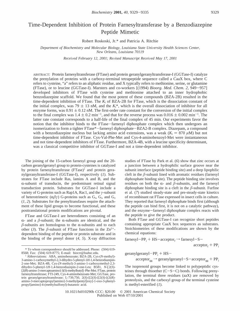
General Characteristics of FTase Inhibition by Peptidesand Peptidomimetics.We performed steady-state kineticexperiments to determine the apparentKi of FTase for BZA-2B, a benzodiazepine peptidomimetic, and we obtainedvalues of 6.5 nM with a 15 min incubation period and 3.4nM with a 30 min incubation. This result prompted us todetermine the rate of product formation, and we found thatthe rate was concave-downward in the presence of BZA-2B(Figure 1). Nonlinear rates of product formation and apparentKi values that decrease as a function of incubation timesuggested that BZA-2B was a slow-binding inhibitor ofFTase. Progress curves initiated by adding enzyme tosubstrates in the presence of slow-binding inhibitors areconcave-down (20). With classical inhibitors, in contrast,reaction rates are linear over the period during which thereis no substrate depletion.
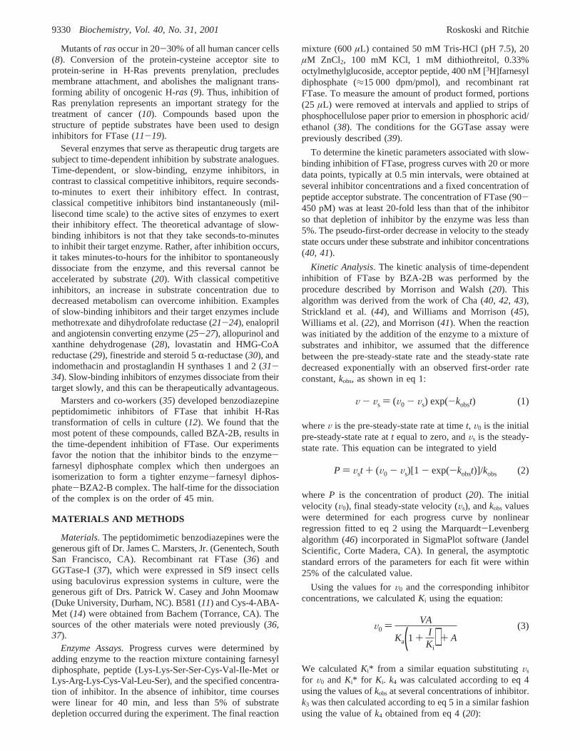
In contrast to BZA-2B, the rate of product formation waslinear in the presence of other inhibitors (Figure 2) includingCys-Val-Phe-Met (47, 48), B581 (11), Cys-4-ABA-Met, orFTI-249 (14), and diazepam, an anxiolytic drug with thebenzodiazepine scaffold of BZA-2B (Figure 3). We foundthat each of these compounds was a linear competitiveinhibitor with respect to the acceptor peptide substrate, Lys-Arg-Lys-Cys-Val-Leu-Ser. Additional experiments wereperformed to more fully understand the slow-binding inhibi-tion process.
Characterization of Time-Dependent Inhibition.There aretwo general mechanisms that describe time-dependent inhibi-tion (20). Mechanism A is represented by the followingequation where E represents the enzyme and I, the inhibitor:
For mechanism B, binding of the inhibitor I (BZA-2B)involves the rapid formation of an initial collision complex(EI) that undergoes a slow isomerization. Binding of inhibitorthus occurs in the following sequence:
where the forward and reverse rate constants for the firstprocess (formation of EI) arek1 and k2, respectively, andfor the second process (formation of EI*) arek3 and k4,respectively. The overall dissociation constant (Ki*) is
whereKi ) k2/k1. In mechanism A, the equilibrium betweenE, I, and EI is reached slowly. In mechanism B, theequilibrium between E, I, and the initial EI complex isattained rapidly, but this EI complex subsequently isomerizesto a tighter EI* complex (41).
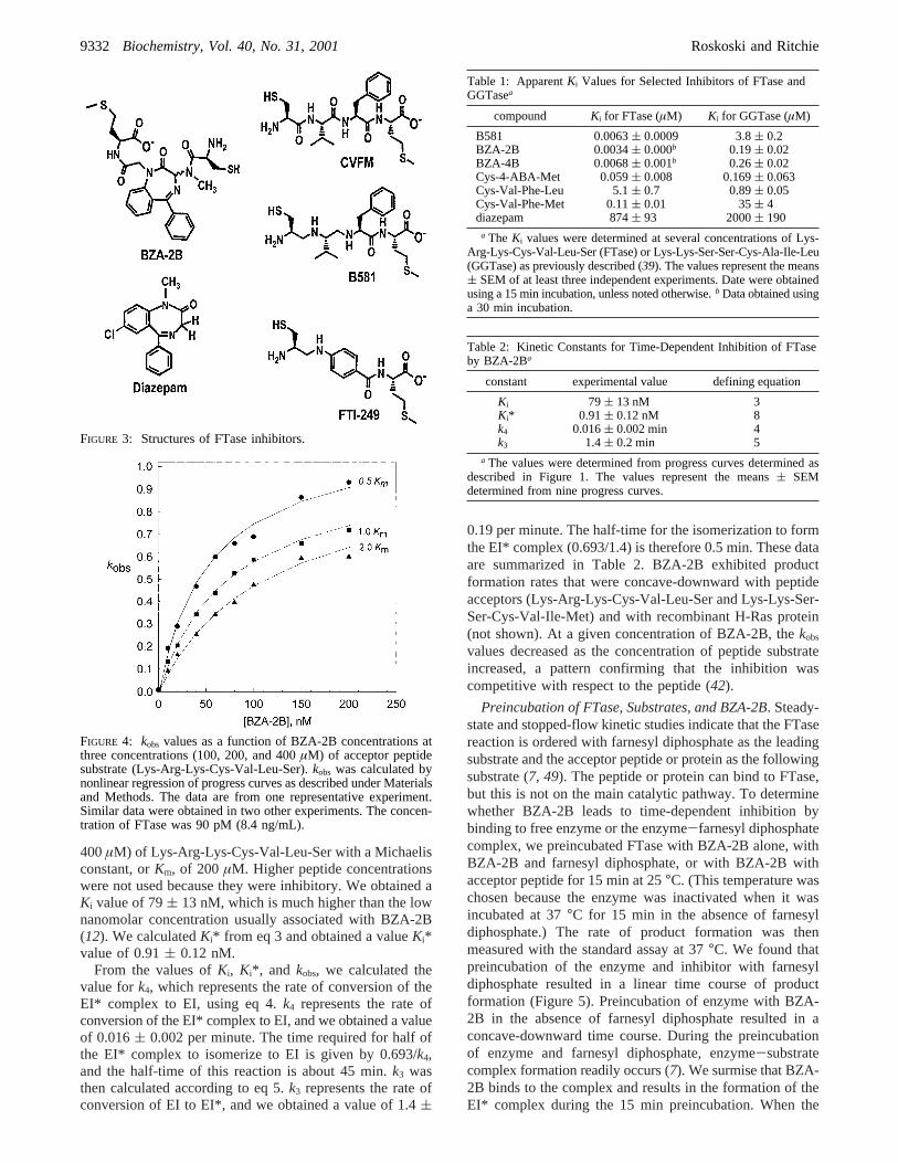
We performed experiments with BZA-2B concentrationsranging from 2 to 600 nM (Figure 1), and we determinedV0, Vs, and kobs by fitting the data to eq 2. The computeranalysis yielded values ofkobs that reached a plateau atincreasing concentrations of BZA-2B (Figure 4), and thisresult indicated that we were most likely dealing with asituation that resembles mechanism B, the two-step slow-binding inhibition pattern.
Using the values forV0 and the corresponding inhibitorconcentrations, we calculatedKi using eq 3. For theseexperiments, we used three concentrations (100, 200, and
kobs) k4[1 + I
Ki*(1 + AKa
)1 + I
Ki(1 + AKa
) ] (4)
kobs) k4 + k3[ IKi
1 + ( AKa
) + ( IKi
)] (5)
E + I h EI (6)
E + I h EI h EI* (7)
FIGURE 1: Progress curves of product formation illustrating time-dependent inhibition of FTase by BZA-2B. The solid lines are thecomputer-generated curves predicted by fitting the data to eq 2.Reactions contained 90 pM FTase (8.4 pg/mL), 100µM Lys-Arg-Lys-Cys-Val-Leu-Ser acceptor peptide, 400 nM farnesyl diphos-phate, the specified concentration of BZA-2B, and the othercomponents given under Materials and Methods. The averages ofduplicate determinations are given for a representative experiment.Similar results were obtained in eight other experiments.
FIGURE 2: Progress curves of product formation in the presence ofclassical inhibitors of FTase. The reaction was performed asdescribed in Figure 1 with the specified concentrations of thedesignated compounds. The data represent the mean of threeindependent experiments.
Ki* ) [E][I]/[EI + EI*] ) Kik4/(k3 + k4) (8)
Inhibition of Protein Farnesyltransferase Biochemistry, Vol. 40, No. 31, 20019331
400µM) of Lys-Arg-Lys-Cys-Val-Leu-Ser with a Michaelisconstant, orKm, of 200µM. Higher peptide concentrationswere not used because they were inhibitory. We obtained aKi value of 79( 13 nM, which is much higher than the lownanomolar concentration usually associated with BZA-2B(12). We calculatedKi* from eq 3 and obtained a valueKi*value of 0.91( 0.12 nM.
From the values ofKi, Ki*, and kobs, we calculated thevalue fork4, which represents the rate of conversion of theEI* complex to EI, using eq 4.k4 represents the rate ofconversion of the EI* complex to EI, and we obtained a valueof 0.016( 0.002 per minute. The time required for half ofthe EI* complex to isomerize to EI is given by 0.693/k4,and the half-time of this reaction is about 45 min.k3 wasthen calculated according to eq 5.k3 represents the rate ofconversion of EI to EI*, and we obtained a value of 1.4(
0.19 per minute. The half-time for the isomerization to formthe EI* complex (0.693/1.4) is therefore 0.5 min. These dataare summarized in Table 2. BZA-2B exhibited productformation rates that were concave-downward with peptideacceptors (Lys-Arg-Lys-Cys-Val-Leu-Ser and Lys-Lys-Ser-Ser-Cys-Val-Ile-Met) and with recombinant H-Ras protein(not shown). At a given concentration of BZA-2B, thekobs
values decreased as the concentration of peptide substrateincreased, a pattern confirming that the inhibition wascompetitive with respect to the peptide (42).
Preincubation of FTase, Substrates, and BZA-2B. Steady-state and stopped-flow kinetic studies indicate that the FTasereaction is ordered with farnesyl diphosphate as the leadingsubstrate and the acceptor peptide or protein as the followingsubstrate (7, 49). The peptide or protein can bind to FTase,but this is not on the main catalytic pathway. To determinewhether BZA-2B leads to time-dependent inhibition bybinding to free enzyme or the enzyme-farnesyl diphosphatecomplex, we preincubated FTase with BZA-2B alone, withBZA-2B and farnesyl diphosphate, or with BZA-2B withacceptor peptide for 15 min at 25°C. (This temperature waschosen because the enzyme was inactivated when it wasincubated at 37°C for 15 min in the absence of farnesyldiphosphate.) The rate of product formation was thenmeasured with the standard assay at 37°C. We found thatpreincubation of the enzyme and inhibitor with farnesyldiphosphate resulted in a linear time course of productformation (Figure 5). Preincubation of enzyme with BZA-2B in the absence of farnesyl diphosphate resulted in aconcave-downward time course. During the preincubationof enzyme and farnesyl diphosphate, enzyme-substratecomplex formation readily occurs (7). We surmise that BZA-2B binds to the complex and results in the formation of theEI* complex during the 15 min preincubation. When the
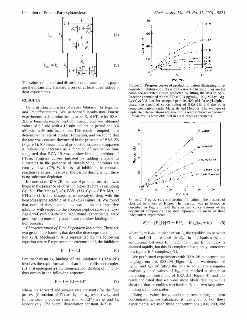
FIGURE 3: Structures of FTase inhibitors.
FIGURE 4: kobs values as a function of BZA-2B concentrations atthree concentrations (100, 200, and 400µM) of acceptor peptidesubstrate (Lys-Arg-Lys-Cys-Val-Leu-Ser).kobs was calculated bynonlinear regression of progress curves as described under Materialsand Methods. The data are from one representative experiment.Similar data were obtained in two other experiments. The concen-tration of FTase was 90 pM (8.4 ng/mL).
Table 1: ApparentKi Values for Selected Inhibitors of FTase andGGTasea
compound Ki for FTase (µM) Ki for GGTase (µM)
B581 0.0063( 0.0009 3.8( 0.2BZA-2B 0.0034( 0.000b 0.19( 0.02BZA-4B 0.0068( 0.001b 0.26( 0.02Cys-4-ABA-Met 0.059( 0.008 0.169( 0.063Cys-Val-Phe-Leu 5.1( 0.7 0.89( 0.05Cys-Val-Phe-Met 0.11( 0.01 35( 4diazepam 874( 93 2000( 190
a The Ki values were determined at several concentrations of Lys-Arg-Lys-Cys-Val-Leu-Ser (FTase) or Lys-Lys-Ser-Ser-Cys-Ala-Ile-Leu(GGTase) as previously described (39). The values represent the means( SEM of at least three independent experiments. Date were obtainedusing a 15 min incubation, unless noted otherwise.b Data obtained usinga 30 min incubation.
Table 2: Kinetic Constants for Time-Dependent Inhibition of FTaseby BZA-2Ba
constant experimental value defining equation
Ki 79 ( 13 nM 3Ki* 0.91 ( 0.12 nM 8k4 0.016( 0.002 min 4k3 1.4( 0.2 min 5
a The values were determined from progress curves determined asdescribed in Figure 1. The values represent the means( SEMdetermined from nine progress curves.
9332 Biochemistry, Vol. 40, No. 31, 2001 Roskoski and Ritchie
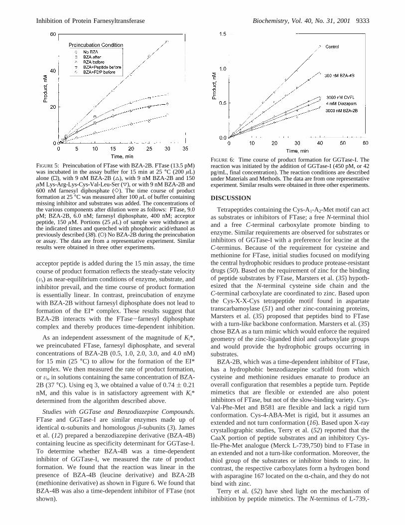
acceptor peptide is added during the 15 min assay, the timecourse of product formation reflects the steady-state velocity(Vs) as near-equilibrium conditions of enzyme, substrate, andinhibitor prevail, and the time course of product formationis essentially linear. In contrast, preincubation of enzymewith BZA-2B without farnesyl diphosphate does not lead toformation of the EI* complex. These results suggest thatBZA-2B interacts with the FTase-farnesyl diphosphatecomplex and thereby produces time-dependent inhibition.
As an independent assessment of the magnitude ofKi*,we preincubated FTase, farnesyl diphosphate, and severalconcentrations of BZA-2B (0.5, 1.0, 2.0, 3.0, and 4.0 nM)for 15 min (25°C) to allow for the formation of the EI*complex. We then measured the rate of product formation,or Vs, in solutions containing the same concentration of BZA-2B (37°C). Using eq 3, we obtained a value of 0.74( 0.21nM, and this value is in satisfactory agreement withKi*determined from the algorithm described above.
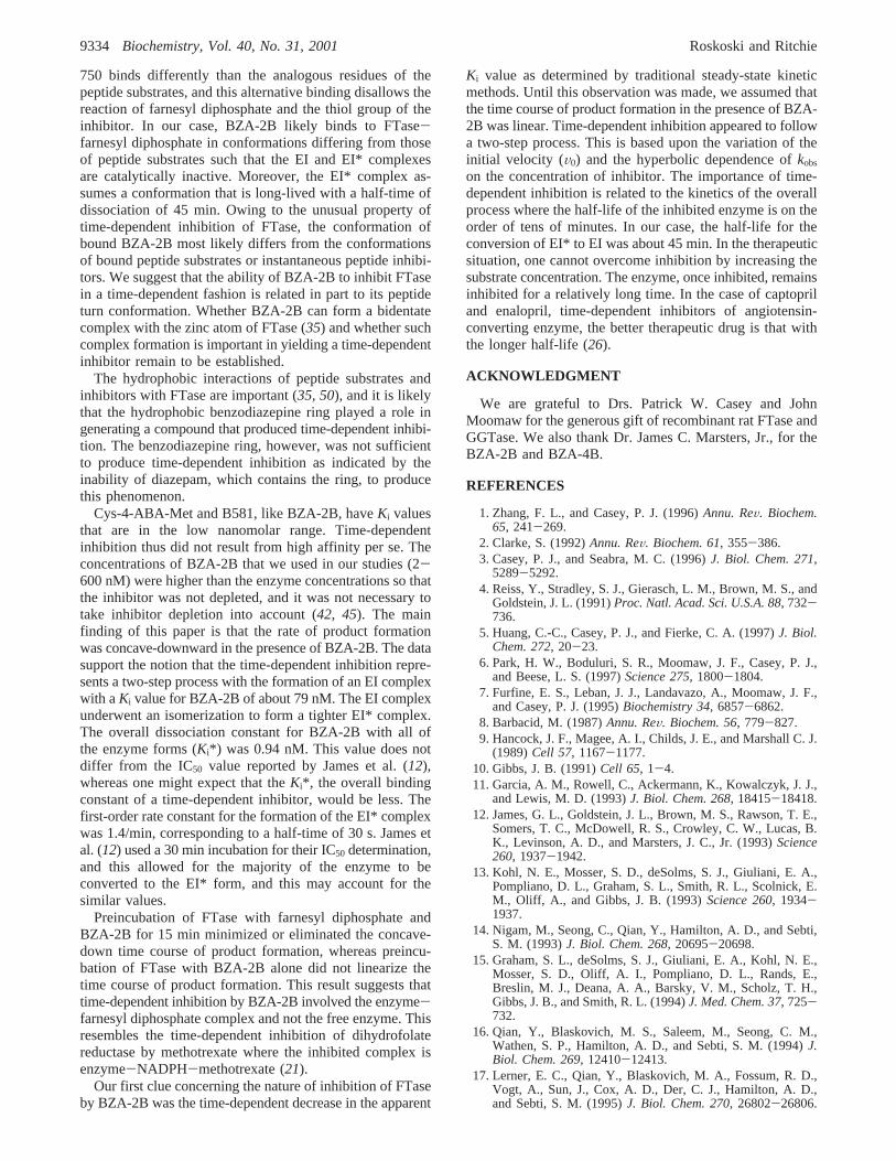
Studies with GGTase and Benzodiazepine Compounds.FTase and GGTase-I are similar enzymes made up ofidenticalR-subunits and homologousâ-subunits (3). Jameset al. (12) prepared a benzodiazepine derivative (BZA-4B)containing leucine as specificity determinant for GGTase-I.To determine whether BZA-4B was a time-dependentinhibitor of GGTase-I, we measured the rate of productformation. We found that the reaction was linear in thepresence of BZA-4B (leucine derivative) and BZA-2B(methionine derivative) as shown in Figure 6. We found thatBZA-4B was also a time-dependent inhibitor of FTase (notshown).
DISCUSSION
Tetrapeptides containing the Cys-A1-A2-Met motif can actas substrates or inhibitors of FTase; a freeN-terminal thioland a freeC-terminal carboxylate promote binding toenzyme. Similar requirements are observed for substrates orinhibitors of GGTase-I with a preference for leucine at theC-terminus. Because of the requirement for cysteine andmethionine for FTase, initial studies focused on modifyingthe central hydrophobic residues to produce protease-resistantdrugs (50). Based on the requirement of zinc for the bindingof peptide substrates by FTase, Marsters et al. (35) hypoth-esized that theN-terminal cysteine side chain and theC-terminal carboxylate are coordinated to zinc. Based uponthe Cys-X-X-Cys tetrapeptide motif found in aspartatetranscarbamoylase (51) and other zinc-containing proteins,Marsters et al. (35) proposed that peptides bind to FTasewith a turn-like backbone conformation. Marsters et al. (35)chose BZA as a turn mimic which would enforce the requiredgeometry of the zinc-liganded thiol and carboxylate groupsand would provide the hydrophobic groups occurring insubstrates.
BZA-2B, which was a time-dependent inhibitor of FTase,has a hydrophobic benzodiazepine scaffold from whichcysteine and methionine residues emanate to produce anoverall configuration that resembles a peptide turn. Peptidemimetics that are flexible or extended are also potentinhibitors of FTase, but not of the slow-binding variety. Cys-Val-Phe-Met and B581 are flexible and lack a rigid turnconformation. Cys-4-ABA-Met is rigid, but it assumes anextended and not turn conformation (16). Based upon X-raycrystallographic studies, Terry et al. (52) reported that theCaaX portion of peptide substrates and an inhibitory Cys-Ile-Phe-Met analogue (Merck L-739,750) bind to FTase inan extended and not a turn-like conformation. Moreover, thethiol group of the substrates or inhibitor binds to zinc. Incontrast, the respective carboxylates form a hydrogen bondwith asparagine 167 located on theR-chain, and they do notbind with zinc.
Terry et al. (52) have shed light on the mechanism ofinhibition by peptide mimetics. TheN-terminus of L-739,-
FIGURE 5: Preincubation of FTase with BZA-2B. FTase (13.5 pM)was incubated in the assay buffer for 15 min at 25°C (200 µL)alone (0), with 9 nM BZA-2B (4), with 9 nM BZA-2B and 150µM Lys-Arg-Lys-Cys-Val-Leu-Ser (3), or with 9 nM BZA-2B and600 nM farnesyl diphosphate (]). The time course of productformation at 25°C was measured after 100µL of buffer containingmissing inhibitor and substrates was added. The concentrations ofthe various components after dilution were as follows: FTase, 9.0pM; BZA-2B, 6.0 nM; farnesyl diphosphate, 400 nM; acceptorpeptide, 150µM. Portions (25µL) of sample were withdrawn atthe indicated times and quenched with phosphoric acid/ethanol aspreviously described (38). (O) No BZA-2B during the preincubationor assay. The data are from a representative experiment. Similarresults were obtained in three other experiments.
FIGURE 6: Time course of product formation for GGTase-I. Thereaction was initiated by the addition of GGTase-I (450 pM, or 42pg/mL, final concentration). The reaction conditions are describedunder Materials and Methods. The data are from one representativeexperiment. Similar results were obtained in three other experiments.
Inhibition of Protein Farnesyltransferase Biochemistry, Vol. 40, No. 31, 20019333
750 binds differently than the analogous residues of thepeptide substrates, and this alternative binding disallows thereaction of farnesyl diphosphate and the thiol group of theinhibitor. In our case, BZA-2B likely binds to FTase-farnesyl diphosphate in conformations differing from thoseof peptide substrates such that the EI and EI* complexesare catalytically inactive. Moreover, the EI* complex as-sumes a conformation that is long-lived with a half-time ofdissociation of 45 min. Owing to the unusual property oftime-dependent inhibition of FTase, the conformation ofbound BZA-2B most likely differs from the conformationsof bound peptide substrates or instantaneous peptide inhibi-tors. We suggest that the ability of BZA-2B to inhibit FTasein a time-dependent fashion is related in part to its peptideturn conformation. Whether BZA-2B can form a bidentatecomplex with the zinc atom of FTase (35) and whether suchcomplex formation is important in yielding a time-dependentinhibitor remain to be established.
The hydrophobic interactions of peptide substrates andinhibitors with FTase are important (35, 50), and it is likelythat the hydrophobic benzodiazepine ring played a role ingenerating a compound that produced time-dependent inhibi-tion. The benzodiazepine ring, however, was not sufficientto produce time-dependent inhibition as indicated by theinability of diazepam, which contains the ring, to producethis phenomenon.
Cys-4-ABA-Met and B581, like BZA-2B, haveKi valuesthat are in the low nanomolar range. Time-dependentinhibition thus did not result from high affinity per se. Theconcentrations of BZA-2B that we used in our studies (2-600 nM) were higher than the enzyme concentrations so thatthe inhibitor was not depleted, and it was not necessary totake inhibitor depletion into account (42, 45). The mainfinding of this paper is that the rate of product formationwas concave-downward in the presence of BZA-2B. The datasupport the notion that the time-dependent inhibition repre-sents a two-step process with the formation of an EI complexwith aKi value for BZA-2B of about 79 nM. The EI complexunderwent an isomerization to form a tighter EI* complex.The overall dissociation constant for BZA-2B with all ofthe enzyme forms (Ki*) was 0.94 nM. This value does notdiffer from the IC50 value reported by James et al. (12),whereas one might expect that theKi*, the overall bindingconstant of a time-dependent inhibitor, would be less. Thefirst-order rate constant for the formation of the EI* complexwas 1.4/min, corresponding to a half-time of 30 s. James etal. (12) used a 30 min incubation for their IC50 determination,and this allowed for the majority of the enzyme to beconverted to the EI* form, and this may account for thesimilar values.
Preincubation of FTase with farnesyl diphosphate andBZA-2B for 15 min minimized or eliminated the concave-down time course of product formation, whereas preincu-bation of FTase with BZA-2B alone did not linearize thetime course of product formation. This result suggests thattime-dependent inhibition by BZA-2B involved the enzyme-farnesyl diphosphate complex and not the free enzyme. Thisresembles the time-dependent inhibition of dihydrofolatereductase by methotrexate where the inhibited complex isenzyme-NADPH-methotrexate (21).
Our first clue concerning the nature of inhibition of FTaseby BZA-2B was the time-dependent decrease in the apparent
Ki value as determined by traditional steady-state kineticmethods. Until this observation was made, we assumed thatthe time course of product formation in the presence of BZA-2B was linear. Time-dependent inhibition appeared to followa two-step process. This is based upon the variation of theinitial velocity (V0) and the hyperbolic dependence ofkobs
on the concentration of inhibitor. The importance of time-dependent inhibition is related to the kinetics of the overallprocess where the half-life of the inhibited enzyme is on theorder of tens of minutes. In our case, the half-life for theconversion of EI* to EI was about 45 min. In the therapeuticsituation, one cannot overcome inhibition by increasing thesubstrate concentration. The enzyme, once inhibited, remainsinhibited for a relatively long time. In the case of captopriland enalopril, time-dependent inhibitors of angiotensin-converting enzyme, the better therapeutic drug is that withthe longer half-life (26).
ACKNOWLEDGMENT
We are grateful to Drs. Patrick W. Casey and JohnMoomaw for the generous gift of recombinant rat FTase andGGTase. We also thank Dr. James C. Marsters, Jr., for theBZA-2B and BZA-4B.
REFERENCES
1. Zhang, F. L., and Casey, P. J. (1996)Annu. ReV. Biochem.65, 241-269.
2. Clarke, S. (1992)Annu. ReV. Biochem. 61, 355-386.3. Casey, P. J., and Seabra, M. C. (1996)J. Biol. Chem. 271,
5289-5292.4. Reiss, Y., Stradley, S. J., Gierasch, L. M., Brown, M. S., and
Goldstein, J. L. (1991)Proc. Natl. Acad. Sci. U.S.A. 88, 732-736.
5. Huang, C.-C., Casey, P. J., and Fierke, C. A. (1997)J. Biol.Chem. 272, 20-23.
6. Park, H. W., Boduluri, S. R., Moomaw, J. F., Casey, P. J.,and Beese, L. S. (1997)Science 275, 1800-1804.
7. Furfine, E. S., Leban, J. J., Landavazo, A., Moomaw, J. F.,and Casey, P. J. (1995)Biochemistry 34, 6857-6862.
8. Barbacid, M. (1987)Annu. ReV. Biochem. 56, 779-827.9. Hancock, J. F., Magee, A. I., Childs, J. E., and Marshall C. J.
(1989)Cell 57, 1167-1177.10. Gibbs, J. B. (1991)Cell 65, 1-4.11. Garcia, A. M., Rowell, C., Ackermann, K., Kowalczyk, J. J.,
and Lewis, M. D. (1993)J. Biol. Chem. 268, 18415-18418.12. James, G. L., Goldstein, J. L., Brown, M. S., Rawson, T. E.,
Somers, T. C., McDowell, R. S., Crowley, C. W., Lucas, B.K., Levinson, A. D., and Marsters, J. C., Jr. (1993)Science260, 1937-1942.
13. Kohl, N. E., Mosser, S. D., deSolms, S. J., Giuliani, E. A.,Pompliano, D. L., Graham, S. L., Smith, R. L., Scolnick, E.M., Oliff, A., and Gibbs, J. B. (1993)Science 260, 1934-1937.
14. Nigam, M., Seong, C., Qian, Y., Hamilton, A. D., and Sebti,S. M. (1993)J. Biol. Chem. 268, 20695-20698.
15. Graham, S. L., deSolms, S. J., Giuliani, E. A., Kohl, N. E.,Mosser, S. D., Oliff, A. I., Pompliano, D. L., Rands, E.,Breslin, M. J., Deana, A. A., Barsky, V. M., Scholz, T. H.,Gibbs, J. B., and Smith, R. L. (1994)J. Med. Chem. 37, 725-732.
16. Qian, Y., Blaskovich, M. S., Saleem, M., Seong, C. M.,Wathen, S. P., Hamilton, A. D., and Sebti, S. M. (1994)J.Biol. Chem. 269, 12410-12413.
17. Lerner, E. C., Qian, Y., Blaskovich, M. A., Fossum, R. D.,Vogt, A., Sun, J., Cox, A. D., Der, C. J., Hamilton, A. D.,and Sebti, S. M. (1995)J. Biol. Chem. 270, 26802-26806.
9334 Biochemistry, Vol. 40, No. 31, 2001 Roskoski and Ritchie
18. Vogt, A., Qian, Y., Blaskovich, M. A., Fossum, R. D.,Hamilton, A. D., and Sebti, S. M. (1995)J. Biol. Chem. 270,660-664.
19. Anthony, N. J., Gomez, R. P., Schaber, M. D., Mosser, S. D.,Hamilton, K. A., O’Neil, T. J., Koblan, K. S., Graham, S. L.,Hartman, G. D., Shah, D., Rands, E., Kohl, N. E., Gibbs, J.B., and Oliff, A. I. (1999)J. Med. Chem. 42, 3356-3368.
20. Morrison, J. F., and Walsh, C. T. (1988)AdV. Enzymol. Relat.Areas Mol. Biol. 61, 201-301.
21. Williams, J. W., Morrison, J. F., and Duggleby, R. G. (1979)Biochemistry 18, 2567-2573.
22. Williams, J. W., Duggleby, R. G., Cutler, R., and Morrison,J. F. (1980)Biochem. Pharmacol. 29, 589-595.
23. Blakley, R. L., and Cocco, L. (1985)Biochemistry 24, 4704-4709.
24. Blakley, R. L., and Cocco, L. (1985)Biochemistry 24, 4772-4777.
25. Reynolds, C. H. (1983)Biochem. Pharmacol. 33, 1273-1276.26. Shapiro, R., and Riordan, J. F. (1984)Biochemistry 23, 5234-
5240.27. Bull, H. G., Thornberry, N. A., Cordes, M. H. J., Patchett, A.
A., and Cordes, E. H. (1985)J. Biol. Chem. 260, 2952-2962.28. Massey, V., Komai, H., Palmer, G., and Elion, G. B. (1970)
J. Biol. Chem. 245, 2837-2844.29. Nakamura, C. E., and Abeles, R. H. (1985)Biochemistry 24,
1364-1376.30. Faller, B., Farley, D., and Nick, H. (1993)Biochemistry 32,
5705-5710.31. Rome, L. H., and Lands, W. E. M. (1975)Proc. Natl. Acad.
Sci. U.S.A. 72, 4863-4865.32. Guo, Q., Wang, L. H., Ruan, K. H., and Kulmacz, R. J. (1996)
J. Biol. Chem. 271, 19134-19139.33. Callan, O. H., So, O. Y., and Swinney, D. C. (1996)J. Biol.
Chem. 271, 3548-3554.34. So, O. Y., Scarafia, L. E., Mak, A. Y., Callan, O. H., and
Swinney, D. C. (1998)J. Biol. Chem. 273, 5801-5807.35. Marsters, J. C., McDowell, R. S., Reynolds, M. E., Oare, D.
A., Somers, T. C., Stanley, M. S., Rawson, T. E., Struble, M.
E., Burdick, D. J., Chan, K. S., Duarte, C. M., Paris, K. J.,Tom, J. Y. K., Wan, D. T., Xue, Y., and Burnier, J. P. (1994)Bioorg. Med. Chem. 2, 949-957.
36. Chen, W.-J., Moomaw, J. F., Overton, L., Kost, T. A., andCasey, P. J. (1993)J. Biol. Chem. 268, 9675-9680.
37. Zhang, F. L., Moomaw, J. F., and Casey, P. J. (1994)J. Biol.Chem. 269, 3175-3180.
38. Roskoski, R., Jr., Ritchie, P., and Gahn, L. G. (1994)Anal.Biochem. 222, 275-280.
39. Roskoski, R., Jr., and Ritchie, P. (1998)Arch. Biochem.Biophys. 356, 167-176.
40. Cha, S. (1976)Biochem. Pharmacol. 25, 2695-2702.41. Morrison, J. F. (1982)Trends Biochem. Sci. 7, 102-104.42. Cha, S. (1975)Biochem. Pharmacol. 24, 2177-2185.43. Cha, S. (1976a)Biochem. Pharmacol. 25, 1561.44. Strickland, S., Palmer, G., and Massey, V. (1975)J. Biol.
Chem. 250, 4048-4052.45. Williams, J. W., and Morrison, J. F. (1979)Methods Enzymol.
63, 437-467.46. Press, W. H., Flannery, B. P., Teukolsky, S. A., and Vetterling,
W. T. (1986)Numerical Recipes, Cambridge University Press,Cambridge, U.K.
47. Goldstein, J. L., Brown, M. S., Stradley, S. J., Reiss, Y., andGierasch, L. M. (1991)J. Biol. Chem. 266, 15575-15578.
48. Brown, M. S., Goldstein, J. L., Paris, K. J., Burnier, J. P., andMarsters, J. C., Jr. (1992)Proc. Natl. Acad. Sci. U.S.A. 89,8313-8316.
49. Pompliano, D. L., Schaber, M. D., Mosser, S. D., Omer, C.A., Shafer, J. A., and Gibbs, J. B. (1993)Biochemistry 32,8341-8347.
50. Sebti, S. M., and Hamilton, A. D. (1996)Biochem. Soc. Trans.24, 692-699.
51. Stevens, R. C., Gouauz, J. E., and Lipscomb, W. N. (1990)Biochemistry 29, 7691-7701.
52. Terry, K. L., Long, S. B., and Beese, L. S. (2001)Enzymes(3rd Ed.) 21, 19-46.
BI010290B
Inhibition of Protein Farnesyltransferase Biochemistry, Vol. 40, No. 31, 20019335