timothy j. carroll . e. paul zehr . david f. collins
TRANSCRIPT

Exp Brain Res (2005) 161: 133–144DOI 10.1007/s00221-004-2050-7
RESEARCH ARTICLE
Timothy J. Carroll . E. Paul Zehr . David F. Collins
Modulation of cutaneous reflexes in human upper limb musclesduring arm cycling is independent of activityin the contralateral arm
Received: 3 February 2004 / Accepted: 2 June 2004 / Published online: 23 October 2004# Springer-Verlag 2004
Abstract The amplitudes and signs of cutaneous reflexesare modulated during rhythmic movements of the armsand legs (during walking and arm or leg cycling forinstance). This reflex modulation is frequently indepen-dent of the background muscle activity and may involvecentral pattern generator (CPG) circuits. The purpose ofthe present study was to investigate the nature and degreeof coupling between the upper limbs during arm cycling,with regard to the regulation of cutaneous reflexes.Responses to electrical stimulations of the right, superfi-cial radial nerve (five 1 ms pulses, 300 Hz) were recordedbilaterally in six arm muscles of eight participants duringarm cycling involving only the limb ipsilateral to thestimulation, only the limb contralateral to the stimulation,and bilateral movement when the limbs were both in-phase and 180° out of phase. The pattern of cutaneousreflex modulation throughout the arm cycle was indepen-dent of the functional state of the limb contralateral to therecording site, irrespective of whether recordings weremade ipsilateral or contralateral to the stimulation.Furthermore, cutaneous reflexes were significantly(p<0.05) modulated with arm position in only 8% ofcases in which the limb containing the responding muscle
was either stationary or being moved passively by theexperimenter. The results show that there is relativelyweak coupling between the arms with regard to theregulation of cutaneous reflexes during rhythmic, cyclicalarm movements. This suggests a loose connection betweenthe CPGs for each arm that regulate muscle activity andreflex amplitude during rhythmic movement.
Keywords Cutaneous reflexes . Arm cycling . Centralpattern generator . CPG . Bilateral movement . Reflexmodulation
Introduction
The organisation and function of cutaneous reflexes in thelegs during rhythmic, cyclical movement has receivedconsiderable attention for tasks such as walking and legcycling (for reviews, see Brooke et al. 1997; Zehr andStein 1999). Less is known regarding the patterns of reflexmodulation during rhythmic, cyclical movement per-formed with the arms. However, it has recently beendemonstrated that some features of cutaneous reflexmodulation observed during cyclical leg movements,such as task and phase dependency, are common torhythmic arm movements, including arm cycling (Zehrand Kido 2001) and walking (Zehr and Haridas 2003). Forexample, in some muscles, the position of the limb duringarm cycling influenced the size of the cutaneous reflexresponses in a manner that was independent of thebackground EMG activity (premotoneuronal gating).There were also cases in which cutaneous stimulationresulted in an excitatory response at some points in thearm cycle, but inhibition at other points of the cycle (reflexreversals). Reflex reversals occurred in some musclesbetween static contractions and rhythmic movements (inother words task dependency; Zehr and Kido 2001).
The accumulating data support the hypothesis that themechanisms of reflex modulation during rhythmic,cyclical movement are similar between the arms andlegs, and may be due to interneuronal circuits that
T. J. Carroll (*)Health and Sports Science, School of Medical Sciences, TheUniversity of New South Wales,LG02M Wallace Wurth Building,2052 Sydney, New South Wales, Australiae-mail: [email protected].: +61-2-9385-8709Fax: +61-2-9385-1059
E. P. ZehrRehabilitation Neuroscience Laboratory, University of Victoria,Victoria, British Columbia, Canada
E. P. ZehrInternational Collaboration on Repair Discoveries (ICORD),Vancouver, British Columbia, Canada
D. F. CollinsHuman Neurophysiology Laboratory, Faculty of PhysicalEducation and Recreation, University of Alberta,Edmonton, Alberta, Canada

comprise central pattern generators (CPGs). The nature ofcoupling between CPGs for the arms is of significantinterest as a basis for further comparison of themechanisms of neural control between arm and legmovements. We recently found that some differencesexist in the regulation of H-reflexes between the arms andlegs, in that H-reflexes are not depressed in a stationaryarm when the contralateral arm is passively moved in arhythmic, cyclical manner (Zehr et al. 2003). Such weakcontralateral effects may be indicative of a relatively“loose coupling” between the CPGs for each arm. This isin contrast to what has been observed for leg cycling tasks(Cheng et al. 1998; Collins et al. 1993; Misiaszek et al.1998), and suggests that the strength of the neuralcoupling between the arms is weaker than between thelegs, at least in terms of the regulation of muscle afferentreflexes. Tax et al. (1995) reported similarities in cutane-ous reflexes at corresponding phases in the step cyclebetween the legs during running and suggested thatcutaneous reflexes elicited during locomotion are deter-mined by the functional context of the limb in which theresponding muscles reside. In the present study, weexamined cutaneous reflexes in both arms during bilateraland unilateral cycling tasks. Our purpose was to add to ourunderstanding of the neural control of the arms duringrhythmic, cyclical movements by using reflex modulationas a “neural probe” (Burke 1999). The working hypothesisin these experiments is that CPG elements are involved inregulating muscle activity and reflex amplitude duringrhythmic arm cycling. The main objectives of this studywere to further elucidate the strength of neural couplingbetween the arms during rhythmic movement, and toprovide evidence regarding the sites of cutaneous reflexmodulation. We systematically assessed how cutaneousreflexes in homologous arm muscles located both ipsilat-eral and contralateral to the stimulation were affected bymovements of each arm. We hypothesized that if there isstrong linkage in neural control between the arms,cutaneous reflexes in muscles both ipsilateral and contra-lateral to the stimulus should be influenced by movementof the opposite arm.
Materials and methods
Subjects
Eight individuals (six males, two females) with nodocumented neurological disease volunteered for thisexperiment. The participants ranged in age from 21 to39 years old. The procedures conformed to the Declarationof Helsinki and were approved by the Human ResearchEthics Board at the University of Alberta.
Protocol
Participants performed rhythmic arm cycling with one orboth limbs using a custom-built, hydraulic arm ergometer
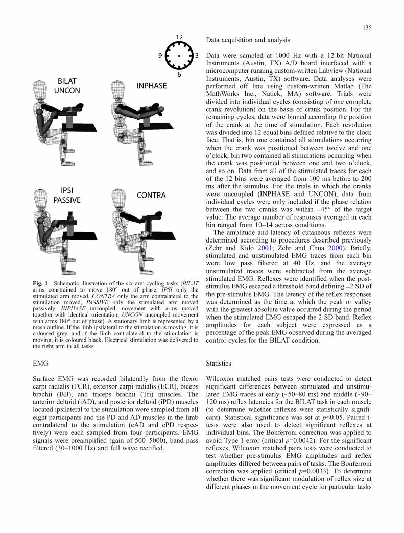
that has been described previously (Zehr et al. 2003; Zehrand Kido 2001). The positions of the ergometer crankswere defined relative to a clock face, with the “top deadcentre” position specified as 12 o’clock and the remainingpositions following the conventions of a clock face whenlooking at the ergometer from the participant’s right (Fig.1). The ergometer could be adjusted so that the two crankswere either coupled or allowed to rotate independently.The participants performed three bilateral and threeunilateral arm cycling tasks. The bilateral tasks were armcycling with: 1) the cranks coupled 180° out of phase(BILAT); 2) the cranks uncoupled but with instructions tomaintain the arms 180° out of phase (UNCON); 3) thecranks uncoupled but with instructions to maintain thearms in phase (INPHASE). The unilateral tasks were armcycling with: 1) the arm ipsilateral to the stimulation activeand the contralateral limb held stationary at the six o’clockposition (IPSI); 2) the arm ipsilateral to the stimulationpassively rotated by an experimenter and the contralaterallimb held stationary at the six o’clock position (PAS-SIVE); 3) the arm contralateral to the stimulation activeand the ipsilateral limb held stationary at the six o’clockposition (CONTRA). During the CONTRA condition, theparticipants maintained a tonic contraction in the ipsilat-eral wrist extensor muscles that was similar in magnitudeto the average level of contraction observed during theBILAT condition.
The participants performed each of the arm cyclingtasks for a total of eight minutes at a comfortable, self-selected rate between 60 and 80 revolutions per minute(rpm). Up to four short rest breaks were taken during trials.The order in which the tasks were performed was variedpseudo-randomly. Mean cycling rate varied from 67 (±6)rpm in the UNCON condition to 76 (±9) rpm in the IPSIand contra conditions. The mean rate of the BILAT (±5),passive (±5) and INPHASE (±9) conditions was approxi-mately 70 rpm.
Nerve stimulation
The superficial radial (SR) nerve, innervating the skin onthe dorso-lateral surface of the hand, was stimulated with atrain of five, 1 ms pulses at 300 Hz using self-adhesive,disposable stimulating electrodes placed on the dorsalsurface of the right forearm just proximal to the radialhead. A Grass S88 (Grass Instruments, AstroMed Inc.,West Warwick, RI) stimulator, connected in series with aGrass SIU5 isolator and a Grass CCU1 constant currentunit, delivered the stimuli at pseudorandom intervals ofapproximately 2.5–3.5 s during the arm cycling trials. Theintensity of stimulation was set at approximately twice theradiating threshold (RT) for each subject and a brace wasworn on the stimulated wrist to minimise electrodemovement, as in previous studies (Zehr and Chua 2000;Zehr and Kido 2001). The participants were regularlyasked to qualitatively rate the sensation induced by thenerve stimulation during the experiment to ensure thatstimulus intensity was consistent.
134
EMG
Surface EMG was recorded bilaterally from the flexorcarpi radialis (FCR), extensor carpi radialis (ECR), bicepsbrachii (BB), and triceps brachii (Tri) muscles. Theanterior deltoid (iAD), and posterior deltoid (iPD) muscleslocated ipsilateral to the stimulation were sampled from alleight participants and the PD and AD muscles in the limbcontralateral to the stimulation (cAD and cPD respec-tively) were each sampled from four participants. EMGsignals were preamplified (gain of 500–5000), band passfiltered (30–1000 Hz) and full wave rectified.
Data acquisition and analysis
Data were sampled at 1000 Hz with a 12-bit NationalInstruments (Austin, TX) A/D board interfaced with amicrocomputer running custom-written Labview (NationalInstruments, Austin, TX) software. Data analyses wereperformed off line using custom-written Matlab (TheMathWorks Inc., Natick, MA) software. Trials weredivided into individual cycles (consisting of one completecrank revolution) on the basis of crank position. For theremaining cycles, data were binned according the positionof the crank at the time of stimulation. Each revolutionwas divided into 12 equal bins defined relative to the clockface. That is, bin one contained all stimulations occurringwhen the crank was positioned between twelve and oneo’clock, bin two contained all stimulations occurring whenthe crank was positioned between one and two o’clock,and so on. Data from all of the stimulated traces for eachof the 12 bins were averaged from 100 ms before to 200ms after the stimulus. For the trials in which the crankswere uncoupled (INPHASE and UNCON), data fromindividual cycles were only included if the phase relationbetween the two cranks was within ±45° of the targetvalue. The average number of responses averaged in eachbin ranged from 10–14 across conditions.
The amplitude and latency of cutaneous reflexes weredetermined according to procedures described previously(Zehr and Kido 2001; Zehr and Chua 2000). Briefly,stimulated and unstimulated EMG traces from each binwere low pass filtered at 40 Hz, and the averageunstimulated traces were subtracted from the averagestimulated EMG. Reflexes were identified when the post-stimulus EMG escaped a threshold band defining ±2 SD ofthe pre-stimulus EMG. The latency of the reflex responseswas determined as the time at which the peak or valleywith the greatest absolute value occurred during the periodwhen the stimulated EMG escaped the 2 SD band. Reflexamplitudes for each subject were expressed as apercentage of the peak EMG observed during the averagedcontrol cycles for the BILAT condition.
Statistics
Wilcoxon matched pairs tests were conducted to detectsignificant differences between stimulated and unstimu-lated EMG traces at early (~50–80 ms) and middle (~90–120 ms) reflex latencies for the BILAT task in each muscle(to determine whether reflexes were statistically signifi-cant). Statistical significance was set at p<0.05. Paired t-tests were also used to detect significant reflexes atindividual bins. The Bonferroni correction was applied toavoid Type 1 error (critical p=0.0042). For the significantreflexes, Wilcoxon matched pairs tests were conducted totest whether pre-stimulus EMG amplitudes and reflexamplitudes differed between pairs of tasks. The Bonferronicorrection was applied (critical p=0.0033). To determinewhether there was significant modulation of reflex size atdifferent phases in the movement cycle for particular tasks
Fig. 1 Schematic illustration of the six arm-cycling tasks (BILATarms constrained to move 180° out of phase, IPSI only thestimulated arm moved, CONTRA only the arm contralateral to thestimulation moved, PASSIVE only the stimulated arm movedpassively, INPHASE uncoupled movement with arms movedtogether with identical orientation, UNCON uncoupled movementwith arms 180° out of phase). A stationary limb is represented by amesh outline. If the limb ipsilateral to the stimulation is moving, it iscoloured grey, and if the limb contralateral to the stimulation ismoving, it is coloured black. Electrical stimulation was delivered tothe right arm in all tasks
135
and muscles, one-way ANOVAs were performed on reflexamplitudes across the 12 movement bins for each task.Statistical significance was set at p<0.05. Pearson productmoment correlation analysis was conducted on therelationship between reflex amplitudes and control EMGfor each muscle to determine the extent to which variationsin reflex size across movement phases were linearlyrelated to changes in background muscle activation. Forour sample size of eight, there were seven degrees offreedom, yielding a critical r of 0.666 at p<0.05.
Results
Muscle activity during the different arm cycling tasks
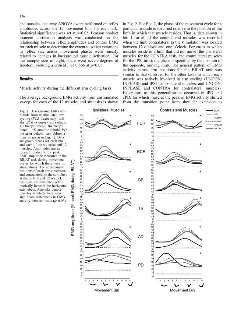
The average background EMG activity from unstimulatedsweeps for each of the 12 muscles and six tasks is shown
in Fig. 2. For Fig. 2, the phase of the movement cycle for aparticular muscle is specified relative to the position of thelimb in which that muscle resides. That is, data shown inbin 1 for all of the contralateral muscles was recordedwhen the limb contralateral to the stimulation was locatedbetween 12 o’clock and one o’clock. For cases in whichmuscles reside in a limb that did not move (the ipsilateralmuscles for the CONTRA task, and contralateral musclesfor the IPSI task), the phase is specified by the position ofthe opposite, moving limb. The general pattern of EMGactivity across arm positions for the BILAT task wassimilar to that observed for the other tasks in which eachmuscle was actively involved in arm cycling (UNCON,INPHASE and IPSI for ipsilateral muscles, and UNCON,INPHASE and CONTRA for contralateral muscles).Exceptions to this generalization occurred in iPD andcPD, for which muscles the peak in EMG activity shiftedfrom the transition point from shoulder extension to
Fig. 2 Background EMG am-plitude from unstimulated armcycling (FCR flexor carpi radi-alis, ECR extensor carpi radialis,Tri triceps brachii, BB bicepsbrachii, AD anterior deltoid, PDposterior deltoid; task abbrevia-tions as given in Fig. 1). Dataare group means for each binand each of the six tasks and 12muscles. Amplitudes are ex-pressed relative to the peakEMG amplitude measured in theBILAT task during movementcycles for which there were nostimulations. The approximatepositions of each arm (ipsilateraland contralateral to the stimulus)at the 3, 6, 9 and 12 o’clockpositions are illustrated sche-matically beneath the horizontalaxis labels. Asterisks denotemuscles in which there weresignificant differences in EMGactivity between tasks (p<0.05)
136
flexion in the BILAT task (bins three, four, and five) toapproximately at the mid-point of the shoulder flexion(bins seven, eight and nine) in the other active tasks foreach arm. Despite the general similarities in the pattern ofEMG activity across the movement cycle, there weresignificant EMG amplitude differences between at leastone pair of tasks for all muscles (p<0.05). There wassignificantly less EMG activity (p<0.05) during the BILATcondition compared with at least one of the otherunconstrained, active tasks (IPSI, INPHASE, UNCONfor ipsilateral muscles; CONTRA, INPHASE, UNCONfor contralateral muscles) for iECR, cECR, iBB, cBB, iTri,cTri, iAD, cAD and iPD. Therefore, it appears that thepattern of muscle activity required to perform arm cyclingmovements for each arm independently is generallysimilar to the pattern required for constrained arm cycling,for all of the muscles sampled in this study other than PD.However, some of the unconstrained tasks involvedsignificantly greater EMG activity than the BILAT taskin ECR, BB, Tri, AD and PD.
Cutaneous reflexes
The first two reflex latencies were analysed in detail in thisstudy and termed early (~50–80 ms) and middle (~90–120ms). The peak latencies of these responses are comparableto those that we have reported previously (Zehr and Kido2001; Zehr and Chua 2000), and are similar across tasks.Reflexes were statistically significant (p<0.05) in all
muscles except for cPD (early latency), iECR (middlelatency), cFCR (middle latency) and iPD (middle latency).Therefore, electrical stimulation of the SR nerve elicitedsignificant cutaneous reflexes in 20 out of 24 cases (12muscles by two latencies).
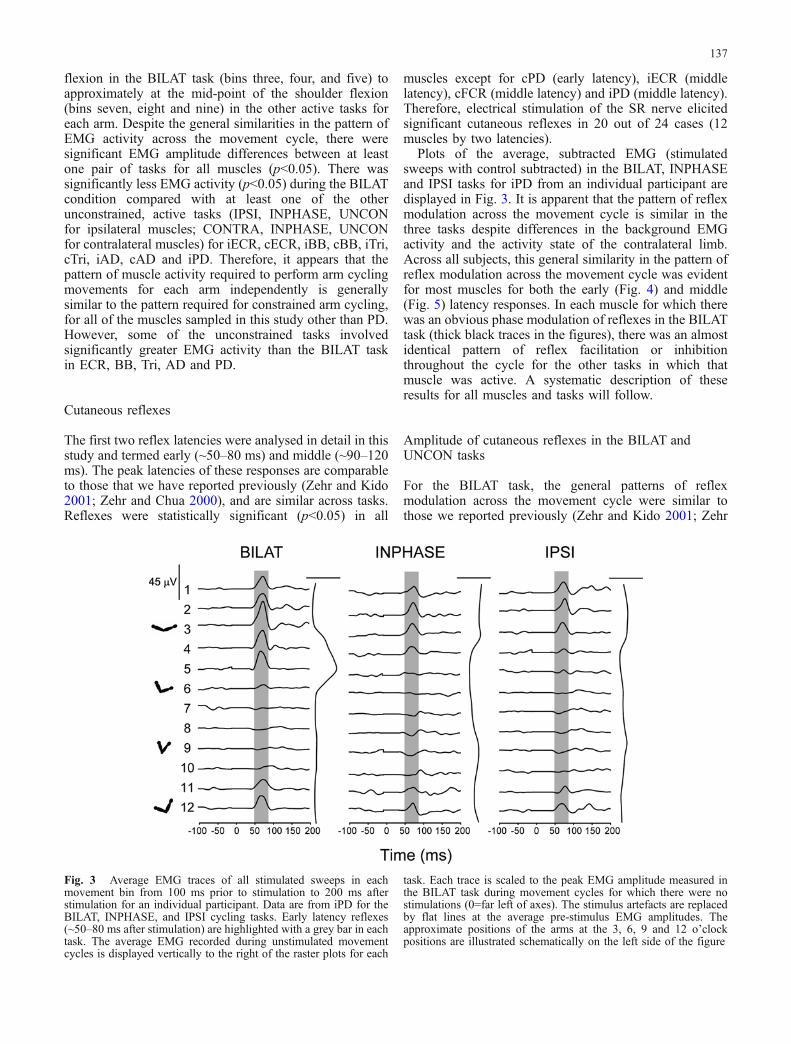
Plots of the average, subtracted EMG (stimulatedsweeps with control subtracted) in the BILAT, INPHASEand IPSI tasks for iPD from an individual participant aredisplayed in Fig. 3. It is apparent that the pattern of reflexmodulation across the movement cycle is similar in thethree tasks despite differences in the background EMGactivity and the activity state of the contralateral limb.Across all subjects, this general similarity in the pattern ofreflex modulation across the movement cycle was evidentfor most muscles for both the early (Fig. 4) and middle(Fig. 5) latency responses. In each muscle for which therewas an obvious phase modulation of reflexes in the BILATtask (thick black traces in the figures), there was an almostidentical pattern of reflex facilitation or inhibitionthroughout the cycle for the other tasks in which thatmuscle was active. A systematic description of theseresults for all muscles and tasks will follow.
Amplitude of cutaneous reflexes in the BILAT andUNCON tasks
For the BILAT task, the general patterns of reflexmodulation across the movement cycle were similar tothose we reported previously (Zehr and Kido 2001; Zehr
Fig. 3 Average EMG traces of all stimulated sweeps in eachmovement bin from 100 ms prior to stimulation to 200 ms afterstimulation for an individual participant. Data are from iPD for theBILAT, INPHASE, and IPSI cycling tasks. Early latency reflexes(~50–80 ms after stimulation) are highlighted with a grey bar in eachtask. The average EMG recorded during unstimulated movementcycles is displayed vertically to the right of the raster plots for each
task. Each trace is scaled to the peak EMG amplitude measured inthe BILAT task during movement cycles for which there were nostimulations (0=far left of axes). The stimulus artefacts are replacedby flat lines at the average pre-stimulus EMG amplitudes. Theapproximate positions of the arms at the 3, 6, 9 and 12 o’clockpositions are illustrated schematically on the left side of the figure
137

and Chua 2000) for the ipsilateral muscles and for cAD(Figs. 4 and 5). Reflexes in cPD were either small (middlelatency) or non-significant (early latency), and showedlittle relationship to the pattern of modulation evident iniPD. For the contralateral BB and Tri muscles, which hadnot previously been studied, cutaneous reflexes weresimilar to those for the homologous ipsilateral muscles atthe same phase of the movement cycle. Cutaneous reflexesfor cFCR and cECR were small and showed no significantmodulation across the movement cycle. Therefore, therewas no consistent cutaneous reflex modulation in three ofthe six muscles contralateral to the stimulus duringconstrained, bilateral arm cycling. For the other threemuscles contralateral to the stimulus, the amplitudes ofcutaneous reflexes at each point in the movement cycle
were strikingly similar to those of the ipsilateral muscles(Figs. 4 and 5).
The amplitudes of cutaneous reflexes were similarbetween the BILAT and UNCON tasks at equivalentpoints in the movement cycle. There were significantdifferences in reflex amplitude (p<0.05) between the twotasks in only 5 of 20 cases (the 20 significant cases fromthe 12 muscles and two latencies). This suggests that anydifferences in motor pattern associated with the indepen-dent control of each arm had a minor effect on themodulation of cutaneous reflexes.
Fig. 4 Mean amplitudes ofearly latency cutaneous reflexes(~50–80 ms post stimulus) eli-cited in each movement bin forthe six tasks and 12 muscles(abbreviations as given in Figs.1 and 2). Reflex amplitudes areexpressed relative to the peakEMG amplitude measured in theBILAT task during movementcycles for which there were nostimulations. The approximatepositions of each arm at eachmovement bin are illustratedschematically beneath the hor-izontal axis labels. Asterisksdenote muscles in which signif-icant reflexes were observed
138
Amplitude of cutaneous reflexes in the unilateralcycling tasks
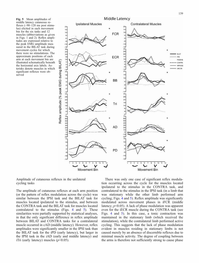
The amplitude of cutaneous reflexes at each arm position(or the pattern of reflex modulation across the cycle) wassimilar between the IPSI task and the BILAT task formuscles located ipsilateral to the stimulus, and betweenthe CONTRA task and the BILAT task for muscles locatedcontralateral to the stimulus (Figs. 4 and 5). Thesesimilarities were partially supported by statistical analyses,in that the only significant difference in reflex amplitudebetween BILAT and CONTRA tasks for a contralateralmuscle occurred in cAD (middle latency). However, reflexamplitudes were significantly smaller in the IPSI task thanthe BILAT task for the iPD (early latency), but larger inthe IPSI task in the iAD (early and middle latency) andiTri (early latency) muscles (p<0.05).
There was only one case of significant reflex modula-tion occurring across the cycle for the muscles locatedipsilateral to the stimulus in the CONTRA task, andcontralateral to the stimulus in the IPSI task (in a limb thatwas stationary while the other limb performed armcycling; Figs. 4 and 5). Reflex amplitude was significantlymodulated across movement phases in iFCR (middlelatency; p<0.05). A lack of phase modulation was apparenteven for the iECR muscle during the CONTRA task (seeFigs. 4 and 5). In this case, a tonic contraction wasmaintained in the stationary limb (which received thestimulation), while the contralateral limb performed activecycling. This suggests that the lack of phase modulationevident in muscles residing in stationary limbs is notcaused merely by an absence of discernible reflexes due tominimal muscle activity. The degree of coupling betweenthe arms is therefore not sufficiently strong to cause phase
Fig. 5 Mean amplitudes ofmiddle latency cutaneous re-flexes (~90–120 ms post stimu-lus) elicited in each movementbin for the six tasks and 12muscles (abbreviations as givenin Figs. 1 and 2). Reflex ampli-tudes are expressed relative tothe peak EMG amplitude mea-sured in the BILAT task duringmovement cycles for whichthere were no stimulations. Theapproximate positions of eacharm at each movement bin areillustrated schematically beneaththe horizontal axis labels. As-terisks denote muscles in whichsignificant reflexes were ob-served
139

modulation of cutaneous reflexes in a stationary limb thatis contralateral to a moving arm, irrespective of whetherthis arm is on the same or the opposite side of the body tothe stimulation.
Amplitude of cutaneous reflexes in the PASSIVEcycling task
Cutaneous reflexes during the PASSIVE condition weresmall and showed little phase modulation for both theearly and middle latency responses (Fig. 4 and 5). Therewas significant modulation of reflex amplitude (p<0.05)across the cycle in only 2 of 20 cases (early and middlelatency reflexes for iAD). In summary, for the inactivelimbs in these three tasks, there was significant reflexmodulation across the cycle in only 3 of 40 cases (8%; 2tasks by 20 significant reflex latencies).
Amplitude of cutaneous reflexes in the INPHASEcycling task
The amplitudes of cutaneous reflexes at each arm positionduring the INPHASE task were similar to those during theBILAT task for both limbs, although reflexes weresignificantly larger in the BILAT task for iPD (earlylatency) and iECR (early latency) and significantly smallerin the BILAT task for iAD (early and middle latency;p<0.05). Importantly, there were no significant differences
in reflex amplitude between these tasks in any of themuscles located contralateral to the stimulation. In thiscase, the position of the limb opposite to the stimulus wasidentical, but the limb that was stimulated was 180° out ofphase between the two conditions.
Pattern of reflex modulation across the movementcycle
The general similarity in the pattern of cutaneous reflexregulation among all of the tasks that involved active armcycling, that has been illustrated across a range of musclesin the upper limbs, can be further appreciated byconsidering the results for individual muscles in detail.For example, in iAD, there were small excitatory earlylatency reflexes in bins one to five, and larger inhibitoryreflexes in bins six to ten for the BILAT, IPSI, INPHASE,and UNCON tasks (Fig. 4). Therefore, the pattern ofcutaneous reflex modulation across the movement cyclewas identical in all of these tasks, even though the limbcontralateral to the stimulation was either stationary,constrained to move out of phase with the ipsilaterallimb, or moving in or out of phase with the ipsilateral limbby volitional control. In contrast, there was much lessmodulation of the early reflexes for the PASSIVE andCONTRA tasks (thin and dashed lines in Fig. 4), althoughthere was reflex modulation that achieved statisticalsignificance for the PASSIVE task (p<0.05). In thesetasks, the limb in which iAD resides was either stationary
Table 1 Pearson correlationcoefficients (r) for regressionsbetween reflex amplitude andbackground EMG
Statistically significant (p<0.05)correlations are indicated by *.Abbreviations: BILAT (armsconstrained to move 180° out ofphase), IPSI (only the stimulatedarm moved), CONTRA (onlythe arm contralateral to thestimulation moved), PASSIVE(only the stimulated arm movedpassively), INPHASE (un-coupled movement with armsmoved together with identicalorientation), UNCON (un-coupled movement with arms180° out of phase). “i” and “c”denote ipsilateral and contralat-eral respectively. FCR=flexorcarpi radialis, ECR=extensorcarpi radialis, Tri=triceps bra-chii, BB=biceps brachii,AD=anterior deltoid, PD=pos-terior deltoid
Muscles r values for cycling task
BILAT UNCON INPHASE IPSI PASSIVE CONTRA
Early latency iFCR 0.30 0.17 0.13 0.21 0.06 0.06iECR −0.02 0.01 −0.19 0.03 −0.27 −0.11iBB −0.76* −0.29 −0.16 −0.28 0.28 −0.52iTri −0.48 −0.60 −0.73* −0.54 0.23 0.69*iAD −0.58 −0.47 −0.65 −0.72* −0.42 −0.46iPD 0.46 0.52 0.03 −0.16 0.35 −0.37cFCR −0.33 0.03 −0.24 0.03 0.35 0.06cECR −0.10 0.00 −0.04 −0.11 0.03 −0.26cBB −0.63 −0.69* −0.67* −0.46 −0.40 −0.48cTri −0.82* −0.82* −0.87* −0.58 −0.21 −0.91*cAD −0.83* −0.38 −0.53 −0.63 0.13 −0.56cPD −0.01 0.09 −0.44 0.65 0.03 −0.49
Middle latency iFCR −0.47 0.39 0.20 −0.31 −0.24 −0.81*iECR 0.13 0.11 0.00 0.20 0.05 0.25iBB 0.40 0.29 0.13 0.01 0.27 0.76*iTri −0.22 0.58 0.02 0.01 0.03 −0.64iAD 0.67* 0.52 0.57 0.60 0.46 0.80iPD −0.24 −0.42 0.28 0.22 0.15 0.60*cFCR −0.14 0.11 0.14 −0.32 0.11 −0.10cECR 0.04 0.04 −0.09 0.12 0.09 0.20cBB 0.65 0.65 0.60 0.11 0.11 0.60cTri 0.83* 0.81* 0.80* 0.66 0.10 0.87*cAD 0.85* 0.61 0.63 0.52 0.11 0.60cPD −0.06 0.44 0.38 −0.13 −0.13 0.77*
140

or being passively moved by the experimenter. A similartrend was also observed in some muscles contralateral tothe stimulated arm. For example, in cTri, there was littleearly latency reflex activation in bins one to eight, and alarge reflex inhibition of the ongoing EMG in bins 8 to 12for the BILAT, UNCON, INPHASE, and CONTRA tasks(Fig. 4). Once again, the pattern was identical in theseconditions regardless of the functional state of the limbipsilateral to the stimulation. There was no significantreflex modulation across the cycle for the IPSI andPASSIVE conditions, in which cases the arm in which cTriresides was stationary.
Relationship between reflex amplitude andbackground muscle activity
Before interpretations regarding the mechanisms of cuta-neous reflex modulation could be made, it was importantto establish that the modulation of reflex amplitudesobserved across the movement cycle was not simply dueto a scaling of reflex size to the background EMG.Qualitative comparison between background EMG ampli-tudes (Fig. 2) and reflex amplitudes (Figs. 4 and 5) revealsimilarities in the general pattern of phase modulation inBB, Tri and AD. These similarities were reflected insignificant linear relationships between background EMGamplitude and cutaneous reflex amplitude for some of thetasks in which these muscles were actively involved incycling (Table 1). Overall, this analysis revealed that only15% (21/144) of correlations showed significant relation-ships between EMG level and reflex amplitude. Further-more, for the FCR, ECR and PD muscles, there was onlyone case of a significant linear relationship betweenbackground EMG and reflex amplitude when a muscleactively contributed to arm cycling (cPD in the CONTRAtask). Therefore, in these three muscles, there wasmodulation of cutaneous reflex magnitude that was notstrongly related to the ongoing muscle activity, whichprovides evidence of pre-motoneuronal gating. There werealso examples of changes in the sign of reflexes fromexcitatory to inhibitory throughout the cycle for iAD (earlylatency response; Fig. 4), and cAD (middle latencyresponse; Fig. 5).
Discussion
In this study we first compared reflex modulation duringbilateral arm cycling with the limbs constrained to move180° out of phase, which we have previously described(Zehr and Kido 2001), with reflex modulation during armcycling when the arms were free to move independently,but the participants intended to keep a 180° phaserelationship between the limbs. This “unconstrained”cycling task was necessary to ensure that any differencesin reflex modulation between the constrained task and theother unconstrained tasks were not due to differences inthe motor pattern required to independently rotate each
limb through the entire range of motion (withoutassistance from the contralateral limb; see Ting et al.1998). We then compared cutaneous reflexes during thesetwo bilateral, out of phase tasks with reflexes during armcycling involving only the limb ipsilateral to the stimu-lation, only the limb contralateral to the stimulation, andduring passive cycling of the ipsilateral limb. Finally, wemeasured reflex responses in both arms during bilateralarm cycling when the limbs were in phase. If the pattern ofreflex responses in the arm contralateral to the stimulationwas phase-locked to the position of the stimulated limb inboth the in-phase and out-of-phase tasks, it would suggestthat the regulation of cutaneous reflexes depends primarilyon the functional state (afferent and/or efferent activity) ofthe stimulated limb. Conversely, if cutaneous reflexes inthe limb contralateral to the stimulation were modulatedaccording to the position of that arm in both tasks, it wouldindicate that the sign and amplitude of cutaneous reflexesis determined according to the functional state of the limbin which a reflex is expressed, regardless of the site ofstimulation. The most striking feature of our results is thatthe pattern of cutaneous reflex modulation in each armduring rhythmic arm cycling is relatively independent ofthe activity of the contralateral limb. That is, the modu-lation of cutaneous reflexes depends mainly on themovement context of the limb in which the reflex isexpressed. This was the case for reflexes in muscles bothipsilateral and contralateral to the stimulation, andoccurred despite differences between tasks in the rate ofcycling and background EMG activity. Furthermore, whenreflexes were recorded from a muscle in a limb that waseither stationary, or being moved passively, there wassignificant phase modulation of cutaneous reflexes in only3 out of 40 cases (8%).
Effect of task constraints on cutaneous reflexmodulation during arm cycling
An important aspect of the present study was theexamination of reflex modulation in both limbs whilethe opposite limb was in various functional states. Therewere no consistent differences in the amplitudes ofcutaneous reflexes between the four active cycling tasksinvolving each arm, despite fundamental differences inposition, movement and muscle activity of the oppositelimb in each of these tasks. Therefore, for most muscles,the amplitude of cutaneous reflexes was not dependent onthe functional state of the opposite limb, or whether thephase relationship between the limbs was mechanicallyconstrained or not. The magnitude and sign of cutaneousreflexes elicited in a particular muscle during arm cyclingappears to be determined by the functional state of thelimb in which that muscle resides, irrespective of whetherthe stimulation is ipsilateral or contralateral to the muscle.This suggests that cutaneous reflexes elicited in aparticular muscle are mostly influenced by neural circuitsthat are directly involved in controlling the limb in whichthat muscle resides (e.g. the CPG for that arm), rather than
141
determined by circuits specifically involved in controllingthe limb in which the afferent volley to electricalstimulation arises.
Changes in the level of EMG activity across themovement cycle are likely involved in the expression ofphase-dependent modulation during arm cycling, sincethere was an association between EMG amplitude andreflex amplitude in some muscles in this study andpreviously (Zehr and Chua 2000; Zehr and Kido 2001).The magnitude of cutaneous reflexes is also related to theEMG activity in upper limb muscles under staticconditions, but not while walking (Zehr and Haridas2003). Changes in background EMG probably make smallcontributions to the reflex effects in the present study,however, because we observed reflex reversals or changesin reflex amplitude that were independent of the ongoingEMG in four of the six muscles studied. Overall, only 15%of reflexes were significantly related to background EMGactivity. Importantly, the similarities between tasks in thepattern of reflex modulation across the cycle were evidenteven in muscles in which there was no strong relationshipbetween background EMG and cutaneous reflex size (iPDfor example). This suggests that the circuits that determinecutaneous reflex sign and amplitude for each limb are notsimply related to the activation of the motoneuron pool.
Comparisons between ipsilateral and contralateralcutaneous reflexes
We have previously described responses evoked in armmuscles by stimulation of cutaneous nerves in thecontralateral arm (Zehr and Haridas 2003; Zehr andKido 2001) and leg (Harridas and Zehr 2003). Similarly,Van Wezel et al. (1997) and Tax et al. (1995) studiedreflexes in leg muscles contralateral to electrical stimula-tion of cutaneous nerves during walking and runningrespectively. In these studies, significant phase-dependentreflex modulation was evident in most of the contralateralmuscles sampled, although the contralateral responseswere often smaller than the ipsilateral reflexes. In ourpresent study, there was significant phase modulation inhalf of the muscles contralateral to the stimulus. Theamplitude and sign of the responses at equivalent positionsin the movement cycle were similar for the two arms forthese muscles. However, during walking, completelydifferent patterns of cutaneous reflexes were expressedin homologous, contralateral, arm muscles at equivalentpoints in the arm-swing cycle (Harridas and Zehr 2003;Zehr and Harridas 2003). These differences in the waysthat reflexes are modulated for each arm between walkingand arm cycling may be due to the involvement of the legsduring walking. Recent data showing the effects of armcycling on soleus H-reflexes (Frigon et al. 2004), as wellas modulation in arm muscles of cutaneous reflexesevoked by stimulation at the ankle (Haridas and Zehr2003) suggest that there are important functional linksbetween the neural circuits underlying reflex organisationfor the legs and the arms.
Passive arm cycling and cutaneous reflexes
We found that passive movement of the arm did not causephase modulation of cutaneous reflexes in the limbcontralateral to the stimulation, or in four of the sixmuscles ipsilateral to the stimulation. A lack of phasemodulation with passive arm cycling is consistent with thedata of Brooke et al. (1999), who found that passivecycling movements of the leg did not cause phasemodulation of cutaneous reflexes. It appears that cutane-ous reflexes, elicited during rhythmic, cyclical movementof either the arms or the legs, are not directly modulated byfeedback from peripheral afferents. Rather, our datasuggest that the central processes associated with activeproduction of rhythmic movement (CPGs for instance) arerequired to modulate cutaneous reflexes in both arms andlegs. However, previous work indicates that reflex orga-nization differs in other respects between the arms andlegs. Active (McIlroy et al. 1992) and passive (Cheng etal. 1998; Collins et al. 1993; Misiaszek et al. 1998) cyclingof the contralateral leg have both been shown todramatically inhibit the soleus H-reflex in the stationaryleg (for a review, see Brooke et al. 1997). In contrast, wefound that active, but not passive, contralateral armcycling inhibited ipsilateral forearm H-reflexes in thestationary arm (Zehr et al 2003). Our current dataregarding the regulation of cutaneous reflexes in theupper limb are also indicative of a relatively weakcoupling between the arms. Therefore, it appears that thestrength of coupling in the neural control of rhythmiccyclical movements is weaker between the arms thanbetween the legs. We have speculated that this reduction incoupling strength may be associated with the morefrequent independent usage of the arms (Zehr et al 2003).
It is important to discuss the extent to which movementsin the PASSIVE condition were truly “passive”, sincethere was significant phase modulation of backgroundEMG in five of the six muscles in the moving limb for thePASSIVE task (all except iTri). Although the origin of thisEMG is unclear, we are confident that it was not generatedby volitional control, because the participants wererepeatedly reminded to relax their arms during the task,and verbally confirmed that their arms were relaxed. It ispossible that the involuntary EMG was brought about byreflex or CPG mechanisms. Nevertheless, the amplitude ofbackground EMG was significantly greater in all of thetasks involving active arm cycling than in the PASSIVEtask for all muscles. Importantly, there was little phasemodulation of cutaneous reflexes in the PASSIVE task,even in most cases in which there was some involuntaryEMG. It therefore seems clear that the afferent informationalone that is derived from passive, rhythmic movements isinsufficient to cause modulation of cutaneous reflexes.
Modulation of early and middle latency responses
The general nature and pattern of reflex modulationdiscussed above was quite similar whether the early or
142

middle latency reflexes were examined. Furthermore, bothearly and middle latency reflexes were usually observed inall subjects. The sign of the response is typically invertedwhen contrasting one latency with another (for instanceearly latency inhibition followed by middle latencyfacilitation). This has been shown before in other workon arm cycling and cutaneous reflexes (Haridas and Zehr2003; Zehr and Chua 2000; Zehr and Kido 2001; Zehr andHaridas 2003). This contrasts with data from the legmuscles during walking in which early latency reflexes arenot ubiquitous (for example, see Haridas and Zehr 2003;Duysens et al. 1990; Van de Crommert et al. 2003; VanWezel et al. 1997) and are not as “stable” as those in armmuscles. Interestingly, the observation that both early andmiddle latency responses are typically evident and phasemodulated in the arm parallels data from the cat hindlimb(Abraham et al. 1985; Duysens and Loeb 1980). Theexplanation and implication for these observations duringrhythmic arm movement is presently unclear.
Neural mechanisms controlling rhythmic humanmovement
Tax et al. (1995) suggested that the pattern of cutaneousreflex modulation in leg muscles during running isdetermined by the functional state of the limb in which amuscle resides, although they were unable to exclude thepossibility that a reciprocal pattern reflex organisationexists for opposite limbs that depends on the position andactivation of the limb that receives the stimulation.Because we tested the regulation of cutaneous reflexesin tasks involving a number of functional relationshipsbetween the two limbs, we can confirm that, at least withinthe context of arm cycling, that reflex amplitude duringrhythmic movement is largely determined according to theposition and activity of the arm in which the responseoccurs. The data here support the concept that there areseparate CPGs for each arm that can be flexibly coupleddepending upon the functional relationship between thearms. This form of neural control seems widespread andcrosses boundaries separating many species. For example,bilateral coordination of the feline hindlimbs has been wellestablished. During split-belt treadmill walking in the lowspinal cat it was clearly shown that the two hindlimbscould maintain coordinated bilateral rhythms even whenone limb was driven at four times the rate of the other limb(Forssberg et al. 1980). This suggests that the feline spinalcord contains the necessary circuitry for coordination bothwithin a hindlimb and between the hindlimbs (Grillner andDubuc 1988). Stride cycle reorganization and bilateraltiming compensation has also been shown between thelegs in infant (Thelen et al. 1987) and adult (Dietz et al.1994; Dietz et al. 1989; Dietz et al. 1995) humans walkingon split-belt treadmills.
Although our data are consistent with the proposal fromTax et al. (1995) that the circuits responsible for cutaneousreflex modulation during rhythmic movement are locatedon the same side of the spinal cord as the responding
muscles, the available data are insufficient to allowidentification of the circuits responsible for this reflexmodulation. It is clear in neonatal mice that some CPGneurons project from one side of the ventro-medial spinalcord to influence the activity of the contralateral motorpools (Butt et al. 2002). Furthermore, in neonatal Xenopustadpoles, interneurons on one side of the spinal cord caninhibit the activity of contralaterally-projecting sensorypathway interneurons (Li et al. 2002). Therefore, despitethe fact that our current data suggest that the modulatingcircuits for cutaneous reflexes are specifically involved incontrolling the responding limb, the locus of cutaneousreflex modulation may be ipsilateral to the cutaneousstimulation. It is also possible that cutaneous reflexmodulation occurs in sensory pathway interneurons (ifthe activity of these neurons can be independentlyinfluenced by the activity state of the limb to which theyproject). Although these possibilities cannot be dis-counted, the recent description of the precise neuronalpathway for a cutaneous flexion reflex in the frog tadpolepresented by Li et al. (2003) indicates that more likelysites of reflex modulation lie ipsilateral to the motor pool.Li et al. (2003) showed that dorso-lateral commissuralinterneurons receive connections from sensory pathwayneurons before crossing the midline and synapsing withboth motoneurons and a range of ascending and descend-ing interneurons. We therefore suggest that the simplestexplanation for our results is that, during rhythmic,cyclical movements, cutaneous reflexes for a particularmuscle are modulated by CPG circuits located on the sameside of the spinal cord as its motor pool.
Acknowledgements We thank Alejandro Ley and NienkeHoogenboom for assistance with data collection, and ZoltanKenwell for technical assistance. The work was supported by grantsfrom the Natural Sciences and Engineering Research Council ofCanada (EPZ), the Heart and Stroke Foundation of Canada (EPZ),and the Alberta Heritage Foundation for Medial Research (AHFMR)(DC). DC is an AHMFR Scholar and TC was supported by a KillamPostdoctoral Fellowship.
References
Abraham LD, Marks WB, Loeb GE (1985) The distal hindlimbmusculature of the cat. Cutaneous reflexes during locomotion.Exp Brain Res 58:594–603
Brooke JD, Cheng J, Collins DF, McIlroy WE, Misiaszek JE,Staines WR (1997) Sensori-sensory afferent conditioning withleg movement: gain control in spinal reflex and ascendingpaths. Prog Neurobiol 51:393–421
Brooke JD, McIlroy WE, Staines WR, Angerilli PA, Peritore GF(1999) Cutaneous reflexes of the human leg during passivemovement. J Physiol–Lond 518:619–628
Burke RE (1999) The use of state-dependent modulation of spinalreflexes as a tool to investigate the organization of spinalinterneurons. Exp Brain Res 128:263–277
Butt SJ, Harris-Warrick RM, Kiehn O (2002) Firing properties ofidentified interneuron populations in the mammalian hindlimbcentral pattern generator. J Neurosci 22:9961–9971
Cheng J, Brooke JD, Misiaszek JE, Staines WR (1998) Crossedinhibition of the soleus H reflex during passive pedallingmovement. Brain Res 779:280–284
143
Collins DF, McIlroy WE, Brooke JD (1993) Contralateral inhibitionof soleus H reflexes with different velocities of passivemovement of the opposite leg. Brain Res 603:96–101
Dietz V, Horstmann GA, Berger W (1989) Interlimb coordination ofleg-muscle activation during perturbation of stance in humans.J Neurophysiol 62:680–693
Dietz V, Zijlstra W, Duysens J (1994) Human neuronal interlimbcoordination during split-belt locomotion. Exp Brain Res101:513–520
Dietz V, Zijlstra W, Prokop T, Berger W (1995) Leg muscleactivation during gait in Parkinson’s disease: adaptation andinterlimb coordination. Electroen Clin Neuro 97:408–415
Duysens J, Loeb GE (1980) Modulation of ipsi- and contralateralreflex responses in unrestrained walking cats. J Neurophysiol44:1024–1037
Duysens J, Trippel M, Horstmann GA, Dietz V (1990) Gating andreversal of reflexes in ankle muscles during human walking.Exp Brain Res 82:351–358
Forssberg H, Grillner S, Halbertsma J, Rossignol S (1980) Thelocomotion of the low spinal cat. II. Interlimb coordination.Acta Physiol Scand 108:283–295
Frigon A, Collins DF, Zehr EP (2004) Effect of rhythmic armmovement on reflexes in the legs: modulation of soleus H-reflexes and somatosensory conditioning. J Neurophysiol91:1516–1523
Grillner S, Dubuc R (1988) Control of locomotion in vertebrates:Spinal and supraspinal mechanisms. In: Waxman SG (ed)Advances in neurology. Functional recovery in neurologicaldisease. Raven, New York, pp 425–453
Haridas C, Zehr EP (2003) Coordinated interlimb compensatoryresponses to electrical stimulation of cutaneous nerves in thehand and foot during walking. J Neurophysiol 90:2850–2861
Li WC, Soffe SR, Roberts A (2002) Spinal inhibitory neurons thatmodulate cutaneous sensory pathways during locomotion in asimple vertebrate. J Neurosci 22:10924–10934
McIlroy WE, Collins DF, Brooke JD (1992) Movement features andH-reflex modulation. II. Passive rotation, movement velocityand single leg movement. Brain Res 582:85–93
Misiaszek JE, Cheng J, Brooke JD, Staines WR (1998) Movement-induced modulation of soleus H reflexes with altered length ofbiarticular muscles. Brain Res 795:25–36
Tax AA, Van Wezel BM, Dietz V (1995) Bipedal reflex coordinationto tactile stimulation of the sural nerve during human running. JNeurophysiol 73:1947–1964
Ting LH, Raasch CC, Brown DA, Kautz SA, Zajac FE (1998)Sensorimotor state of the contralateral leg affects ipsilateralmuscle coordination of pedaling. J Neurophysiol 80:1341–1351
Thelen E, Ulrich BD, Niles D (1987) Bilateral coordination inhuman infants: stepping on a split-belt treadmill. J Exp PsycholHuman 13:405–410
Van de Crommert HW, Steijvers PJ, Mulder T, Duysens J (2003)Suppressive musculocutaneous reflexes in tibialis anteriorfollowing upper leg stimulation at the end of the swingphase. Exp Brain Res 149:405–412
Van Wezel BM, Ottenhoff FA, Duysens J (1997) Dynamic control oflocation-specific information in tactile cutaneous reflexes fromthe foot during human walking. J Neurosci 17:3804–3814
Zehr EP, Chua R (2000) Modulation of human cutaneous reflexesduring rhythmic cyclical arm movement. Exp Brain Res135:241–250
Zehr EP, Haridas C (2003) Modulation of cutaneous reflexes in armmuscles during walking: further evidence of similar controlmechanisms for rhythmic human arm and leg movements. ExpBrain Res 149:260–266
Zehr EP, Kido A (2001) Neural control of rhythmic, cyclical humanarm movement: task dependency, nerve specificity and phasemodulation of cutaneous reflexes. J Physiol–Lond 537:1033–1045
Zehr EP, Stein RB (1999) What functions do reflexes serve duringhuman locomotion? Prog Neurobiol 58:185–205
Zehr EP, Collins DF, Frigon A, Hoogenboom N (2003) Neuralcontrol of rhythmic human arm movement: phase dependenceand task modulation of Hoffmann reflexes in forearm muscles.J Neurophysiol 89:12–21
144