tips and techniques for enhancing your cell culture · tips and techniques for enhancing your cell...
TRANSCRIPT
Tips and Techniques for Enhancing Your Cell CultureShabana Islam, Ph.D.
BD Biosciences
September 15, 2009
Introduction
• An essential tool in the study of cell biology is the use of in vitro cell culture.
• Variables in cell culture include cell source, isolation techniques, growth conditions such as matrix proteins and soluble factors, and cell age.
• Basic laboratory practices are sometimes overlooked as a source of discrepancy in data; however the application of fastidious and reproducible technique can reduce cell culture as a source of data variation.
• In this presentation we will identify and discuss basic principles of in vitro mammalian cell culture that influence the quality of experimental results.
To Study:• Intracellular activities
– DNA replication– Transcription (RNA)– Translation (protein)– Signal Transduction
• Cell-environment interaction– Drug action, infection, cytotoxicity, receptor/ligand interaction
• Cell-cell interaction• Genomics and proteomicsTo Produce:• Cells, cell products, and cell secreted products
– Protein, DNA, RNA, antibody, transfected protein product
Why Culture Living Cells?
• Cultivation in a more physiological environment== increased value of experimental dataincreased value of experimental data
• Cultivation in a controlled environment== defined parameters that can be changed/measureddefined parameters that can be changed/measured
• Cultivation in an optimized environment== increased signal/noise ratioincreased signal/noise ratio
Why Standardize Techniques?
Why Standardize Techniques?
As an example:It is estimated that it takes ~12 to 15 years and ~$800 million - $1.7B to develop a new drug
– Drug Development Process: A Review (Pharmainfo.net)http://www.pharmainfo.net/reviews/drug-development-process-review
Failure of drug candidate is 10% when moving from ADME to Clinical Testing
– Chris Bode, PhD, Vice President, Corporate Development, Absorption Systems, L.P. (January 12, 2009) Going In Vitro. Drug Discovery & Development http://www.dddmag.com/articles-in-vitro-ADME-models.aspx
A 10% improvement in predicting compound failure before clinical trial could save up to 100 million $ in development costs per drug

Experimental Data is Influenced by:1) Cell Source2) Initial Growth Conditions3) Medium Composition4) Cell Counting Procedure5) Physiological Parameters6) Characterization
• Age, Karyotype7) Substrate, Matrix , or Not
• Morphology8) Dissociation Method9) Contamination Control
• Aseptic Technique10) Cryopreservation
BD™ Human Umbilical Vein Endothelial Cells (cat. No. 354151)
Experimental Data
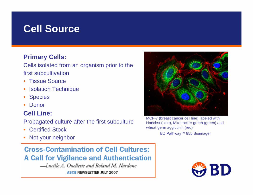
Primary Cells:Cells isolated from an organism prior to thefirst subcultivation• Tissue Source• Isolation Technique• Species• DonorCell Line:Propagated culture after the first subculture• Certified Stock• Not your neighbor
MCF-7 (breast cancer cell line) labeled with Hoechst (blue), Mitotracker green (green) and wheat germ agglutinin (red)
BD Pathway™ 855 Bioimager
Cell Source
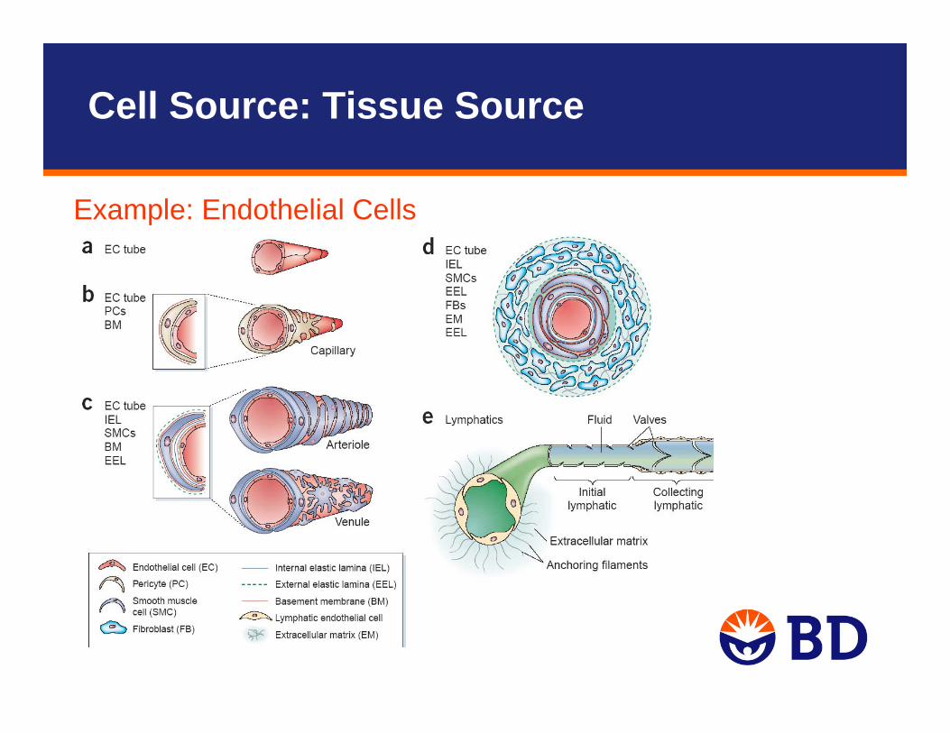
Example: Endothelial Cells
Cell Source: Tissue Source
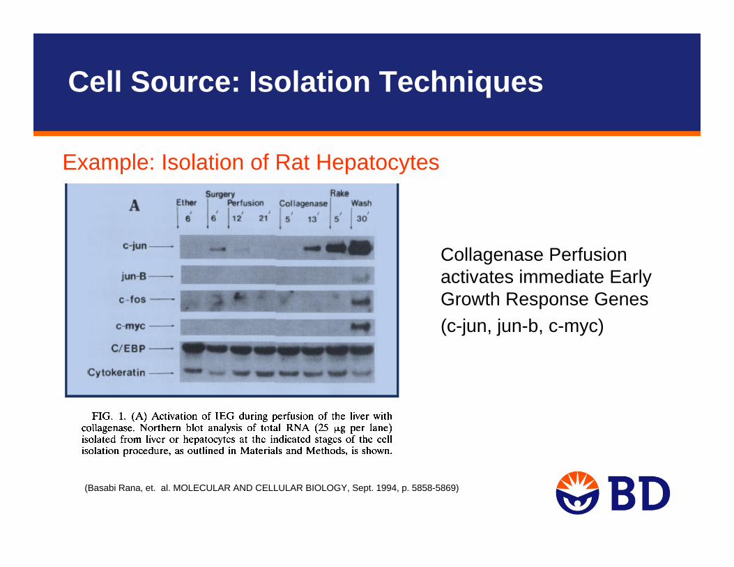
(Basabi Rana, et. al. MOLECULAR AND CELLULAR BIOLOGY, Sept. 1994, p. 5858-5869)
Collagenase Perfusion activates immediate Early Growth Response Genes (c-jun, jun-b, c-myc)
Example: Isolation of Rat Hepatocytes
Cell Source: Isolation Techniques

• Know what you want in experimental data
• Example: In Vitro Testing of Drug Compound Bioavailability
(Taipalensuu, J. et.al. JPET 299:164-170, 2001)
– MDCK Cells, Dog Kidney– MDCK2 Cells, Dog Kidney transfected with MDR-1– Caco2 Cells, Human intestinal epithelial
Cell Source: Species
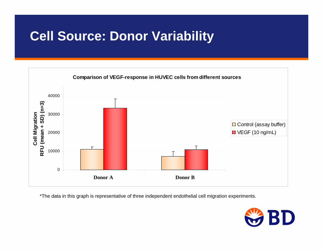
Cell Source: Donor Variability
Comparison of VEGF-response in HUVEC cells from different sources
0
10000
20000
30000
40000
Cel
l Mig
ratio
nR
FU (m
ean
+ SD
) (n=
3)
Control (assay buffer)VEGF (10 ng/mL)
*The data in this graph is representative of three independent endothelial cell migration experiments.
Donor A Donor B
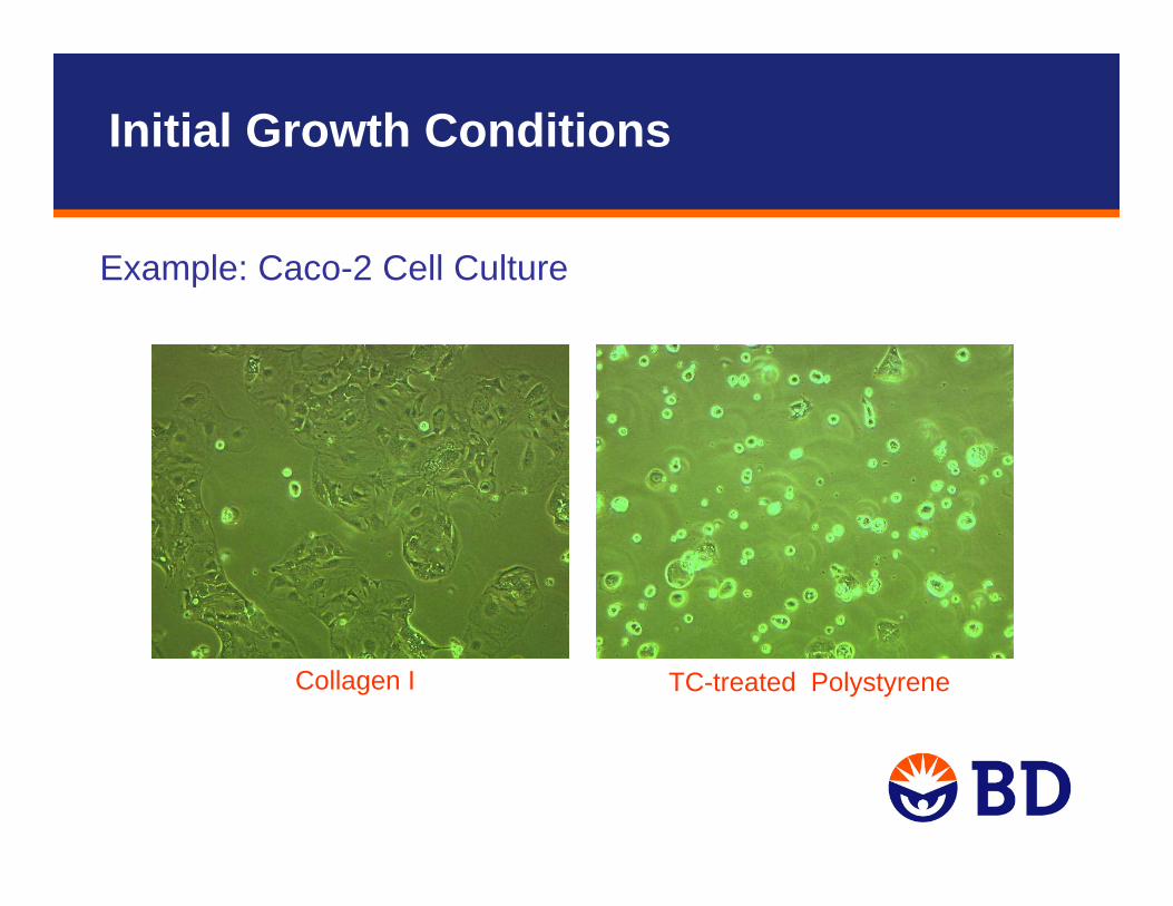
Collagen I TC-treated Polystyrene
Example: Caco-2 Cell Culture
Initial Growth Conditions

D ig o x in P erm eab ility C o m p ariso n
C aco -2 P o re P ap p P ap pL in e S ize A -B B -A R atio
10% F B S 1 uM m ean : 2.9 9 .7 3 .6SD : 0 .8 1 .8
% C V : 27 19(n=4)
20% F B S 1 uM m ean : 1 .3 17 .3 14SD : 0 .24 2 .50
% C V : 19 14(n=8)
Example: Caco-2 Cell Culture
Initial Growth Conditions
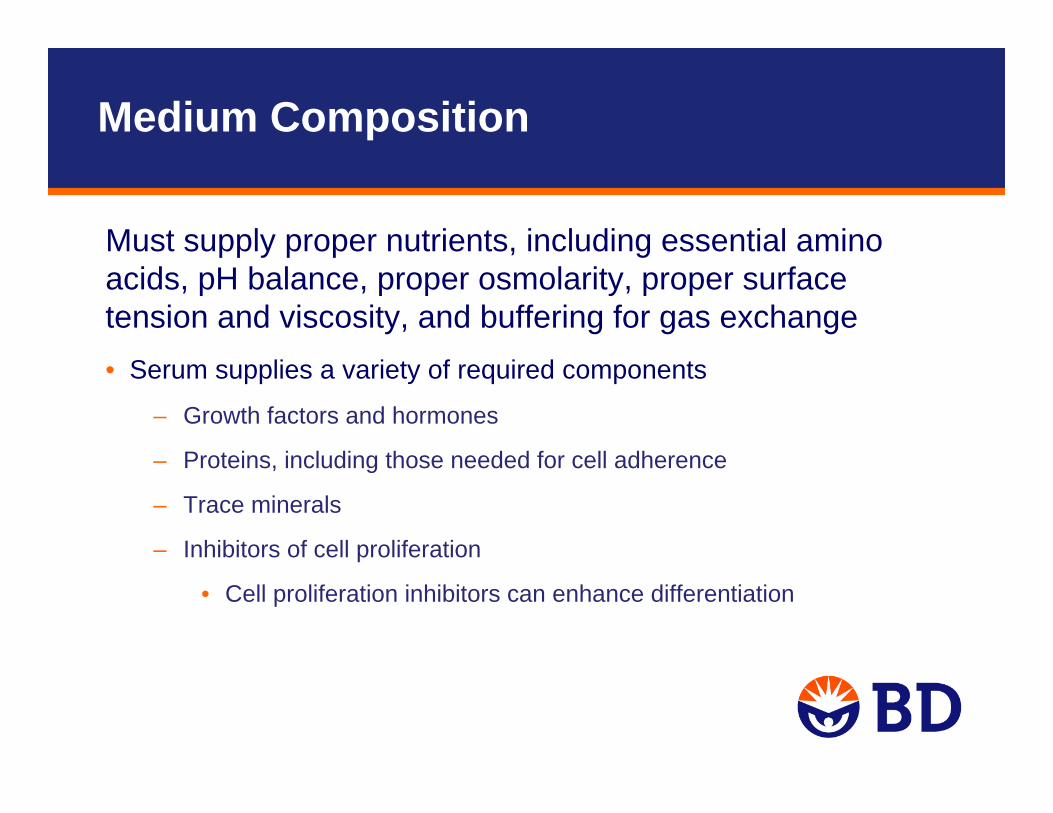
Must supply proper nutrients, including essential amino acids, pH balance, proper osmolarity, proper surface tension and viscosity, and buffering for gas exchange• Serum supplies a variety of required components
– Growth factors and hormones
– Proteins, including those needed for cell adherence
– Trace minerals
– Inhibitors of cell proliferation
• Cell proliferation inhibitors can enhance differentiation
Medium Composition
• Control for water source and reagent preparation• FBS
– Lot to lot (uncontrolled) variation in hormone and growth factor levels
– Test FBS or use serum free• Antibiotics or Not• Feed cells every 2 days, warm media to 37°C• Store media at 4°C, in the dark, observe expiration dates
Media Watch-outs
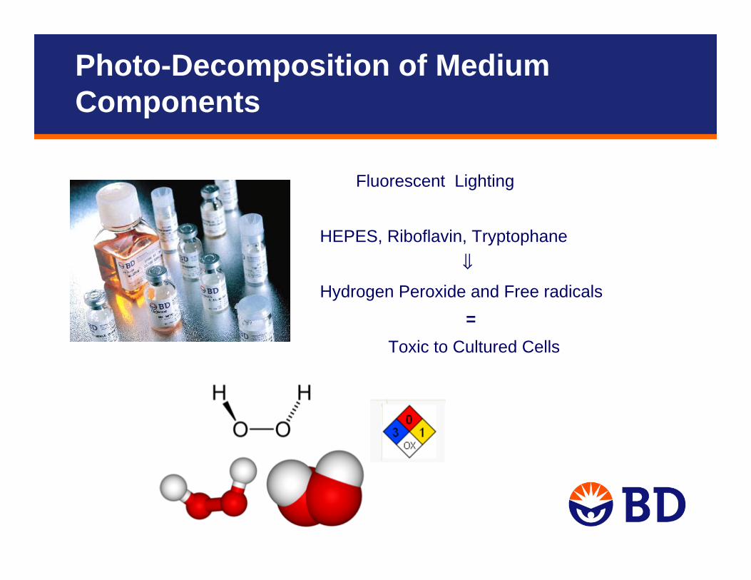
Fluorescent Lighting
HEPES, Riboflavin, Tryptophane⇓
Hydrogen Peroxide and Free radicals=
Toxic to Cultured Cells
Photo-Decomposition of Medium Components
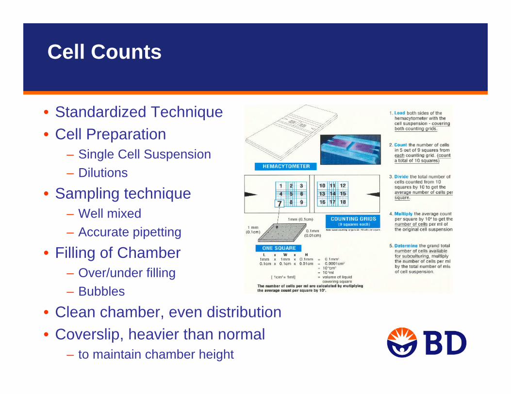
• Standardized Technique• Cell Preparation
– Single Cell Suspension– Dilutions
• Sampling technique– Well mixed– Accurate pipetting
• Filling of Chamber– Over/under filling– Bubbles
• Clean chamber, even distribution• Coverslip, heavier than normal
– to maintain chamber height
Cell Counts
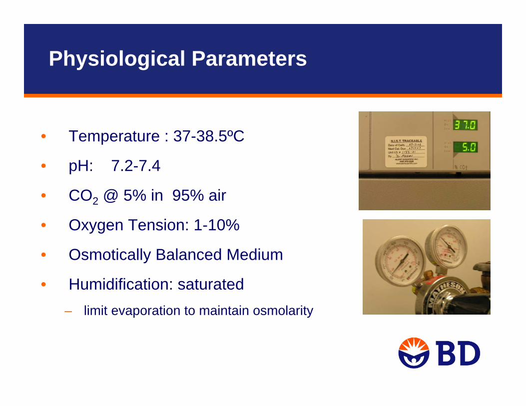
• Temperature : 37-38.5ºC
• pH: 7.2-7.4
• CO2 @ 5% in 95% air
• Oxygen Tension: 1-10%
• Osmotically Balanced Medium
• Humidification: saturated – limit evaporation to maintain osmolarity
Physiological Parameters
• Most Mammalian Cells grow well at pH 7.4.
• Cells produce waste products that tend tobe acidic.
• Thus, generally require a buffered medium.
• Sodium bicarbonate is usually the buffering agent.
• The pH is governed by the reaction
– H2O + CO2 <-> H2CO3 <-> H+ + HCO3-
• Cultures must be incubated in a CO2 environment in equilibrium with the H2CO3 in the medium
Physiological Parameters: pH
A1 A6
A12 B2 B5
B9 C3
C7
D1
D5
D7
D9
D12 E3
E6
E11 F7
F10
G3
G6
G9
H1
H4
H9
H11
H12
1
4
70
75
80
85
90
95
100
95-10090-9585-9080-8575-8070-75
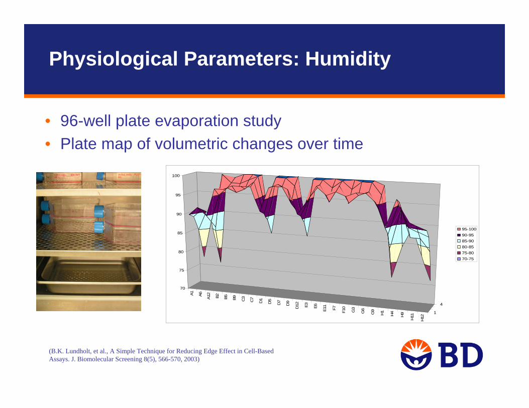
• 96-well plate evaporation study• Plate map of volumetric changes over time
Physiological Parameters: Humidity
(B.K. Lundholt, et al., A Simple Technique for Reducing Edge Effect in Cell-Based Assays. J. Biomolecular Screening 8(5), 566-570, 2003)
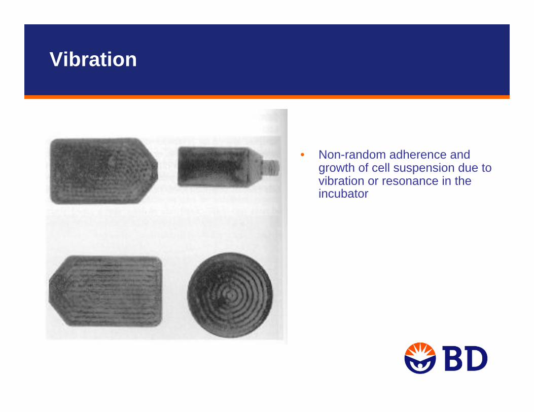
• Non-random adherence and growth of cell suspension due to vibration or resonance in the incubator
Vibration

• Cell Characteristics can vary over time
• Use Standardized Protocols to control and understand the age of your cells
Population Doublings, Generation Number :
1 2 4 8 16 number of cells in vessel at subculture
0 1 2 3 4 …n, number of generations
n = 3.32(logN - logX)
N= final population, and X=initial population
Passage Number: • Number of times a culture has been subcultured or split• Does not reflect population age unless there is a consistent time
between splits and a unvarying split ratio
(Kruse and Patterson, Tissue Culture Methods and Applications, 1973)
Cell Age
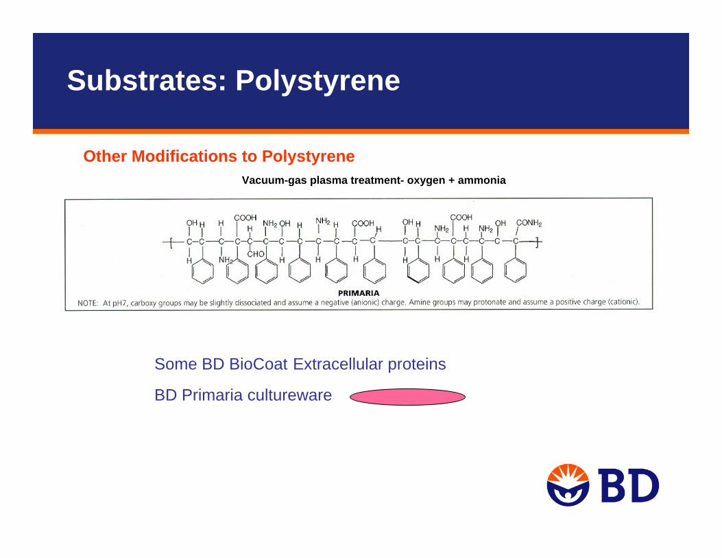
Vertebrate cells have negative surface charges, and can be cultured on either positive or negative charged surfaces
– Glass and Tissue culture treated polystyrene plastic carry a negative charge
– BD Primaria™ cultureware carries both positive and negative charges– Poly-lysine surfaces have positive charges
Extracellular matrix components (2D or 3D)– Cells spread using the ECM they lay down and the serum or media
derived attachment factors– Fibronectin is key attachment factor derived from serum– BD BioCoat™ cultureware
Microporous membranes (cell culture inserts)
Substrates
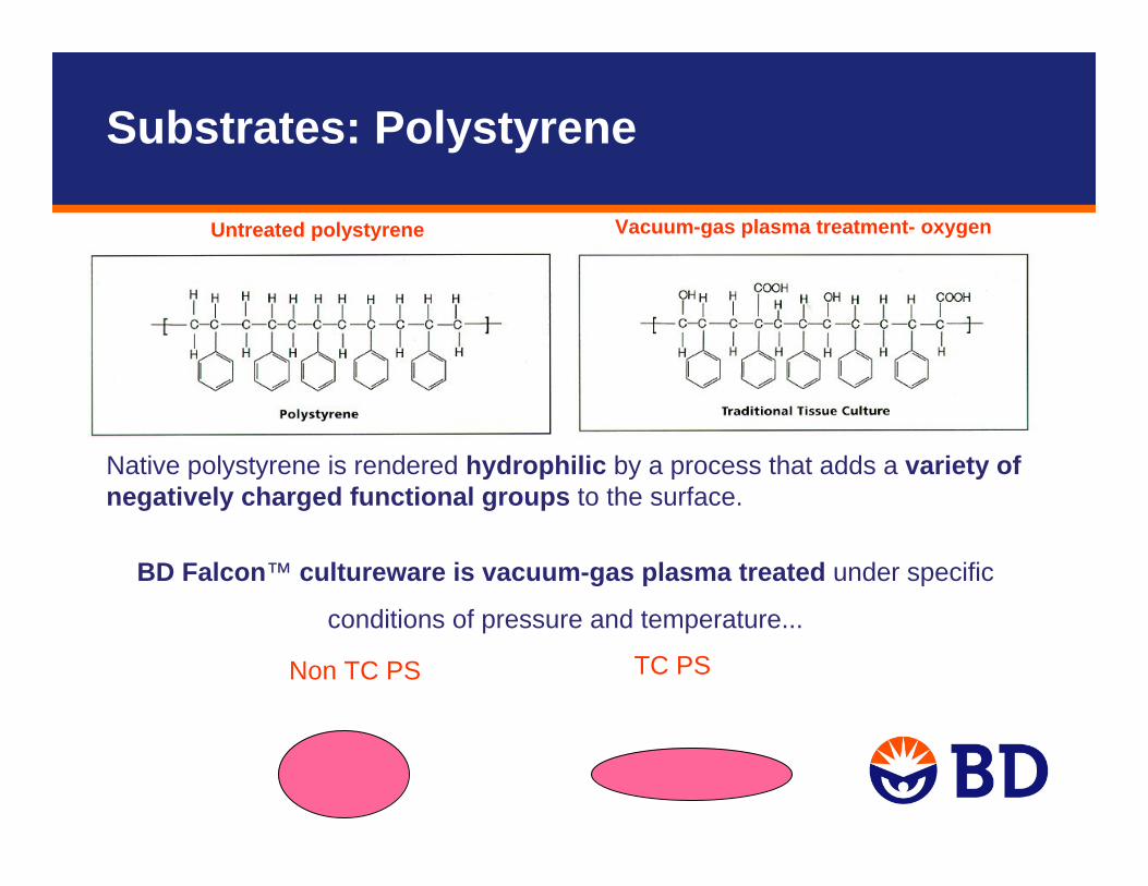
Vacuum-gas plasma treatment- oxygenUntreated polystyrene
Native polystyrene is rendered hydrophilic by a process that adds a variety of negatively charged functional groups to the surface.
BD Falcon™ cultureware is vacuum-gas plasma treated under specific
conditions of pressure and temperature...
Non TC PS TC PS
Substrates: Polystyrene
Substrates: Polystyrene
Other Modifications to PolystyreneVacuum-gas plasma treatment- oxygen + ammonia
Some BD BioCoat Extracellular proteins
BD Primaria cultureware
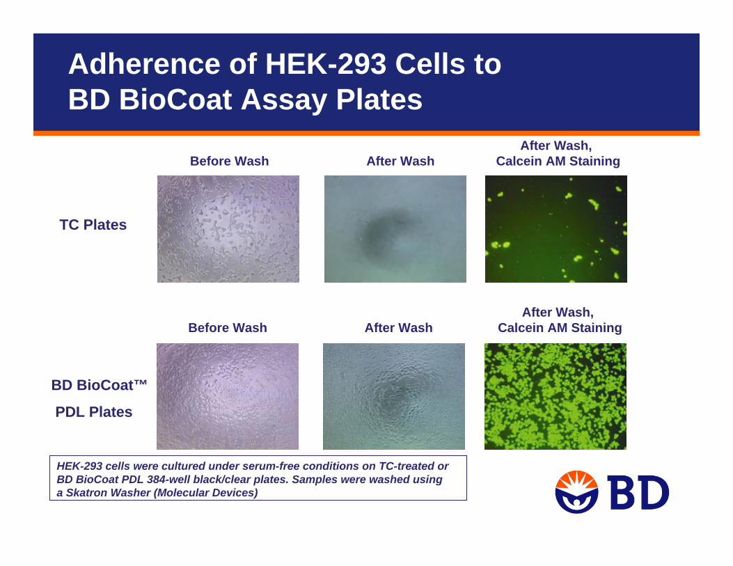
TC Plates
BD BioCoat™
PDL Plates
After Wash, Before Wash After Wash Calcein AM Staining
After Wash, Before Wash After Wash Calcein AM Staining
HEK-293 cells were cultured under serum-free conditions on TC-treated or BD BioCoat PDL 384-well black/clear plates. Samples were washed using a Skatron Washer (Molecular Devices)
Adherence of HEK-293 Cells to BD BioCoat Assay Plates
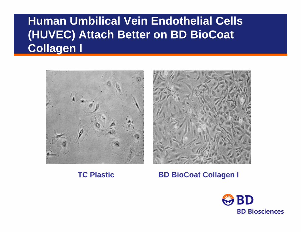
Human Umbilical Vein Endothelial Cells (HUVEC) Attach Better on BD BioCoat Collagen I
TC Plastic BD BioCoat Collagen I
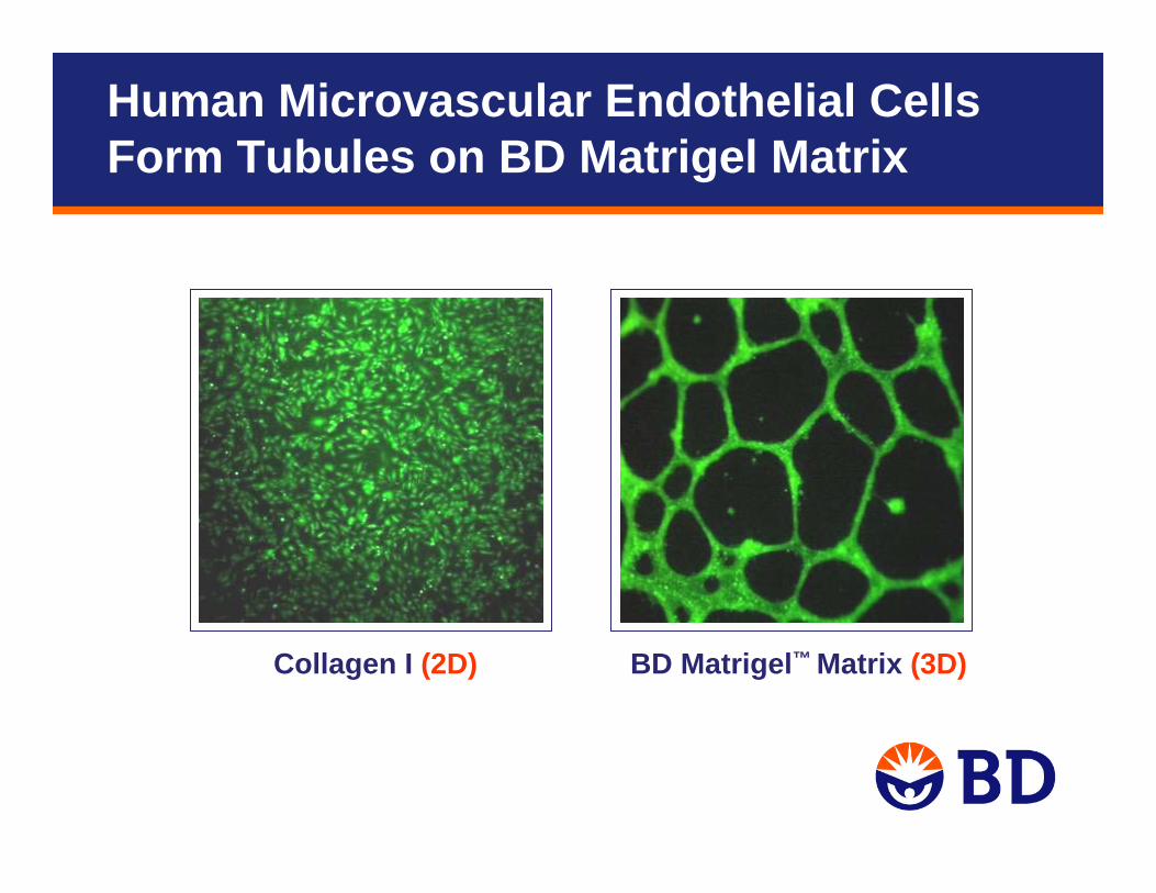
Collagen I (2D) BD Matrigel™ Matrix (3D)
Human Microvascular Endothelial Cells Form Tubules on BD Matrigel Matrix
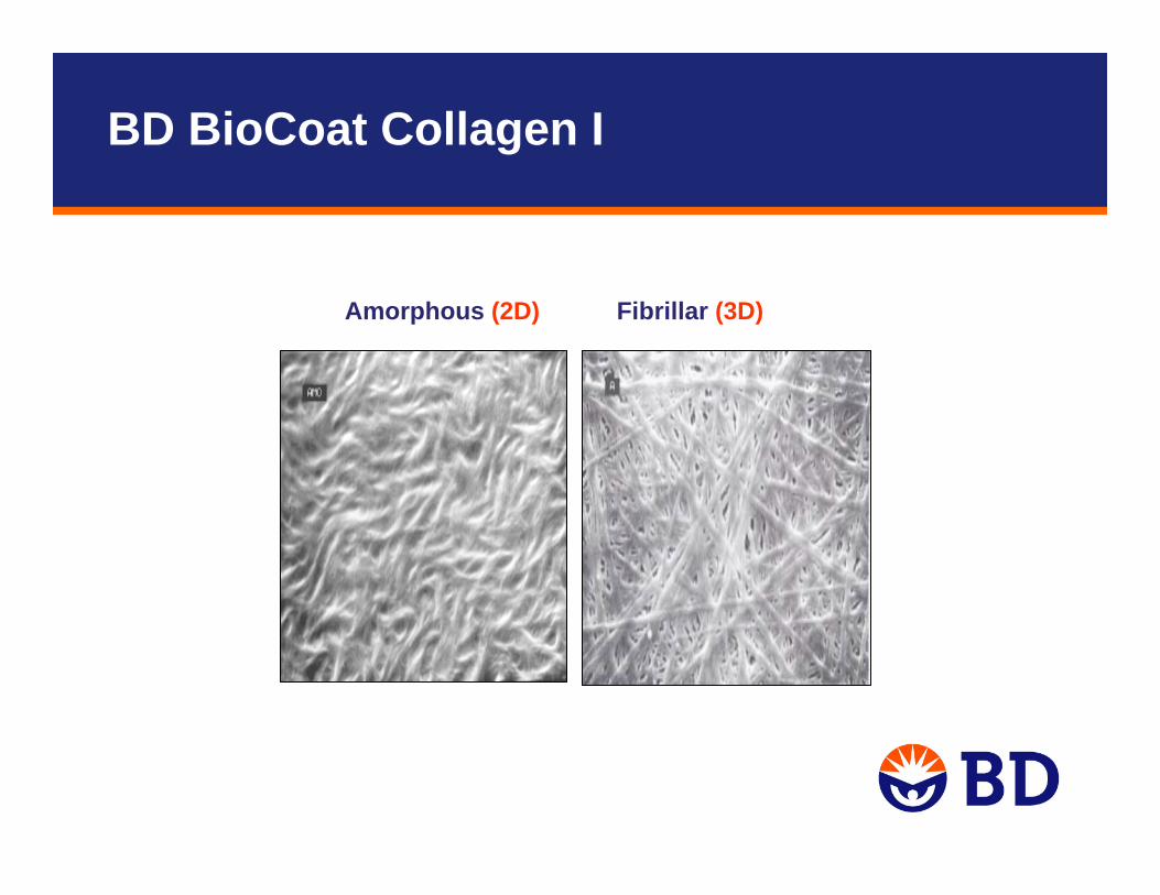
Amorphous (2D) Fibrillar (3D)
BD BioCoat Collagen I
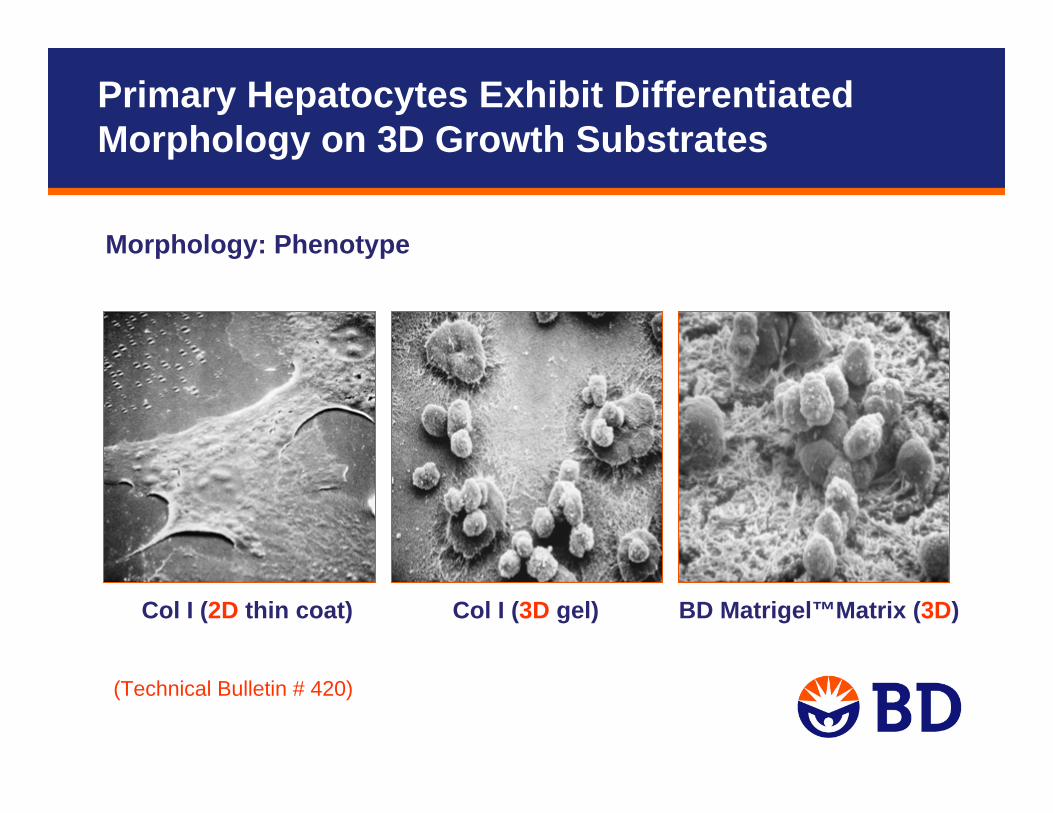
Col I (2D thin coat) Col I (3D gel) BD Matrigel™Matrix (3D)
(Technical Bulletin # 420)
Morphology: Phenotype
Primary Hepatocytes Exhibit Differentiated Morphology on 3D Growth Substrates
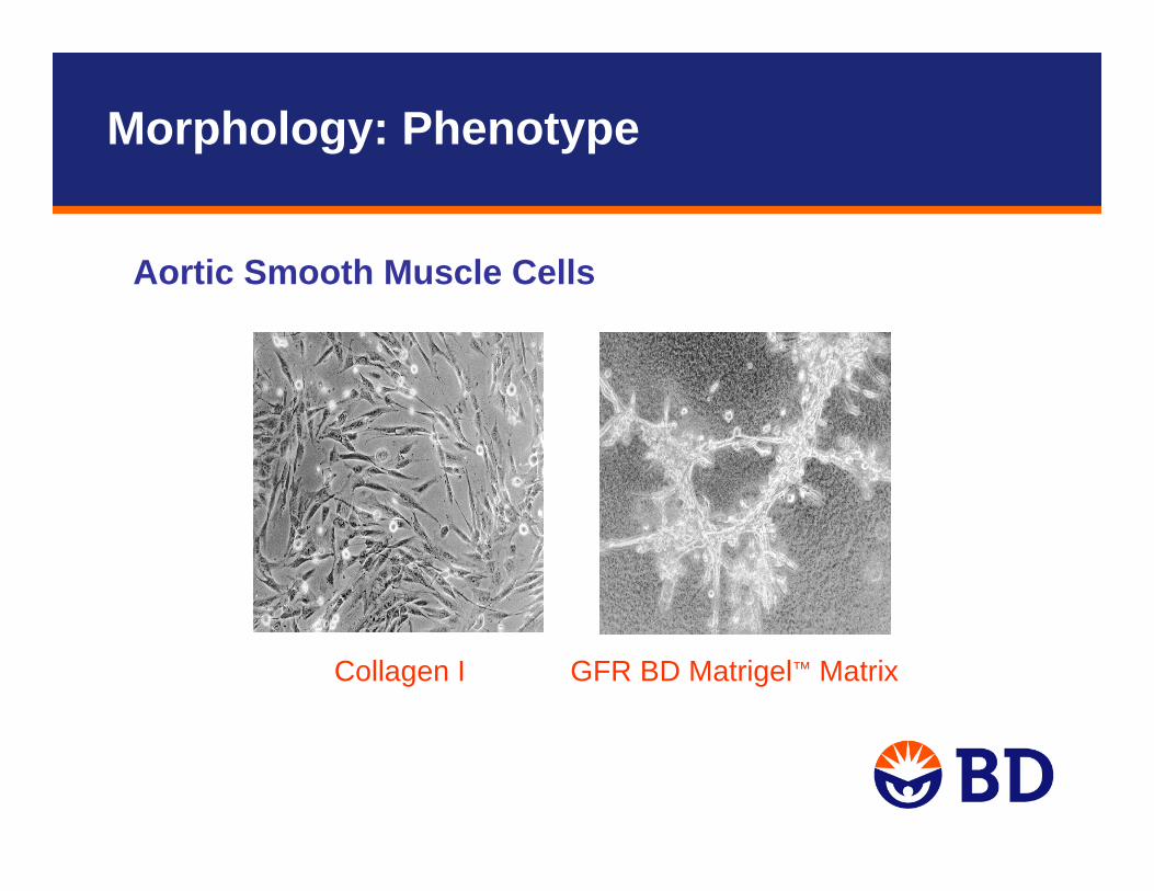
GFR BD Matrigel™ MatrixCollagen I
Aortic Smooth Muscle Cells
Morphology: Phenotype

• Chemically defined, animal-free surfaces
• Manufactured by a proprietary thin-film coating technology
• BD PureCoat™ Carboxyl
– Negatively charged surface
• BD PureCoat Amine
– Positively charged surface
The Next Generation of Advanced Cell Culture Surfaces
What is BD PureCoat Cultureware ?
Webinar scheduled on October 22, 2009
Application Note No. 466
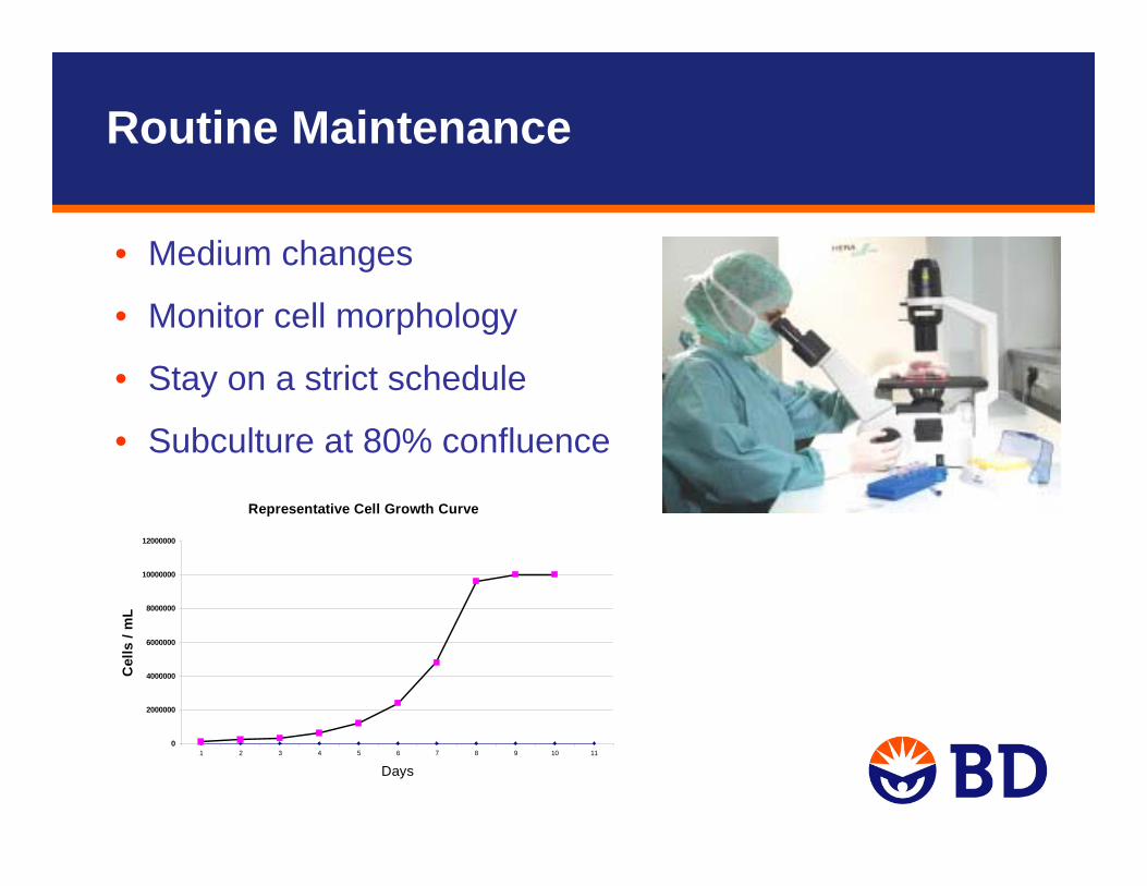
Representative Cell Growth Curve
0
2000000
4000000
6000000
8000000
10000000
12000000
1 2 3 4 5 6 7 8 9 10 11
Days
Cel
ls /
mL
• Medium changes
• Monitor cell morphology
• Stay on a strict schedule
• Subculture at 80% confluence
Routine Maintenance
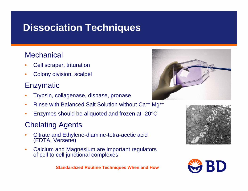
Dissociation Techniques
Mechanical• Cell scraper, trituration• Colony division, scalpel
Enzymatic• Trypsin, collagenase, dispase, pronase• Rinse with Balanced Salt Solution without Ca++ Mg++
• Enzymes should be aliquoted and frozen at -20°C
Chelating Agents • Citrate and Ethylene-diamine-tetra-acetic acid
(EDTA, Versene)• Calcium and Magnesium are important regulators
of cell to cell junctional complexes
Standardized Routine Techniques When and How
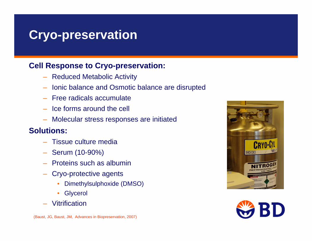
Cryo-preservation
(Baust, JG, Baust, JM, Advances in Biopreservation, 2007)
Cell Response to Cryo-preservation:– Reduced Metabolic Activity– Ionic balance and Osmotic balance are disrupted– Free radicals accumulate– Ice forms around the cell– Molecular stress responses are initiated
Solutions:– Tissue culture media– Serum (10-90%)– Proteins such as albumin– Cryo-protective agents
• Dimethylsulphoxide (DMSO)• Glycerol
– Vitrification
Contamination Sources:– Chemical: endotoxins, plasticizers, disinfectants, fluorescent lighting
– Biological: Bacterial, viral, fungal,mycoplasmal, cellular
Contamination
• Endotoxins: lipopolysaccaride containing by-product of gram-negative bacteria– Water, sera, culture additives
• Plasticizers: in storage containers, tubing
• Reused storage containers
• Disinfectants: deposits left from washing glassware
• Germicides: used to disinfect Incubators
• CO2: Use medical grade
Contamination: Chemical
• Bacteria, Mold, Yeast– Easily detected: pH, turbidity, cell morphology, better to avoid
antibiotics
• Viral, Mycoplasma– Alter host cell function, hard to detect
• Cell Cross-contamination– Technique, don’t borrow cells
Contamination: Biological
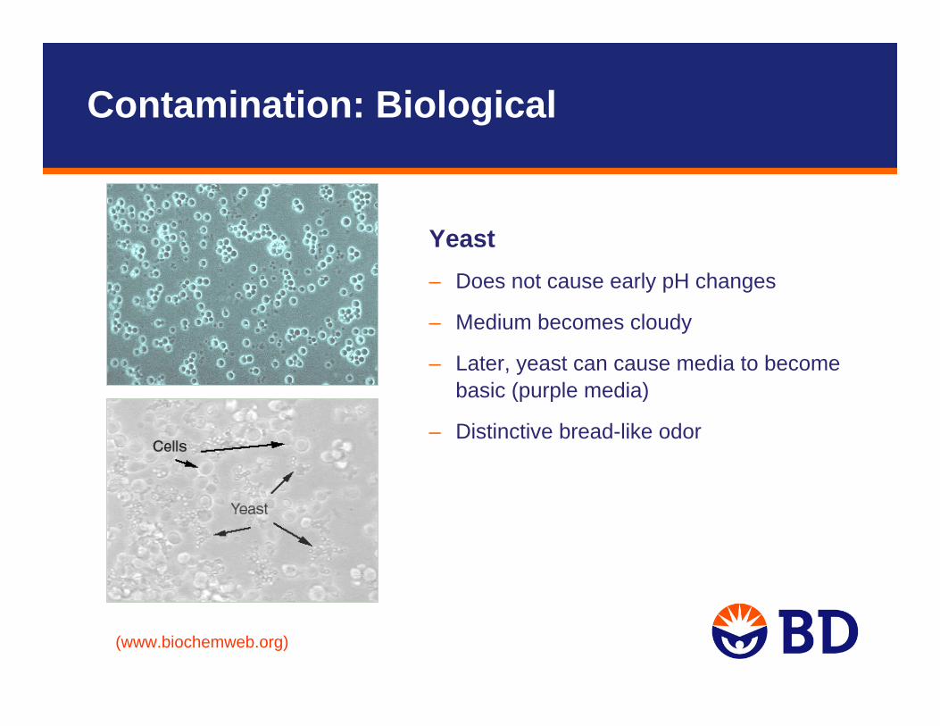
Yeast– Does not cause early pH changes
– Medium becomes cloudy
– Later, yeast can cause media to become basic (purple media)
– Distinctive bread-like odor
(www.biochemweb.org)
Contamination: Biological

Bacterial Causes changes in pH of Medium– Medium becomes acidic, – Cloudy and bright yellow– Often produce toxins that destroy cells
The Microbial World, Jim DeaconInstitute of Cell and Molecular Biology, The University of Edinburgh
Contamination: Biological

MoldDoes not cause immediate pH changes• Often is not cytotoxic• Easy to observe under a low power microscope
and can even be seen without magnification in advanced stages of contamination.
• Tough to catch early• Appears whiteish, yellowish, or black in culture
Contamination: Biological
Mycoplasma Cannot be seen under normal magnification
• No overt effects in culture, only subtle ones• The only way to confirm mycoplasma contamination
is by routine testing
Mycoplasma can cause changes in:• Cell growth characteristics• Cell metabolism• Disruption of nucleic acid synthesis• Chromosomal aberrations• Changes in cell membrane antigenicity• Can alter transfection rates and virus susceptibility.
Contamination: Biological
Control– Certified seed stock
– Aseptic technique
– Periodic testing
– Antibiotic-free media
– Sterile equipment, glassware and media
– Quality reagents
– Use frozen cell stock
Most Dangerous:– Undetected contaminants
Contamination: Control
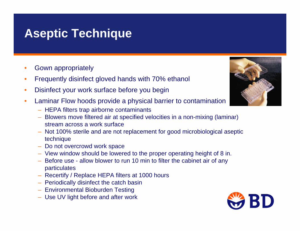
• Gown appropriately• Frequently disinfect gloved hands with 70% ethanol • Disinfect your work surface before you begin• Laminar Flow hoods provide a physical barrier to contamination
– HEPA filters trap airborne contaminants – Blowers move filtered air at specified velocities in a non-mixing (laminar)
stream across a work surface– Not 100% sterile and are not replacement for good microbiological aseptic
technique– Do not overcrowd work space– View window should be lowered to the proper operating height of 8 in.– Before use - allow blower to run 10 min to filter the cabinet air of any
particulates– Recertify / Replace HEPA filters at 1000 hours– Periodically disinfect the catch basin– Environmental Bioburden Testing– Use UV light before and after work
Aseptic Technique
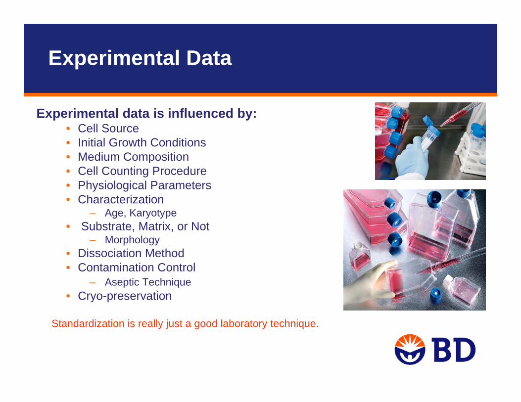
Experimental data is influenced by:• Cell Source• Initial Growth Conditions • Medium Composition• Cell Counting Procedure• Physiological Parameters• Characterization
– Age, Karyotype• Substrate, Matrix, or Not
– Morphology• Dissociation Method• Contamination Control
– Aseptic Technique• Cryo-preservation
Standardization is really just a good laboratory technique.
Experimental Data

Contact Us
Questions?Contact information:Shabana Islam, Ph.D.e-mail: [email protected]
Technical Support:In the U.S.tel: 877.232.8995 or or 978-901-7491e-mail: [email protected] the U.S.Contact your local distributor or visitbdbiosciences.com/offices to locateyour nearest BD Biosciences office.For research use only. Not intended for use in diagnostic or therapeutic procedures. BD, BD Logo, and all other trademarks are the property of Becton, Dickinson and Company. ©2009 BD