tips & techniques in preanalytics - sarstedt 5 “preanalytics encompasses any and all...
TRANSCRIPT
Tips & Techniquesin Preanalytics
32
Preanalytics & S-Monovette®
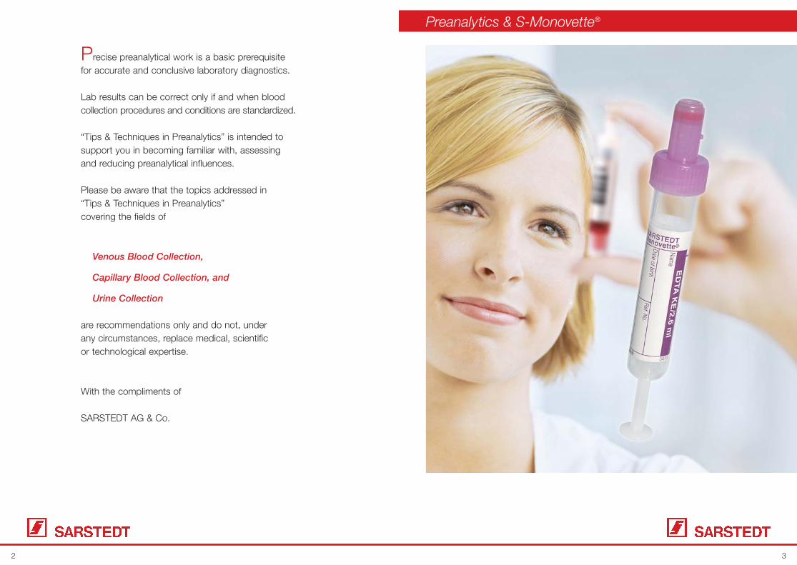
Precise preanalytical work is a basic prerequisite for accurate and conclusive laboratory diagnostics.
Lab results can be correct only if and when bloodcollection procedures and conditions are standardized.
“Tips & Techniques in Preanalytics” is intended to support you in becoming familiar with, assessing and reducing preanalytical influences.
Please be aware that the topics addressed in“Tips & Techniques in Preanalytics”covering the fields of
Venous Blood Collection,
Capillary Blood Collection, and
Urine Collection
are recommendations only and do not, underany circumstances, replace medical, scientificor technological expertise.
With the compliments of
SARSTEDT AG & Co.

4 5
“Preanalytics encompasses any and allprocedures prior to laboratory work”.
Note:Preanalytical issues can never be settled by individuals alone but requirethe cooperation of physicians, nurses and laboratory personnel involvedin the overall procedure.
Preanalytical Influence Factors
Population (race)There are significant differences between the African-American compared to theCaucasian population:
- Significantly lower leucocyte count
- Vitamin B12 concentration is 1.35 times higher
- Higher CK and -amylases
SexApart from individual sex-related co mponents (hormones), a person’s muscular mass, for example, is one of the factors that determine pertinent parameters.
- The proportion of CK and creatinine depends on the muscular mass.
AgeAs a person grows older, the cholesterol level frequently increases in both menand women (although this depends primarily on nutrition).
300
250
200
150
100
50
0m
g/dl
15 25 35 45 55 65years
Cholesterol
LDL
HDL
Fig.: Guder, Narayanan, Wisser, Zawta ”Samples: From the Patient to the Laboratory“ GIT-VERLAG GmbH & Co. KG, Darmstadt
Permanent
Population (race)
Sex
Long-term
Age
Pregnancy
Nicotine / illegal drugs / alcohol
Short-term
Circadian fluctuations
Posture
Physical strain
Patient-related influence
Permanent influence
Long-term influence

6 7
Nicotine
Chronic nicotine abuse is known to increase leucocyte count, a number of enzyme values and tumor markers (particularly the CEA value).
Alcohol
Chronic alcohol abuse causes an increase in liver enzymes, e.g. GT, ALT (GPT) and AST (GOT) while folic acid and vitamin B6 values decrease.
Fig.: Guder, Narayanan, Wisser, Zawta ”Samples: From the Patient to the Laboratory“ GIT-VERLAG GmbH & Co. KG, Darmstadt
PostureIncrease in concentration when moving from the horizontal to an upright position.Parameters increase in %Hematocrit 13 %Erythrocytes 15 %HDL Cholesterol 10 %Aldosterone 15 %Epinephrines 48 %Renin 60 %
Effects of constriction timeComparison: 1 min. vs. 3 min.Parameter deviation in%Bilirubin + 8Cholesterol + 5Creatinine - 9Creatine kinase - 4Iron + 7Glucose - 9y-Glutamyltransferase - 10Potassium - 5
Fig.: Guder, Narayanan, Wisser, Zawta ”Samples: From the Patient to the Laboratory“ GIT-VERLAG GmbH & Co. KG, Darmstadt
Increase in values x times the amount
CreatinkinasePyruvatkinase
ASATBilirubin
UreaUric acid
Inorganic phosphateGlucoseAlbuminCalcium
Alcaline phosphataseSodium
1 2 3 4 5
Physical strainIncrease in various analytes after extreme physical strain, e.g. a marathon
Preanalytical Influence Factors
-50 0 50 150 250 350 450
+ 1000 % GTASATNoradrenalineAdrenalineCortisolTriglyceridesCHOLVMSLDL
Deviations in %
Fig.: Guder, Narayanan, Wisser, Zawta ”Samples: From the Patient to the Laboratory“ GIT-VERLAG GmbH & Co. KG, Darmstadt
-60 -40 -20 0 20 40 60 80
ACEProlactin
-CarotinoidsPyridoxalphosphateSeleniumHDL-CholesterolLDL-CholesterolCholesterolHematocritMCVFibrinogenCopperMCHCCadmiumMonocytesLymphocytesGranulocytesCEA
Deviations in %
Short-term influence

8 9
®
Name
®
Name Name
®
Name
Informing the patient
Inform the patient of the forthcoming procedure in order
to alleviate possible anxiety and stress.
Explaining particular preconditionsthat must be observed is an essential part of this information, e.g.
Consumption of pharmaceutical drugs
Restriction to a special diet
Sample collection on an empty stomach (except for emergency diagnostics)
Precise instructions for useshould be given to explain the use of urine and faeces collection containers.
Carefully explain the forthcoming procedures to children using terms which are easy for them to understand in order to prevent unnecessary distress.
Never analyze sample tubes which are not clearly identified.
Barcode labels enable reliable sample identification.
The identification should always be provided on the primary tube.
Only use water-proof felt tip pens on glass or plastic tubes.
Additives (anticoagulants, clotting activators, gel) are identified by a color code on the sample tubes. Due to the absence of international standardization, additional identification may be required from case to case.
Never provide sample identification on the cap,outer packaging or transport container.
Barcode line
they enable unrestricted visibility of the tube content.
they enable confirmation of the filling volume.
the screw cap can be easily removed.
the tube and label do not get jammed or stuck in the centrifuge.
Clearly mark the tube and request formif the material is known to be infectious.
Preparation and Identification
Correct patient identificationis a fundamental necessity (surname, first name/s, date of birth, admission number, department, room number).
Errors do not only occur with popular names.
Patients should always identify themselves when directly addressed. Partially/completely deaf or cognitively impaired patients might answer questions like “You are Mr. Miller, aren‘t you?” with an affirmative nod.
A person sitting on the edge of the bed might just as well be a visitor.
If the patient‘s identity is not entirely clear, the phlebotomist should refuse to perform blood collection until the problem is resolved.
Patient identification
Sample identificationPatient preparation
10 11
The identity of the phlebotomist should be ascertainable for each sample taken and,
if possible, also noted on the request form.
Problems concerning the type and time of blood collection, the patient’s condition, and other important issues should be noted and will be helpful in the event of un-clear analysis results.
Preparation and Identification
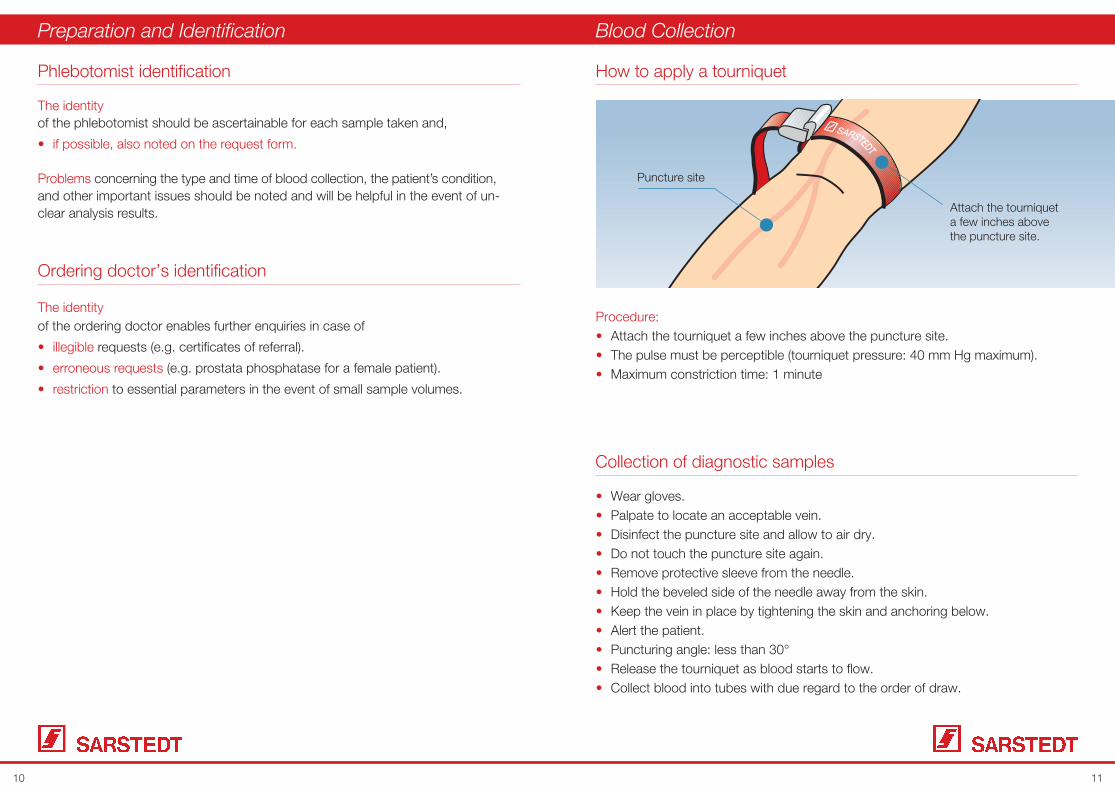
Procedure:Attach the tourniquet a few inches above the puncture site.The pulse must be perceptible (tourniquet pressure: 40 mm Hg maximum).Maximum constriction time: 1 minute
How to apply a tourniquet
Wear gloves.Palpate to locate an acceptable vein.Disinfect the puncture site and allow to air dry.Do not touch the puncture site again.Remove protective sleeve from the needle.Hold the beveled side of the needle away from the skin.Keep the vein in place by tightening the skin and anchoring below.Alert the patient.Puncturing angle: less than 30°Release the tourniquet as blood starts to flow.Collect blood into tubes with due regard to the order of draw.
Collection of diagnostic samples
SARSTEDT
Puncture site
Attach the tourniquet a few inches above the puncture site.
The identityof the ordering doctor enables further enquiries in case of
illegible requests (e.g. certificates of referral).
erroneous requests (e.g. prostata phosphatase for a female patient).
restriction to essential parameters in the event of small sample volumes.
Ordering doctor’s identification
Phlebotomist identification
Blood Collection

12 13
Poor vein conditions:
Select new puncture site.
Apply thermo-pad or pre-heated cloth.
Use Safety-Multifly®-Needle (set for difficult veins).
Use aspiration method for collection.
Penetrating the vein:
Slightly withdraw the needle.
Interruption of blood flow during collection:
Needle position has been changed.
Vein has collapsed.
Blood Collection S-Monovette® - Aspiration Principle
Open the Safety-Needle packaging at the tear-off line
Attach the needle
Remove the protective sleeve
IMPORTANT:Do not lock the Safety-Needle of the S-Monovette® into place by slightly turning
clockwise until immediately prior to venipuncture.
Problems prior to / during blood collection:
”Pumping“ the fist to enhance blood flow leads to a rise in K+ and Mg2+ due to increased muscle activity.
Extended constriction changes parameters, e.g. K+, -GT.”Bending“ the needle is not necessary when using the S-Monovette® system
because of a generally very flat penetration angle. Lumen changes caused by bent needles may damage the cells (hemolysis).
Hemolysis can also be the result of a needle that is too thin.
Mistakes during blood collection:
Insufficient mixing of the sample (micro clots)
Excessive mixing (shaking) causes hemolysis
Ensure compliance with coagulation times prior to centrifuging serum samples
(approximately 30 minutes after sampling) to prevent post-coagulation
(gelatinization).
Ensure observation of the centrifugation recommendations for improved
sample quality
Incorrect handling after blood collection:

14 15
Use the thumb of your free hand to tighten the skin and hold the vein in place. “Alert” the patient and puncture the vein.
S-Monovette® – Aspiration Principle
Loosen the tourniquet and slowly withdraw the plunger. Wait until the blood flow stops.
For multiple sampling, remove S-Monovettes from the needle by slightly twisting counterclockwise. The needle remains in the vein.
the S-Monovette® from the needle and then withdraw the needle from the vein.
IMPORTANT:When blood collection is complete, withdraw the plunger into the “click” position and break off.
After blood collection
click
16 17
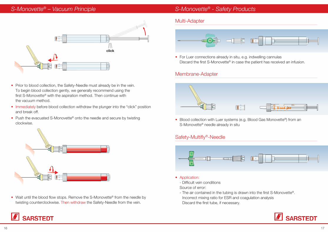
Prior to blood collection, the Safety-Needle must already be in the vein. To begin blood collection gently, we generally recommend using the first S-Monovette® with the aspiration method. Then continue with the vacuum method.
before blood collection withdraw the plunger into the “click” position and break off.
Push the evacuated S-Monovette® onto the needle and secure by twisting clockwise.
S-Monovette® – Vacuum Principle
click
Wait until the blood flow stops. Remove the S-Monovette® from the needle by twisting counterclockwise. Then withdraw the Safety-Needle from the vein.
For Luer connections already in situ, e.g. indwelling cannulas Discard the first S-Monovette® in case the patient has received an infusion.
S-Monovette® - Safety Products
Blood collection with Luer systems (e.g. Blood Gas Monovette®) from an S-Monovette® needle already in situ
Multi-Adapter
Membrane-Adapter
Safety-Multifly®-Needle
- Difficult vein conditions Source of error: - The air contained in the tubing is drawn into the first S-Monovette®. Incorrect mixing ratio for ESR and coagulation analysis Discard the first tube, if necessary.

18 19
S-Monovette® - Safety Products S-Monovette® - User Guide
Centrifugation
Centrifuged sampleclotted in a
vertical position
Centrifuged sampleclotted in ahorizontal position
Nomograph to convert the number of revolutions per minute into g-force
r n
RCF
r = Centrifuge rotor radius in cm (tube holders in swing-out position)
n = Number of revolutions per minute (min-1)
RCF = relative centrifugal force =̂ g force (g = 9.81 m/s2 gravity acceleration)
Example: Centrifuge radius r = 16 cm and number of revolutions per minute n = 4,000 min-1
results in RCF 2,860 x g
This nomograph is based on the following formulag = 0.00001118 x r x n2
org = 11.18 x r x ( )2n
1.000
Before Centrifugation
Hold the Safety-Needle at the adapter, place the needle protector on a stable, flat surface and slightly press the needle downwards until it noticeably and audibly locks into the needle protector.
After activating the protective mechanism:
Discard the safely locked Safety-Needle in a disposal box.
After blood collection:
Detach the last S-Monovette® from the Safety-Needle and then withdraw the Safety-Needle from the vein.
Safety-Multifly®-Needle
...until the needle is noticeably and audibly locked into the protective casing.
After activating the protective mechanism:
Discard the safely locked Safety-Multifly®-Needle into a disposal box.
click
Hold the needle protector at its back end with your thumb on top and your forefinger below and withdraw the Safety-Multifly®-Needle from the vein. Stabilize the tubing by pressing it slightly against the palm of your hand and push the needle protector over the needle …
Safety-Needle
click
click
Alternatively, you can also activate the needle protector using your index finger.For safe operation, make sure to press against the lower end of the protection device.
1
2a
2b
3

20 21
Difference between fixed-angle and swing-out rotors
Please refer to the information provided by the centrifuge manufacturer for the centrifuge rotor radius (r max ) required for calculation or determine the radius by means of the following illustrations.
Fixed-angle rotor Swing-out rotor
S-Monovette® – Centrifugation conditions
*We recommend processing of S-Monovettes with gel preparation in swing-out rotors only.
Refer to the nomograph on the previous page or the centrifugation calculator at www.sarstedt.com/User information/Centrifugation to convert the g-force into the number of revolutions per minute.
S-Monovette® Serum 10 min. 2,000 x g 20°C
S-Monovette® Serum-Gel* 10 min. 2,500 x g 20°C
S-Monovette® Li-Heparin 10 min. 2,000 x g 20°C
S-Monovette® Li-Heparin-Gel* 10 min. 3,000 x g 20°C or 15 min. 2,500 x g 20°C
S-Monovette® EDTA-Gel* 10 min. 2,500 x g 20°C
S-Monovette® Citrate 10 min. 1,800 x g 22°C
Blood samples should be taken to the laboratory for analysis as soon as possible.
After centrifugation, separating gels or filters prevent the diffusion of substances from the erythrocytes into the serum/plasma.
Whole blood without serum/plasma separation must not be frozen under any circumstances as this process would result in a total hemolysis.
For long-term storage, the serum should be stored in enclosed containers at 2 to 4°C.
Serum or plasma samples can be stored at -20°C for extended periods of time.
Special cool transport containers should be used to protect samples during prolonged transit.
Some analytes must be transported to the laboratory without delay (e.g. Ammonia: within 15 min.).
S-Monovette® - General Information
Sample storage and transport
S-Monovette® - User Guide
22 23
As a rule, analysis results should be communicated to the requesting institution in writing only.
Exception: emergency diagnostics
The communication of results by phone should remain an exception. Results should be disclosed to the doctor in charge only. Conveying the analysis results to the patient as well as their interpretation
is restricted to the doctor in charge.
The patient decides if results are to be disclosed to third parties. If the patient is unable to do so, the decision should be left to the doctor in charge.
A patient’s laboratory data is private and it isa doctor’s duty to maintain confidentiality.
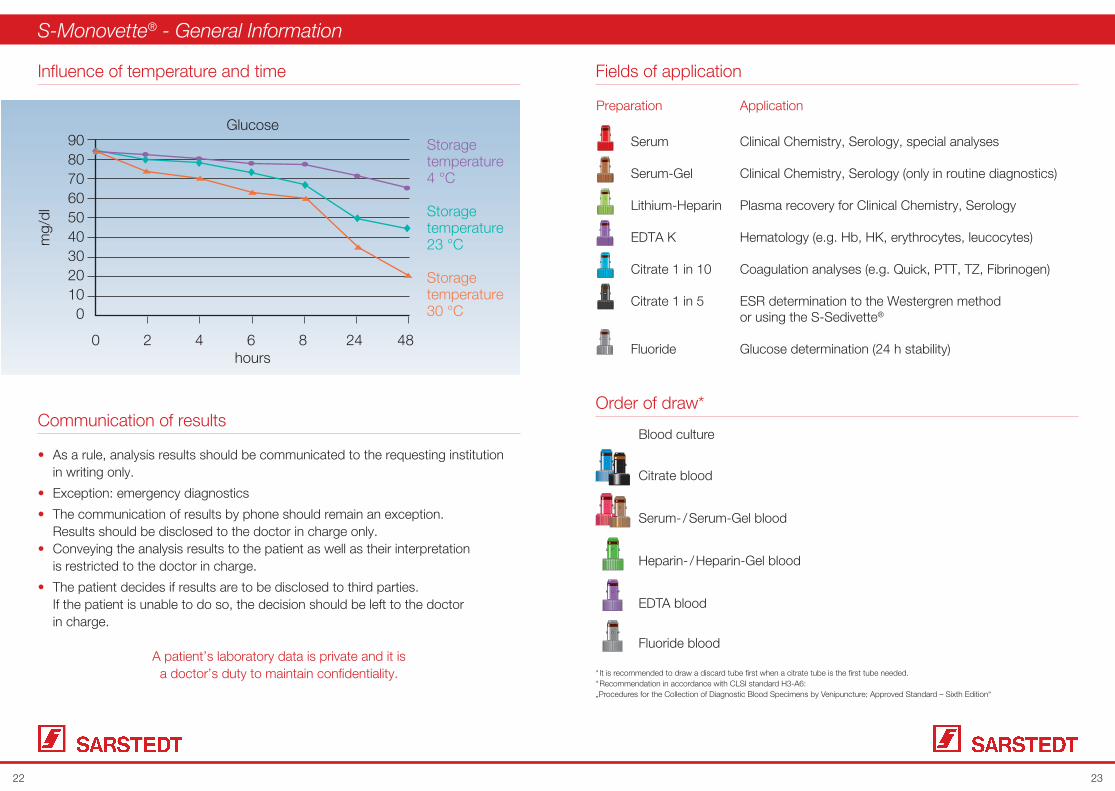
Influence of temperature and time
Communication of results
908070605040302010
0
mg/
dl
0 2 4 6 8 24 48hours
GlucoseStoragetemperature4 °C
Storagetemperature23 °C
Storagetemperature30 °C
Preparation Application
Serum Clinical Chemistry, Serology, special analyses
Serum-Gel Clinical Chemistry, Serology (only in routine diagnostics)
Lithium-Heparin Plasma recovery for Clinical Chemistry, Serology
EDTA K Hematology (e.g. Hb, HK, erythrocytes, leucocytes)
Citrate 1 in 10 Coagulation analyses (e.g. Quick, PTT, TZ, Fibrinogen)
Citrate 1 in 5 ESR determination to the Westergren method or using the S-Sedivette®
Fluoride Glucose determination (24 h stability)
S-Monovette® - General Information
Fields of application
Order of draw*
Blood culture
Citrate blood
Serum- / Serum-Gel blood
Heparin- / Heparin-Gel blood
EDTA blood
Fluoride blood
* It is recommended to draw a discard tube first when a citrate tube is the first tube needed.* Recommendation in accordance with CLSI standard H3-A6: „Procedures for the Collection of Diagnostic Blood Specimens by Venipuncture; Approved Standard – Sixth Edition“

24 25
Capillary Blood Collection & Microvette®

26 27
A mixture of blood from arterioles, venules and capillaries as well as interstitial and
intra-cellular fluids.
Note: Capillary blood collection does not necessarily involve the use of anend-to-end capillary.
Capillary Blood Collection
Pediatrics
Geriatrics
Adults: blood gas analysis, glucose and lactate determination
Point-of-Care tests
Fields of application
Volumes > 1 ml (e.g. blood culture)
Coagulation analysis
Inflammations
Patient in shock
Preparation
- Material
- Patient
- Puncture site
Puncture
Sampling
Capillary blood collection
Criteria that exclude capillary blood collectionWhat is capillary blood?

28 29
Gloves
Swab
Disinfectant
Semi-automatic disposable lancet (Safety-Lancet)
Sample tube (blood gas capillary, Microvettes, bilirubin capillaries etc.)
Sharps container
A bandage, if required (not advisable with small children – risk of swallowing)
Patient identification
Explain the procedure.
Select the puncture site.
Prewarm the puncture site for enhanced blood circulation, if required.
Finger tip Heel Earlobe
Preparation
Puncture sites
Enhanced blood flow by up to seven times the normal volume
Precondition for capillary blood gas analyses
Enhancing the blood circulation leads to an arterialization of the capillary blood and, as a result, to an acceptable comparability with the analyses obtained from arterial blood.
Advantages of warming the puncture site
Wrap the patient’s foot or hand with a cloth heated to 39° – 40°C. For best result, pull on a rubber glove. Duration: 3 to 5 min.
For capillary Blood Gas analyses from adults, rub a hyperaemisating ointment into the earlobe.
How to warm the puncture site
Wear gloves.
Disinfect skin and allow to air dry.
Hold the finger or foot securely.
Puncture with a Safety-Lancet.
Puncturing and sampling
Material required:
Patient preparation

30 31
Safety-Lancet
Product range
5 options
Version for heel puncturing
User Guide
1. Twist the cap until it separates from the Safety-Lancet.
2. Press the Safety-Lancet firmly against the chosen and cleaned puncture site and press the firing button.
3. Discard the Safety-Lancet into a suitable sharps container.
4. Collect blood
Wipe away the first drop of blood.
Angle the puncture site downward.
Avoid blood smears.
Ensure that the sample tube is held in the correct position.
Avoid repeated strong pressure (milking).
This causes hemolysis and contamination of samples with tissue fluid.
Important information
Product features
Primed system ready for use - one step less
The sterile, disposable product cannot be reused.
Easy handling – secure firing button avoids the risk of unintentional activation and deactivation of the system
Ridged lancet body ensures safe grip
Small contact face for precise puncturing
Variety of options
1 2
3 4
Pon
Thca
Ethde
R
Description Mini Normal Extra Super Neonatal
Penetration Depth 1.6 mm 1.8 mm 1.8 mm 1.6 mm 1.2 mm
Needle size 28 G 21 G 18 G Blade 1.5 mm Blade 1.5 mm
Blood volume Low Medium Medium to high High Medium to high

32 33
200ED
TA K
200EDTA K
200
200ED
TA K
200ED
TA K
EDTA200
EDTA200
EDTA
Lithium-Heparin /Lithium-Heparin-Gel
Fluoride
Serum /Serum-Gel
Microvette®
Microvette® – Order of draw*
Product features:
Capillary method using the end-to-end capillary
Sampling with the collection rim
Note: Letting blood drip into a capillary tube by means of a Luer needle does not constitute capillary blood collection.
2. Collection is complete when the capillary is entirely filled with blood.
3. Hold the Microvette® upright to allow blood to flow into the collection tube.
4. Twist to remove the cap including the capillary and discard as a complete unit.
5. Remove the cap from the tube base
and close the tube (”click“ position).
6. Mix samples thoroughly and gently.
1. Hold the Microvette® in a horizontal or slightly inclined position and collect the blood sample with the end-to-end capillary.
1
2
3 4
5 6
Capillary method - Microvette® 100 and 200
1. Capillary method
For the collection of even the smallest blood volumes from 100 μl to 500 μl
Different inner tube options – conical tube for a high supernatant after centrifugation or cylindrical tube for enhanced mixing results
Range of collection techniques
The special cap design minimizes aerosol effect when the tube is opened.
* Recommendation in accordance with CLSI standard H4 - A6:„Procedures and Devices for the Collection of Diagnostic Capillary Blood Specimens ; Approved Standard – Sixth Edition“

34 35
500EDTA K
500ED
TA K
500ED
TA K
500
EDTA
KED
TA K
500ED
TA K
500ED
TA K
Microvette®
Microvette® – Centrifugation conditions
Microvette® Serum 5 min. 10,000 x g 20°C
Microvette® Serum-Gel 5 min 10,000 x g 20°C
Microvette® Heparin 5 min. 2,000 x g 20°C
Microvette® Heparin-Gel 5 min. 10,000 x g 20°C
Microvette® Fluoride 5 min. 2,000 x g 20°C
Information on the centrifugation conditions is also printed on the inner box label.
1 2
3
4 5
1. Slightly twist the cap to detach.
2. Attach the cap to the tube base.
3. Use the special rim to collect the blood drops.
4. Remove the cap from the tube base and close the Microvette® (“click” position).
5. Mix samples thoroughly and gently.
2. Sampling with the collection rim

36 37
Preanalytics & Urine-Monovette®

38 39
Types of urine samples
Preanalytics:
Reliable results in urine analysis are subject to carefully observed collection, transport and storage conditions.
Depending on the time and type of urine collection, we differentiate between:
Mid-stream urine - First morning urine - Second morning urine - Spontaneous urine
Bladder puncture urine
Catheter urine Urine collection in cases of one-time catheterization or a permanent catheter
24 h urine
Mid-stream urine
Correct sampling:
Thorough cleaning of external genital area
Once urine has been flowing for approximately 3 seconds, 10 to 20 ml of urine are passed into a sterile collection container without interrupting the urinary stream. Be sure to avoid contamination. Note:
Especially important for microbiological analysesPrecondition: Patient must be able to cooperate
First morning urine
The constituents contained in the first morning urine are of particularly high concentration.
Application: Suited for bacterial analyses, test strips, sediment, clinical-chemical analyses, protein diagnostics
Due to the extended retention time in the bladder, morning urine is ideally suited for nitrite and protein determination.
40 41
Types of urine samples
The constituents in the second urine passed after the first morning urine are of a medium concentration.
Test strips, glucose, protein
Not suited for nitrite testing
Spontaneous urine
Urine collected at any given time
Entirely sufficient for many chemical and microscopic parameters
Easy to collect
Dilution error – always take into account the specific weight (density) for correct determination
Bladder puncture urine
Urine collected through bladder puncture is suited for bacterial analysis, primarily in case of infants and small children.
Note:Reduced risk of infection compared to catheterization
Catheter urine
One-time catheterization:Collecting urine by means of one-time catheterization is very rarely done as it is painful for the patient and involves a high risk of infection.
Permanent catheter:If collecting urine from a permanent catheter is an absolute requirement, this must be done through aseptic access of the tubing sampling port.
Note:For diagnostic purposes, urine should not be collected from the urine drainage bags.
Second morning urine

42 43
24h Urine Collection
The term ”Urine Collection” generally describes any urine volume collected over a particular period of time, while the 24 h period is the interval most frequently applied.
e.g. catecholamines, creatinine clearance
Eliminates any fluctuation in the parameters as may be caused by a difference in concentration
Extended collection periods, sufficiently dimensioned collection containers, precise patient instructions, correct stabilizer
Urine container volumes
Studies have revealed that a 3,000 ml container accommodated 91.4 % of all 24 h urines collected.
A 2,000 ml container can hold only 60 %.
How to collect 24 h urine
1. Discard the first morning urine.
Note the collection time, e.g. 7:00 a.m.
2. Pass the second morning urine into the container
and add stabilizer, if required.
3. Collect
each
urine
and mix.
4. Collect the first morning urine on the following day at the same time as the day before, e.g. 7:00 a.m.
START
END(24 hours)
.
.
.
UriSet 24 - “The complete set”
or

44 45
Urinalysis Urine-Monovette®
Dry chemical urine analysis by means of a test strip to determine early symptoms (screening test)
Note:The screening result alone is not sufficient for a direct diagnosis as it only renders an indication of the existence and approximate amount of a particular substance.The results obtained serve as a basis for more detailed microscopic, bacterial or clinical-chemical analyses.
Sediment analysis in case of unusual results obtained in dry chemistry
Urinalysis and preanalytics
Use fresh, non-stabilized and non-centrifuged mid-stream urine (not stored for longer than 2 hours).
Extended storage may cause the following changes, e.g. - Disintegration of leucocytes and erythrocytes - Bacterial growth - Glycolysis caused by bacteria
Mix urine thoroughly immediately before using the test strips.
Overall wetting of all test fields
Ensure observation of the incubation time.
Correct centrifugation to obtain the urine sediment (5 min. 400 x g)
Urine-Monovette® User Guide
Hold the Urine-Monovette®
with the suction tip upwards and withdraw the plunger in downward direction until the suction tip is empty.
Remove the tip, break off the plunger and attach cap.
Insert the suction tip into the cup and and draw up the Urine-Monovette® pistonto the base line.
The Urine-Monovette® is suitable for urine sampling, transport, analysis and centrifugation.
1 2 3
SAMo
Bacterio
log
y10/06
Urin
e Z
8
6
4
2
10 ml
SM
Bac
teri
olo
gy
Uri
ne
Z
8
6
4
2
10 ml
Waste
SM
Bac
teri
olo
gy
Uri
ne
Z
8
6
4
2
10 ml

46 47
Microbiological urine diagnostics
Note:
Native urine is the ideal material to determine the bacteria causing urinary infections provided the sample is analyzed within 2 hours when stored at ambient temperature, and within 4 hours when kept in a cool environment.
We suggest the use of morning urine (mid-stream urine). During the day, advise patients not to pass urine at least 4 hours before collection.
No antibiotic treatment 2 to 3 days prior to collection
In a filling volume of 10 ml, the boric acid concentration is 1.5 %.Micro organisms are stabilized for up to 48 hourswhen stored at room temperature.
Important:Observe the nominal volume.
Mix thoroughly after urine has been aspirated into the Urine-Monovette®.
Not suitable for clinical-chemical analyses, test strips, etc.
Recommendations for urine collection
Analyze urine within two hours.
If possible, use mid-stream urine for analysis.
Proficient sampling
Use clean, disposable containers.
Correct identification of containers prior to sampling
Errors in preanalytical work are the figures in front of the decimal.
Errors in laboratory analytics arethe figures behind the decimal.
Urine-Monovette® with boric acid
In a f1.5 %Microwhen
ImpoOb
Mth
Note
48 49
Notes

50 51
Notes

453a
-501
Th
is d
ocum
ent m
ay c
onta
in in
form
atio
n on
pro
duct
s th
at m
ay n
ot b
e av
aila
ble
in p
artic
ular
cou
ntrie
s
Tech
nica
l mod
ifica
tions
res
erve
d
SARSTEDT Inc.1025 St. James Church RoadP.O. Box 468Newton, NC 28658-0468Tel. (800) 257-5101Fax (828) [email protected]