tissue barrier formation pulpotomized monkey teeth
TRANSCRIPT

Hard Tissue Barrier Formation in Pulpotomized Monkey TeethCapped with Cyanoacrylate or Calcium Hydroxide for 10 and 60 Minutes
M. CVEK4, L. GRANATH', P. CLEATON-JONES2, and J. AUSTIN3
Department of Pedodontics, Eastmaninstitutet, Dalagatan 11, S-113 24 Stockholm, Sweden; 'Department of Pedodontics, University of Lund,School of Dentistry, Malm&, Sweden; 2MRC/University of the Witwatersrand Dental Research Institute and 3Central Animal Service, Universityof the Witwatersrand, Johannesburg, South Africa
Monkey incisor teeth were pulpotomized in groups of 10. After phys-iological hemostasis, the pulps of group I were covered with isobutylcyanoacrylate, and those of groups 11 and III with calcium hydroxidefor 10 and 60 minutes, respectively, thereafter this compound waswashed away and the wound surfaces covered with Teflon. In groupIV, calcium hydroxide was used as a positive control, and Teflon asa negative control in group V. The animals were killed after 12 weeksand the teeth removed in tissue blocks. The material was processedand evaluated histologically with respect to location and continuity ofa hard tissue barrier, type of newly formed hard tissue, state of thepulp, and presence of stainable bacteria in the coronal cavity.
Seven of nine teeth in group I showed a hard tissue barrier. Thecorresponding figure for group HI was eight out of 10 teeth. All teethin groups III and IV had a barrier. The incidence of a continuousbarrier increasedfrom group I through group IV, as did the incidenceof its location below the level of the original wound surface. Thecondition of the pulp was related to the presence of bacteria and thecontinuity of the barrier to the presence of inflammation. There wasno bridging in group V.
The results support the theory that a low-grade irritation is re-sponsible for the formation of a hard tissue barrier in exposed pulps.
J Dent Res 66(6):1166-1174, June, 1987
Introduction.
In 1971, Schr6der and Granath described the effects of calciumhydroxide on surgically treated human pulps and noted thatthe compound caused a multilayered necrosis, followed bysubjacent mineralization. They postulated that the lowermostlayer, a zone of firm necrosis, generates a low-grade irritationof the underlying tissue sufficient to produce a matrix thatmineralizes. The mechanism is obscure, but it is unlikely thatcalcium ions released from calcium hydroxide would play adecisive role (for review, see Granath, 1982, and Schr6der,1985).
Support for this low-grade irritation theory comes from theclaim that isobutyl cyanoacrylate induces hard tissue formationwithout an obvious zone of firm necrosis acting as an inducer(Berkman et al., 1971). Agreement on this, however, is notcomplete. Nixon and Hannah (1972) have reported that n-butylcyanoacrylate failed to produce satisfactory dentin barriers,while, with regard to calcium hydroxide, Holland et al. (1982)have speculated on whether calcium or other cations are in-volved.
Because of an incomplete understanding of the mechanismof barrier formation, we started to look for alternatives to hy-droxides for pulpal wound dressings, one of which was cy-anoacrylate. Isobutyl cyanoacrylate, a cyanoacrylic ester{CH2 = C(C-N)COOC4H9}, is an odorless, colorless, or paleyellow liquid with a boiling point of 820C at 6 mm Hg addi-tional pressure and a density of 0.990 (The Merck Index, 1976).It polymerizes in the presence of water and is not soluble inwater, but is degradable in a biological environment. Thera-
peutically, it is used in surgery as a tissue adhesive.The literature offers no explanation as to why pulp tissue
might respond with formation of hard tissue to cyanoacrylatethat lacks both calcium and hydroxyl ions. However, the fol-lowing hypothesis has been suggested (Granath, 1982): "Thematerial is apparently biocompatible as such. On degradationin contact with the pulp tissue, some substances) is releasedthat either causes a low-grade degeneration in the nearest cel-lular layer or alters the physical state in the tissue layer whichin turn changes the chemical equilibrium locally. In both in-stances calcium is attracted. Mineralization of newly formedcollagenous substance is initiated from the calcified foci."
In this connection, it is interesting to note that hard tissueformation has been found directly against some calcium-hy-droxide-containing hard-setting cements in experimentally ex-posed monkey pulps, i.e., without a visible zone of necrosisin the light microscope (Tronstad, 1974; Heys et al., 1980,1981; Cox et al., 1982). The mechanism can be assumed tobe the same as with pure calcium hydroxide, while a lowerpH and a varying release of hydroxyl ions might explain thelack of visible necrosis.
One way to elucidate the mechanism of hard tissue induc-tion following application of calcium hydroxide would be toremove the compound from the surgically treated pulp tissueonce an initial caustic effect has been established. This occurssoon after exposure to calcium hydroxide (Schroder and Gran-ath, 1971). Such a treatment would minimize the effect ofionized calcium and would thus be a useful comparison withcalcium-free cyanoacrylate.
The aim of this study, therefore, was to elucidate furtherthe morphological appearance of a hard tissue barrier in pul-potomized teeth with calcium hydroxide and cyanoacrylate aswound dressings. Teflon was used as a control substance be-cause it is biologically inert.
Materials and methods.Operative technique. - Fifty-two mature permanent incisor
teeth from 14 monkeys (Cercopithecus aethiops pygerythrus)were used for the experiments. The animals were first tran-quilized with Ketamine hydrochloride (Ketalar, Parke-DavisLaboratories, Ltd., Johannesburg) and then anesthetized withpentobarbitone sodium (Sagatal, Maybaker, Ltd., Johannes-burg). A rubber dam was used to isolate the maxillary teeth,while the mandibular teeth were isolated with cotton rolls. Theteeth were mechanically cleaned with a rubber cup containingwater-mixed pumice and thereafter washed, together with therubber dam or the surrounding mucosa, with 0.5% chlorhex-idine in 70% ethanol.The operative procedure was performed with diamond in-
struments according to the technique of Granath and Hagman(1971). In order to facilitate cavity preparation and coolingduring pulpotomy, we removed incisal enamel for about one-third of the crown length. Subsequently, a round and then acylindrical diamond instrument were used to cut a hole throughdentin but without perforating the roof of the pulp chamber.The final cut was made with a cylindrical instrument with
Received for publication September 3, 1986Accepted for publication December 18, 19864To whom correspondence and reprint requests should be addressed
1166

PULP CAPPING WITH CYANOACRYLA TI AND CALCIUM HYDROXIDE
TABLE 1HARD TISSUE BARRIER FORMATION IN 49 PUtLPOTOMIZED MONKEY INCISORS CAPPED WITH ISOBUTYL CYANOACRYLATE OR
CALCIUM HYDROXIDE FOR DIFFERENT TIME PERIODS OR LEFT WITHOUT AN ACTIVE WOUND DRESSING. IN RELATION TO STATEOF PULP AND PRESENCE OF BACTERIA; OBSERVATIONS AFTER 12 WEEKS
Wound Dressing Hard Tissue Barrier' State of Pulp"Group No. Material Time d c Is's bsss sI m se ne Bacteria*'
1 9 cyano- 12 weeks 6 7 4(4) 5acrylate
1 1(0 calciumhydroxide
IIl 10 calciumhydroxide
IV If) calciLuhydroxide
V 1(0 no
10 min
60 11 i E
5 3 4 4
7 3 2 8
12 wccks 9
5(3) 1(1) 4
3(3)
10
4(4) 2(2) 6diressine,
id discontinuous barrier, chai-acterized bh a ltaer of boni-like and a SubsequeCInt layr of dcntin likc tissue: C = continuous bairier. otherwise as in
d1 Iws at level and bws - below level of oaieinal wound surface.sl slight scatteredd inflammatoryy cells). m = moderate and se = sevcc Inflammation (accumillation1 of inflammatory cells, abscess): ne
necrosis, numbeI of cases with stainable bacteria within paienthesis.*-;<*Presence of stainable bacteria in coronal activity or in dentin walls form-ing the cavity.
smooth sides and diamond particles only on its base and of adiameter that just exceeded the radius of the access cavity. Therotating instrument was gradually lowered into the tooth withcircular movements until pulp tissue, surrounded by dentin,was removed to about the level of the largest cross-section ofthe pulp.
During all steps of the operative procedure, the tooth andcutting instruments were irrigated with physiological saline so-lution. A high-speed handpiece running at about 100.000 rpmwas used, and the CLtting was performed intermittently in orderto keep the dentin moist and to avoid overheating the pulp.After completing the pulpotomy, we irrigated the wound sur-face continuously with saline until bleeding ceased. Thereafter,
10 m
each of the following procedures was performed in groups of10 teeth, selected according to a randomized system:
I. Isobutyl cyanoacrylate (Ethicon Bucrylat, EthiconGmnbH, Nordestedt, W. Germany) was placed overthe pulps. Thereafter, two or more sterilized Teflondiscs, 2 mm in diameter and previously punched froma 0.08-mm-thick Teflon tape (USA standard BS 4375,1978), were placed over the cyanoacrylate and moldedagainst the surrounding dentin shelf with gentle pres-sure, by mcans of a small round instrument placedtoward the cavity corner.
11-111. Calcium hydroxide paste (Calasept, Scania DentalAB. Knivsta, Sweden) was placed over the pulps and
p
.tLit
N'a W"reog4Se0#vFig. (A) Cyanoacrylate. Discontinuous barrier. inlamed pulp tissue (p): "tunnel' (t) between irritation (secondary) dentin (sd) and true barrier
(b); hard tissue deposited on dentinal shelf (arrow).(B) Laycrs of bonc-like (b) and dentin-like (d) tissue.
Vol/. )60No 0 11657
4ft,
.1

1168 CVEKet al.
0/ALm 30JAm
Fig. 2 (A) Cyanoacrylate. Discontinuous barrier; inflamed tissue (p) in upper right corner adjacent to a resorption area magnified in (B); depositionof bone-like tissue on dentinal shelf and coronal surface of barrier (arrows).
(B) Rcsorption of dentinal cavity wall.(C) Deposition of bone-like (b) and ostcoid-like (o) tissue on dentinal shelf.(D) Another section of the same tooth; more extensive apposition of bone-like tissue on dentinal shelf.
molded with light pressure by means of dry cottonpellets for either 10 or 60 minutes. Subsequently, thecalcium hydroxide was washed away with a streamof physiological saline and the pulps were coveredwith Teflon discs as described above.
IV. As a positive control, calcium hydroxide paste wasplaced over the pulps and molded with light pressureby means of dry cotton pellets in such a way that alayer of compressed calcium hydroxide, approxi-mately 2 mm in thickness, remained over the pulps.No Teflon was placed in these specimens.
V. As a negative control, only Teflon discs were placedover the exposed pulps.
The remaining part of the cavity in each crown of all teethwas filled with a fortified zinc-oxide/eugenol cement (IRM,
L.D. Caulk, Milford, DE). Finally, two teeth were used as acontrol for determining the effects of the operative procedures.These were extracted immediately after pulpotomy and pre-pared histologically.
After 12 weeks, the monkeys were killed with an overdoseof pentobarbitone sodium (Euthanaze, Centaur Laboratories,Ltd., Johannesburg) and the head and neck retrogradely per-fused with physiological saline, followed by 10% neutral buff-ered formalin (Retief and Austin, 1973). The teeth were removedin tissue blocks, decalcified in 10% EDTA at pH 6.8, andembedded in celloidin-paraffin. The blocks were serially sec-tioned at 5 yim. Every tenth section was stained with hema-toxylin and eosin and every ninth or eleventh section with amodified Gram stain according to the method of Brown andBrenn (1931).
J Dent Re2s June2 1987

PULP CAPPIN\( WITH CYAN'OA('RYI A7T ANI) (zAI(IL AMHYD)ROXlI)1
Fig. 3 (A) Cyanoacrvlate. Discontinulous haliriei-, inflamed puLlp tissue(p)- bone-like (b) and dentin-likc (d) tissuC.
(B) Unidentitied torcin material. possibly trapped remnants ol eappinoiateiiial (arrows).(C) I)'position 1obone-litke tiSuLC On the lcft-hand xv all oi1oronal cavitv
(arrows).
The remaining 60 incisor teeth of the 14 monkeys were usedfor study of the effects of other pulp-capping materials notreported here.
Histological assessment: Hard tissue barrier. Hard tissuebarriers were classified as continuous or discontinuous. Thecriteria used to assess barrier formation were derived from thetypical appearance of a barrier induced through the effect ofcalcium hydroxide. Characteristically, such a barrier consistsof a layer of bone-like tissue and a subsequent layer of dentin-like tissue (Schrbder and Granath, 1971) located at a certaindistance (about 1 mm) from the original level of the woundsurface. When the barrier was continuous through all sectionsand no vital tissue was seen above, it was classified as contin-uous; the barrier was termed discontinuous if these criteriawere not met.
Condition of the pulp. - Pathologic changes in the pulpwere denoted as slight (scattered inflammatory cells), moderateor severe (accumulation of cells, abscess) inflammation, or
necrosis. The condition recorded was that seen in the section
with the most advanced degree of inflammation. The changeswere notcd separately for the pulp proper and for the tissuethat had proliferated from the pulp into the coronal cavity.
Bacteria. Specimens were examined for the presence orabsence of stainable bacteria either in the coronal cavity or inthe dentin forming this cavit\.
Results.The results are listed in Table 1. Seven of-i the nine teeth
capped with cyanoacrylate (roup 1: one tooth JO this groupwas lost during preparation) showed formation of a ord tissuebarrier (Figs. 1-3). The corresponding numbers for calciumhydroxide applied for 10 minutes (group 11) were eight out of10 teeth (Figs. 4 and 5). for 60 minutes (group 111) 10 of' 10(Figs. 6 and 7), and for 12 weeks (group IV) 10 of 10. Theincidence of continuous barrier formation increased from groupI through group IV, as did the incidence of barricr formationbelow the original wound surface.
100 Am__-.-
Volf. 00f No. 6) 1/e'9
1170 C VIAx c( IaI.
XSi\100 vm which formation of a true barrier wAas not seen. The amountwas proportional to the size of the dentinal shelf and consisted
mostly of two layers. One layer resembled bone, the otherlayer resembled dentin (Fig. 9). The pulpal surface in theseteeth consisted of a dense, collagen-rich connective tissue withcells arranged lngitudinally ag inst the surface of the Teflondiscs (Figs. 8 and 9).
V In the teeth of groups I-Ill, with a barber formed at the levelof pulpal amputation. the barrier seemed, in most instances,to be a combination of hard tissue induced by the medicament
5J D and opposition of hard tissue on dentinal walls (Figs. 1.2,4,4|̂;Vx*#z_and 6). Discontinutiies in the form of so-called tunnels" (Cox
et di., 1985) were often Tbund between the two types of hardON ~~~~~~~~~~~tissue(Figs. 1,2, and 4). These tunnels contained pulp tissue
with numerous fibroblast-like cells, probably original tissuethat had proliferated into the space between the coronal surfaceof the barrier and the Teflon discs. In five teeth capped withcyanoacrylate. hard tissue was also deposited on the dentinalshelf and the vertical walls of the coronal cavity. In three of
these teeth, hard tissue was deposited on the coronal surfacedIof the barrier as well. This hard tissue on the shelf consisted
t,,vt~p warily of a bone-like layer and a layer of osteoid like tissue,4,4,f,ii wounded by proliferated pulp tissue (Figs. 1-3). Further-more in two of these teeth, there was resorption of cavity
4liC walls adjacent to the proliferated and inflamed tissue (Fig. 2).
None of the teeth with a barrier located at the level of pulpal.t,,@rp sf; ; amputation contained necrotic tissue between the barrier andthe Teflon discs.
0.11 OW The proliferation of pulp tissue into the coronal cavity above201M the level of the original wound surface was also seen in most
W9 of the teeth in which the pulp was covered with Teflon alone(group V). In two of these teeth, hard tissue was deposited toanlimted extent on the dentinal shelf (Fig. 8).
In teeth from groups II- V. in which there was a barrierbelow the level of the original wound surface, none of thebarriers included hard tissue deposited on the dentinal wall inthe manner described above (Figs. 5 and 7). In two teeth fromgroup II, such hard tissue was deposited on the dentinal wallsbelow and separate from the barrier. It was found adjacent tothe dentinal tubules which, at the site of the dentinal shelf ofthe coronal cavity, were covered with an amorphous, necrotic
4~lit;0;0w-X0Y;sWt3like tissue (Fig. 5).No dressing material was seen above the hard tissue barrier
A:i any of the teeth. In some sections of one tooth in which theV I pulp had been capped with cyanoacrylate. however, the cor-
t onal, bone-like layer of the hard tissue included spaces thatcontained unidentified foreign material, possibly remnants ofthe capping material (Fig. 3).
The association between the presence of stainable bacteriaand the status of the pulps is demonstrated in Table I. Inpractically all teeth with necrosis or moderate-to-severe pulpal
4~g. r--(A)inflammation, bacteria were demonstrated. In Table 2, the re-ICrriclXloFtlteelatthclevel()l the evrsglnalrelationship between the state of the pulp and presence of a hard
tissue barrier is presented for groups I-IV. It can be seen thatthe pulps of all teeth without a barrier were inflamed. In teethwithout inflam-nmation, the proportion of those with a contin-uous barrier increased from group I through group IV.
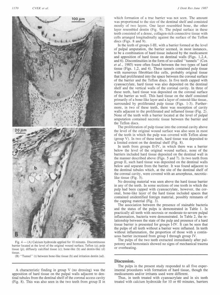
Fig.4 (A) Caliehydroxideapupaledr 10MinutesDcontto The pulps of the two teeth extracted immediately after pulFrig. 4o(a)e Cathlcum thydoieo'napleO11d It)fac Discfoninuous potomny and hemnostasis showed no signs ofnmechanical traumabarrcilcatdattheleve ofIhorgiaWCudsrceTe Io (a): 1pul or overheating.
tissue (p). diffuLsely calcificd tissue (c)-, hone-like (h) and dentin -like (d)USSLie.
(13) 'Tunnel" (t) between hone-like tissue (h) and inritation dent in (sd)._________________ ___ ____ ___ ___ ____ ___ ___ Discussion .
The pulps in the present study responded to all five exper-A characteristic finding in group V (no dressing) was the imiental procedures with formation of hard tissue, though the
apposition of hard tissue on the pulpal walls adjacent to den- medicaments and/or irritants used were different.tinal tubules from the dentinal shelf of the coronal access cavity Thus, in teeth treated with cyanoacrylate and in six teeth(Fig. 8). This was also seen in the two teeth from group 11 inI treated with calcium hydroxide for 10 or 60 minutes, barriers
J Deolt Resd Jlune 19<S7

66LlAJP)CA/PING 1411-1 ( }A\()1(1111 All ANI) C'Af C' HYDROAII)117
Fi-. 5 - (A) Calcium hxdroxidc applied tor 10 minutes. A continuousbiarier (b) located about 1.5 mm below the level of the oiiginal woundsurface: Teflon (a); necrotic tissue (ni)
20fO m (3) Deposittio of irritation dentin (sd) beloss and sepai-ate froan thebarrier: bone-like (b) and dentin-likc (d) tissuLC.
(C) Necrotic tiSSUeC ()aIaOss) on dentiatal shell. covering tLihuls coIn-spondi nu to secondlarv dentin formation.
were f rmed at thc surface of the original pulpal wound. Thesebarriers consisted of hard tissue that formed in response to thepredicament that was placed on the surface of' the pulp. Inaddition, hard tissue deposited on the dentinal walls appearedto have been induced by the irritation trom the dentinal shellof the pulpotony access cavity. Both types of tissue seem tohave been formed simultaneously. Inosone cacsen, they werein contact with each other, and in others they were separatedby 'tunnels" containing soft tissue. With respect to the teethtreated with calcium hydroxide, presumably the caustic effecttAf the medicament was responsible lor the formation of thehard tissue barrier at the level of the pulpsal wound. It was notstrong enough to destroy pulp tissue completely but was astimulant sufficicnt to induce hard tissue formation.
Apposition of hard tissue to dentinal walls was a character-istic finding in teeth in which the pulps were covered only withTeflon. The source of irritation may have been the operationaltrauma together with debris and/or necrotic tissue, which was
seen coverinL the dentinal shelf in Sonme cases. In these teeth.the pulp had formed a laycr of dense, collagen-rich tissue thatwas in contact with the Teflon. This ability of the pulp to forma fibrous barrier in response to a foreign, biologically inertsubstance has been demonstrated previously by Granath andHagman (1971). In two teeth treated with calcium hydroxidefor 10 miultCS. opposition of' hard tissue to dentinal walls wasseen below and separate from a continuous barrier. It couldhave been due to the wide dentinal shelf which wias connectedwith deeper parts of the pulp and eventually the presence ofbacterial in the coronal cavity.
The present findings concerning the ability of cyanoacrylateto induce hard tissue corroborate the results reported by Berk-man et al. (1971). Trhe barriers induced in this study were,however, discontinuous and permitted proliferation of pulp tis-sue into the coronal cavity. This clearly indicates that cyanoac-rylates cannot be regarded as an adequate therapeutic alternativeto calcium hydroxide.
Vo}l. 66t NotS. () 11 71

1172 ('Il-,'K !I tLle9.
100 Am
I
4
4eN..i W.
I AP4d42/tw
ft
7 Calcium hydroxide applied for 60 m11inc1ites. ConatinuouLLIs bar--ci. located bMlows the evci Ol the01t cinal OLuIlnd Surtace; lavis otfnecrotic (n). bone-likc (h) and dentin-like (d) tiSSUCe intlaiiLiIIItiontice-cpul1p (p1 ibis section IS also tVypical ot tCettl xVith PLilps cxCied vWithcltI uILIm hv-dioxide for I2 xnecks.
b
IS
30/,m
Fiel. 6 -(A) CalICiLumII hydroxide applied fr 6011)m1inuLtes. Discontilluosharricr, located at the leN'el oft thre (ViiIlil WoUnd SLcIItfCC. Tetlon (a):iirritation dentin (sd): bonc-like (b) and dlentin-like (di) tsscle-
(t3) DemarIcatiol linl (ariows) between bonc-like tissuLe (hi and irritationdentil (Sci).
In a num11bCr of cases, pi-oliftcrationl o1f pulp tissue Wxas fol-
lowed by the opposition of bone-like tissue On the walls of thecoional. cavity. 1his findings is intcresting1 from the standpointof hard tissue induction. It has been shown that dentin, treated
in different ways. possesses a capacity to induce formation ofectopic hone (Urist et c(i., 1968: Bang. 1973). Therefore, onCmay speculate that when in contact with vital tissue, non-vitaldentin may induce the dif lerentiation of odontoprogenitor cellswhich results in apposition of bone-like tissue on the dentinalcavity walls. A parallel can be drawn with opposition of hardtissue on the walls of the pulp chamber in re implanted teeth,after the necrotic pulp has been replaced by ingrowing mes-
enchymal tissue (Ohman. 1965: Kling et ait., 1986).After treatment with calcium hydroxide, the pulps responded
with formation of a hard tissue barrier in all but two teeth.regardless of' the treatment duration. i.e.. after 10 or 60 min-utes. or after 12 weeks ot pulpal contact. Most of the barrierswere formed below the level of the original Wound surface andwere typical, with respect to hard tissue layers (Schroder andGranath. 1971). These results show. first, that a short treat-ment with calcium hydroxide is enough to induce formationof hard tissue, and second, that it is not necessary to place thecompoLund over a pulpal wound for a longer interval in orderto induce the formation of a complete hard tissue barrier. Thisobservation supports the finding of Pisantl anti Sciaky (1 964)that calcium, which is needed for mineraliziation, is derivedfrom tissue fluid. It is also probable that the beneficial effectof calcium hydroxide. which is left over the exposure site tor
ai longer time, is due to its antibacterial property.AlthoLugh a particular inductive effect of calcium ions cannot
be excluded, the short treatmcnit experiments, together withthose using cy anoaciy late as a wxfound dressing. support thetheory that a low-grade irritation is responsible for the for-
mition ot the hard tissue barrier. SuIch a view iS also supportedby recent finldings that the effects of calciLum11 hydroxide arenon-specific and highly pH-dependent (Gordon et at.. 1985).Moreover our results support the concept that any low-grade
200gmf iV
JI )e w1 Res .Junlt, 1'<7
VUo6P CAPIIINAG WITH ( YANOA( RYI.AJI7l' AN!) C HAYDICH4HYIROXIDIE
a 100 Pm
20/m,low
(p4 ( :
s~s
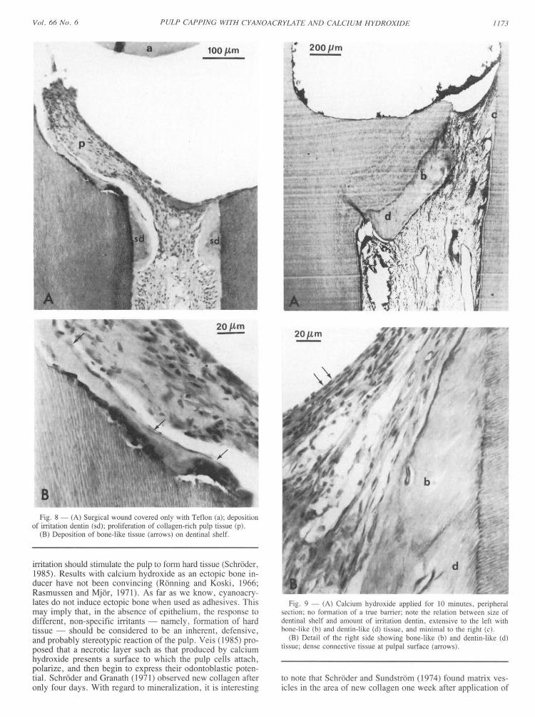
B.Fi. 8 (IN) Surgical wound covered only with Teflon (a): deposition
of irTitation dentin (sd), proliferation of collagen-rich pulp tissue (p).(B) Deposition of bo.nc-lilkc tissue (arrows) on dentinal shelf.
irritation should stimulate the pulp to form hard tissue (Schrdder,1985). Results with calcium hydroxide as an ectopic bone inlducer have not been convincing (Rdnning and Koski, 1966;Rasmussen and Mj6ri 1971). As far as we know. cyanoacry-lates do not induce ectopic bone when used as adhesives. Thismay imply that, in the absence of epithelium. the response todifferent, non specific irritants -namely. formation of hardtissue should be considered to be an inherent, defensive,and probably stereotypic reaction of the pulp. Vels (1985) pro-posed that a necrotic layer such as that produced by calciumhydroxide presents a surface to which the pulp cells attach,polarize, and then begin to express their odontoblastic poten-tial. Schrdder and Granath ( 1971) observed new collagen afteronly four days. With regard to mineralization. it is interesting
.,Z
i#:I/-b
BExi} .g tS~~~~~~~~~~~~~~~~~~~~~~~~~~~~~~~~~~~~~~~~~~~~~~~~~~~~~~~~~~~~~~~~f~~~~~~~~~~~~~~~~~~~~~~~~~~j...........
Fi.i 9 (A) CalciLum hsdroxidc applied for 1If nin ties. eCI-iphetalsection- no formation of a true barrier note the relation between size offdentinal shelf and aniount of irritation dentin. extcnsixe to the left SNvithhone-like (b) and dentin-like (d) tissue, and minimal to the rtiht (e).
(B) Detail of the rimht side shosinLc bone-like (b) and dentin-likeC (d)tissuLe, deCnSC conee tiCe tissue at pulpal surface (arrows).
to note that Schrdder and Sundstr6m (1974) found matrix x Csicles in the area of new' collagen one week after application of
Vol/. 00& NoJ. 0 I173
(e0

1174 CVEK et al.
TABLE 2CROSS-TABULATION OF HARD TISSUE BARRIER FORMATIONAND PULP STATE IN 39 PULPOTOMIZED MONKEY INCISORSCAPPED WITH ISOBUTYL CYANOACRYLATE OR CALCIUM
HYDROXIDE FOR DIFFERENT PERIODS OF TIME.OBSERVATIONS AFTER 12 WEEKS
Hard Tissue Barrier
Group I Groups II + III Group IV
Pulpal n = 9 n = 20 n = 10Inflammation none d c none d c none d c
none 4 1 4 6 1 9*sI 1**
m 1 4***se 2 2 1 3*Six cases, **one case, and ***two cases with no stainable bacteria.For definitions see Table 1.
calcium hydroxide on the exposed pulp. Such vesicles arethought to play a role in the initial calcification process ofdentin and other mineralizing tissues (Ten Cate and Torneck,1982). The type of tissue to be formed may depend on thedegree of irritation and on the genetic make-up of progenitorcells rather than on the irritant itself. Hence, Cvek and Sund-str6m (1974) found the apical closure of immature roots ofnon-vital permanent incisors treated with calcium hydroxide toconsist of a bone-like layer followed by cementum-like tissue.
In the present study, inflammatory changes in pulps wereclearly related to the presence of bacteria in the coronal cavity,and the continuity of the barriers was in turn related to thecondition of the pulp. This emphasizes the importance of bac-teria in pulpal (Cox et al., 1985) as well as periapical (Fabri-cius et al., 1982) inflammatory reactions. The role ininflammatory pulp disease is currently receiving a great dealof attention in the dental literature.
Acknowledgments.The authors wish to express their sincere gratitude to Drs.
Henry Trowbridge and Edward Sweeney, University of Penn-sylvania School of Dental Medicine, Philadelphia, for theirconstructive comments in the preparation of this manuscript.
REFERENCES
BANG, G. (1973): Induction of Heterotopic Bone Formation by De-mineralized Dentine: An Experimental Model in Guinea Pigs, ScandJ Dent Res 81:240-250.
BERKMAN, M.D.; CUCOLO, F.A.; LEVIN, M.P.; and BRU-NELLE, L.J. (1971): Pulpal Response to Isobutyl Cyanoacrylatein Human Teeth, J Am Dent Assoc 83:140-145.
BROWN, J. H. and BRENN, L.A. (1931): A Method for the Dif-ferential Staining of Gram-positive and Gram-negative Bacteria inTissue Sections, Bull Johns Hopkins Hosp 48:69-73.
COX, C.F.; BERGENHOLTZ, G.; FITZGERALD, M.; HEYS, D.R.;HEYS, R.J.; AVERY, J.K.; and BAKER, J.A. (1982): Cappingof the Dental Pulp Mechanically Exposed to the Oral Microflora
A 5-week Observation of Wound Healing in the Monkey, JOral Pathol 11:327-339.
COX, C.F.; BERGENHOLTZ, G.; HEYS, D.R.; SYED, S.A.; FITZ-GERALD, M.; and HEYS, R.J. (1985): Pulp Capping of DentalPulp Mechanically Exposed to Oral Microflora: A 1-2-year Ob-
servation of Wound Healing in the Monkey, J Oral Pathol 14: l56168.
CVEK, M. and SUNDSTROM, B. (1974): Treatment of Non-vitalPermanent Incisors with Calcium Hydroxide. V. Histologic Ap-pearance of Roentgenographically Demonstrable Apical Closureof Immature Roots, Odont Revy 25:379-391.
FABRICIUS, L.; DAHLEN. G.: HOLM, S.E.; and MOLLER, A.J.R.(1982): Influence of Combinations of Oral Bacteria on PeriapicalTissues of Monkeys. Scand J Dent Res 90:200-206.
GORDON, T.M.; RANLY, D.M.: and BOYAN, B.D. (1985): TheEffects of Calcium Hydroxide on Bovine Pulp Tissue: Variationsin pH and Calcium Concentration, J Endodont 11: 156-160.
GRANATH, L. (1982): Pulp Capping Materials. In: Biocompatibilityof Dental Materials, Vol. II, D.C. Smith and D.F. Williams,Eds., Boca Raton. FL: C.R.C. Press. Inc., pp. 253-267.
GRANATH, L.-E. and HAGMAN, G. (1971): Experimental Pulpo-tomy in Human Bicuspids with Reference to Cutting Technique,Acta Odontol Scand 29:155- 163.
HEYS, D.R.; COX, C.F.; HEYS, R.J.; and AVERY, J.K. (1981):Histological Considerations of Direct Pulp-capping Agents, J DentRes 60:1371-1379.
HEYS, D.R.; HEYS, R.J.; COX, C.F.; and AVERY, J.K. (1980):The Response of Four Calcium Hydroxides on Monkey Pulps, JOral Pathol 9:372-379.
HOLLAND, R.; PINHEIRO, C.E.; DE MELLO, W.; NERY, M.J.:and DE SOUZA, V. (1982): Histochemical Analysis of the Dog'sDental Pulp after Pulp Capping with Calcium, Barium, and Stron-tium Hydroxides, J Endodont 8:444-447.
KLING, M.; CVEK, M.; and MEJARE, I. (1986): Rate and Pre-dictability of Pulp Revascularization in Therapeutically Reim-planted Permanent Incisors. Endodont Dent Traumatol 2:83-89.
NIXON, G.S. and HANNAH, C. M. (1972): N-butyl Cyanoacrylateas a Pulp Capping Agent, Br Dent J 133:14-18.
OHMAN, A. (1965): Healing and Sensitivity to Pain in Young Re-planted Human Teeth. An Experimental, Clinical and HistologicalStudy. Odont Tidskr 73:165-228.
PISANTI, S. and SCIAKY, I. (1964): Origin of Calcium in the RepairWall after Pulp Exposure in the Dog, J Dent Res 43:641-644.
RASMUSSEN, P. and MJOR, I.A. (1971): Calcium Hydroxide as anEctopic Bone Inductor in Rats, Scand J Dent Res 79:24-30.
RETIEF, D.H. and AUSTIN, J.C. (1973): The Vervet Monkey (Cer-copithecus aethiops) as an Experimental Model for Pulpal Studies,J Dent Assoc S Afr 28:98-103.
RONNING, 0. and KOSKI, K. (1966): The Fate of Anorganic Im-plants in the Subcutaneous Tissue of the Rat, Plast Reconstr Surg37:121-124.
SCHRODER, U. (1985): Effects of Calcium Hydroxide-containingPulp-capping Agents on Cell Migration, Proliferation, and Differ-entiation. J Dent Res 64 (Sp. Iss.): 541-548.
SCHRODER, U. and GRANATH, L.-E. (1971): Early Reaction ofIntact Human Teeth to Calcium Hydroxide Following Experimen-tal Pulpotomy and Its Significance to the Development of HardTissue Barrier, Odont Revy 22:379-396.
SCHRODER, U. and SUNDSTROM, B. (1974): Transmission Elec-tron Microscopy of Tissue Changes Following Experimental Pul-potomy of Intact Human Teeth and Capping with CalciumHydroxide, Odont Revy 25:57-67.
TEN CATE, A.R. and TORNECK, C.D. (1982): The Dentin-PulpComplex: Development, Structure, and Repair. In: Biocompati-bility of Dental Materials, Vol. I, D.C. Smith and D.F. Wil-liams, Eds., Boca Raton, FL: C.R.C. Press, Inc., pp. 85-86.
TRONSTAD, L. (1974): Reaction of the Exposed Pulp to DycalTreatment, Oral Surg 38:945-953.
URIST, M.R.; DOWELL, T.A.; HAY, P.H.; and STRATES, B.S.(1968): Inductive Substrates for Bone Formation, Clin Orthop 59:59-96.
VEIS, A. (1985): The Role of Dental Pulp-Thoughts on the Sessionon Pulp Repair Processes, J Dent Res 64(Sp. Iss.): 552-554.
J Denlt Re~s Junbe 198'7