tissue identification - suffolk county community … hints on finding tissues on slides: keep in...
TRANSCRIPT

Tissue Identification Exercise 6A/6 (begins page 67 in 9th, 10th, 11th and 12th editions) Lab 4 Objectives Read lab Exercise 6A/6 Do Activities 1-4 Histology: identify the following tissue types using the complete name: 1. Simple squamous epithelium 2. Simple cuboidal epithelium 3. Simple columnar epithelium 4. Pseudostratified columnar epithelium (ciliated) 5. Stratified squamous epithelium non-keratinized (mucosa) 6. Stratified squamous epithelium keratinized (epidermis) (see figures 7.2 a & 7.6b) 7. Transitional epithelium 8. Areolar connective tissue 9. Adipose tissue 10. Reticular connective tissue 11. Dense regular connective tissue 12. Dense irregular connective tissue 13. Elastic connective tissue (not in 9th edition- see page 2 of this handout) 14. Hyaline cartilage 15. Elastic cartilage 16. Fibrocartilage 17. Compact bone / Osseous tissue 18. Blood 19. Skeletal muscle 20. Cardiac muscle 21. Smooth muscle 22. Nervous tissue Optional Computer Activity:
PhysioEx Exercise 6B (On the PhysioEx CD-ROM packaged with the lab book) pages PEx-21 to PEx-22 (back of the book) in 9th and10th editions (not in 11th or 12th editions)
For Study: Review Sheet Exercise 6A/6 pages 85-90 in 9th and 10th editions
pages 87-92 in 11th and 12th editions
Answers in the Instructors Manual at the Eastern Campus Library on reserve
Amy Warenda Czura, Ph.D. 1 SCCC BIO130 Lab 4 Tissues
Amy Warenda Czura, Ph.D. 2 SCCC BIO130 Lab 4 Tissues
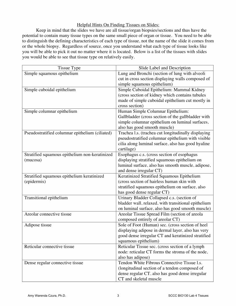
Helpful Hints On Finding Tissues on Slides: Keep in mind that the slides we have are all tissue/organ biopsies/sections and thus have the
potential to contain many tissue types on the same small piece of organ or tissue. You need to be able to distinguish the defining characteristics of each type of tissue, not the name of the slide it comes from or the whole biopsy. Regardless of source, once you understand what each type of tissue looks like you will be able to pick it out no matter where it is located. Below is a list of the tissues with slides you would be able to see that tissue type on relatively easily.
Tissue Type Slide Label and Description Simple squamous epithelium Lung and Bronchi (section of lung with alveoli
cut in cross section displaying walls composed of simple squamous epithelium)
Simple cuboidal epithelium Simple Cuboidal Epithelium: Mammal Kidney (cross section of kidney which contains tubules made of simple cuboidal epithelium cut mostly in cross section)
Simple columnar epithelium Human Simple Columnar Epithelium: Gallbladder (cross section of the gallbladder with simple columnar epithelium on luminal surfaces, also has good smooth muscle)
Pseudostratified columnar epithelium (ciliated) Trachea l.s. (trachea cut longitudinally displaying pseudostratified columnar epithelium with visible cilia along luminal surface, also has good hyaline cartilage)
Stratified squamous epithelium non-keratinized (mucosa)
Esophagus c.s. (cross section of esophagus displaying stratified squamous epithelium on luminal surface, also has smooth muscle, adipose, and dense irregular CT)
Stratified squamous epithelium keratinized (epidermis)
Keratinized Stratified Squamous Epithelium (cross section of hairless human skin with stratified squamous epithelium on surface, also has good dense regular CT)
Transitional epithelium Urinary Bladder Collapsed c.s. (section of bladder wall, relaxed, with transitional epithelium on luminal surface, also has good smooth muscle)
Areolar connective tissue Areolar Tissue Spread Film (section of areola composed entirely of areolar CT)
Adipose tissue Sole of Foot (Human) sec. (cross section of heel displaying adipose in dermal layer, also has very good dense irregular CT and keratinized stratified squamous epithelium)
Reticular connective tissue Reticular Tissue sec. (cross section of a lymph node: reticular CT forms the stroma of the node, also has adipose)
Dense regular connective tissue Tendon White Fibrous Connective Tissue l.s. (longitudinal section of a tendon composed of dense regular CT, also has good dense irregular CT and skeletal muscle
Amy Warenda Czura, Ph.D. 3 SCCC BIO130 Lab 4 Tissues

Dense irregular connective tissue Human Scalp sec. With Hair (cross section through scalp skin displaying dense irregular CT in the dermal layer, also has good keratinized stratified squamous epithelium)
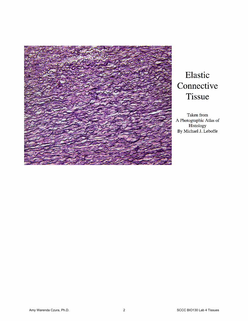
Elastic connective tissue Aortic Elastic Tissue Human (longitudinal section of aorta, with the bulk of wall composed of elastic CT)
Hyaline cartilage Trachea c.s. (cross section through trachea showing C-shaped rings of hyaline cartilage, also has good adipose, pseudostratified ciliated columnar epithelium, and simple cuboidal epithelium
Elastic cartilage Elastic Cartilage: Epiglottis sec. (longitudinal section of epiglottis displaying elastic cartilage in the center, also has good non-keratinized stratified squamous epithelium)
Fibrocartilage White Fibro-cartilage: Pubic Symphysis sec. (cross section of the junction of the pubic bones, the pubic symphysis, composed of fibrocartilage, also has skeletal muscle and adipose)
Compact bone / Osseous tissue Bone Dry Ground, Human c.s. (section of compact bone)
Blood Human Blood Smear Wright Stain (stained human blood smear)
Skeletal muscle Skeletal Muscle Teased (skeletal muscle cells pulled apart, not sectioned, appear 3 dimensional)
Cardiac muscle Intercalated Discs, Heart sec. (cross section of the heart wall showing all cardiac muscle cells)
Smooth muscle Duodenum c.s. (cross section of small intestine displaying smooth muscle deep to the epithelium, also has good columnar epithelium)
Nervous tissue Giant Multipolar Neurons Smear (smear of central nervous system tissue showing large star-shaped neurons)
Photo pages can be found in front of each slide tray indicating the various tissue types that are visible on each slide. It would be very helpful for you to orient yourself one slide at a time using these photos as a guide. Tissue types are generally best observed at 400X so that the cellular details that distinguish them are evident. The easiest slides to start with, since they have only one tissue type on them are: Areolar Connective Tissue Blood Bone Cardiac Muscle Skeletal Muscle Giant Multipolar Neuron
Amy Warenda Czura, Ph.D. 4 SCCC BIO130 Lab 4 Tissues