tissues. hierarchy of organization –atoms –molecules –cells –tissues –organs –organ...
TRANSCRIPT
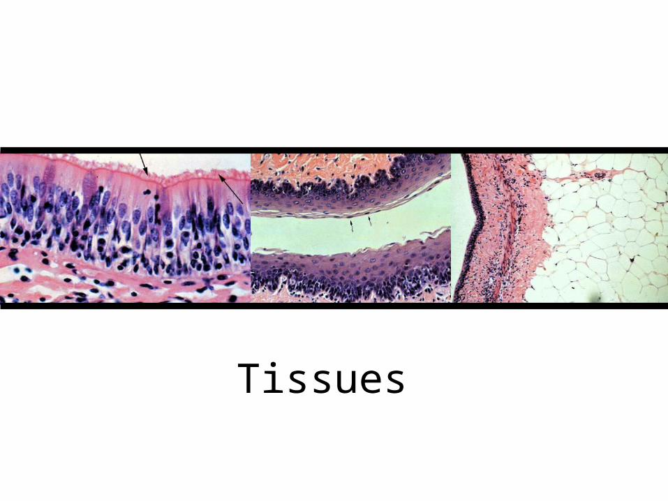
Tissues
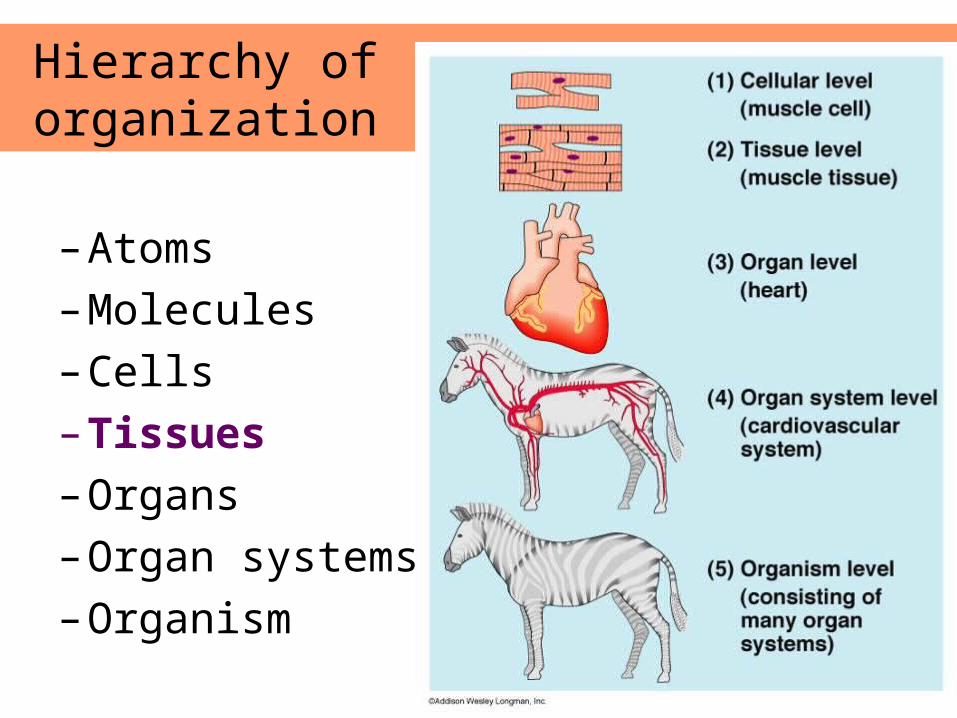
Hierarchy of organization
– Atoms
– Molecules
– Cells
– Tissues
– Organs
– Organ systems
– Organism
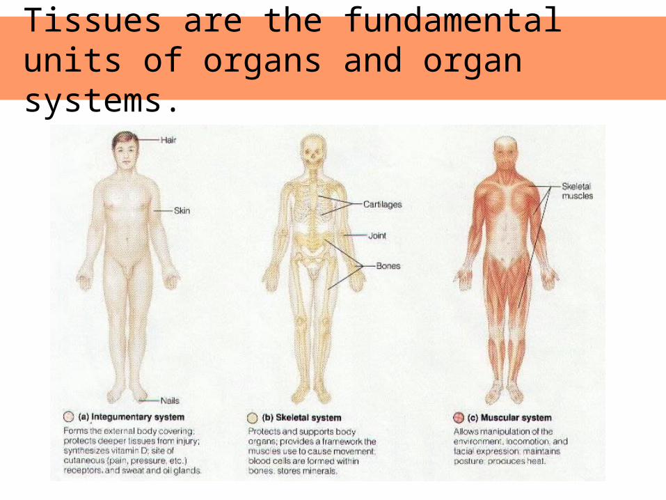
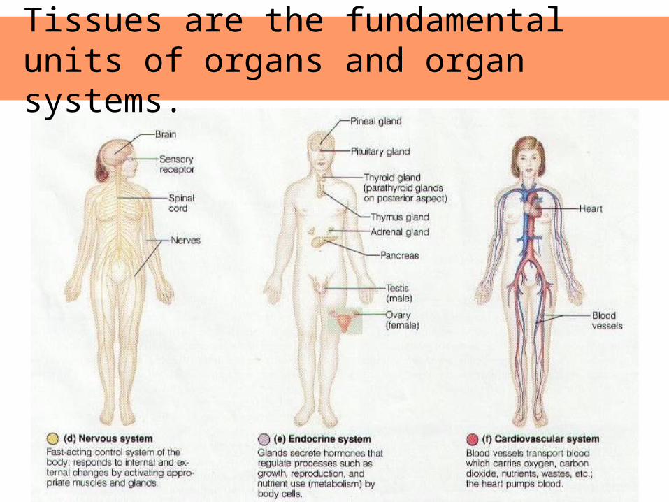
Tissues are the fundamental units of organs and organ systems.
Tissues are the fundamental units of organs and organ systems.
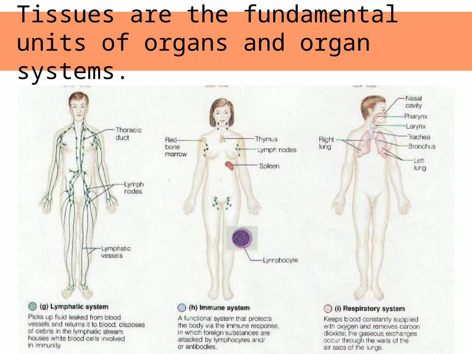
Tissues are the fundamental units of organs and organ systems.
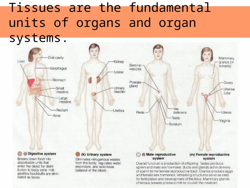
Tissues are the fundamental units of organs and organ systems.
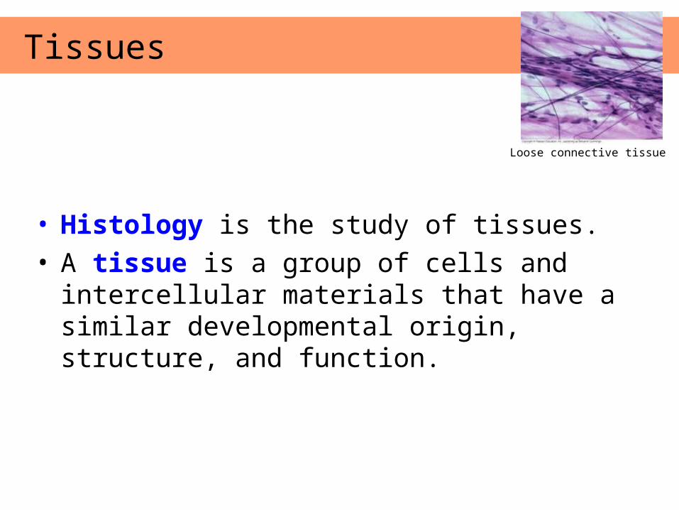
• Histology is the study of tissues.• A tissue is a group of cells and intercellular
materials that have a similar developmental origin, structure, and function.
Tissues
Loose connective tissue
• Know the structure and be able to identify it from a slide.
• Know the function.
• Know location in the body it is likely to be found.
• Know any special attributes of that tissue.
What you should know about a tissue
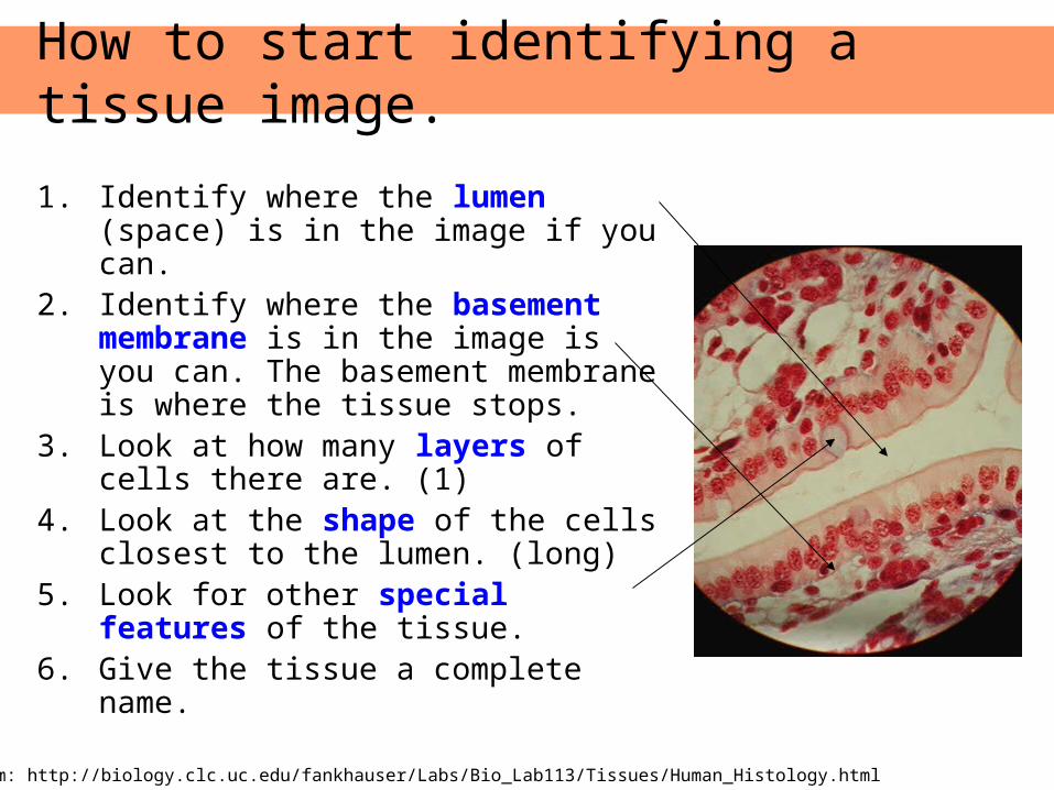
1. Identify where the lumen (space) is in the image if you can.
2. Identify where the basement membrane is in the image is you can. The basement membrane is where the tissue stops.
3. Look at how many layers of cells there are. (1)
4. Look at the shape of the cells closest to the lumen. (long)
5. Look for other special features of the tissue.
6. Give the tissue a complete name.
How to start identifying a tissue image.
Image from: http://biology.clc.uc.edu/fankhauser/Labs/Bio_Lab113/Tissues/Human_Histology.html
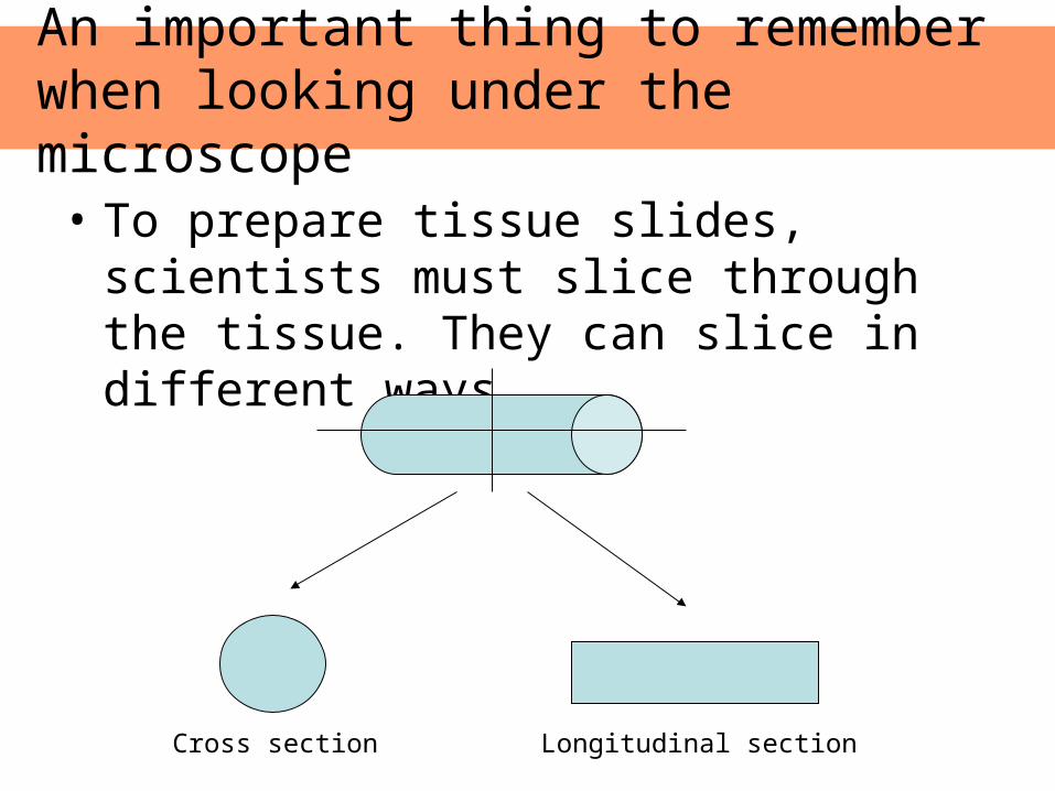
An important thing to remember when looking under the microscope
• To prepare tissue slides, scientists must slice through the tissue. They can slice in different ways.
Cross section Longitudinal section
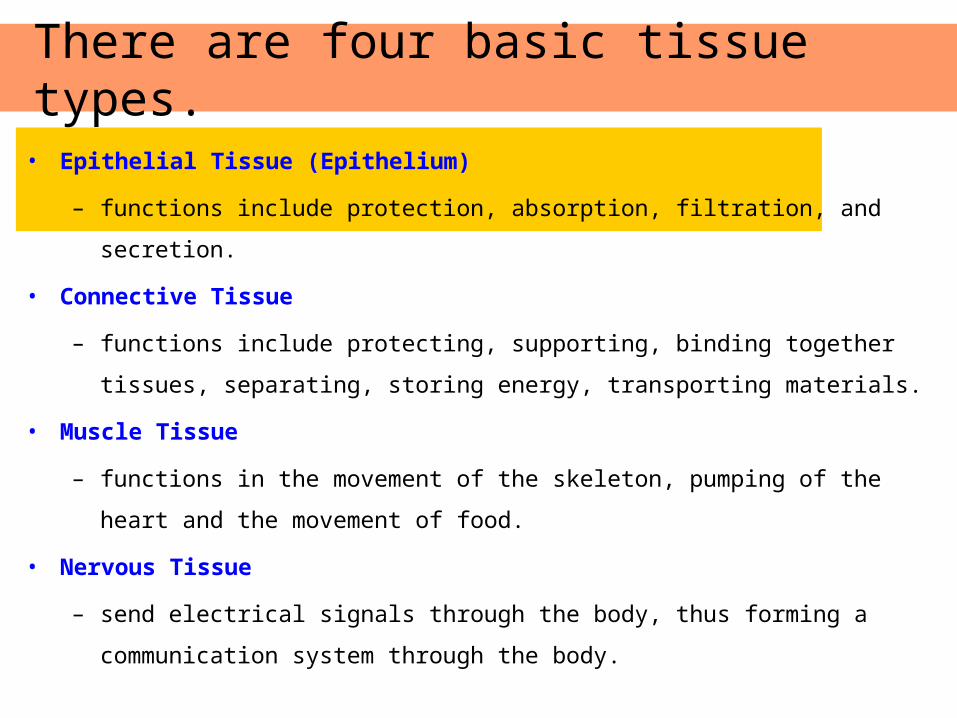
• Epithelial Tissue (Epithelium)
– functions include protection, absorption, filtration, and secretion.
• Connective Tissue
– functions include protecting, supporting, binding together tissues, separating,
storing energy, transporting materials.
• Muscle Tissue
– functions in the movement of the skeleton, pumping of the heart and the
movement of food.
• Nervous Tissue
– send electrical signals through the body, thus forming a communication
system through the body.
There are four basic tissue types.
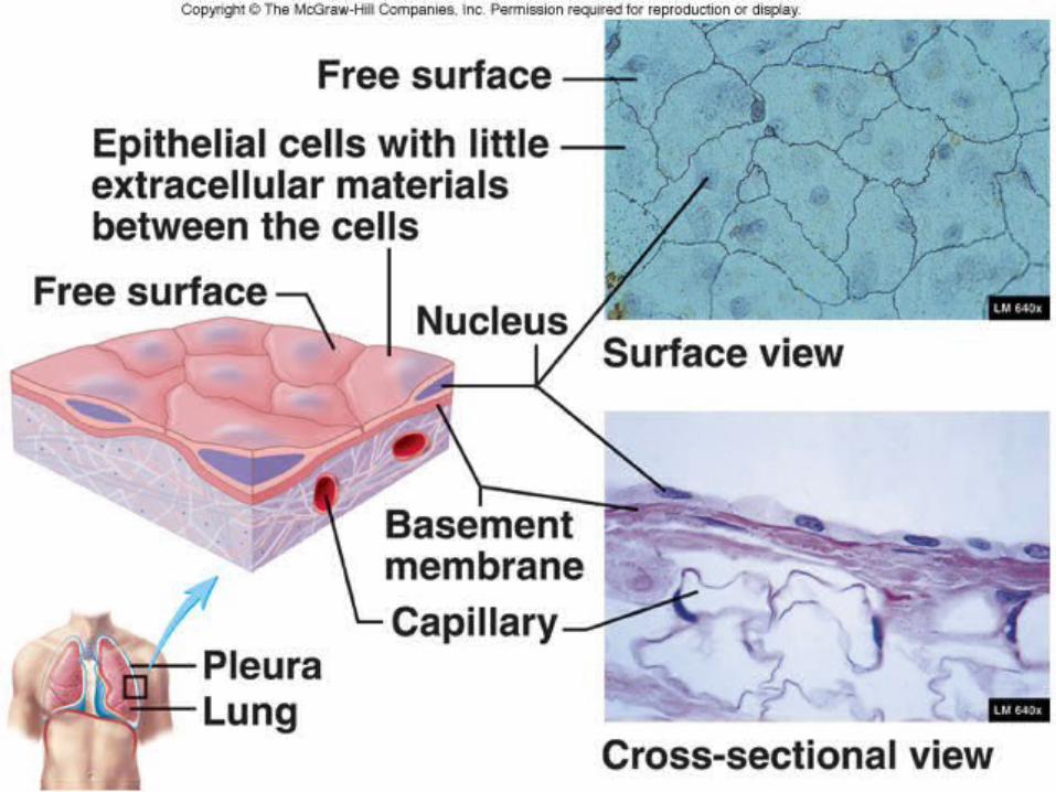
• Cells fit closely together to form continuous sheets and are bound together at many points by cell junctions.
• Cells have one free surface or edge. This apical surface is exposed to the body’s exterior or to a cavity (the lumen)
• The lower cell surface rests on a basement membrane, a structureless material secreted by the cells.
• These tissues are avascular, meaning that they have no blood supply and depend on diffusion from capillaries in the underlying connective tissue
• If well nourished, they can regenerate easily.
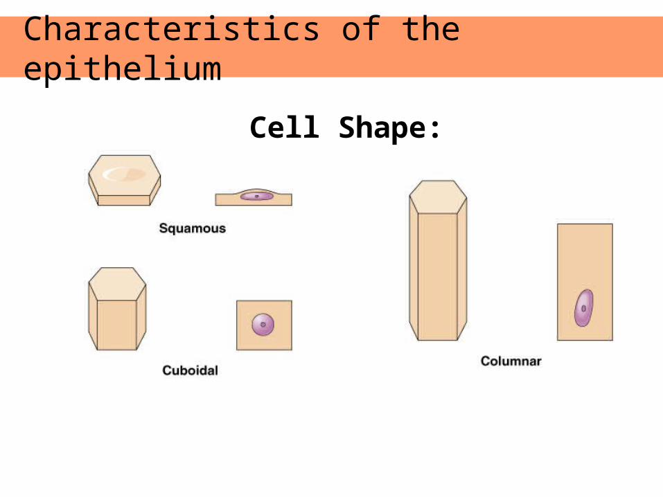
Characteristics of the epithelium
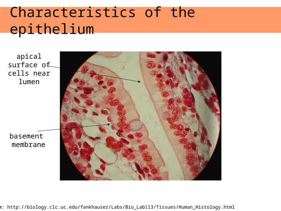
Characteristics of the epithelium
apical surface of cells near
lumen
basement membrane
Image from: http://biology.clc.uc.edu/fankhauser/Labs/Bio_Lab113/Tissues/Human_Histology.html
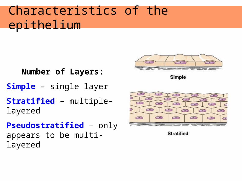
Number of Layers:
Simple – single layer
Stratified – multiple-layered
Pseudostratified – only appears to be multi-layered
Characteristics of the epithelium
Characteristics of the epithelium
Cell Shape:
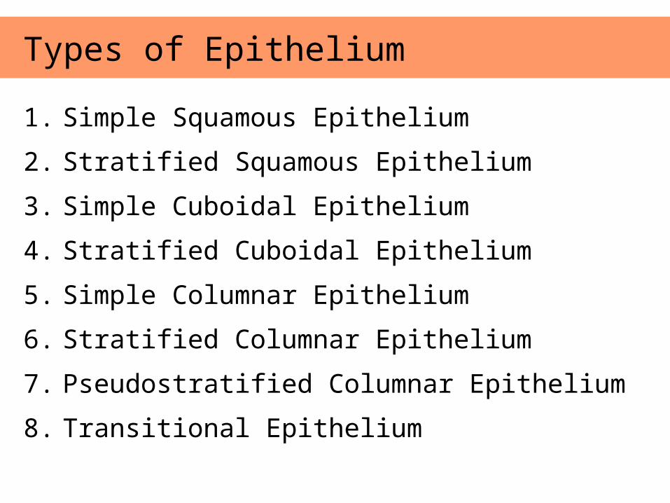
1. Simple Squamous Epithelium
2. Stratified Squamous Epithelium
3. Simple Cuboidal Epithelium
4. Stratified Cuboidal Epithelium
5. Simple Columnar Epithelium
6. Stratified Columnar Epithelium
7. Pseudostratified Columnar Epithelium
8. Transitional Epithelium
Types of Epithelium
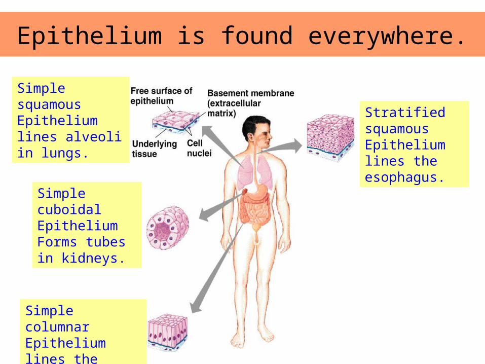
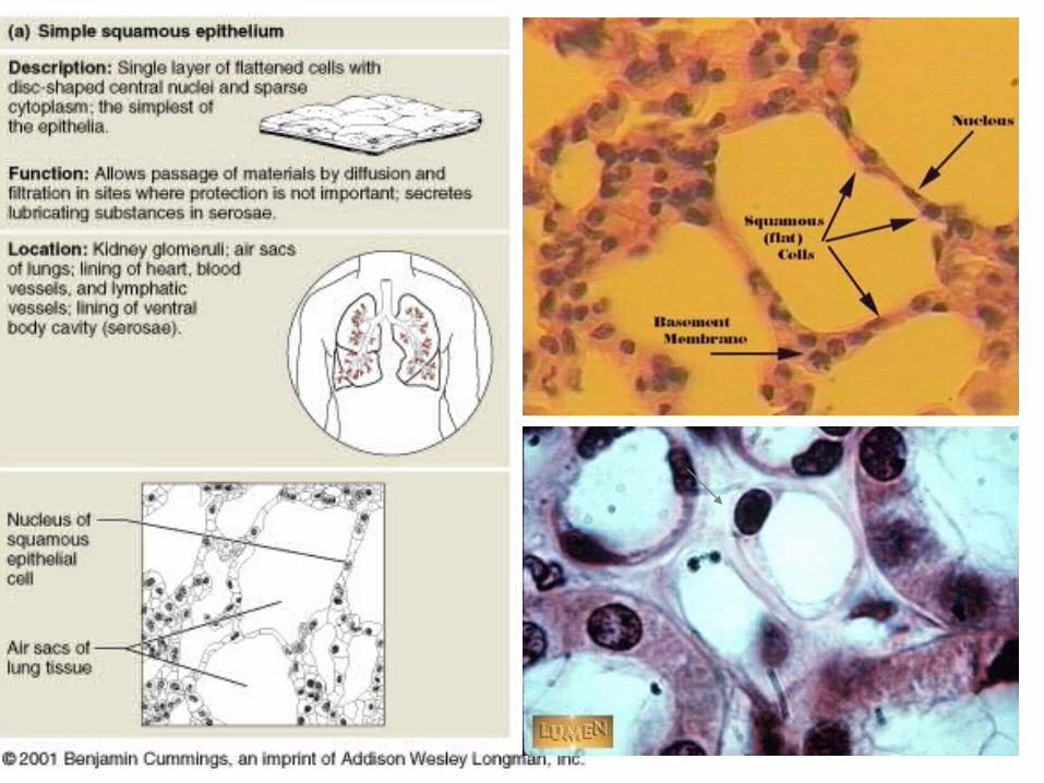
Simple squamous Epithelium lines alveoli in lungs.
Simple cuboidal EpitheliumForms tubes in kidneys.
Simple columnar Epithelium lines the intestine.
Stratified squamous Epithelium lines the esophagus.
Epithelium is found everywhere.
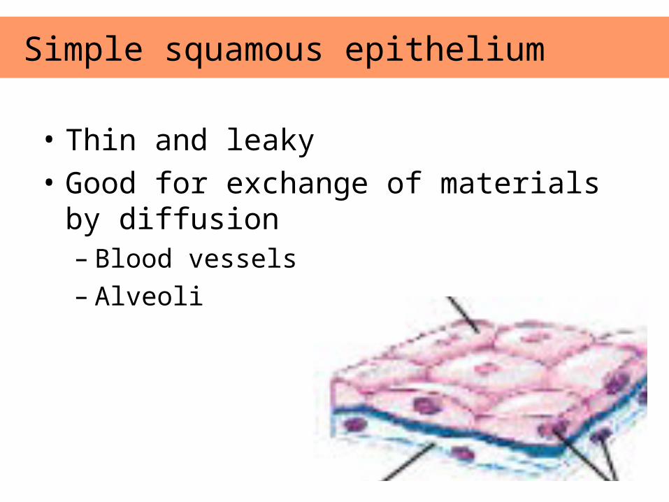
• Thin and leaky
• Good for exchange of materials by diffusion– Blood vessels– Alveoli
Simple squamous epithelium
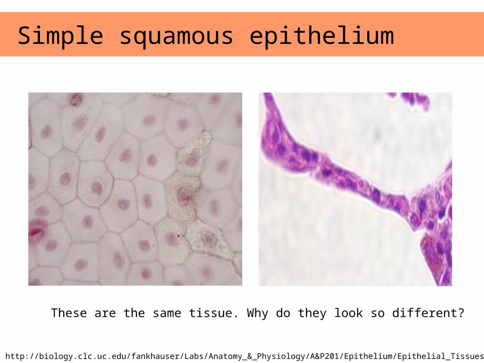
Simple squamous epithelium
Images from: http://biology.clc.uc.edu/fankhauser/Labs/Anatomy_&_Physiology/A&P201/Epithelium/Epithelial_Tissues.htm
These are the same tissue. Why do they look so different?
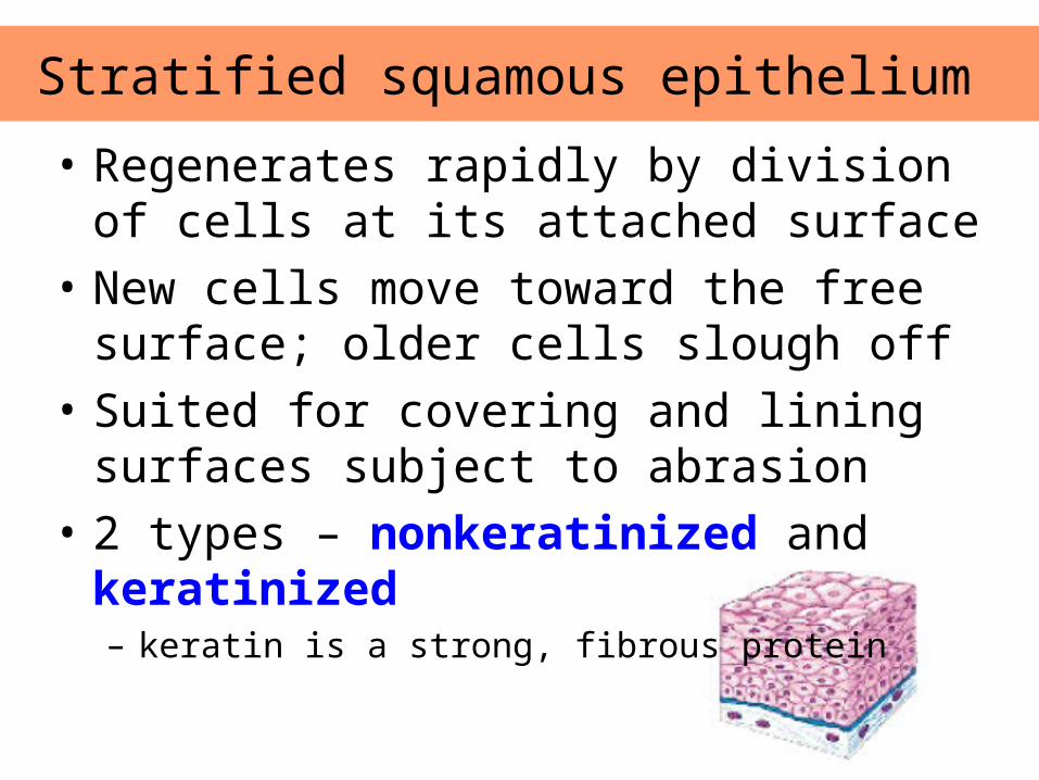
• Regenerates rapidly by division of cells at its attached surface
• New cells move toward the free surface; older cells slough off
• Suited for covering and lining surfaces subject to abrasion
• 2 types – nonkeratinized and keratinized– keratin is a strong, fibrous protein
Stratified squamous epithelium
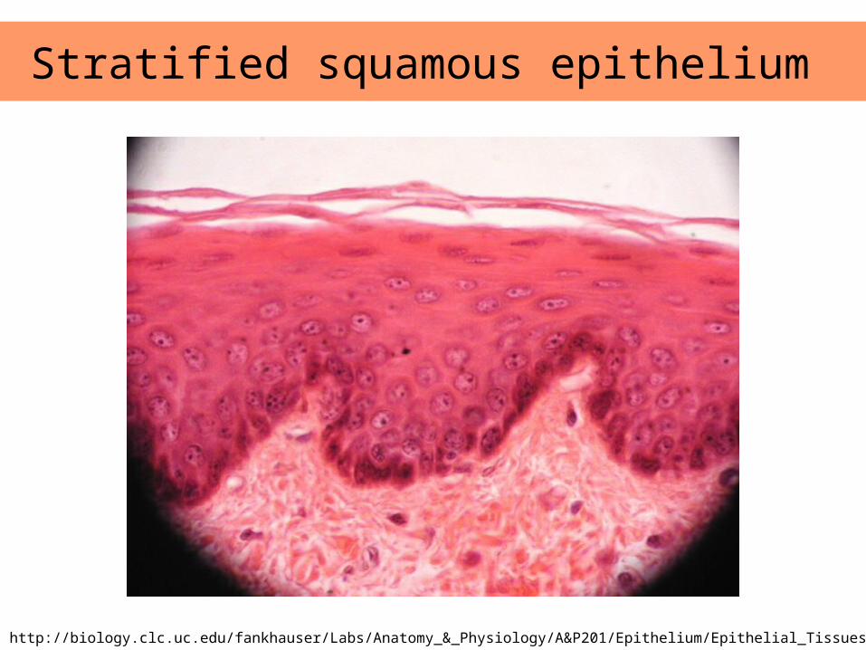
Stratified squamous epithelium
Images from: http://biology.clc.uc.edu/fankhauser/Labs/Anatomy_&_Physiology/A&P201/Epithelium/Epithelial_Tissues.htm
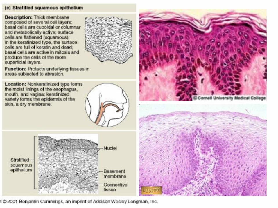
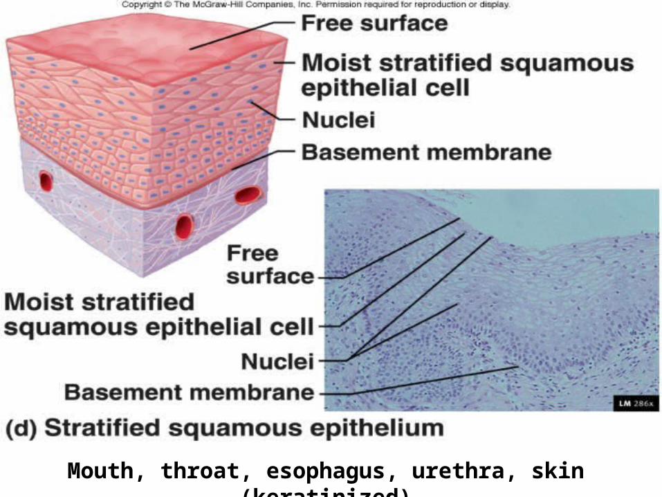
Mouth, throat, esophagus, urethra, skin (keratinized)
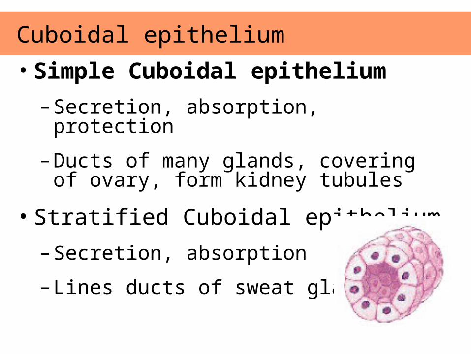
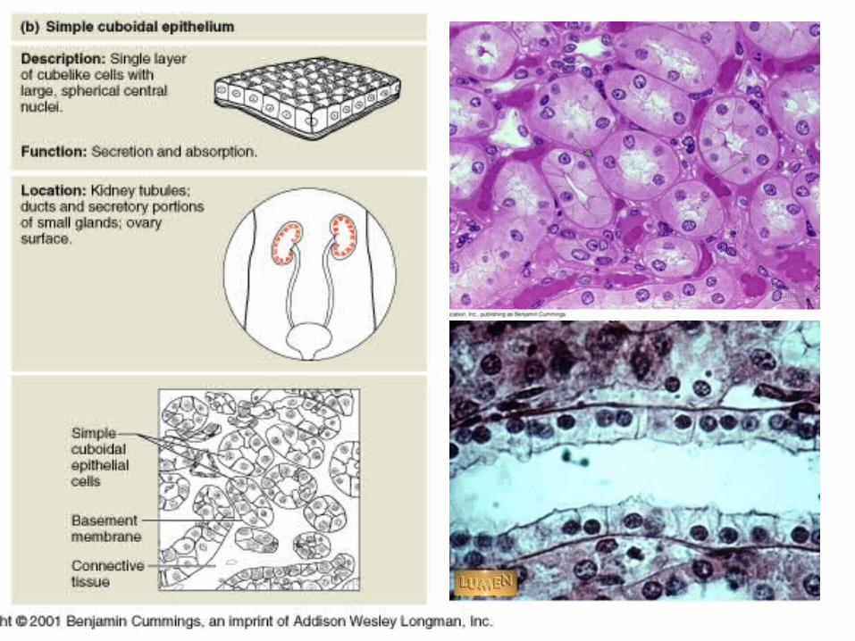
• Simple Cuboidal epithelium
– Secretion, absorption, protection
– Ducts of many glands, covering of ovary, form kidney tubules
• Stratified Cuboidal epithelium
– Secretion, absorption
– Lines ducts of sweat glands.
Cuboidal epithelium
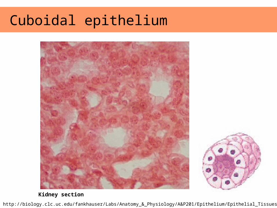
Cuboidal epithelium
Images from: http://biology.clc.uc.edu/fankhauser/Labs/Anatomy_&_Physiology/A&P201/Epithelium/Epithelial_Tissues.htm
Kidney section
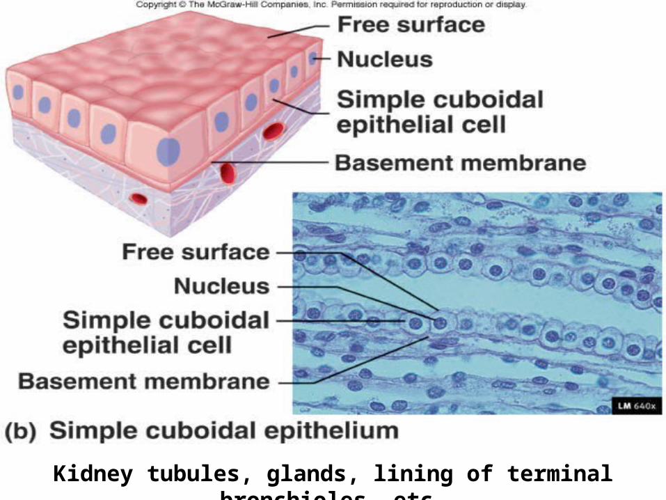
Kidney tubules, glands, lining of terminal bronchioles, etc.
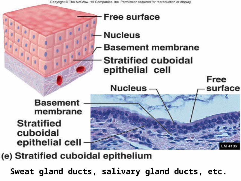
Sweat gland ducts, salivary gland ducts, etc.
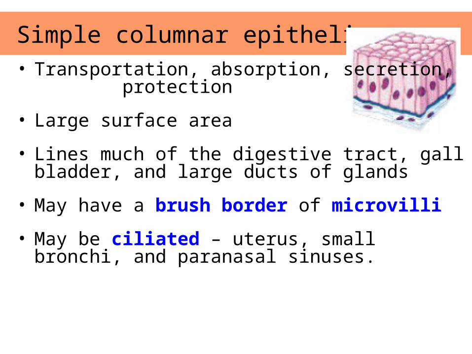
Simple columnar epithelium • Transportation, absorption, secretion,
protection
• Large surface area
• Lines much of the digestive tract, gall bladder, and large ducts of glands
• May have a brush border of microvilli
• May be ciliated – uterus, small bronchi, and paranasal sinuses.
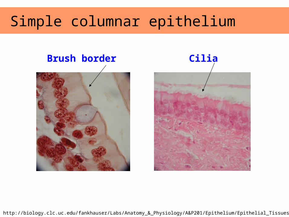
Simple columnar epithelium
Brush border Cilia
Images from: http://biology.clc.uc.edu/fankhauser/Labs/Anatomy_&_Physiology/A&P201/Epithelium/Epithelial_Tissues.htm
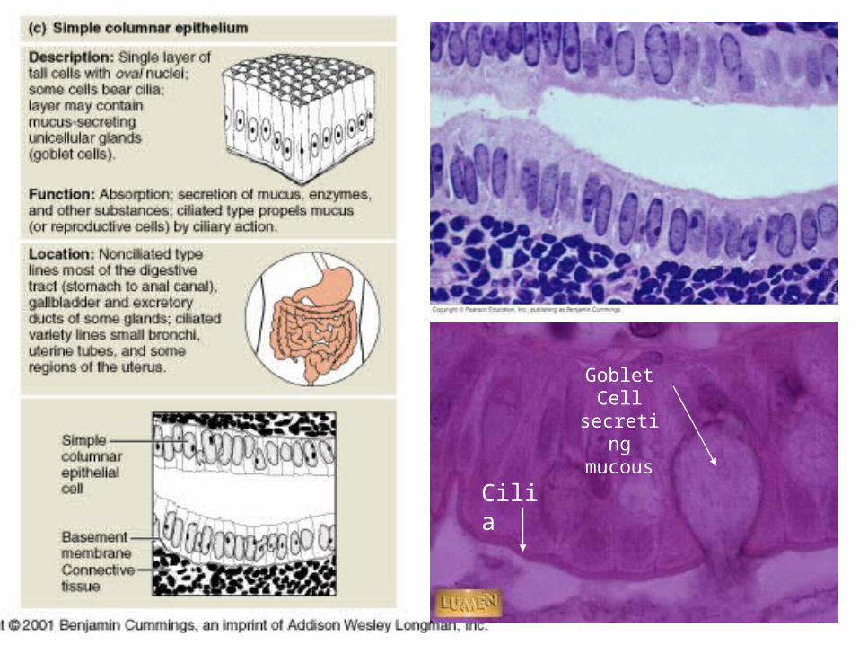
Goblet Cell secreting mucous
Cilia
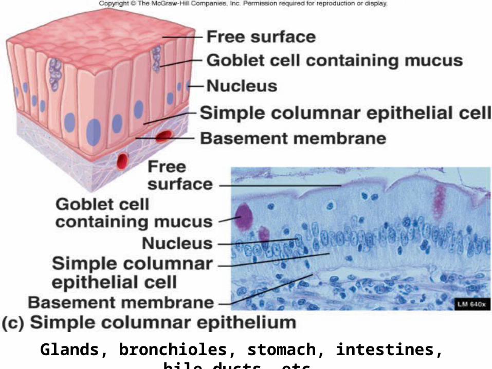
Glands, bronchioles, stomach, intestines, bile ducts, etc.
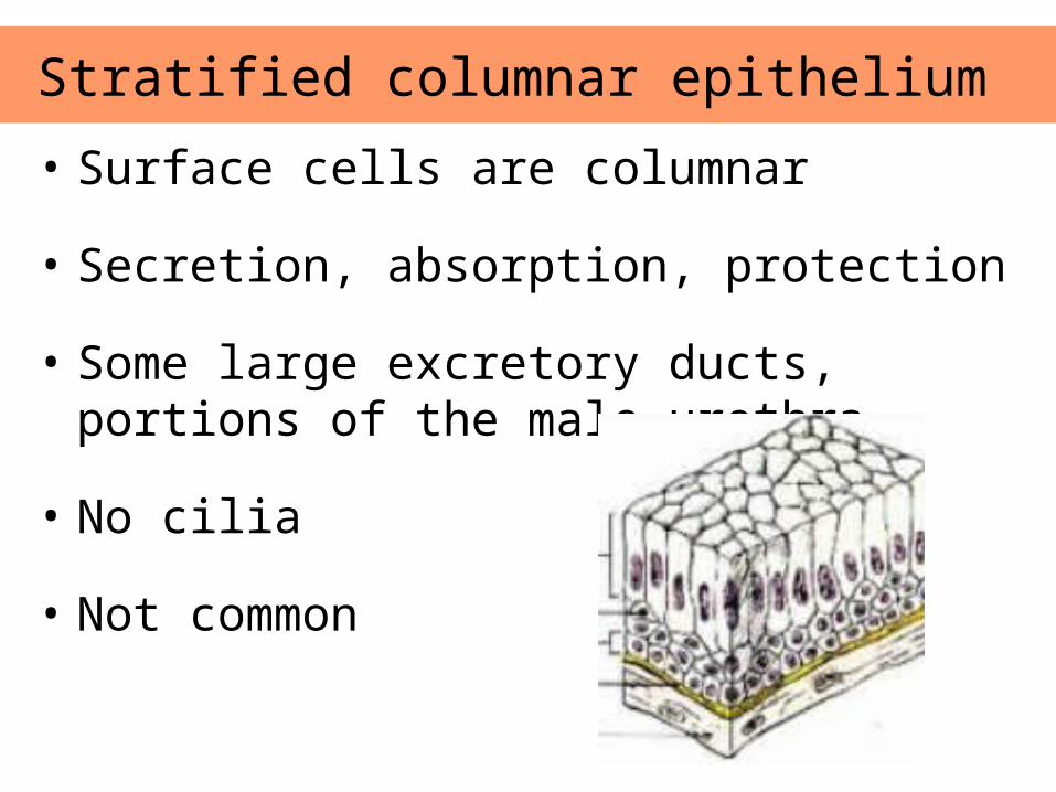
• Surface cells are columnar
• Secretion, absorption, protection
• Some large excretory ducts, portions of the male urethra
• No cilia
• Not common
Stratified columnar epithelium
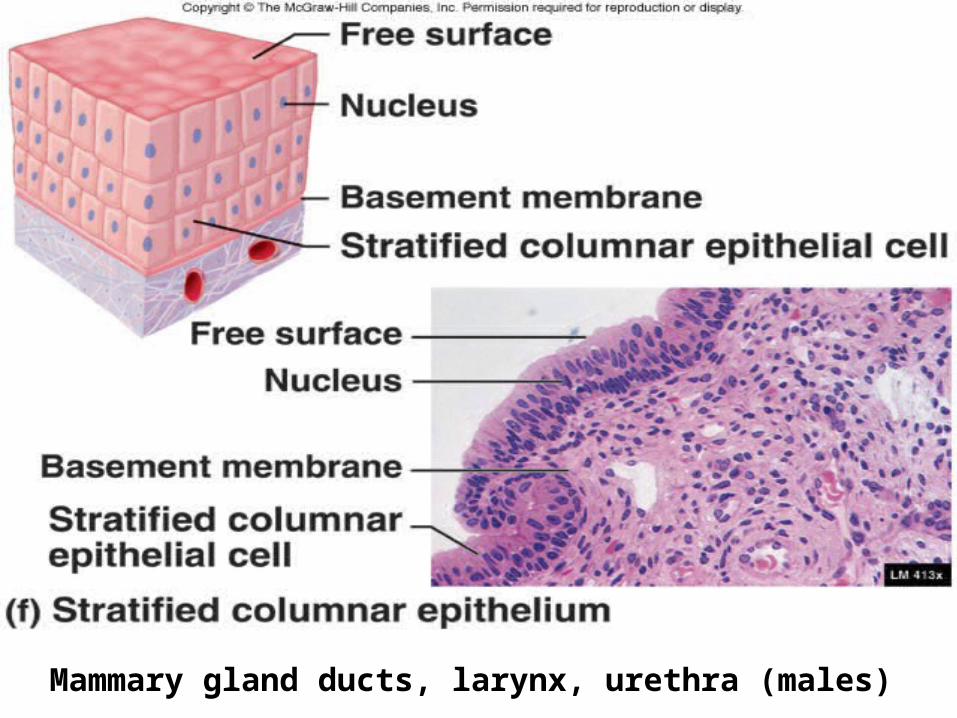
Mammary gland ducts, larynx, urethra (males)
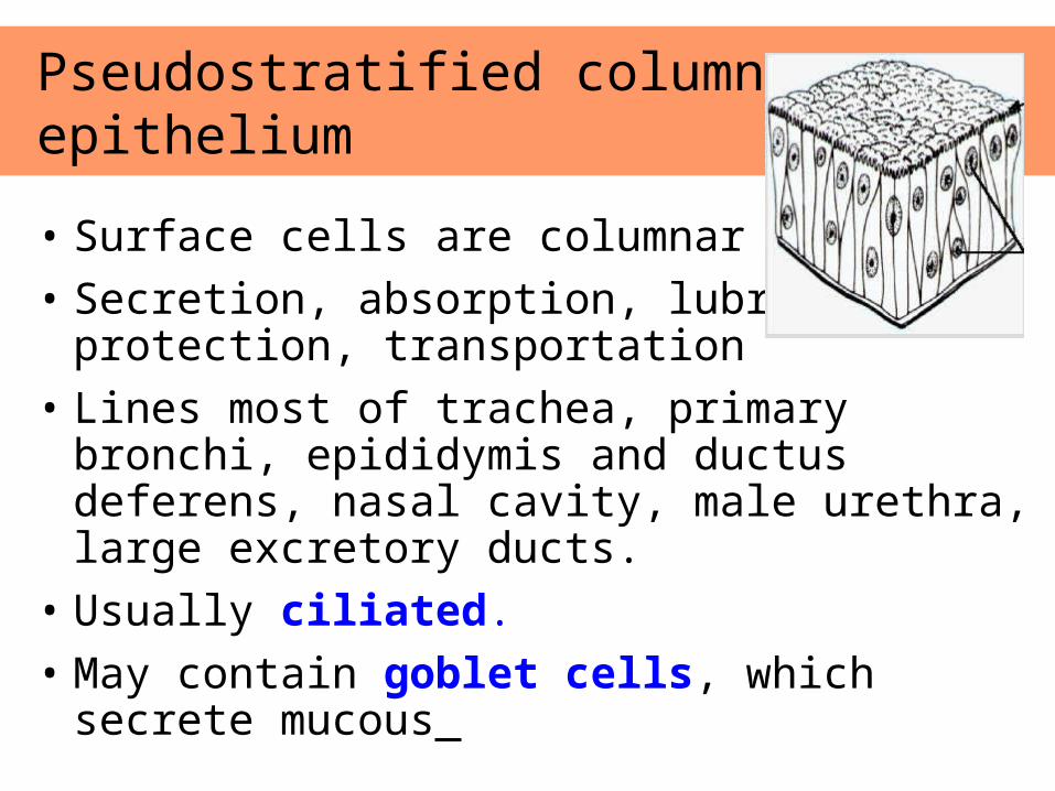
• Surface cells are columnar
• Secretion, absorption, lubrication, protection, transportation
• Lines most of trachea, primary bronchi, epididymis and ductus deferens, nasal cavity, male urethra, large excretory ducts.
• Usually ciliated.
• May contain goblet cells, which secrete mucous
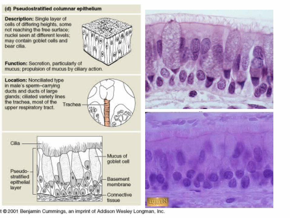
Pseudostratified columnar epithelium
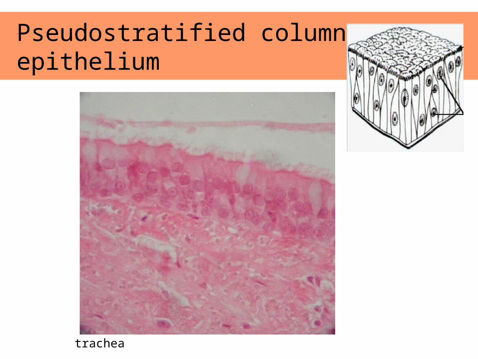
Pseudostratified columnar epithelium
trachea
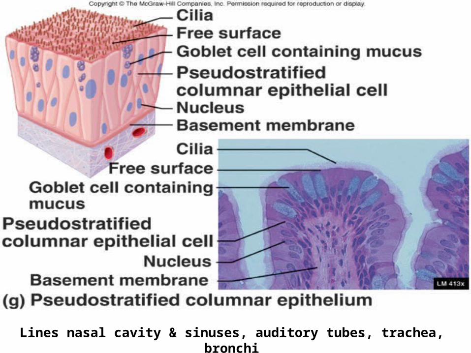
Lines nasal cavity & sinuses, auditory tubes, trachea, bronchi
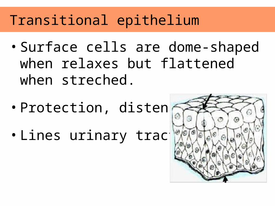
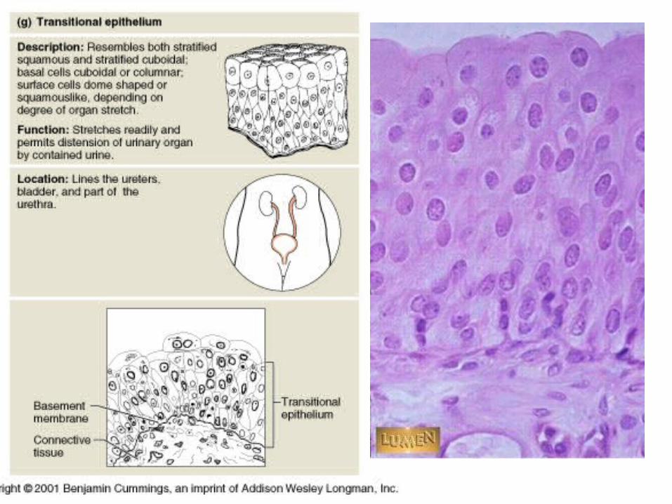
• Surface cells are dome-shaped when relaxes but flattened when streched.
• Protection, distensible
• Lines urinary tract
Transitional epithelium
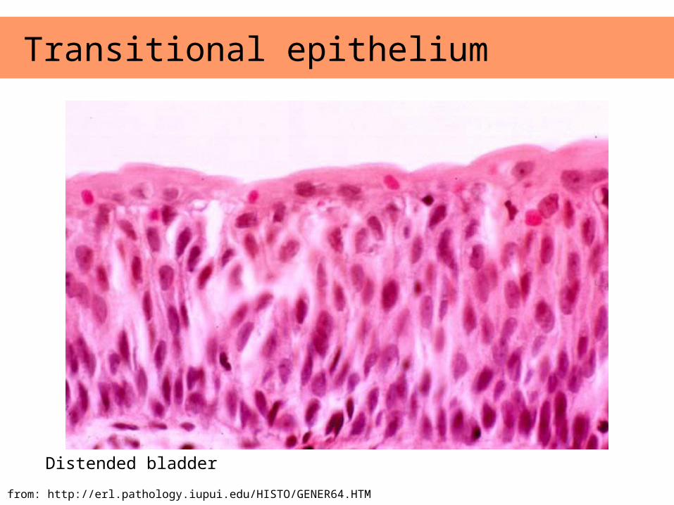
Transitional epithelium
Image from: http://erl.pathology.iupui.edu/HISTO/GENER64.HTM
Distended bladder
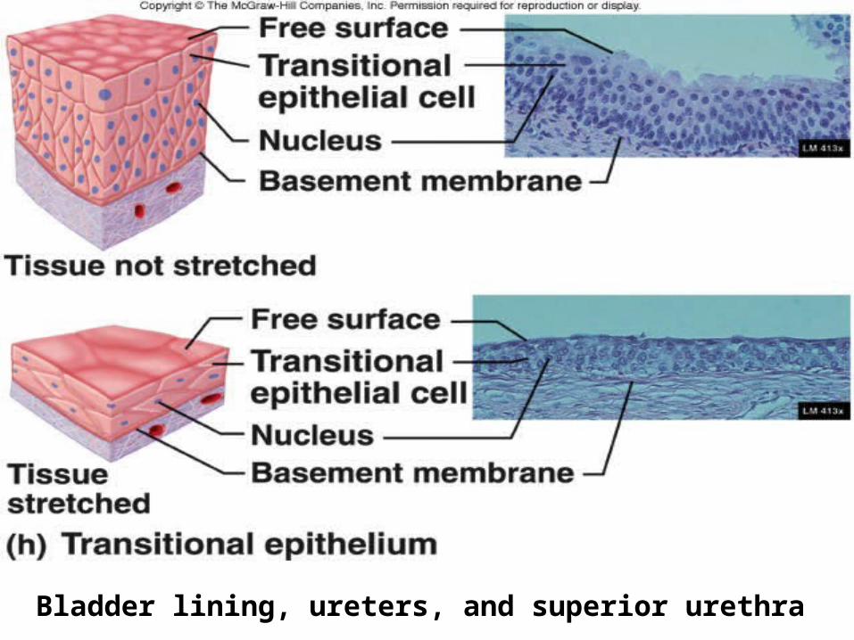
Bladder lining, ureters, and superior urethra
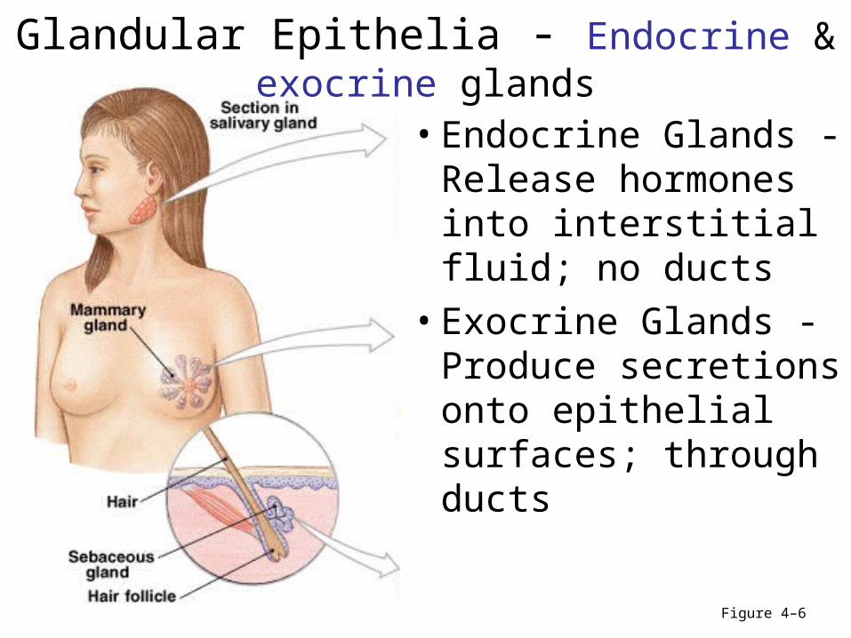
Glandular Epithelia - Endocrine & exocrine glands
Figure 4–6
• Endocrine Glands - Release hormones into interstitial fluid; no ducts
• Exocrine Glands - Produce secretions onto epithelial surfaces; through ducts

Modes of Secretion• Merocrine secretion
Figure 4–6a
• Apocrine secretion*Are produced in Golgi apparatus
*Are released by shedding cytoplasm
*e.g., mammary glands
– Are produced in Golgi apparatus– Are released by vesicles
(exocytosis) – e.g., sweat glands
• Holocrine secretion• Are released by cells bursting, killing
gland cells• Gland cells replaced by stem cells• e.g., sebaceous gland
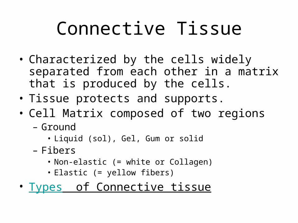
Connective Tissue
• Characterized by the cells widely separated from each other in a matrix that is produced by the cells.
• Tissue protects and supports.• Cell Matrix composed of two regions
– Ground• Liquid (sol), Gel, Gum or solid
– Fibers• Non-elastic (= white or Collagen)• Elastic (= yellow fibers)
• Types of Connective tissue
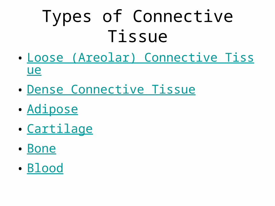
Types of Connective Tissue
• Loose (Areolar) Connective Tissue
• Dense Connective Tissue
• Adipose
• Cartilage
• Bone
• Blood
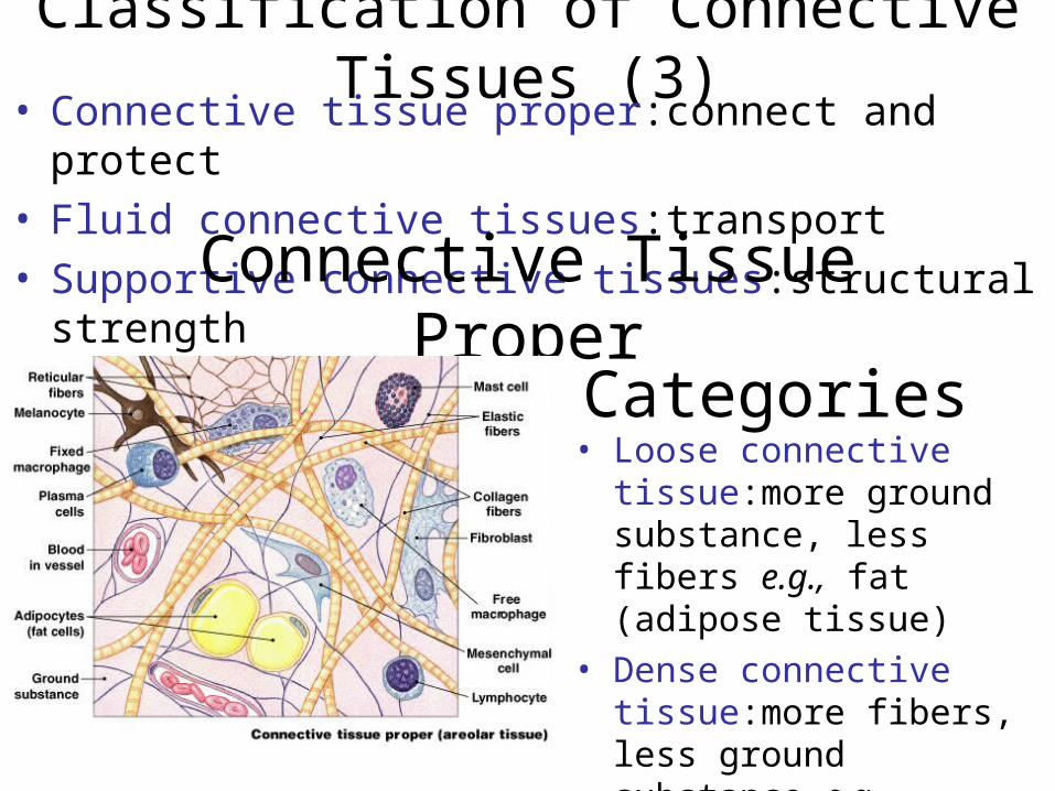
Classification of Connective Tissues (3)• Connective tissue proper:connect and protect• Fluid connective tissues:transport• Supportive connective tissues:structural strength
Connective Tissue Proper
Categories• Loose connective
tissue:more ground substance, less fibers e.g., fat (adipose tissue)
• Dense connective tissue:more fibers, less ground substance e.g., tendons
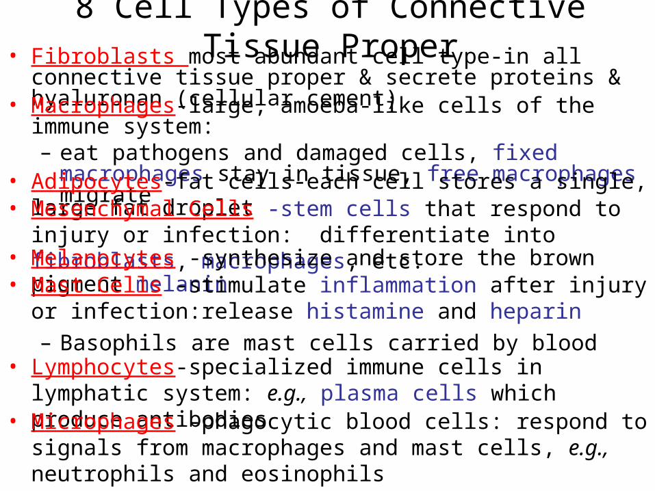
8 Cell Types of Connective Tissue Proper• Fibroblasts most abundant cell type-in all connective tissue
proper & secrete proteins & hyaluronan (cellular cement)• Macrophages-large, amoeba-like cells of the immune system:
– eat pathogens and damaged cells, fixed macrophages stay in tissue, free macrophages migrate
• Adipocytes-fat cells-each cell stores a single, large fat droplet• Mesenchymal Cells -stem cells that respond to injury or
infection: differentiate into fibroblasts, macrophages, etc.• Melanocytes -synthesize and store the brown pigment melanin• Mast Cells -stimulate inflammation after injury or
infection:release histamine and heparin – Basophils are mast cells carried by blood
• Lymphocytes-specialized immune cells in lymphatic system: e.g., plasma cells which produce antibodies
• Microphages -phagocytic blood cells: respond to signals from macrophages and mast cells, e.g., neutrophils and eosinophils
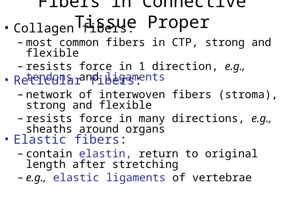
Fibers in Connective Tissue Proper• Collagen fibers:
– most common fibers in CTP, strong and flexible– resists force in 1 direction, e.g., tendons and
ligaments• Reticular fibers:
– network of interwoven fibers (stroma), strong and flexible
– resists force in many directions, e.g., sheaths around organs
• Elastic fibers: – contain elastin, return to original length after
stretching– e.g., elastic ligaments of vertebrae
Ground Substance in Connective Tissue Proper
• In connective tissue proper and ground substance: – is clear, colorless, and viscous– fills spaces between cells and slows
pathogens
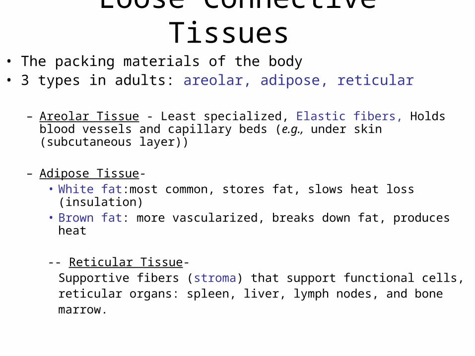
Loose Connective Tissues
• The packing materials of the body • 3 types in adults: areolar, adipose, reticular
– Areolar Tissue - Least specialized, Elastic fibers, Holds blood vessels and capillary beds (e.g., under skin (subcutaneous layer))
– Adipose Tissue-• White fat:most common, stores fat, slows heat loss (insulation) • Brown fat: more vascularized, breaks down fat, produces heat
-- Reticular Tissue-Supportive fibers (stroma) that support functional cells,reticular organs: spleen, liver, lymph nodes, and bone marrow.
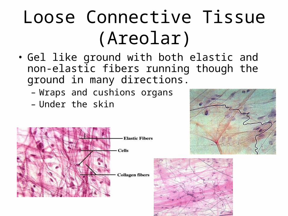
Loose Connective Tissue (Areolar)
• Gel like ground with both elastic and non-elastic fibers running though the ground in many directions.– Wraps and cushions organs– Under the skin
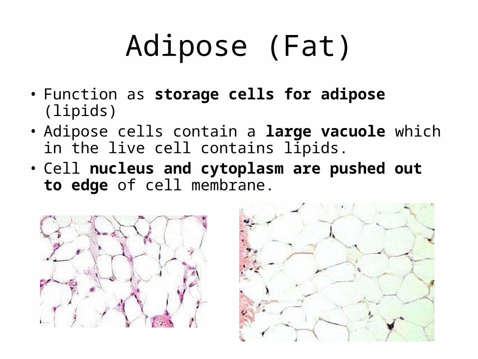
Adipose (Fat)
• Function as storage cells for adipose (lipids)• Adipose cells contain a large vacuole which in
the live cell contains lipids.• Cell nucleus and cytoplasm are pushed out
to edge of cell membrane.
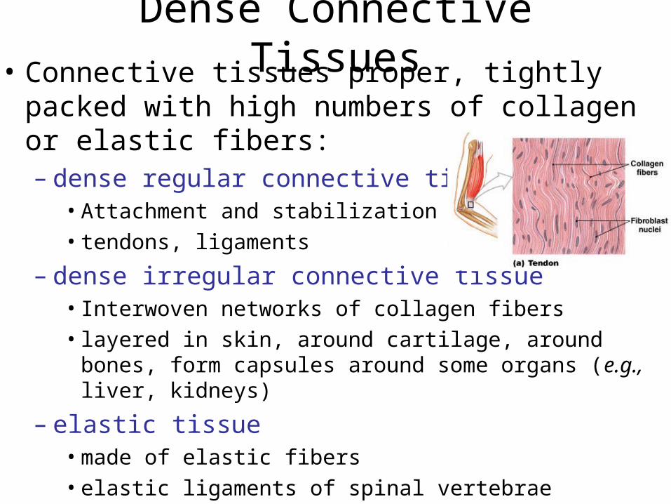
Dense Connective Tissues• Connective tissues proper, tightly packed with
high numbers of collagen or elastic fibers: – dense regular connective tissue
• Attachment and stabilization• tendons, ligaments
– dense irregular connective tissue• Interwoven networks of collagen fibers • layered in skin, around cartilage, around bones, form
capsules around some organs (e.g., liver, kidneys)
– elastic tissue • made of elastic fibers• elastic ligaments of spinal vertebrae
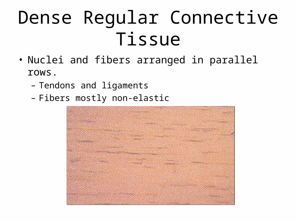
Dense Regular Connective Tissue
• Nuclei and fibers arranged in parallel rows.– Tendons and ligaments– Fibers mostly non-elastic
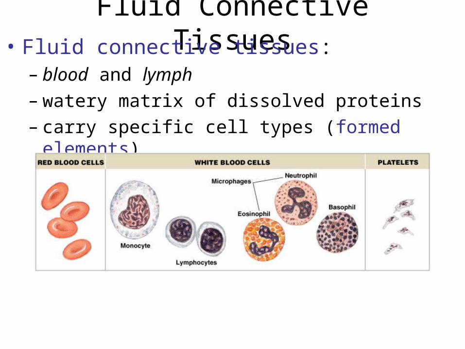
Fluid Connective Tissues• Fluid connective tissues:
– blood and lymph– watery matrix of dissolved proteins– carry specific cell types (formed elements)
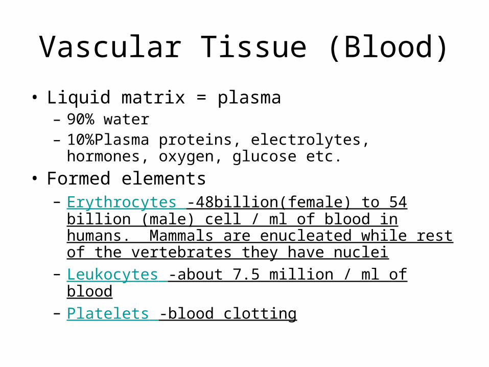
Vascular Tissue (Blood)
• Liquid matrix = plasma – 90% water– 10%Plasma proteins, electrolytes, hormones, oxygen,
glucose etc.
• Formed elements– Erythrocytes -48billion(female) to 54 billion (male) cell
/ ml of blood in humans. Mammals are enucleated while rest of the vertebrates they have nuclei
– Leukocytes -about 7.5 million / ml of blood– Platelets -blood clotting
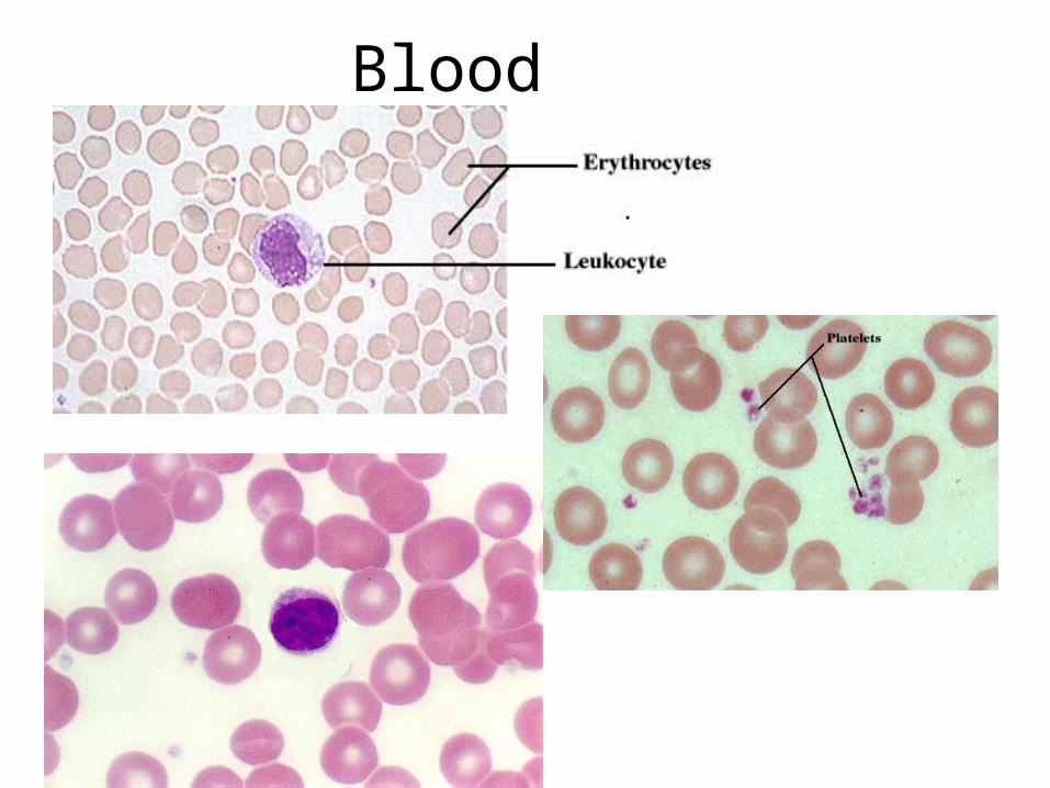
Blood
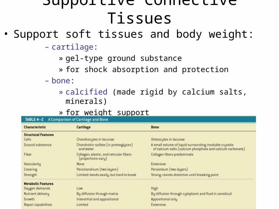
Supportive Connective Tissues
• Support soft tissues and body weight:– cartilage:
» gel-type ground substance» for shock absorption and protection
– bone: » calcified (made rigid by calcium salts, minerals)» for weight support
• Cartilage Matrix – Proteoglycans, ground substance proteins, cells (chondrocytes) surrounded by lacunae (chambers)
• Cartilage Structure – No blood vessels:chondrocytes produce
antiangiogenesis factor– Perichondrium:outer, fibrous layer (for strength),
inner, cellular layer (for growth and maintenance)
Cartilage
• Ground of matrix is gum like.• Cells are found in Lacunae within the matrix.• Fibers may be elastic or non-elastic, or a form
of non-elastic called reticular(where the non-elastic fibers of very thin)– Hyaline Cartilage-example on the ends of bones– Elastic Cartilage- example ear cartilage– Non-elastic Cartilage- example nose cartilage.
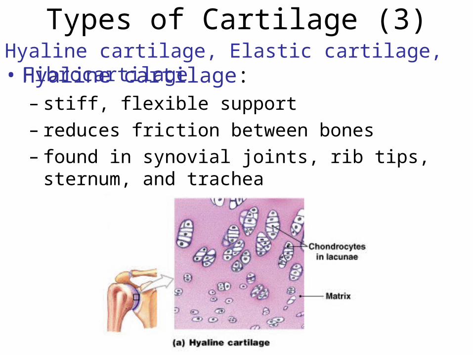
Types of Cartilage (3)Hyaline cartilage, Elastic cartilage, Fibrocartilage• Hyaline cartilage:
– stiff, flexible support– reduces friction between bones– found in synovial joints, rib tips, sternum, and
trachea
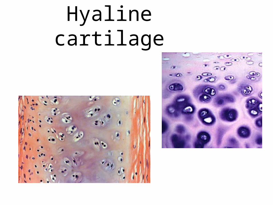
Hyaline cartilage
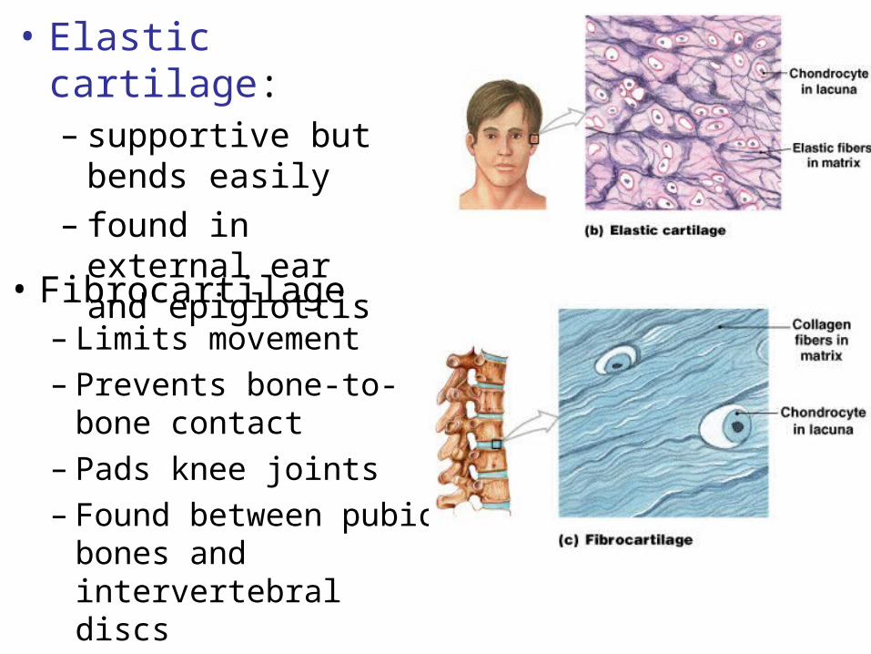
• Elastic cartilage:– supportive but
bends easily– found in external
ear and epiglottis
• Fibrocartilage– Limits movement– Prevents bone-to-bone
contact– Pads knee joints– Found between pubic
bones and intervertebral discs
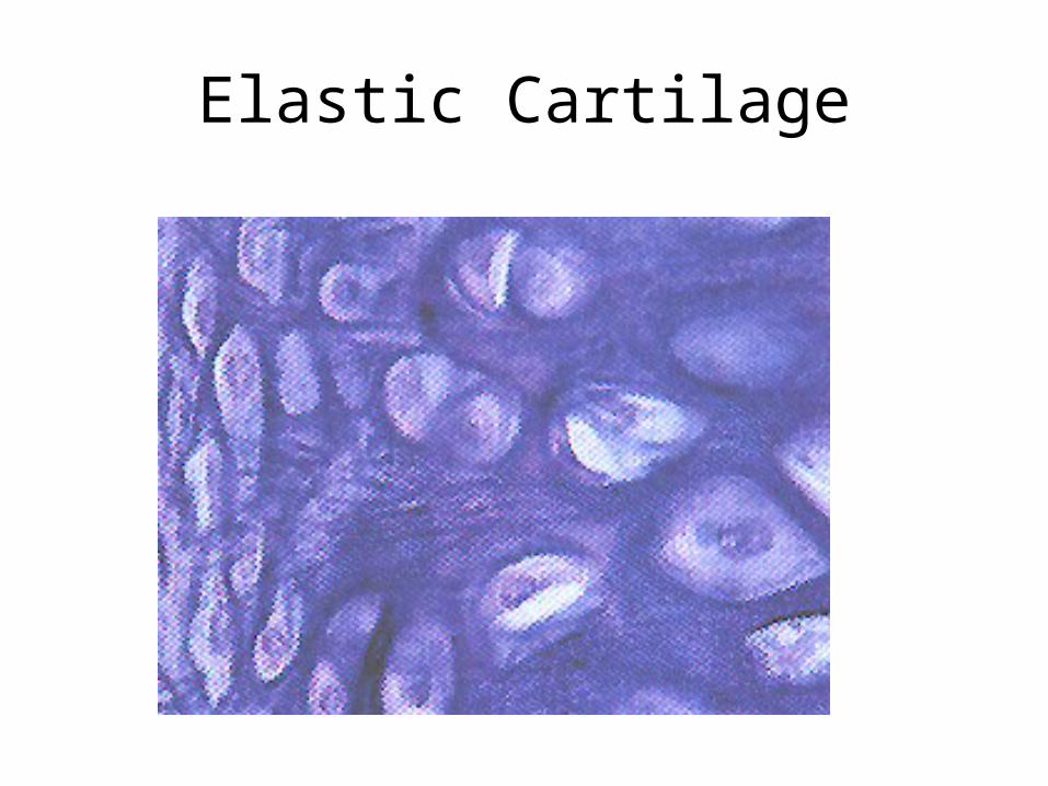
Elastic Cartilage
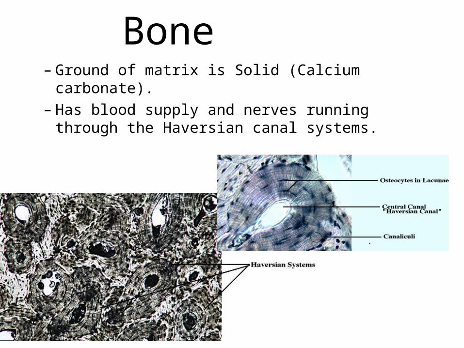
Bone– Ground of matrix is Solid (Calcium carbonate).– Has blood supply and nerves running through
the Haversian canal systems.
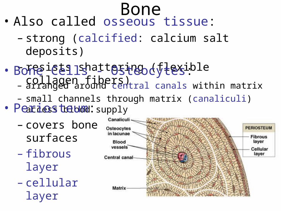
Bone• Also called osseous tissue:
– strong (calcified: calcium salt deposits)– resists shattering (flexible collagen fibers)
• Bone Cells - Osteocytes:– arranged around central canals within matrix
– small channels through matrix (canaliculi) access blood supply• Periosteum:
– covers bone surfaces
– fibrous layer– cellular layer
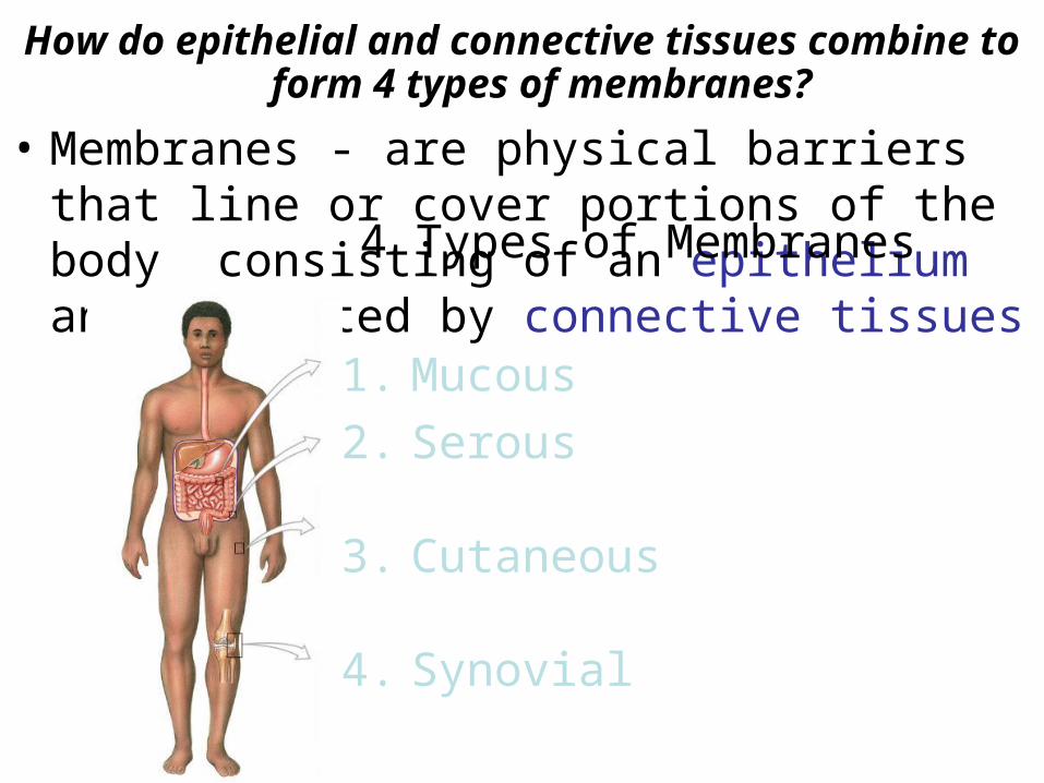
How do epithelial and connective tissues combine to form 4 types of membranes?
• Membranes - are physical barriers that line or cover portions of the body consisting of an epithelium and supported by connective tissues4 Types of Membranes
1. Mucous
2. Serous
3. Cutaneous
4. Synovial
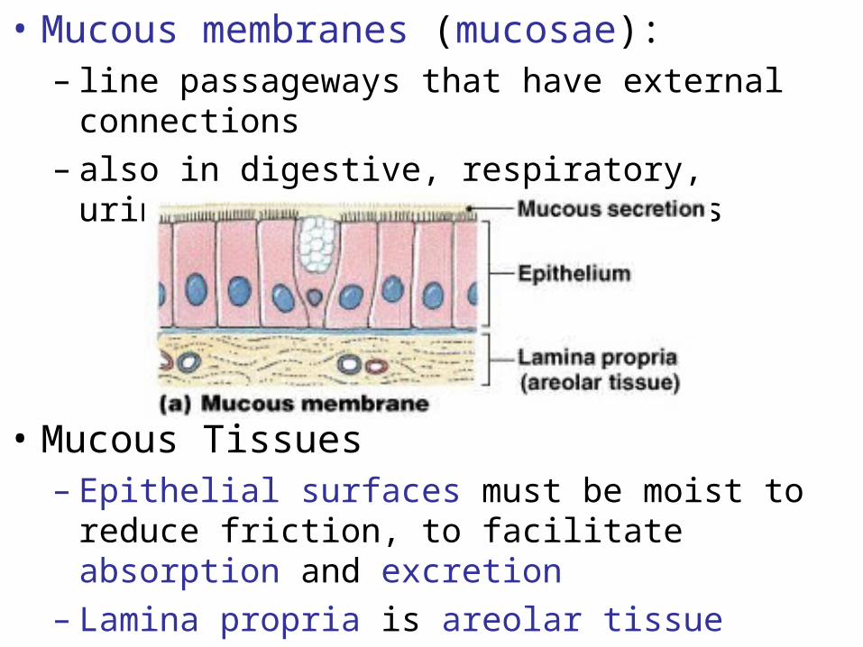
• Mucous membranes (mucosae):– line passageways that have external connections – also in digestive, respiratory, urinary, and
reproductive tracts
• Mucous Tissues – Epithelial surfaces must be moist to reduce friction,
to facilitate absorption and excretion – Lamina propria is areolar tissue

Serous Membranes• Line cavities not open to the outside
• Are thin but strong
• Have fluid transudate to reduce friction• Serous
membranes: double, have a parietal portion covering the cavity and a visceral portion (serosa) covering the organs
• Pleural membrane lines pleural cavities covers lungs
• Peritoneum lines peritoneal cavity covers abdominal organs
• Pericardium lines pericardial cavity covers heart
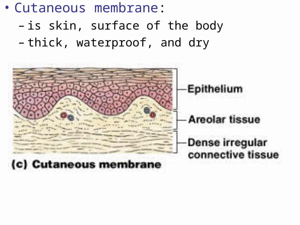
• Cutaneous membrane:– is skin, surface of the body– thick, waterproof, and dry
How do connective tissues form the framework of the body?
• Connective tissues:– provide strength and stability– maintain positions of internal organs– provides routes for blood vessels, lymphatic
vessels, and nerves• fascia:
– the body’s framework of connective tissue – layers and wrappings that support or surround
organs
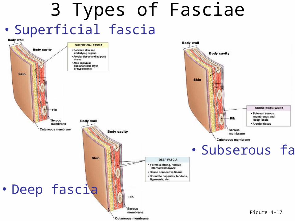
3 Types of Fasciae• Superficial fascia
Figure 4–17
• Deep fascia
• Subserous fascia
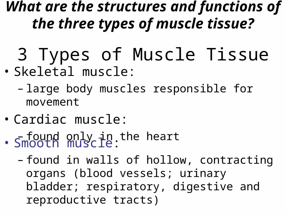
What are the structures and functions of the three types of muscle tissue?
3 Types of Muscle Tissue• Skeletal muscle:
– large body muscles responsible for movement
• Cardiac muscle:– found only in the heart
• Smooth muscle:– found in walls of hollow, contracting organs (blood
vessels; urinary bladder; respiratory, digestive and reproductive tracts)
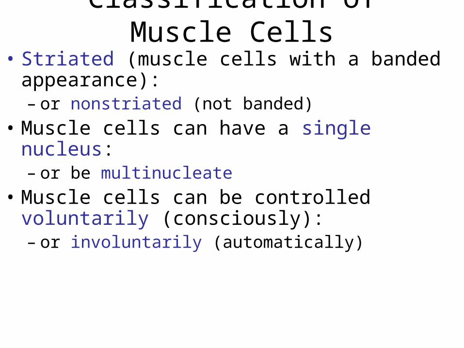
Classification of Muscle Cells
• Striated (muscle cells with a banded appearance): – or nonstriated (not banded)
• Muscle cells can have a single nucleus: – or be multinucleate
• Muscle cells can be controlled voluntarily (consciously):– or involuntarily (automatically)
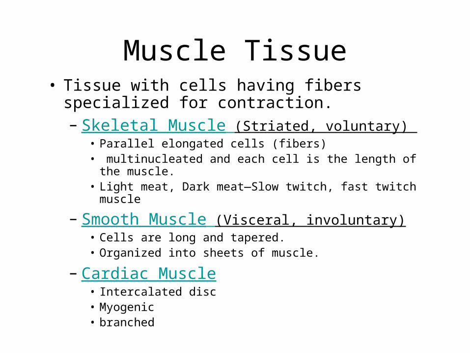
Muscle Tissue• Tissue with cells having fibers specialized for
contraction.– Skeletal Muscle (Striated, voluntary)
• Parallel elongated cells (fibers) • multinucleated and each cell is the length of the
muscle.• Light meat, Dark meat—Slow twitch, fast twitch muscle
– Smooth Muscle (Visceral, involuntary)• Cells are long and tapered. • Organized into sheets of muscle.
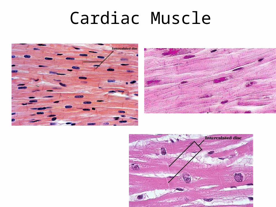
– Cardiac Muscle• Intercalated disc• Myogenic• branched
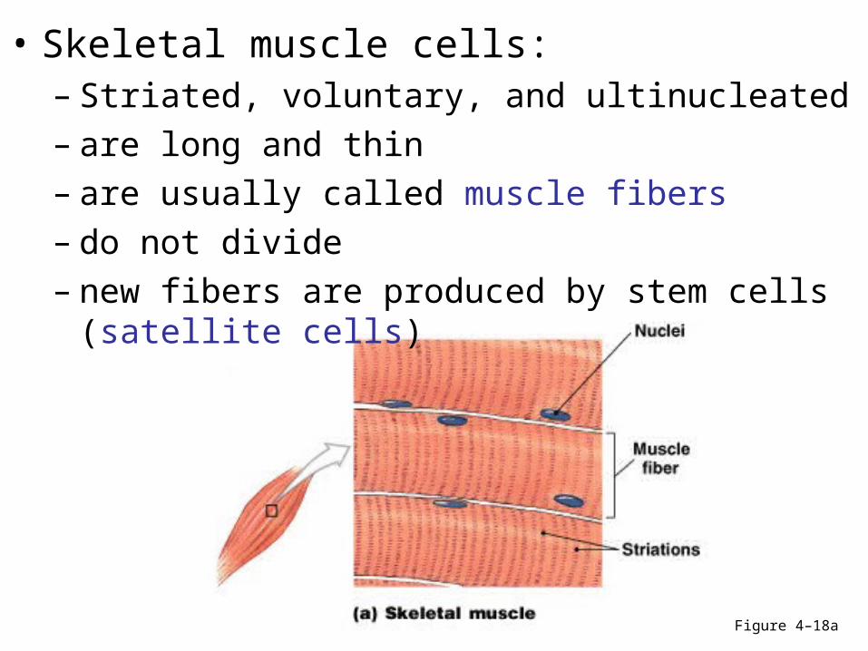
Figure 4–18a
• Skeletal muscle cells:– Striated, voluntary, and ultinucleated– are long and thin– are usually called muscle fibers– do not divide– new fibers are produced by stem cells (satellite
cells)
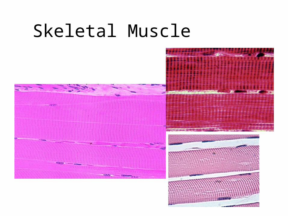
Skeletal Muscle
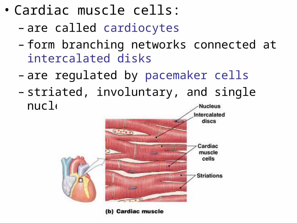
• Cardiac muscle cells:– are called cardiocytes– form branching networks connected at intercalated
disks– are regulated by pacemaker cells– striated, involuntary, and single nucleus
Cardiac Muscle
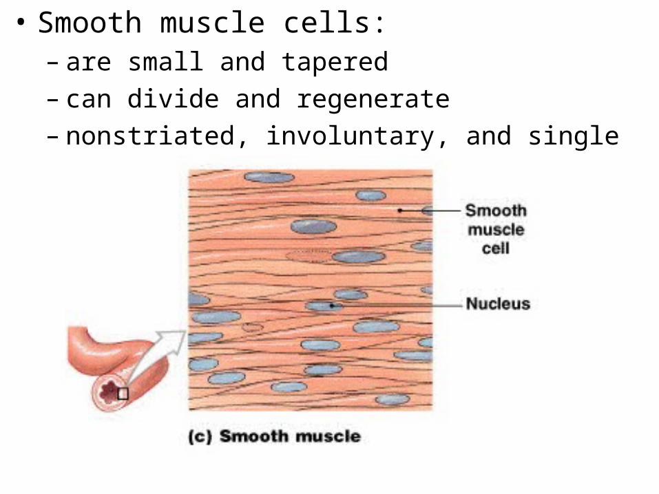
• Smooth muscle cells:– are small and tapered– can divide and regenerate – nonstriated, involuntary, and single nucleus
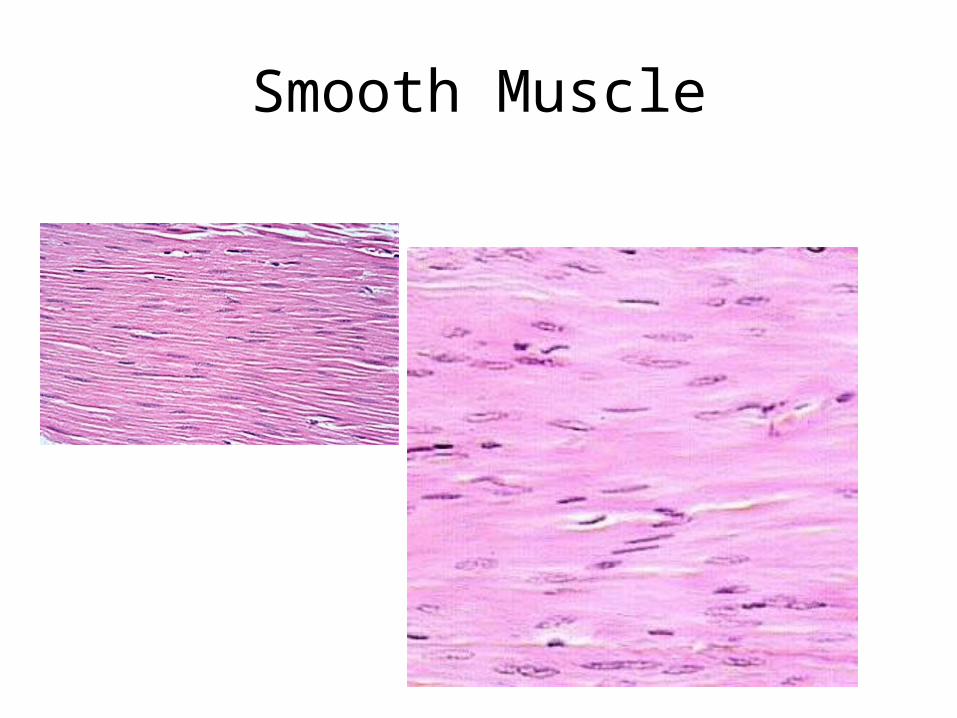
Smooth Muscle
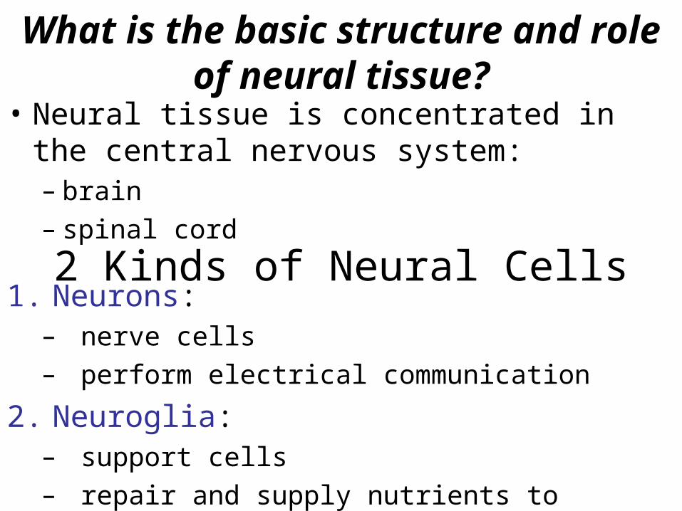
What is the basic structure and role of neural tissue?
• Neural tissue is concentrated in the central nervous system:– brain– spinal cord
2 Kinds of Neural Cells1. Neurons:
– nerve cells – perform electrical communication
2. Neuroglia:– support cells– repair and supply nutrients to neurons
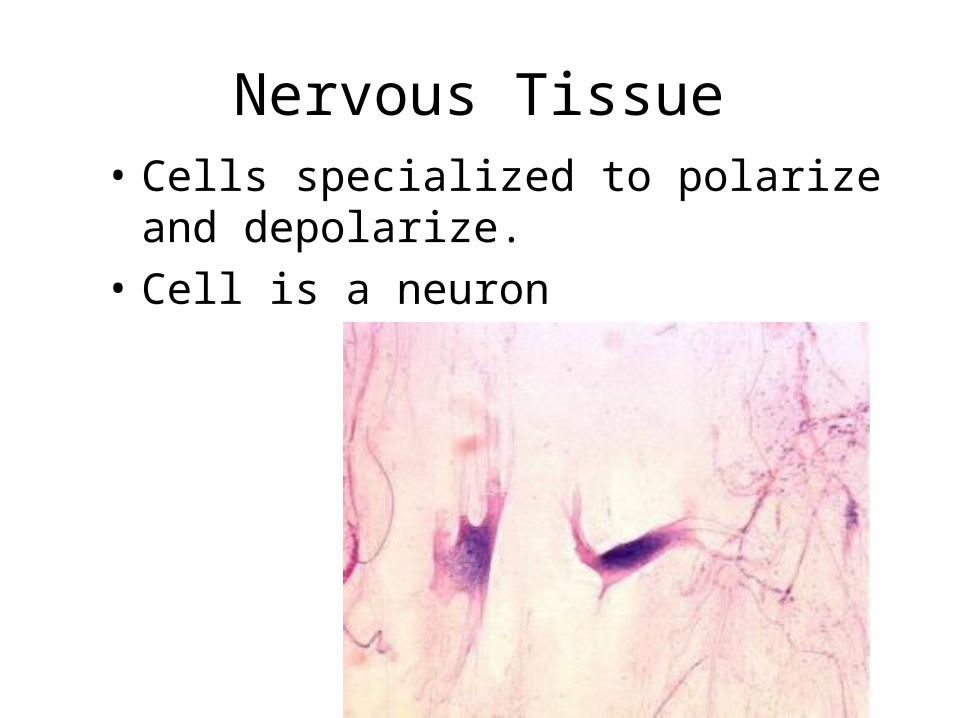
Nervous Tissue• Cells specialized to polarize and
depolarize.
• Cell is a neuron
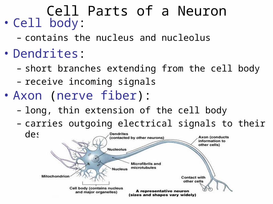
Cell Parts of a Neuron• Cell body:
– contains the nucleus and nucleolus
• Dendrites:– short branches extending from the cell body– receive incoming signals
• Axon (nerve fiber):– long, thin extension of the cell body– carries outgoing electrical signals to their destination
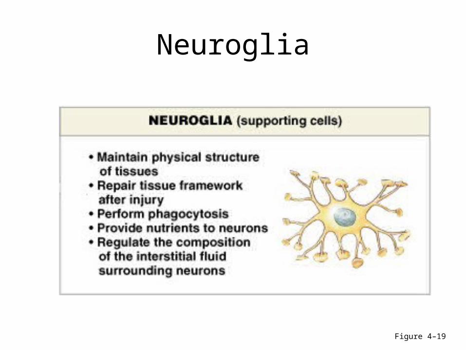
Neuroglia
Figure 4–19
End of Tissue presentation