title: interplay between clathrin and rab5 controls the early
TRANSCRIPT

1
Title: Interplay Between Clathrin and Rab5 Controls the Early Phagocytic Trafficking and
Intracellular Survival of Brucella abortus within HeLa cells
Authors: Jin Ju Leea, Dae Geun Kima, Dong Hyeok Kima, Hannah Leah Simborioa, Wongi Mina, Hu
Jang Leea, Moon Herb, Suk Chan Jungb, Masahisa Wataraic, Suk Kima,d*
Complete names of institutions: aCollege of Veterinary Medicine, Gyeongsang National University,
Jinju 660-701, Republic of Korea, bAnimal, Plant and Fisheries Quarantine and Inspection
Agency, Anyang, Gyeonggi-do, 430-824, Republic of Korea, cDepartment of Veterinary
Public Health, Faculty of Agriculture, Yamaguchi University, Yamaguchi 753-8515, Japan,
dInstitute of Agriculture and Life Science, Gyeongsang National University, Jinju, 660-701,
Republic of Korea
Running title: Interplay of Clathrin/Rab5 during B. abortus Infection
Key words: Brucella abortus, phagocytosis, clathrin, Rab5, lipid rafts
* To whom correspondence should be addressed: College of Veterinary Medicine, Gyeongsang
National University, Jinju, 660-701, Republic of Korea, Tel: +82–55–772–2359; Fax: +82–55–772–
2349; E-mail address: [email protected].
http://www.jbc.org/cgi/doi/10.1074/jbc.M113.491555The latest version is at JBC Papers in Press. Published on August 12, 2013 as Manuscript M113.491555
Copyright 2013 by The American Society for Biochemistry and Molecular Biology, Inc.
by guest on April 1, 2018
http://ww
w.jbc.org/
Dow
nloaded from

2
Capsule
Background: Phagocytic mechanisms are
important for understanding the host-pathogen
interactions of intracellular parasitic pathogens.
Results: The entry and early intracellular
trafficking of B. abortus are dependent on
clathrin cooperation with Rab5.
Conclusions: Clathrin influences phagocytic
pathways of B. abortus associating with Rab5 at
a host subcellular site allowing replication.
Significance: This study promotes the early
control of B. abortus infection.
Abstract
Lipid raft-associated clathrin is essential for
host-pathogen interactions during infection.
Brucella abortus is an intracellular pathogen that
circumvents host defenses, but little is known
about the precise infection mechanisms that
involve interaction with lipid raft-associated
mediators. The aim of the present study was to
elucidate the clathrin-mediated phagocytic
mechanisms of B. abortus. The clathrin
dependency of B. abortus infection in HeLa cells
was investigated using an infection assay and
immunofluorescence microscopy. The
redistribution of clathrin in the membrane and in
phagosomes was investigated using sucrose
gradient fractionation of lipid rafts and the
isolation of B. abortus-containing vacuoles
(BCVs), respectively. Clathrin and dynamin
were concentrated into lipid rafts during B.
abortus infection, and the entry and intracellular
survival of B. abortus within HeLa cells were
abrogated by clathrin inhibition. Clathrin
disruption decreased actin polymerization and
the colocalization of BCVs with clathrin and
Rab5 but not LAMP-1. Thus, our data
demonstrated that clathrin plays a fundamental
role in the entry and intracellular survival of B.
abortus via interaction with lipid rafts and actin
rearrangement. This process facilitates the early
intracellular trafficking of B. abortus to safe
replicative vacuoles.
Introduction
Brucella species are Gram-negative,
facultative intracellular bacteria and the
etiological agent of brucellosis in many animals
and humans (1, 2). The ability of these bacteria
to escape killing within phagocytes is
hypothesized to be involved in their virulence by
promoting invasion and chronic infections (3, 4),
by guest on April 1, 2018
http://ww
w.jbc.org/
Dow
nloaded from

3
but the exact molecular mechanisms are
unknown.
Previous studies explored the mechanisms
that underlie intracellular survival by
investigating the trafficking of Brucella-
containing vacuoles (BCVs); these studies
revealed that BCVs interact with early endocytic
compartments before acquiring the lysosome-
associated membrane protein-1 (LAMP-1) and
progressively exclude themselves from
endocytic compartments to circumvent fusion
with terminal lysosomes (5, 6). However, a
recent study that used live cell imaging to
investigate BCV trafficking confirmed that
fusion with lysosomes occurs in the intermediate
stages of BCV trafficking and is required for the
maturation of BCVs into endoplasmic reticulum
(ER)-derived replicative organelles (7).
Lipid rafts are specialized membrane
microdomains enriched in cholesterol,
glycosylphosphatidylinositol (GPI)-anchored
proteins and GM1 gangliosides (8). There is
growing evidence concerning the potential role
of lipid rafts in host-pathogen interactions, and
lipid rafts have been implicated as portals of
entry for intracellular pathogens (9, 10). Several
studies have implicated lipid rafts in the entry
and endocytic pathway of B. abortus in host
cells. These studies indicated that lipid raft-
associated molecules, such as GPI-anchored
proteins, GM1 gangliosides and cholesterol, are
selectively integrated into Brucella-containing
macropinosomes following the internalization of
Brucella into macrophages and continuously
sustain a dynamic state of the phagosomal
membrane (11-13). In addition, the route by
which Brucella is internalized into phagocytic
cells determines the intracellular fate of this
bacterium, and this event is modulated by lipid
rafts (11, 14).
Clathrin is an endocytic coat protein that
mediates the internalization of a variety of
transmembrane receptors and their ligands, and
there is evidence that lipid rafts play a role in
clathrin-dependent uptake mechanisms (15).
Additionally, clathrin facilitates the entry of a
variety of zippering bacteria, such as Listeria
and Chlamydia trachomatis, into non-phagocytic
cells (16, 17).
Dynamin, a member of the large (100 kD)
GTPase family, participates in several endocytic
processes. This protein is required for endocytic
events during membrane fusion and selectively
regulates the assembly of endocytic vacuoles (18,
by guest on April 1, 2018
http://ww
w.jbc.org/
Dow
nloaded from

4
19). In particular, dynamin plays an important
role in both clathrin-dependent endocytosis (20,
21) and caveolae-dependent uptake and
endocytosis (22). In addition, dynamin is not
only involved in the formation of endosomes at
the plasma membrane but is also required for the
vesiculation of endosome tubules (23).
Despite extensive study, the roles of clathrin
and dynamin in B. abortus infection remain
unclear. Although B. abortus interacts with host
cells such as macrophages and epithelial cells,
through lipid rafts (1, 4, 12-14), little is known
about the precise mechanisms by which this
pathogen exploits the host entry and intracellular
trafficking apparatus, including the mediators
that enable this interaction, for its intracellular
survival.
Here, we elucidated the mechanism
underlying the clathrin-dependent entry and
early intracellular trafficking of B. abortus
following association with lipid rafts. Clathrin
critically influenced the intracellular trafficking
of B. abortus, which was associated with Rab5.
Experimental procedures
Cell Culture and Transfection
Experiments―HeLa cells (ATCC 229) were
grown at 37°C in a 5% CO2 atmosphere in
DMEM containing 10% heat-inactivated fetal
bovine serum (FBS), 2 mм L-glutamine, 100
U/ml penicillin, and 100 μg/ml streptomycin (all
from Gibco). For all assays, cells were seeded (2
× 104 cells/well) in cell culture plates and
incubated for 24 h before infection.
For transfection, siRNA targeting the clathrin
heavy chain (HC oligo I) (AAC CUG CGG
UCU GGA GUC AAC) and the non-targeting
firefly (Photinus pyralis) luciferase siRNA
(AAC GTT ACC GCG GAA TAC TTC GA)
were obtained from Dharmacon (Lafayette).
RNA targeting and formation of an siRNA
duplex were performed as previously described
(24). HeLa cells were transfected with clathrin
heavy chain siRNA (60 pmol of RNA duplex)
using Lipofectamine 2000 (Invitrogen)
according to the manufacturer's instructions. As
a control, firefly luciferase siRNA was
transfected into cells in parallel. The knockdown
efficiency was determined by comparing protein
levels in cells transfected with clathrin siRNA
and control siRNA, as indicated by Western blot
analysis using antibodies against the clathrin
by guest on April 1, 2018
http://ww
w.jbc.org/
Dow
nloaded from

5
heavy chain (BD Transduction Laboratories) and
β-actin (Cell Signaling). Western blot signals
were quantified using the NIH ImageJ software
program.
Bacterial Strains and Culture
Conditions―Brucella abortus strains were
derived from 544 (ATCC 23448), a smooth,
virulent B. abortus biovar 1 strains. B. abortus
organisms were stored as frozen aliquots in 80%
(v/v) glycerol at -70°C. Bacteria were grown in
Brucella broth (Difco, Becton Dickinson) at
37°C with shaking incubation until they reached
the stationary phase, and then the viable
counting of bacteria was assessed by plating
serial dilutions on Brucella agar.
Bacterial Infection―To analyze bacterial
internalization efficiency, cultured cells were
treated with various inhibitors or transfected
with target siRNA prior to infection. Following
treatment, bacteria were deposited onto cells at a
multiplicity of infection (MOI) of 10,
centrifuged at 150 × g for 10 min and incubated
at 37°C in 5% CO2 for 0, 15 and 30 min. Cells
were incubated in DMEM containing 10% FBS
and gentamicin (30 µg/ml, Sigma-Aldrich) for
30 min to kill any remaining extracellular
bacteria. To determine the intracellular
replication efficacy, infected cells were
incubated at 37°C for 1 h, cultured in DMEM
containing 10% FBS and gentamicin and
incubated with various inhibitors. After 2, 24
and 48 h, infected cells were lysed and spread on
Brucella agar plates in triplicate, and the number
of viable bacteria was determined by counting
CFUs.
Inhibitor Studies―Inhibitors targeting
different lipid raft-associated molecules were
selectively used. The viability of drug-treated
cells was evaluated by staining single cell
preparations with trypan blue. Prior to infection,
cultured cells were incubated at 37°C with the
following inhibitors for the indicated times: 12.5
µм chloropromazine (CPZ), a clathrin inhibitor,
for 45 min; 80 µм dynasore, a dynamin inhibitor,
for 30 min; and 5 mм MβCD, a lipid raft-
associated cholesterol-depleting drug, for 30 min
(all from Sigma-Aldrich). The viability values of
cells treated with the above indicated
concentrations of all drugs showed more than
98% compared with the untreated cells (100%).
by guest on April 1, 2018
http://ww
w.jbc.org/
Dow
nloaded from

6
Isolation of Lipid Rafts―Lipid rafts were
isolated on ice as previously described (25), with
some modifications. Briefly, the cells were
scraped and lysed in base buffer and centrifuged
at 1,000 × g for 10 min. The post-nuclear
supernatant (PNS) (0.84 ml) was adjusted to
35% OptiPrep (Sigma-Aldrich) by adding 60%
OptiPrep (1.16 ml); subsequently, 2 ml each of
30, 25 and 20% OptiPrep and 1 ml of base buffer
(0%) were overlaid on top of the lysate (35%
OptiPrep). The gradients were centrifuged at
52,000 × g for 3 h using an SW-41 rotor in a
Beckman ultracentrifuge (Beckman Coulter) and
fractionated into nine fractions (1 ml per
fraction). Fractionated proteins were analyzed by
Western blotting.
Purification of Bacteria-Containing
Phagosomes (BCPs)―BCPs were purified as
previously described, with slight modifications
(26). Infected cells (1 × 10⁸ cells) were washed
by centrifugation at 300 × g for 5 min at 4°C and
suspended and lysed in 1 ml of ice-cold
homogenization buffer (HB). Nuclei and intact
cells were removed from the homogenate by
centrifugation at 800 × g for 5 min at 4°C. The
supernatant was laid on top of a discontinuous
sucrose gradient consisting of 2 ml 50% sucrose,
4 ml 37% sucrose and 4 ml 25% sucrose and
centrifuged at 100,000 × g for 1 h at 4°C using
an SW-41 rotor in a Beckman ultracentrifuge.
The band between the 50% and 37% sucrose
layers, which contained the BCPs, was collected
and resuspended in 12 ml of cold PBS. The
BCPs were pelleted by centrifugation at 40,000
× g for 15 min at 4°C, and the resulting pellets
were resuspended in 0.1 ml HB and subjected to
Western blotting.
Western Blotting―Protein concentrations
were determined using the Bradford protein
assay (Richmond, CA), and proteins were loaded
and separated by SDS-PAGE before transfer to
PVDF membranes (Millipore). Blots were
blocked for 1 h with 5% bovine serum albumin
(BSA) in Tris-buffered saline containing 0.1%
Tween-20 (TBS-T) and washed three times with
TBS-T for 20 min. Blots were incubated with the
appropriate primary antibodies overnight at 4°C
with gentle shaking and washed as described
above. Binding of primary antibodies was
visualized using HRP-conjugated secondary
antibodies, and immunolabeling was detected
by guest on April 1, 2018
http://ww
w.jbc.org/
Dow
nloaded from

7
using enhanced chemiluminescence (ECL)
(SurModics), according to the manufacturer's
instructions, and exposure to X-ray film
(FUJIFILM).
Immunofluorescence Staining and
Microscopy―HeLa cells were treated with
inhibitors before infection as described above.
After 10, 15, 30 and 60 min of infection with
Alexa Fluor 405-conjugated B. abortus, the cells
were fixed in 4% paraformaldehyde in PBS for
30 min at 37°C, washed three times with PBS,
and permeabilized with 0.1% TritonX-100 for 10
min at 22°C. After 30 min of incubation with a
blocking buffer (2% goat serum in PBS), the
preparations were stained with different
antibodies in blocking buffer. To stain F-actin,
the cells were incubated with 0.5 μM phalloidin-
TRITC for 30 min at 22°C. To detect
intracellular protein localization, the cells were
incubated with primary antibodies against
clathrin, dynamin (BD Transduction
Laboratories) and Rab5 (Cell Signaling) and the
corresponding fluorescence-conjugated
secondary antibodies. For LAMP-1 staining,
after 2, 4, 8, 24 and 48 h of infection with
unconjugated B. abortus, fixed cells were
stained with an anti-B. abortus polyclonal
antibody produced in rabbits immunized with B.
abortus (12) and an anti-LAMP-1 antibody
(Invitrogen). Finally, the preparations were
washed and mounted with fluorescent mounting
medium (DakoCytomation). Fluorescent images
were collected using an Olympus FV1000 laser
scanning confocal microscope. Images were
processed using Adobe Photoshop and NIH
ImageJ software. For LAMP-1 staining, one
hundred bacteria within macrophages were
selected randomly, and the extent of bacterial
LAMP-1 acquisition was determined.
Statistical Analysis―Data are expressed as
the mean ± standard deviation (SD) for replicate
experiments. Statistical analysis was performed
using GraphPad Prism software, version 4.00
(GraphPad Software). Student’s t-test or a one-
way ANOVA followed by the Newman-Keuls
test was used for statistical comparisons between
groups. The results for which P < 0.05 were
considered statistically significant.
Results
by guest on April 1, 2018
http://ww
w.jbc.org/
Dow
nloaded from

8
Clathrin Affects the Entry and Intracellular
Survival of B. abortus―Several infection
processes are dependent on functional clathrin
(16, 17, 27), but the interaction between B.
abortus and clathrin has yet to be elucidated. To
investigate the role of clathrin in the entry and
intracellular survival of B. abortus, HeLa cells
were treated with 12.5 μм CPZ, a clathrin
inhibitor. The entry and intracellular replication
of B. abortus in CPZ-treated cells were
significantly diminished in comparison with
untreated cells (p < 0.001, Fig. 1A and B). We
used 60 pmol of clathrin-specific siRNA to
silence clathrin expression, and the transfection
efficiency in HeLa cells was adequate (98.32 ±
3.74%) (Fig. 1C). The entry and intracellular
replication of B. abortus in clathrin-knockdown
cells were also significantly decreased compared
to control cells (p < 0.01, Fig. 1D and E). These
results suggested that clathrin was essential for
the entry and intracellular survival of B. abortus
in epithelial cells.
The Lipid Raft-Associated Molecules
Dynamin and Cholesterol Play a Prominent
Role in the Entry and Intracellular Survival of B.
abortus ―To confirm the role of lipid rafts in B.
abortus infection, HeLa cells were pretreated
with the dynamin inhibitor dynasore (80 µм) or
the lipid raft-associated cholesterol-depleting
drug MβCD (5 mм) and infected with B. abortus.
The results indicated that the inhibition of
dynamin (p < 0.001, Fig. 2A and B) and
cholesterol (p < 0.001, Fig. 2C and D) reduced
bacterial entry and intracellular replication
compared to control cells.
Clathrin Cooperates with Dynamin to
Facilitate B. abortus Entry into
Phagocytes―Because B. abortus internalization
has been associated with the membrane sorting
of lipid rafts within host cells (12, 13), we
assessed whether clathrin and dynamin
cooperated in the entry of B. abortus via lipid
rafts. The results indicated that both clathrin and
dynamin were recruited and concentrated into
lipid rafts at 10 min post-infection. Additionally,
the accumulation of clathrin in lipid raft
fractions in both clathrin- and cholesterol-
inhibited cells was reduced compared to
untreated cells (Fig. 3A). The distribution of
dynamin was similar in clathrin-, dynamin-, and
cholesterol-inhibited cells (Fig. 3B). Consistent
with evidence indicating that the overall
by guest on April 1, 2018
http://ww
w.jbc.org/
Dow
nloaded from

9
recruitment of clathrin and dynamin to the
plasma membrane is increased by infection with
pathogens (17, 18, 35, 36), we observed that in
infected cells, although clathrin and dynamin
were mainly redistributed to lipid rafts (fraction
2-3) compared to uninfected controls, these
molecules were also partially distributed to non-
lipid rafts (fractions 7-9) (Fig. 3A and B). Taken
together, these findings clearly demonstrated
that in B. abortus-infected phagocytes, clathrin
and dynamin were associated with lipid rafts via
membrane sorting and modification.
Clathrin Associated with Lipid Rafts
Contributes to Actin-Dependent B. abortus
Entry―Actin filaments interact with membrane-
associated coat, adaptor and accessory proteins
to assist carrier biogenesis in endocytic
pathways (28). The collaboration of clathrin with
the actin cytoskeleton is a critical event for
bacterial entry into non-professional phagocytes
(17, 28, 29). Thus, we investigated whether the
actin rearrangements needed for B. abortus entry
mediated the action of lipid raft-associated
clathrin. To test this premise, the recruitment of
clathrin and F-actin during the entry of B.
abortus into clathrin-depleted cells was assessed
using confocal microscopy. The results indicated
that F-actin polymerization in clathrin- and
cholesterol-inhibited cells was attenuated
compared to untreated cells and that clathrin
recruitment at sites where B. abortus was bound
was reduced in clathrin-inhibited cells (Fig. 4).
These observations suggested that the entry of B.
abortus through a lipid raft- and clathrin-
dependent pathway might be accompanied by
actin polymerization.
The Interaction of Rab5 with B. abortus-
Containing Vacuoles (BCVs) Requires the
Clathrin-Dependent Pathway―The small
GTPase Rab5 is commonly linked with early
endosomes and plays a fundamental role in the
intracellular trafficking of several pathogens,
including Brucella (30, 31). In the endocytic
pathway, Rab5 plays a key role in the transport
of vesicles to their acceptor compartments (32).
We determined whether clathrin was recruited to
Rab5-associated BCVs and affected the
interaction of Rab5 with BCVs. High levels of
clathrin and Rab5 were colocalized in BCVs at
15 min p.i. in control cells (Fig. 5A), but clathrin
recruitment to Rab5-associated BCVs was
dramatically reduced at later time points (Fig. 5B
by guest on April 1, 2018
http://ww
w.jbc.org/
Dow
nloaded from

10
and C) (49.5 ± 2.59%, 13.75 ± 1.75% and 10.25
± 1.25% colocalization at 15, 30 and 60 min p.i.,
respectively) (Fig. 5D). In contrast, clathrin-
inhibited cells had markedly lower levels of
colocalization than control cells at 15 min p.i.
(Fig. 5A), but BCVs continued to fuse with early
endosomes, and clathrin colocalized with Rab5-
associated BCVs at later time points (Fig. 5B
and C) (21.75 ± 1.93%, 21.00 ± 1.29% and
20.25 ± 1.65% colocalization at 15, 30 and 60
min p.i., respectively) (Fig. 5D). These findings
suggest that B. abortus was contained within
clathrin-coated vesicles in association with Rab5.
However, 30 min after infection, there was a
considerable reduction of clathrin and Rab5
association with the Brucella-containing vesicles.
In addition, we determined whether clathrin
was involved in the interaction of BCVs with
late endosomal/lysosomal glycoproteins. In
control cells, LAMP-1-positive BCVs increased
until 4 h p.i. and gradually declined thereafter; in
contrast, the BCVs of clathrin-inhibited cells
displayed higher levels of LAMP-1 than control
cells after 8 h p.i. Indeed, they were
continuously retained within late endosomes,
showing significant increases of 1.41- and 1.67-
fold at 24 and 48 h p.i., respectively (p < 0.001,
Fig. 5E).
Clathrin Affects the Recruitment of Functional
Proteins Associated with the Endocytic Pathway
of BCVs―We investigated whether clathrin-
associated dynamin, lipid rafts and actin
cytoskeletal proteins collaborated in the
establishment of BCVs by investigating the
association of these molecules with Rab5 during
the early stages of the endocytic entry of B.
abortus. We isolated BCVs after infection, and
the presence of target proteins in BCVs was
evaluated by immunoblotting (Fig. 6A). High
levels of dynamin, caveolin (22) and clathrin
were detected in BCVs at 15 min p.i. (2.11-,
5.76- and 2.88-fold higher than uninfected cells,
respectively (Fig. 6B). In addition, BCVs from
infected cells also contained 2.43-fold more
Rab5 than those of uninfected cells at 15 min p.i.
(Fig. 6B).
Because F-actin polymerization is
associated with clathrin-dependent endocytosis
(28, 33), we evaluated the levels of F-actin and
Arp2/3, which is a protein necessary for the actin
filament network (34). Levels of F-actin (3.00-
fold) and Arp2/3 (1.63-fold) were also increased
by guest on April 1, 2018
http://ww
w.jbc.org/
Dow
nloaded from

11
in BCVs at 15 min p.i., which is consistent with
microscopic observations and results from
previous studies (33, 35). However, the observed
levels of clathrin and Rab5 but not dynamin,
caveolin, F-actin or Arp2/3 were reduced in
BCVs at 30 min p.i. compared to BCVs at 15
min p.i. In contrast, the levels of all tested
proteins in the BCVs of clathrin-inhibited cells
at 15 min p.i. were lower than those of control
cells (p < 0.001, Fig. 6B). Collectively, these
findings indicated that clathrin cooperates with
functional proteins, which are necessary for the
formation of lipid rafts and the function of the
actin cytoskeleton, in the early endocytic events
that lead to the establishment of BCVs.
Discussion
Clathrin-mediated endocytosis (CME) is a
primary endocytic pathway that is mediated
through clathrin-coated pits, which are
assembled from cytosolic coat proteins that
subsequently invaginate and pinch off of the
membrane to form a clathrin-coated vesicle (36).
During CME, dynamin is highly associated with
clathrin, and both protein-dependent
internalization processes are essential for the
entry of large particles, including pathogenic
bacteria, fungi, and large viruses (16, 17, 27, 37).
However, few studies have addressed the
interaction of B. abortus with clathrin, and the
roles of clathrin coats and clathrin-associated
functional proteins in the intracellular trafficking
of B. abortus have remained elusive.
The interaction of bacteria with host cells is
based on the recruitment of multiple signaling
molecules to lipid rafts (38). The exploitation of
lipid rafts is a common mechanism for immune
subversion by pathogens, suggesting that the
lipid raft-mediated pathway may allow for
escape from the endocytic pathway and
subsequent lysosomal degradation (39, 40).
Although several studies have described lipid
raft-dependent B. abortus pathogenesis (12, 13),
the interaction of clathrin with B. abortus
remains unclear. Consistent with a previous
report (41), we demonstrated that clathrin was
concentrated in lipid rafts during B. abortus
entry. In addition, clathrin inhibition caused a
shift of clathrin and dynamin out of raft fractions.
These findings suggested that B. abortus
internalization via clathrin- and dynamin-
dependent pathways proceeded via lipid rafts.
Thus, we propose that the primary pathway that
by guest on April 1, 2018
http://ww
w.jbc.org/
Dow
nloaded from

12
mediates phagocytosis of B. abortus is lipid raft-
mediated entry via clathrin-coated pits.
B. abortus invades a variety of cell types,
including macrophages (3, 5), epitheloid HeLa
cells, NIH3T3 fibroblasts, Vero cells and
MDBK cells (4, 6). The phagocytic strategies
used by this bacterium have been elucidated by
analyzing internalization and BCV trafficking in
macrophage and epithelial cell models (1-7).
Previous studies with B. abortus have
illuminated the diverse processes involved in the
invasion of non-professional phagocytic cells,
such as HeLa cells (1, 6, 7). Several studies
investigated the role of actin in clathrin-
mediated endocytosis, primarily in eukaryotic
cells (35), and demonstrated that the actin
cytoskeleton participates directly in membrane
dynamics during clathrin-mediated endocytosis.
In this study, we found that clathrin inhibition
disrupted the recruitment of actin and clathrin to
sites of bacteria-host cell interaction. Our results
are consistent with previous evidence indicating
that clathrin recruitment is necessary for actin
polymerization during bacterial entry (16, 17)
and suggest that clathrin plays an important role
in actin polymerization-dependent phagocytosis
during B. abortus entry into phagocytes.
Intracellular bacterial pathogens interact
with Rab GTPases on the membranes of the host
cell vacuoles that they occupy and manipulate
Rab functions to exploit host cell trafficking
pathways and establish their replicative niches
(42). Studies exploring the intracellular
trafficking mechanisms of these bacteria
reported that Rab5 modulates fusion events
between bacteria-containing vacuoles and early
endosomes (43, 44). Although the association of
B. abortus with early endosomes is an essential
prerequisite for escaping phagosome-lysosome
fusion (1, 5), no evidence is currently available
concerning the involvement of clathrin in the
interaction of Rab5 with B. abortus in non-
professional phagocytes. In B. abortus-infected
cells, the recruitment of both Rab5 and clathrin
was observed as early as 15 min p.i., with a
significant difference compared to clathrin-
inhibited cells, but the majority of BCVs lacked
both proteins by 30 min. These observations
indicated that BCVs were concentrated in
clathrin-coated vesicles associated with early
endosomes, but this association was transient
and BCVs rapidly segregated from the early
endocytic pathway. We propose that clathrin,
together with Rab5, ensured proper endocytic
by guest on April 1, 2018
http://ww
w.jbc.org/
Dow
nloaded from

13
sorting to promote BCV escape from fusion with
lysosomes. Our findings are consistent with
reported temporary interactions between various
intracellular bacteria (45, 46) and Rab5 in early
endosomes that facilitate pathogen survival
within phagocytes.
The early stage of BCV formation following
B. abortus entry is a critical event that
determines the progression to late replicative
BCVs (13, 31). In addition, it has been suggested
that the bilayered clathrin coats on early
endosomes are involved in protein sorting to
lysosomes (47). Consistent with these data, we
observed the presence of differing levels of
clathrin in the endosomal vacuoles of normal
and B. abortus-infected cells. In B. abortus-
infected cells, there was a transient accumulation
of a high level of clathrin at 15 min p.i., and
clathrin levels were subsequently reduced in
BCVs. In chlorpromazine-treated cells, however,
the level of clathrin in BCVs was decreased at
15 min p.i., and only minor differences were
observed at later time points. This finding is
consistent with a previous study demonstrating
that chlorpromazine prevented the uncoating of
the clathrin coat after vesicle formation (48).
The pattern of clathrin association with
BCVs appears to be similar to that of Rab5
during the early stages of infection. Clathrin
coats associated with early endosomes appear to
contribute to BCV formation and trafficking. In
addition, dynamin, actin and the Arp2/3 complex
have all been shown to be transiently associated
with clathrin-coated pits in mammalian cells (33,
35). We also found concomitant relative
increases in clathrin-associated dynamin and
lipid rafts, as well as actin cytoskeletal proteins,
in BCVs at 15 min p.i. Unlike the transient
interaction of clathrin and Rab5 with BCVs,
increased levels of dynamin, caveolin, F-actin
and Arp2/3 were observed in BCVs for an
extended period of time. These results may
indicate that these components have a variety of
functions related to the transport and sorting of
endocytic vesicles in addition to their roles in the
formation of clathrin-coated vesicles (49, 50).
The association of BCVs with early endosomes
is dependent on Rab5 and clathrin and
accompanied by interactions with lipid rafts,
dynamin and cytoskeletal components.
We conclude that clathrin is a fundamental
molecule that facilitates the interaction of BCVs
with Rab5, thereby regulating fusion between
by guest on April 1, 2018
http://ww
w.jbc.org/
Dow
nloaded from

14
BCVs and intracellular compartments and
allowing bacteria to reside within a safe
replicative subcellular location.
Footnotes:
Potential conflict of interest: none reported
Financial support: This research was supported by Basic Science Research Program through the
National Research Foundation of Korea (NRF) funded by the Ministry of Education,
Science and Technology (2012-1032, 2010-0009080) and National Veterinary Research
and Quarantine Service (0468-2010002), Korea.
Abbreviations
The abbreviations used are: BCVs, B. abortus-containing vacuoles; LAMP-1, lysosome associated
membrane protein-1; ER, endoplasmic reticulum; GPI, glycosylphosphatidylinositol, FBS, fetal
bovine serum; MOI, multiplicities of infection; CME, clathrin-mediated endocytosis; CPZ,
chloropromazine.
References
1. Gorvel, J. P., and Moreno, E. (2002) Brucella intracellular life: from invasion to intracellular
replication. Vet. Microbiol. 90, 281-297
2. Garnham, P. C. (1958) Zoonoses or infections common to man and animals. J. Trop. Med.
Hyg. 61, 92-94
3. Baldwin, C. L., and Winter, A. J. (1994) Macrophages and Brucella. Immunol. Ser. 60, 363-
380
4. Detilleux, P. G., Deyoe, B. L., and Cheville, N. F. (1990) Penetration and intracellular growth
of Brucella abortus in nonphagocytic cells in vitro. Infect. Immun. 58, 2320-2328
5. Celli, J., de Chastellier, C., Franchini, D. M., Pizarro-Cerda, J., Moreno, E., and Gorvel, J. P.
by guest on April 1, 2018
http://ww
w.jbc.org/
Dow
nloaded from

15
(2003) Brucella evades macrophage killing via VirB-dependent sustained interactions with
the endoplasmic reticulum. J. Exp. Med. 198, 545-556
6. Comerci, D. J., Martinez-Lorenzo, M. J., Sieira, R., Gorvel, J. P., and Ugalde, R. A. (2001)
Essential role of the VirB machinery in the maturation of the Brucella abortus-containing
vacuole. Cell. Microbiol. 3, 159-168
7. Starr, T., Ng, T. W., Wehrly, T. D., Knodler, L. A., and Celli, J. (2008) Brucella intracellular
replication requires trafficking through the late endosomal/lysosomal compartment. Traffic 9,
678-694
8. Brown, D. A., and London, E. (1998) Functions of lipid rafts in biological membranes. Annu.
Rev. Cell Dev. Biol. 14, 111-136
9. Gatfield, J., and Pieters, J. (2000) Essential role for cholesterol in entry of mycobacteria into
macrophages. Science 288, 1647-1650
10. Wang, M., and Hajishengallis, G. (2008) Lipid raft-dependent uptake, signalling and
intracellular fate of Porphyromonas gingivalis in mouse macrophages. Cell. Microbiol. 10,
2029-2042
11. Naroeni, A., and Porte, F. (2002) Role of cholesterol and the ganglioside GM(1) in entry and
short-term survival of Brucella suis in murine macrophages. Infect. Immun. 70, 1640-1644
12. Watarai, M., Makino, S., Fujii, Y., Okamoto, K., and Shirahata, T. (2002) Modulation of
Brucella-induced macropinocytosis by lipid rafts mediates intracellular replication. Cell.
Microbiol. 4, 341-355
13. Kim, S., Watarai, M., Makino, S., and Shirahata, T. (2002) Membrane sorting during
swimming internalization of Brucella is required for phagosome trafficking decisions. Microb.
Pathog. 33, 225-237
14. Kim, S., Watarai, M., Suzuki, H., Makino, S., Kodama, T., and Shirahata, T. (2004) Lipid raft
microdomains mediate class A scavenger receptor-dependent infection of Brucella abortus.
Microb. Pathog. 37, 11-19
15. Abrami, L., Liu, S., Cosson, P., Leppla, S. H., and van der Goot, F. G. (2003) Anthrax toxin
by guest on April 1, 2018
http://ww
w.jbc.org/
Dow
nloaded from

16
triggers endocytosis of its receptor via a lipid raft-mediated clathrin-dependent process. J.
Cell. Biol. 160, 321-328
16. Veiga, E., Guttman, J. A., Bonazzi, M., Boucrot, E., Toledo-Arana, A., Lin, A. E., Enninga, J.,
Pizarro-Cerda, J., Finlay, B. B., Kirchhausen, T., and Cossart, P. (2007) Invasive and adherent
bacterial pathogens co-Opt host clathrin for infection. Cell Host Microbe 2, 340-351
17. Veiga, E., and Cossart, P. (2006) The role of clathrin-dependent endocytosis in bacterial
internalization. Trends Cell Biol. 16, 499-504
18. Praefcke, G. J., and McMahon, H. T. (2004) The dynamin superfamily: universal membrane
tubulation and fission molecules? Nat. Rev. Mol. Cell Biol. 5, 133-147
19. Sever, S., Damke, H., and Schmid, S. L. (2000) Garrotes, springs, ratchets, and whips: putting
dynamin models to the test. Traffic 1, 385-392
20. Loerke, D., Mettlen, M., Yarar, D., Jaqaman, K., Jaqaman, H., Danuser, G., and Schmid, S. L.
(2009) Cargo and dynamin regulate clathrin-coated pit maturation. PLoS Biol. 7, e57
21. Mettlen, M., Pucadyil, T., Ramachandran, R., and Schmid, S. L. (2009) Dissecting dynamin's
role in clathrin-mediated endocytosis. Biochem. Soc. Trans. 37, 1022-1026
22. Henley, J. R., Krueger, E. W., Oswald, B. J., and McNiven, M. A. (1998) Dynamin-mediated
internalization of caveolae. J. Cell Biol. 141, 85-99
23. Nicoziani, P., Vilhardt, F., Llorente, A., Hilout, L., Courtoy, P. J., Sandvig, K., and van Deurs,
B. (2000) Role for dynamin in late endosome dynamics and trafficking of the cation-
independent mannose 6-phosphate receptor. Mol. Biol. Cell 11, 481-495
24. Hinrichsen, L., Harborth, J., Andrees, L., Weber, K., and Ungewickell, E. J. (2003) Effect of
clathrin heavy chain- and alpha-adaptin-specific small inhibitory RNAs on endocytic
accessory proteins and receptor trafficking in HeLa cells. J. Biol. Chem. 278, 45160-45170
25. Macdonald, J. L., and Pike, L. J. (2005) A simplified method for the preparation of detergent-
free lipid rafts. J. Lipid Res. 46, 1061-1067
26. Luhrmann, A., and Haas, A. (2000) A method to purify bacteria-containing phagosomes from
infected macrophages. Methods Cell Sci. 22, 329-341
by guest on April 1, 2018
http://ww
w.jbc.org/
Dow
nloaded from

17
27. Veiga, E., and Cossart, P. (2005) Listeria hijacks the clathrin-dependent endocytic machinery
to invade mammalian cells. Nat. Cell Biol. 7, 894-900
28. Anitei, M., and Hoflack, B. (2012) Bridging membrane and cytoskeleton dynamics in the
secretory and endocytic pathways. Nat. Cell Biol. 14, 11-19
29. Clemente, R., and de la Torre, J. C. (2009) Cell entry of Borna disease virus follows a
clathrin-mediated endocytosis pathway that requires Rab5 and microtubules. J. Virol. 83,
10406-10416
30. Alvarez-Dominguez, C., Barbieri, A. M., Beron, W., Wandinger-Ness, A., and Stahl, P. D.
(1996) Phagocytosed live Listeria monocytogenes influences Rab5-regulated in vitro
phagosome-endosome fusion. J. Biol. Chem. 271, 13834-13843
31. Chaves-Olarte, E., Guzman-Verri, C., Meresse, S., Desjardins, M., Pizarro-Cerda, J., Badilla,
J., Gorvel, J. P., and Moreno, E. (2002) Activation of Rho and Rab GTPases dissociates
Brucella abortus internalization from intracellular trafficking. Cell. Microbiol. 4, 663-676
32. McLauchlan, H., Newell, J., Morrice, N., Osborne, A., West, M., and Smythe, E. (1998) A
novel role for Rab5-GDI in ligand sequestration into clathrin-coated pits. Curr. Biol. 8, 34-45
33. Yarar, D., Waterman-Storer, C. M., and Schmid, S. L. (2005) A dynamic actin cytoskeleton
functions at multiple stages of clathrin-mediated endocytosis. Mol. Biol. Cell 16, 964-975
34. Weaver, A. M., Young, M. E., Lee, W. L., and Cooper, J. A. (2003) Integration of signals to
the Arp2/3 complex. Curr. Opin. Cell Biol. 15, 23-30
35. Merrifield, C. J., Feldman, M. E., Wan, L., and Almers, W. (2002) Imaging actin and
dynamin recruitment during invagination of single clathrin-coated pits. Nat. Cell Biol. 4, 691-
698
36. Ehrlich, M., Boll, W., Van Oijen, A., Hariharan, R., Chandran, K., Nibert, M. L., and
Kirchhausen, T. (2004) Endocytosis by random initiation and stabilization of clathrin-coated
pits. Cell 118, 591-605
37. Hernaez, B., and Alonso, C. (2010) Dynamin- and clathrin-dependent endocytosis in African
swine fever virus entry. J. Virol. 84, 2100-2109
by guest on April 1, 2018
http://ww
w.jbc.org/
Dow
nloaded from

18
38. Triantafilou, M., Miyake, K., Golenbock, D. T., and Triantafilou, K. (2002) Mediators of
innate immune recognition of bacteria concentrate in lipid rafts and facilitate
lipopolysaccharide-induced cell activation. J. Cell Sci. 115, 2603-2611
39. Lafont, F., Abrami, L., and van der Goot, F. G. (2004) Bacterial subversion of lipid rafts.
Curr. Opin. Microbiol. 7, 4-10
40. Manes, S., del Real, G., and Martinez, A. C. (2003) Pathogens: raft hijackers. Nat. Rev.
Immunol. 3, 557-568
41. Shen-Tu, G., Schauer, D. B., Jones, N. L., and Sherman, P. M. (2010) Detergent-resistant
microdomains mediate activation of host cell signaling in response to attaching-effacing
bacteria. Lab. Invest. 90, 266-281
42. Brumell, J. H., and Scidmore, M. A. (2007) Manipulation of rab GTPase function by
intracellular bacterial pathogens. Microbiol. Mol. Biol. Rev. 71, 636-652
43. Sturgill-Koszycki, S., Schaible, U. E., and Russell, D. G. (1996) Mycobacterium-containing
phagosomes are accessible to early endosomes and reflect a transitional state in normal
phagosome biogenesis. EMBO J. 15, 6960-6968
44. Perskvist, N., Roberg, K., Kulyte, A., and Stendahl, O. (2002) Rab5a GTPase regulates fusion
between pathogen-containing phagosomes and cytoplasmic organelles in human neutrophils.
J. Cell Sci. 115, 1321-1330
45. Baldeon, M. E., Ceresa, B. P., and Casanova, J. E. (2001) Expression of constitutively active
Rab5 uncouples maturation of the Salmonella-containing vacuole from intracellular
replication. Cell. Microbiol. 3, 473-486
46. Rzomp, K. A., Scholtes, L. D., Briggs, B. J., Whittaker, G. R., and Scidmore, M. A. (2003)
Rab GTPases are recruited to chlamydial inclusions in both a species-dependent and species-
independent manner. Infect. Immun. 71, 5855-5870
47. Sachse, M., Urbe, S., Oorschot, V., Strous, G. J., and Klumperman, J. (2002) Bilayered
clathrin coats on endosomal vacuoles are involved in protein sorting toward lysosomes. Mol.
Biol. Cell 13, 1313-1328
by guest on April 1, 2018
http://ww
w.jbc.org/
Dow
nloaded from

19
48. Wang, L. H., Rothberg, K. G., and Anderson, R. G. (1993) Mis-assembly of clathrin lattices
on endosomes reveals a regulatory switch for coated pit formation. J. Cell Biol. 123, 1107-
1117
49. Holtta-Vuori, M., Vainio, S., Kauppi, M., Van Eck, M., Jokitalo, E., and Ikonen, E. (2012)
Endosomal actin remodeling by coronin-1A controls lipoprotein uptake and degradation in
macrophages. Circ. Res. 110, 450-455
50. Gopaldass, N., Patel, D., Kratzke, R., Dieckmann, R., Hausherr, S., Hagedorn, M., Monroy,
R., Kruger, J., Neuhaus, E. M., Hoffmann, E., Hille, K., Kuznetsov, S. A., and Soldati, T.
(2012) Dynamin A, Myosin IB and Abp1 couple phagosome maturation to F-actin binding.
Traffic 13, 120-130
Figure legends
FIGURE 1. The role of clathrin in the entry and intracellular survival of B. abortus in non-
professional phagocytes. A and B, HeLa cells were pretreated with 12.5 µм chloropromazine (CPZ),
a clathrin inhibitor, for 45 min prior to infection with B. abortus at an MOI of 10 for the indicated
times. C to E, HeLa cells were transiently transfected with control or clathrin-siRNA, whose optimal
conditions were evaluated by Western blotting (C), and subsequently infected according to the
procedure described above (D and E). Bacterial internalization and intracellular survival efficiency
were determined by evaluating the protection of internalized bacteria from gentamicin killing and
calculating the log10 CFU, respectively. The data represent the mean ± SD of triplicate trials from
three independent experiments. Differences that were statistically significant compared to untreated
samples are indicated by asterisks (*, p < 0.05, **, p < 0.01, ***, p < 0.001).
FIGURE 2. The role of lipid raft-associated dynamin and cholesterol in the entry and
intracellular survival of B. abortus in non-professional phagocytes. A to D, HeLa cells were
pretreated with 80 µм dynasore (Dyn), a dynamin inhibitor (A and B), or 5 mм MβCD, a lipid raft-
by guest on April 1, 2018
http://ww
w.jbc.org/
Dow
nloaded from

20
associated cholesterol-depleting drug (C and D), for 30 min prior to infection with B. abortus at an
MOI of 10 for the indicated times. Bacterial internalization and intracellular survival efficiency were
determined by evaluating the protection of internalized bacteria from gentamicin killing and
calculating the log10 CFU, respectively. Data represent the mean ± SD of triplicate trials from three
independent experiments. Differences that were statistically significant compared to untreated
samples are indicated by asterisks (*, p < 0.05, **, p < 0.01, ***, p < 0.001).
FIGURE 3. Cooperation of clathrin with dynamin during membrane sorting and the
modification of lipid rafts in B. abortus-infected phagocytes. HeLa cells were pretreated with 12.5
µм chloropromazine (CPZ), 5 mм MβCD, 80 µм dynasore (Dyn) or transiently transfected with
control or clathrin-siRNA prior to infection with B. abortus at an MOI of 10 for 10 min. Cell
membranes were fractionated using sucrose density gradient ultracentrifugation. A and B, Individual
fractions were analyzed for clathrin (A) and dynamin (B) by immunoblotting. The lipid raft fractions
are indicated by * in fraction 2.
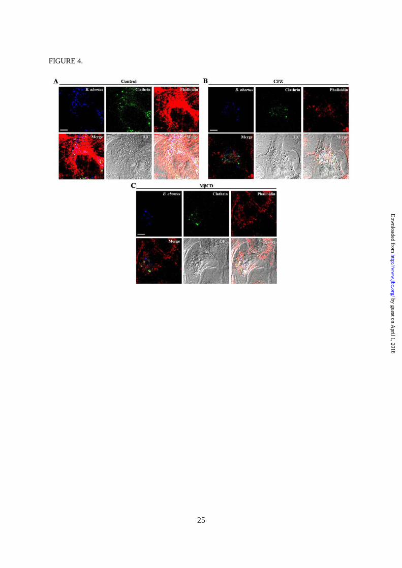
FIGURE 4. Actin polymerization and clathrin rearrangement associated with lipid rafts during
the entry of B. abortus into non-professional phagocytes. A to C, F-actin polymerization, bacterial
co-localization and clathrin rearrangement were observed in HeLa cells pretreated with medium
(Control) (A), 12.5 µм chloropromazine (CPZ) (B) or 5 mм MβCD (C) at 10 min p.i. The cells were
fixed and stained with rhodamine-conjugated phalloidin to visualize F-actin (red), Alexa Fluor 405-
labeled B. abortus (blue) and a FITC-labeled clathrin antibody (green). All images shown are
representative of three separate experiments. Scale bars = 5 µm, as indicated.
FIGURE 5. Clathrin-dependent interaction of Rab5 with B. abortus-containing vacuoles (BCVs)
during the early stage of infection. HeLa cells were pretreated with 12.5 µм chloropromazine (CPZ)
and infected with B. abortus at an MOI of 10 for the indicated times. A to C, At 15 (A), 30 (B) and 60
(C) min p.i., the cells were fixed and stained with Texas red-conjugated Rab5 (red), Alexa Fluor 405-
by guest on April 1, 2018
http://ww
w.jbc.org/
Dow
nloaded from

21
labeled B. abortus (blue) and FITC-labeled clathrin (green) antibodies. All images shown are
representative of three separate experiments. Scale bars = 5 µm, as indicated. D and E, The number of
BCVs that colocalized with clathrin and/or Rab5 (D) or LAMP-1 (E) was determined by analyzing
one hundred BCVs (combined from at least five random fields) from three separate monolayers for
each time point. Data represent the mean ± SD of triplicate trials from three independent experiments.
Differences that were statistically significant compared to untreated samples are indicated by asterisks
(*, p < 0.05, **, p < 0.01, ***, p < 0.001).
FIGURE 6. The role of clathrin in the recruitment of functional proteins associated with the
endocytic pathway to BCVs. A, Immunoblot analysis of clathrin, dynamin, caveolin, Rab5, F-actin,
Arp2/3 and β-actin in BCVs isolated from HeLa cells pretreated with or without 12.5 µм
chloropromazine (CPZ) at the indicated times p.i. The images shown are representative of three
independent experiments. B, Immunoblot ECL signals were quantified using NIH Image J software,
and the densitometry ratios of the β-actin signal to individual target protein signals are shown. Data
represent the mean ± SD of triplicate trials from three independent experiments. Differences that were
statistically significant compared to untreated samples are indicated by asterisks (**, p < 0.01, ***, p
< 0.001).
by guest on April 1, 2018
http://ww
w.jbc.org/
Dow
nloaded from

22
Figures
FIGURE 1.
by guest on April 1, 2018
http://ww
w.jbc.org/
Dow
nloaded from


Jang Lee, Moon Her, Suk Chan Jung, Masahisa Watarai and Suk KimJin Ju Lee, Dae Geun Kim, Dong Hyeok Kim, Hannah Leah Simborio, Wongi Min, Hu
and Intracellular Survival of Brucella abortus within HeLa cellsInterplay Between Clathrin and Rab5 Controls the Early Phagocytic Trafficking
published online August 12, 2013J. Biol. Chem.
10.1074/jbc.M113.491555Access the most updated version of this article at doi:
Alerts:
When a correction for this article is posted•
When this article is cited•
to choose from all of JBC's e-mail alertsClick here
by guest on April 1, 2018
http://ww
w.jbc.org/
Dow
nloaded from