title lamellar macular hole formation in chronic...
TRANSCRIPT

Title Lamellar macular hole formation in chronic cystoid macularedema associated with retinal vein occlusion.
Author(s) Tsukada, Kayoko; Tsujikawa, Akitaka; Murakami, Tomoaki;Ogino, Ken; Yoshimura, Nagahisa
Citation Japanese journal of ophthalmology (2011), 55(5): 506-513
Issue Date 2011-09
URL http://hdl.handle.net/2433/150930
Right
The final publication is available at www.springerlink.com; この論文は出版社版でありません。引用の際には出版社版をご確認ご利用ください。This is not the published version.Please cite only the published version.
Type Journal Article
Textversion author
Kyoto University

Lamellar Macular Hole Formation in Retinal Vein Occlusion. Tsukada, et al. Page 1
Title page
Title: Lamellar Macular Hole Formation in Chronic Cystoid Macular Edema
Associated with Retinal Vein Occlusion
5
Authors: Kayoko Tsukada, MD, Akitaka Tsujikawa, MD, Tomoaki Murakami, MD, Ken
Ogino, MD, Nagahisa Yoshimura, MD.
Institutions: Department of Ophthalmology and Visual Sciences, Kyoto University
Graduate School of Medicine, Kyoto, Japan 10
Running title: Lamellar Macular Hole Formation in RVO
Correspondence to: Akitaka Tsujikawa, Department of Ophthalmology and Visual
Sciences, Kyoto University Graduate School of Medicine, 54 Shogoin-Kawahara-cho, 15
Sakyo-ku, Kyoto 606-8507, Japan.
fax: +81-75-752-0933; tel: +81-75-751-4875; e-mail: [email protected]
The authors have no financial interest in the materials or devices mentioned in this
article. 20
Manuscript

Lamellar Macular Hole Formation in Retinal Vein Occlusion. Tsukada, et al. Page 2
Abstract
Purpose: To report the formation of a lamellar macular hole (LMH) in four eyes with
chronic cystoid macular edema (CME) associated with retinal vein occlusion (RVO).
Methods: We reviewed retrospectively the medical records of four patients with 5
chronic CME associated with RVO, in whom LMH formation was observed by a series
of examinations with optical coherence tomography.
Results: All eyes showed a large chronic cystoid space in the fovea. Three eyes
showed an epiretinal membrane and one eye showed traction of a posterior hyaloid
membrane to the fovea. The chronic cystoid space changed into an LMH by rupture 10
of its inner wall due to traction. After formation of the LMH, mean total foveal
thickness decreased from 590 ± 131 µm to 95 ± 22 µm, which was equal to the
thickness of the foveal photoreceptor layer before formation of the LMH (mean, 100 ±
23 µm). Visual acuity did not change substantially from before to after formation of
the LMH. 15
Conclusions: Chronic CME associated with RVO can transform into an LMH by
rupture of the inner wall of the foveal cystoid space. While this transformation is
accompanied by a substantial reduction in macular thickness, it does not lead to
change in visual function.
20
Key words: cystoid macular edema; lamellar macular hole; optical coherence
tomography; retinal vein occlusion

Lamellar Macular Hole Formation in Retinal Vein Occlusion. Tsukada, et al. Page 3
Introduction
Cystoid macula edema (CME) is the most common vision-threatening complication
associated with retinal vein occlusion (RVO) [1-3]. To date, various treatments have
been reported to reduce macular edema from RVO, including grid laser
photocoagulation [1, 4-6], pars plana vitrectomy combined with internal limiting 5
membrane peeling [7], and intravitreal injection of triamcinolone acetonide [8-11] or
bevacizumab [12]. In spite of these treatments, however, some patients have
persistent CME, and never achieve full visual recovery [13].
A lamellar macular hole (LMH) is occasionally seen in eyes with old RVO [14].
Using only fundus examination, in many cases it is difficult to distinguish the LMH 10
seen associated with old RVO from chronic CME [15]. So far, very limited
information is available on the pathogenesis of LMH associated with RVO, or on its
clinical significance [15, 16]. Based on histopathologic specimens, Gass suggested
that LMH formation may be related to CME and epiretinal membrane (ERM) [17].
Recent technological advances in optical coherence tomography (OCT) allow a more 15
detailed observation of CME and contribute to the elucidation of the pathomorphology
of this condition without entailing histopathologic methods [18, 19]. Herein, with the
use of successive examinations by OCT, we report four cases of spontaneous LMH
formation that occurred in eyes with chronic CME associated with RVO.
20

Lamellar Macular Hole Formation in Retinal Vein Occlusion. Tsukada, et al. Page 4
Material and Methods
In the current study, we retrospectively reviewed the medical records of four eyes of
four patients with chronic CME associated with RVO, in whom OCT examinations
showed transformation of the cystoid space to an LMH. Two eyes had branch retinal
vein occlusion (BRVO) and two eyes had central retinal vein occlusion (CRVO). All 5
eyes included in this study had failed to respond to medical or to surgical treatment,
including intravitreal injections of triamcinolone acetonide and bevacizumab, focal
laser or panretinal photocoagulation, or pars plana vitrectomy. For each patient, a
comprehensive medical interview was conducted regarding the presence of systemic
diseases such as diabetes mellitus, hypertension, and hyperlipemia. Each patient 10
underwent a comprehensive ophthalmologic examination, including determination of
best-corrected visual acuity, testing of intraocular pressure, indirect ophthalmoscopy,
slitlamp biomicroscopy with a contact lens, fundus photography, fluorescein
angiography, and OCT. Visual acuity was measured with a Landolt chart. OCT
scanning was performed using time-domain OCT (Stratus OCT3000, Carl Zeiss, 15
Dublin, CA) or spectral-domain OCT (3DOCT-1000, Topcon, Tokyo, Japan, Spectralis
HRA+OCT, Heidelberg Engineering, Heidelberg, Germany). Using these OCT
images, we measured total foveal thickness and thickness of the foveal photoreceptor
layer. Total foveal thickness was defined as the distance between the vitreoretinal
interface and retinal pigment epithelium in the fovea. Thickness of the foveal 20
photoreceptor layer was defined as distance between the posterior surface of the
cystoid space and retinal pigment epithelium in the fovea. This study was performed
according to the tenets of the Declaration of Helsinki; for this retrospective study,

Lamellar Macular Hole Formation in Retinal Vein Occlusion. Tsukada, et al. Page 5
Institutional Review Board/Ethics Committee approval was not required.

Lamellar Macular Hole Formation in Retinal Vein Occlusion. Tsukada, et al. Page 6
Results
In the current study, four eyes of four patients (two men and two women) with chronic
macular edema associated with RVO, ranging in age from 66 to 74 years (median,
70.5 years), were examined. Table 1 shows the characteristics of patients who were
eligible for inclusion in this study. At the initial visit, three eyes showed marked CME. 5
One eye showed macular thickening without cystoid spaces. Total foveal thickness
at the initial visit ranged from 385 µm to 986 µm (mean, 698 ± 256 µm). Best
corrected visual acuity at the initial visit ranged from 0.02 to 0.3 (median, 0.125).
While one eye did not have any surgical treatment, the remaining three eyes had
undergone various treatments for the macular edema. The two patients with CRVO 10
had undergone panretinal photocoagulation, and one of the patients with BRVO had
undergone focal photocoagulation to the affected area. Three patients had
undergone pars plana vitrectomy to remove either an ERM or posterior hyaloid
membrane.
In spite of the various types of treatment, all four eyes in the current study had 15
chronic CME before formation of the LMH. Three patients had an ERM involving the
foveal cystoid space, and one patient showed traction of the posterior hyaloid
membrane to the fovea. OCT examinations revealed transformation of the foveal
cystoid space to an LMH (Figs. 1-4). No patient, however, noticed a change in visual
function during this transformation. The inner wall of the foveal cystoid space 20
appeared to rupture with increased traction, and, after formation of the LMH,
numerous cystoid spaces remained visible in parafoveal and extrafoveal locations.
After formation of the LMH, the mean total foveal thickness decreased from 590 ± 131

Lamellar Macular Hole Formation in Retinal Vein Occlusion. Tsukada, et al. Page 7
µm to 95 ± 22 µm, which was equivalent to thickness of the foveal photoreceptor layer
before formation of the LMH (mean, 100 ± 23 µm). Even the more precise OCT
examinations showed no change in structure of the foveal photoreceptor layer, which
was the base of the LMH. The mean duration from the occurrence of RVO to the
detection of the LMH was 43.3 ± 31.6 months. Median visual acuity before and after 5
detection of the LMH was 0.15 and 0.175, so visual acuity was essentially unchanged
between before LMH formation and after LMH formation.
Case reports
Case 1. 10
A 66-year-old woman visited our clinic with a visual disturbance of the left eye (0.3
OS). At the initial visit, she had a retinal hemorrhage associated with BRVO, as well
as marked macular edema with a large cystoid space beneath the fovea, which was
623 µm in thickness (Fig. 1a). Detachment of the posterior hyaloid membrane was
not seen. She refused all surgical treatment and had follow-up examinations for only 15
the persistent CME. At 17 months after the initial visit, she still showed chronic CME
in the left eye; total foveal thickness was now 755 µm and thickness of the foveal
photoreceptor layer was 74 µm (Fig. 1b). OCT revealed the posterior hyaloid
membrane, which was attached to the fovea, and the inner wall of the foveal cystoid
space, which seemed to be being torn off by traction of this posterior hyaloid 20
membrane. Visual acuity remained 0.5 in the left eye. At 32 months, however,
OCT showed a complete LMH just beneath the fovea (Fig. 1c). The extrafoveal
cystoid spaces were still seen primarily in the outer plexiform layer. Foveal thickness

Lamellar Macular Hole Formation in Retinal Vein Occlusion. Tsukada, et al. Page 8
had decreased to 78 µm, although visual acuity in the left eye remained at 0.5.
Case 2.
A 73-year-old man visited our clinic with a sudden decrease of visual acuity in the left
eye (0.02 OS). Examination of this eye showed an extensive retinal hemorrhage 5
with severe macular edema associated with CRVO; foveal thickness was 986 µm.
Panretinal photocoagulation was performed to the extensive nonperfused area of the
left eye. Eight weeks after this initial visit for treatment of the macular edema, he
received an intravitreal injection of bevacizumab, after which, to remove the thick
ERM, pars plana vitrectomy with ERM peeling was performed. At 31 months after 10
the initial visit, he had persistent CME with a recurrent ERM. Visual acuity in the left
eye was 0.08. Total foveal thickness was 436 µm and thickness of the foveal
photoreceptor layer was 129 µm (Fig. 2a). At 37 months after the first visit, OCT
showed a marked decrease in foveal thickness. The inner wall of the foveal cystoid
space was not seen but a complete LMH was noted at the centre of the fovea (Fig. 2b). 15
There was no remarkable change of the photoreceptor layer beneath the fovea but
foveal thickness had decreased to 110 µm; visual acuity was essentially unchanged at
0.07 OS.
Case 3. 20
A 68-year-old woman was seen in our clinic with decreased visual acuity in the right
eye (0.1 OD). She showed extensive retinal hemorrhage with severe macular
edema associated with CRVO in the right eye. Total foveal thickness was 796 µm.

Lamellar Macular Hole Formation in Retinal Vein Occlusion. Tsukada, et al. Page 10
edema associated with BRVO; foveal thickness was 986 µm. With pars plana
vitrectomy and laser photocoagulation to the affected retina, macular edema was
resolved and visual acuity recovered to 0.5 OD. Six years later, he had visual
disturbance due to BRVO in the right eye (0.2 OD). He had a retinal hemorrhage
associated with BRVO, as well as marked macular edema with large foveal cystoid 5
spaces with fine ERM in the right eye. Total foveal thickness was 596 µm and
thickness of the foveal photoreceptor layer was 98 µm (Fig. 4a). At 89 months after
the first visit, OCT showed a marked decrease in foveal thickness. The inner wall of
the foveal cystoid space was not seen but a complete LMH was noted (Fig. 4b).
There was no remarkable change of the photoreceptor layer beneath the fovea but 10
foveal thickness had decreased to 116 µm; visual acuity was unchanged at 0.2 OD.
Lamellar Macular Hole Formation in Retinal Vein Occlusion. Tsukada, et al. Page 11
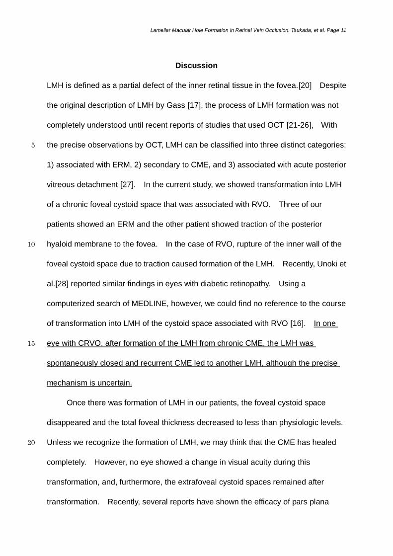
Discussion
LMH is defined as a partial defect of the inner retinal tissue in the fovea.[20] Despite
the original description of LMH by Gass [17], the process of LMH formation was not
completely understood until recent reports of studies that used OCT [21-26], With
the precise observations by OCT, LMH can be classified into three distinct categories: 5
1) associated with ERM, 2) secondary to CME, and 3) associated with acute posterior
vitreous detachment [27]. In the current study, we showed transformation into LMH
of a chronic foveal cystoid space that was associated with RVO. Three of our
patients showed an ERM and the other patient showed traction of the posterior
hyaloid membrane to the fovea. In the case of RVO, rupture of the inner wall of the 10
foveal cystoid space due to traction caused formation of the LMH. Recently, Unoki et
al.[28] reported similar findings in eyes with diabetic retinopathy. Using a
computerized search of MEDLINE, however, we could find no reference to the course
of transformation into LMH of the cystoid space associated with RVO [16]. In one
eye with CRVO, after formation of the LMH from chronic CME, the LMH was 15
spontaneously closed and recurrent CME led to another LMH, although the precise
mechanism is uncertain.
Once there was formation of LMH in our patients, the foveal cystoid space
disappeared and the total foveal thickness decreased to less than physiologic levels.
Unless we recognize the formation of LMH, we may think that the CME has healed 20
completely. However, no eye showed a change in visual acuity during this
transformation, and, furthermore, the extrafoveal cystoid spaces remained after
transformation. Recently, several reports have shown the efficacy of pars plana

Lamellar Macular Hole Formation in Retinal Vein Occlusion. Tsukada, et al. Page 12
vitrectomy for LMH associated with ERM [29-32]. However, It is still uncertain
whether additional treatment, such as pars plana vitrectomy combined with internal
limited membrane peeling, is necessary for the type of LMH seen in our patients. In
these patients, each of whom had an old RVO, the recovery of visual function may be
limited, even with pars plana vitrectomy, because of damage to the foveal 5
photoreceptor cells caused by the long-standing CME.
Recently, integrity of the foveal photoreceptor layer, especially its outer aspect,
has been suggested to be essential to visual acuity [33]. In eyes with resolved
macular edema associated with BRVO, Murakami et al.[34] suggested that, to
achieve good visual recovery, a simple reduction in foveal thickness is insufficient, 10
and that restoration of structure of the photoreceptors to a more physiologic condition
is needed. In addition, Ota et al.[13] reported that thickness of the foveal
photoreceptor layer is correlated closely with visual function in eyes with persistent or
recurrent CME associated with BRVO. Even in eyes with a large cystoid space in
the fovea, if the foveal outer photoreceptor layer beneath the cystoid space is intact, 15
visual acuity can be preserved [13]. In the current study, mean total foveal thickness
after the formation of LMH was equivalent to the thickness of the foveal photoreceptor
layer before transformation. In addition, even the more precise OCT examinations
showed no remarkable change in structure of the foveal photoreceptor layer. A
previous report by Ota et al.[13] may explain our finding that visual acuity did not 20
change during the formation of LMH.
From our findings, we can say that the inner wall of the foveal cystoid space
plays a minor role in visual function. Singh et al.[35] have reported that, although

Lamellar Macular Hole Formation in Retinal Vein Occlusion. Tsukada, et al. Page 13
surgical puncture of the CME caused the structural cystoid changes of the retina to
resolve, it failed to improve visual acuity. Our findings in the current study may
explain the efficacy of puncture of the CME in eyes with RVO, but to treat CME
associated with long-standing RVO, it may be essential to retain integrity of the foveal
photoreceptor layer. 5
Limitations of the current study are its retrospective nature and small number of
cases. Although LMH is a rare complication of old RVO, we have shown that chronic
CME in RVO can, in fact, transform into LMH. In the formation of the LMH, traction
of posterior hyaloid membrane or ERM causes rupture of the inner wall of the foveal
cystoid space. Although this transformation is accompanied by a substantial 10
reduction in macular thickness, it does not necessarily lead to change in visual
function. In addition, the current findings support the importance of the foveal
photoreceptor layer in chronic CME that accompanies RVO, although the perfusion
status around the fovea was not evaluated sufficiently.
15
Lamellar Macular Hole Formation in Retinal Vein Occlusion. Tsukada, et al. Page 9
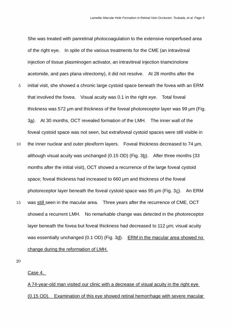
She was treated with panretinal photocoagulation to the extensive nonperfused area
of the right eye. In spite of the various treatments for the CME (an intravitreal
injection of tissue plasminogen activator, an intravitreal injection triamcinolone
acetonide, and pars plana vitrectomy), it did not resolve. At 28 months after the
initial visit, she showed a chronic large cystoid space beneath the fovea with an ERM 5
that involved the fovea. Visual acuity was 0.1 in the right eye. Total foveal
thickness was 572 µm and thickness of the foveal photoreceptor layer was 99 µm (Fig.
3a). At 30 months, OCT revealed formation of the LMH. The inner wall of the
foveal cystoid space was not seen, but extrafoveal cystoid spaces were still visible in
the inner nuclear and outer plexiform layers. Foveal thickness decreased to 74 µm, 10
although visual acuity was unchanged (0.15 OD) (Fig. 3b). After three months (33
months after the initial visit), OCT showed a recurrence of the large foveal cystoid
space; foveal thickness had increased to 660 µm and thickness of the foveal
photoreceptor layer beneath the foveal cystoid space was 95 µm (Fig. 3c). An ERM
was still seen in the macular area. Three years after the recurrence of CME, OCT 15
showed a recurrent LMH. No remarkable change was detected in the photoreceptor
layer beneath the fovea but foveal thickness had decreased to 112 µm; visual acuity
was essentially unchanged (0.1 OD) (Fig. 3d). ERM in the macular area showed no
change during the reformation of LMH.
20
Case 4.
A 74-year-old man visited our clinic with a decrease of visual acuity in the right eye
(0.15 OD). Examination of this eye showed retinal hemorrhage with severe macular

Lamellar Macular Hole Formation in Retinal Vein Occlusion. Tsukada, et al. Page 14
References
1. The Branch Vein Occlusion Study Group. Argon laser photocoagulation for
macular edema in branch vein occlusion. Am J Ophthalmol. 1984;98:271-82.
2. Glacet-Bernard A, Coscas G, Chabanel A, Zourdani A, Lelong F, Samama MM.
Prognostic factors for retinal vein occlusion: prospective study of 175 cases. 5
Ophthalmology. 1996;103:551-60.
3. Tso MOM. Pathology of cystoid macular edema. Ophthalmology. 1982;89:902-15.
4. Esrick E, Subramanian ML, Heier JS, Devaiah AK, Topping TM, Frederick AR, et al.
Multiple laser treatments for macular edema attributable to branch retinal vein
occlusion. Am J Ophthalmol. 2005;139:653-7. 10
5. Ohashi H, Oh H, Nishiwaki H, Nonaka A, Takagi H. Delayed absorption of macular
edema accompanying serous retinal detachment after grid laser treatment in
patients with branch retinal vein occlusion. Ophthalmology. 2004;111:2050-6.
6. Arnarsson A, Stefánsson E. Laser treatment and the mechanism of edema
reduction in branch retinal vein occlusion. Invest Ophthalmol Vis Sci. 15
2000;41:877-9.
7. Mandelcorn MS, Nrusimhadevara RK. Internal limiting membrane peeling for
decompression of macular edema in retinal vein occlusion: a report of 14 cases.
Retina. 2004;24:348-55.
8. Tsujikawa A, Fujihara M, Iwawaki T, Yamamoto K, Kurimoto Y. Triamcinolone 20
acetonide with vitrectomy for treatment of macular edema associated with branch
retinal vein occlusion. Retina. 2005;25:861-7.
9. Karacorlu M, Ozdemir H, Karacorlu SA. Resolution of serous macular detachment

Lamellar Macular Hole Formation in Retinal Vein Occlusion. Tsukada, et al. Page 15
after intravitreal triamcinolone acetonide treatment of patients with branch retinal
vein occlusion. Retina. 2005;25:856-60.
10. Çekiç O, Chang S, Tseng JJ, Barile GR, Del Priore LV, Weissman H, et al.
Intravitreal triamcinolone injection for treatment of macular edema secondary to
branch retinal vein occlusion. Retina. 2005;25:851-5. 5
11. Chen SD, Sundaram V, Lochhead J, Patel CK. Intravitreal triamcinolone for the
treatment of ischemic macular edema associated with branch retinal vein
occlusion. Am J Ophthalmol. 2006;141:876-83.
12. Rabena MD, Pieramici DJ, Castellarin AA, Nasir MA, Avery RL. Intravitreal
bevacizumab (Avastin) in the treatment of macular edema secondary to branch 10
retinal vein occlusion. Retina. 2007;27:419-25.
13. Ota M, Tsujikawa A, Murakami T, Yamaike N, Sakamoto A, Kotera Y, et al. Foveal
photoreceptor layer in eyes with persistent cystoid macular edema associated with
branch retinal vein occlusion. Am J Ophthalmol. 2008;145:273-80.
14. Trempe CL, Takahashi M, Topilow HW. Vitreous changes in retinal branch vein 15
occlusion. Ophthalmology. 1981;88:681-7.
15. Leibovitch I, Azmon B, Pianka P, Alster Y, Loewenstein A. Macular hole secondary
to branch retinal vein occlusion diagnosed by Retinal Thickness Analyzer.
Ophthalmic Surg Lasers Imaging. 2003;34:53-6.
16. Ophir A, Fatum S. Cystoid foveal oedema in symptomatic inner lamellar macular 20
holes. Eye (Lond). 2009;23:1781-5.
17. Gass JD. Lamellar macular hole: a complication of cystoid macular edema after
cataract extraction. Arch Ophthalmol. 1976;94:793-800.
Lamellar Macular Hole Formation in Retinal Vein Occlusion. Tsukada, et al. Page 16
18. Yamaike N, Tsujikawa A, Ota M, Sakamoto A, Kotera Y, Kita M, et al.
Three-dimensional imaging of cystoid macular edema in retinal vein occlusion.
Ophthalmology. 2008;115:355-62.
19. Catier A, Tadayoni R, Paques M, Erginay A, Haouchine B, Gaudric A, et al.
Characterization of macular edema from various etiologies by optical coherence 5
tomography. Am J Ophthalmol. 2005;140:200-6.
20. Guyer DR, Green WR, de Bustros S, Fine SL. Histopathologic features of
idiopathic macular holes and cysts. Ophthalmology. 1990;97:1045-51.
21. Takahashi H, Kishi S. Tomographic features of a lamellar macular hole formation
and a lamellar hole that progressed to a full-thickness macular hole. Am J 10
Ophthalmol. 2000;130:677-9.
22. Theodossiadis PG, Grigoropoulos VG, Emfietzoglou I, Nikolaidis P, Vergados I,
Apostolopoulos M, et al. Evolution of lamellar macular hole studied by optical
coherence tomography. Graefes Arch Clin Exp Ophthalmol. 2009;247:13-20.
23. Haouchine B, Massin P, Gaudric A. Foveal pseudocyst as the first step in macular 15
hole formation: a prospective study by optical coherence tomography.
Ophthalmology. 2001;108:15-22.
24. Haouchine B, Massin P, Tadayoni R, Erginay A, Gaudric A. Diagnosis of macular
pseudoholes and lamellar macular holes by optical coherence tomography. Am J
Ophthalmol. 2004;138:732-9. 20
25. Bottoni F, Carmassi L, Cigada M, Moschini S, Bergamini F. Diagnosis of macular
pseudoholes and lamellar macular holes: is optical coherence tomography the
"gold standard"? Br J Ophthalmol. 2008;92:635-9.

Lamellar Macular Hole Formation in Retinal Vein Occlusion. Tsukada, et al. Page 17
26. Chen JC, Lee LR. Clinical spectrum of lamellar macular defects including
pseudoholes and pseudocysts defined by optical coherence tomography. Br J
Ophthalmol. 2008;92:1342-6.
27. Androudi S, Stangos A, Brazitikos PD. Lamellar macular holes: tomographic
features and surgical outcome. Am J Ophthalmol. 2009;148:420-6. 5
28. Unoki N, Nishijima K, Kita M, Oh H, Sakamoto A, Kameda T, et al. Lamellar
macular hole formation in patients with diabetic cystoid macular edema. Retina.
2009;29:1128-33.
29. Hirakawa M, Uemura A, Nakano T, Sakamoto T. Pars plana vitrectomy with gas
tamponade for lamellar macular holes. Am J Ophthalmol. 2005;140:1154-5. 10
30. Kokame GT, Tokuhara KG. Surgical management of inner lamellar macular hole.
Ophthalmic Surg Lasers Imaging. 2007;38:61-3.
31. Garretson BR, Pollack JS, Ruby AJ, Drenser KA, Williams GA, Sarrafizadeh R.
Vitrectomy for a symptomatic lamellar macular hole. Ophthalmology.
2008;115:884-6. 15
32.Michalewska Z, Michalewski J, Odrobina D, Pikulski Z, Cisiecki S, Dziegielewski K,
et al. Surgical treatment of lamellar macular holes. Graefes Arch Clin Exp
Ophthalmol. 2010;248:1395-400.
33. Costa RA, Calucci D, Skaf M, Cardillo JA, Castro JC, Melo LAJr., et al. Optical
coherence tomography 3: Automatic delineation of the outer neural retinal 20
boundary and its influence on retinal thickness measurements. Invest Ophthalmol
Vis Sci. 2004;45:2399-406.
34. Murakami T, Tsujikawa A, Ohta M, Miyamoto K, Kita M, Watanabe D, et al.

Lamellar Macular Hole Formation in Retinal Vein Occlusion. Tsukada, et al. Page 18
Photoreceptor status after resolved macular edema in branch retinal vein
occlusion treated with tissue plasminogen activator. Am J Ophthalmol.
2007;143:171-3.
35. Singh RP, Margolis R, Kaiser PK. Cystoid puncture for chronic cystoid macular
oedema. Br J Ophthalmol. 2007;91:1062-4. 5

Lamellar Macular Hole Formation in Retinal Vein Occlusion. Tsukada, et al. Page 19
Figure legends
Figure 1. Lamellar macular hole formation by traction of the posterior hyaloid
membrane to the chronic foveal cystoid space associated with branch retinal vein
occlusion. Horizontal (left) and vertical (right) optical coherence tomographic images
centered on the fovea. (a) At 17 months after the initial visit, chronic cystoid edema 5
is seen. Total foveal thickness at this time was 755 µm and thickness of the foveal
photoreceptor layer was 74 µm. (b) The inner wall of the foveal cystoid space seems
to be being torn off by traction of the posterior hyaloid membrane. Visual acuity was
still 0.5. (c) A lamellar macular hole has formed just beneath the fovea. Extrafoveal
cystoid spaces are still seen in the outer plexiform layer. Foveal thickness 10
decreased to 78 µm but visual acuity remained at 0.5.
Figure 2. Lamellar macular hole formation by traction of the epiretinal membrane to
the foveal cystoid space associated with central retinal vein occlusion. Horizontal
(upper) and vertical (lower) optical coherence tomographic images centered on the 15
fovea. (a) At 31 months after the initial visit, a large foveal cystoid space is seen, as
is a recurrent epiretinal membrane. Visual acuity was 0.08. Total foveal thickness
was 436 µm and thickness of the foveal photoreceptor layer was 129 µm. (b) A
lamellar macular hole has been formed by the defect of the inner wall of the foveal
cystoid space. No remarkable change is detected in the photoreceptor layer 20
beneath the fovea. Foveal thickness decreased to 110 µm but visual acuity was
essentially unchanged (0.07 OS).
Lamellar Macular Hole Formation in Retinal Vein Occlusion. Tsukada, et al. Page 20
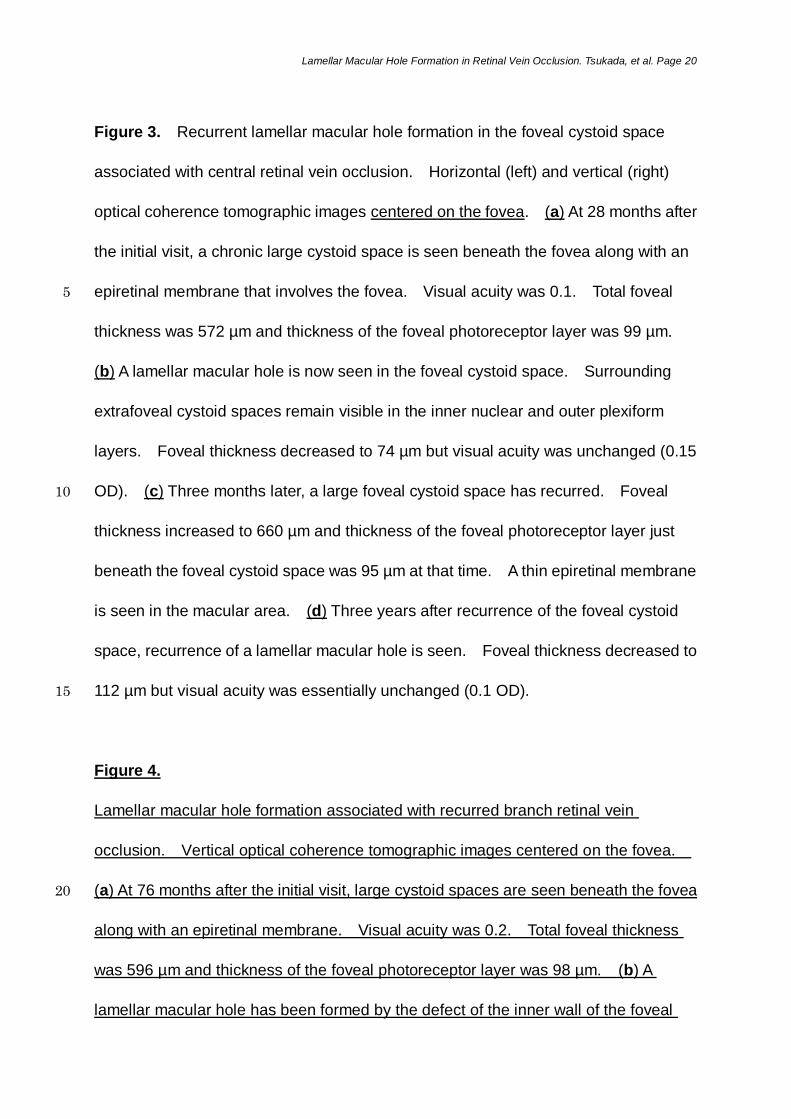
Figure 3. Recurrent lamellar macular hole formation in the foveal cystoid space
associated with central retinal vein occlusion. Horizontal (left) and vertical (right)
optical coherence tomographic images centered on the fovea. (a) At 28 months after
the initial visit, a chronic large cystoid space is seen beneath the fovea along with an
epiretinal membrane that involves the fovea. Visual acuity was 0.1. Total foveal 5
thickness was 572 µm and thickness of the foveal photoreceptor layer was 99 µm.
(b) A lamellar macular hole is now seen in the foveal cystoid space. Surrounding
extrafoveal cystoid spaces remain visible in the inner nuclear and outer plexiform
layers. Foveal thickness decreased to 74 µm but visual acuity was unchanged (0.15
OD). (c) Three months later, a large foveal cystoid space has recurred. Foveal 10
thickness increased to 660 µm and thickness of the foveal photoreceptor layer just
beneath the foveal cystoid space was 95 µm at that time. A thin epiretinal membrane
is seen in the macular area. (d) Three years after recurrence of the foveal cystoid
space, recurrence of a lamellar macular hole is seen. Foveal thickness decreased to
112 µm but visual acuity was essentially unchanged (0.1 OD). 15
Figure 4.
Lamellar macular hole formation associated with recurred branch retinal vein
occlusion. Vertical optical coherence tomographic images centered on the fovea.
(a) At 76 months after the initial visit, large cystoid spaces are seen beneath the fovea 20
along with an epiretinal membrane. Visual acuity was 0.2. Total foveal thickness
was 596 µm and thickness of the foveal photoreceptor layer was 98 µm. (b) A
lamellar macular hole has been formed by the defect of the inner wall of the foveal

Lamellar Macular Hole Formation in Retinal Vein Occlusion. Tsukada, et al. Page 21
cystoid space. No remarkable change is detected in the photoreceptor layer
beneath the fovea. Foveal thickness decreased to 116 µm but visual acuity was
unchanged (0.2 OD).

Table 1. Patient characteristics before and after formation of the LMH associated with RVO
Initial visit Before formation of the LMH After formation of the LMH
Patien
t
Age
(years)
/gende
r
Type
of
RVO
Visual
acuity
Total
foveal
thickness
(µm)
Treatments
Visual
acuity
Total
foveal
thickness
(µm)
Thickness of
foveal
photoreceptor
layer (µm)
Duration
to LMH
formation
(month)
Visual
acuity
Total
foveal
thickness
(µm)
Follow-
up
(month)
1 66/F
BRVO 0.3 623 None 0.5
755
74
17
0.5
78
39
2 73/M
CRVO 0.02 385 PPV, PRP, an intravitreal
injection of bevacizumab
0.08
436
129
37
0.07
110
37
3 68/F
CRVO 0.1 796 PPV, PRP, an intravitreal
injection of t-PA, an
intravitreal injection of TA
0.1
572 99 30
0.15
74 -
- - 0.1 660 97 69 0.1 112 96
4 74/M
BRVO 0.15 986 PPV, focal PC 0.2
596
98
89
0.2
116
92
LMH, lamellar macular hole; RVO, retinal vein occlusion; BRVO, branch retinal vein occlusion; CRVO, central retinal vein occlusion; PPV, pars plana vitrectomy; PRP,
panretinal photocoagulation; t-PA, tissue plasminogen activator; TA, triamcinolone acetonide; PC, photocoagulation.
Visual acuity was measured with a Landolt chart.
Table 1.

Figure 1.

Figure 2.

Figure 3.

Figure 4.