title: pgrmc1 localization and putative function in the
TRANSCRIPT
1
Title:
PGRMC1 localization and putative function in the nucleolus of bovine
granulosa cells and oocytes
Authors and Affiliations: 5
Laura Terzaghi1, Alberto Maria Luciano1, Priscila C. Dall’Acqua2, Silvia C.
Modina1, John J. Peluso3 and Valentina Lodde1*
1Reproductive and Developmental Biology Laboratory, Department of Health,
Animal Science and Food Safety, University of Milan, Italy 10
3 School of Agricultural and Veterinarian Sciences, São Paulo State
University (UNESP), Jaboticabal, Brazil.
3Department of Obstetrics and Gynecology, University of Connecticut Health
Center, Farmington, Connecticut, USA
*Correspondence: Valentina Lodde, Dipartimento di Scienze Veterinarie per 15
la Salute, la Produzione Animale e la Sicurezza Alimentare, Università degli
Studi di Milano, Via Celoria, 10 - 20133 Milano, Italy - Phone (+39) 02
50317987 - [email protected]
Short title: PGRMC1 function in the nucleolus 20
Grant support:
This work was supported by CIG - Marie Curie Actions FP7-Reintegration-
Grants within the 7th European Community Framework Programme (Contract:
303640,"Pro-Ovum") awarded to V.L., ‘Fondo Piano di sviluppo UNIMI linea B 25
Page 1 of 34 Reproduction Advance Publication first posted on 16 January 2018 as Manuscript REP-17-0534
Copyright © 2018 by the Society for Reproduction and Fertility.
2
- Giovani ricercatori’ - Grant n.:15-6-3027000-54, awarded to V.L. and Fondo
Linea 2B - 2016 - Grant n.:15-6-3027000-81, awarded to A.M.L.; P.C.D.A is
supported by Scholarship from Capes foundation /PDSE/88881.134900/2016-
01
30
Conflict of interest: Authors declare no conflict of interest.
Keywords (Up to 5): PGRMC1, nucleolin, nucleolus, granulosa cells, oocyte.
Abstract
Progesterone Receptor Membrane Component-1 (PGRMC1) is a 35
highly conserved multifunctional protein that is found in numerous systems,
including reproductive system. Interestingly, PGRMC1 is expressed at several
intracellular locations, including the nucleolus. The aim of this study is to
investigate the functional relationship between PGRMC1 and nucleolus.
Immunofluorescence experiments confirmed PGRMC1’s nucleolar localization 40
in cultured bovine granulosa cells (bGC) and oocytes. Additional experiments
conducted on bGC revealed that PGRMC1 co-localizes with nucleolin (NCL),
a major nucleolar protein. Furthermore, small interfering RNA (RNAi)
mediated gene-silencing experiments showed that when PGRMC1 expression
was depleted, NCL translocated from the nucleolus to the nucleoplasm. 45
Similarly, oxidative stress induced by hydrogen peroxide (H2O2) treatment,
reduced PGRMC1 immunofluorescent signal in the nucleolus and increased
NCL nucleoplasmic signal, when compared to non-treated cells. Although
PGRMC1 influenced NCL localization, a direct interaction between these two
proteins was not detected using in situ proximity ligation assay. This suggests 50
Page 2 of 34
3
the involvement of additional molecules in mediating the co-localization of
PGRMC1 and nucleolin. Since nucleolin translocates into the nucleoplasm in
response to various cellular stressors, PGRMC1’s ability to regulate its
localization within the nucleolus is likely an important component of
mechanism by which cells response to stress. This concept is consistent with 55
PGRMC1’s well-described ability to promote ovarian cell survival and provides
a rationale for future studies on PGRMC1, NCL and the molecular mechanism
by which these two proteins protect against the adverse effect of cellular
stressors, including oxidative stress.
60
Introduction
Progesterone Receptor Membrane Component-1 (PGRMC1) is a
multifunctional protein that is highly conserved in eukaryotes. It belongs to the
membrane associated progesterone receptor (MAPR) family and is expressed 65
in several mammalian organs and tissues (Runko et al. 1999, Raza et al.
2001, Sakamoto et al. 2004, Bali et al. 2013a, Bali et al. 2013b), including
those of the reproductive system (Zhang et al. 2008, Luciano et al. 2010,
Aparicio et al. 2011, Luciano et al. 2011, Keator et al. 2012, Saint-Dizier et al.
2012, Tahir et al. 2013, Kowalik et al. 2016). Specifically, PGRMC1 is 70
expressed by granulosa and luteal cells of human, rodent, bovine and canine
ovaries (Engmann et al. 2006, Peluso 2006, Aparicio et al. 2011, Luciano et
al. 2011, Tahir et al. 2013, Griffin et al. 2014, Terzaghi et al. 2016), as well as
oocytes (Luciano et al. 2010, Luciano et al. 2013, Terzaghi et al. 2016).
Page 3 of 34
4
Multiple functions are attributed to PGRMC1 (reviewed in (Cahill 2007, 75
Brinton et al. 2008, Neubauer et al. 2013, Peluso & Pru 2014, Cahill et al.
2016, Ryu et al. 2017) as reflected by it being localized to numerous sub-
cellular compartments. As predicted by the presence of a transmembrane
domain, PGRMC1 localizes in several membranous compartments, such as
the endoplasmic reticulum, the Golgi apparatus, the nuclear and plasma 80
membranes, the endosomes, and the secretory vesicles (Meyer et al. 1996,
Raza et al. 2001, Bramley et al. 2002, Hand & Craven 2003, Shin et al. 2003,
Sakamoto et al. 2004, Min et al. 2005, Peluso et al. 2006, Zhang et al. 2008,
Neubauer et al. 2009, Ahmed et al. 2010, Roy et al. 2010, Wu et al. 2011, Xu
et al. 2011, Mir et al. 2012, Mir et al. 2013, Thomas et al. 2014). Interestingly, 85
PGRMC1 is also detected in the nucleus (Beausoleil et al. 2004, Peluso et al.
2008, Zhang et al. 2008, Ahmad et al. 2009, Peluso et al. 2009, Luciano et al.
2010, Peluso et al. 2010a, Peluso et al. 2012), specifically to the nucleolus
(Ahmad et al. 2009, Luciano et al. 2010, Boisvert et al. 2012, Thul et al. 2017;
http://www.proteinatlas.org). This high compartmentalization suggests that at 90
each site PGRMC1 participates in the control of precise cellular processes.
In order to shed light into the intricate story of PGRMC1’s biological
significance, it is important to dissect the function of PGRMC1 at each sub-
cellular compartment. To this end, we have started to address whether
PGRMC1 has a role in regulating the nucleolar function. Although the 95
nucleolus’ main function involves ribosome subunits production, recent
advances describe it as a multifunctional subnuclear compartment. It appears
that the nucleolus is a dynamic structure, which disassembles during mitosis
and responds to signaling events during interphase. As such, it is involved in
Page 4 of 34
5
cell cycle control, especially regulating protein modifications such as 100
sumoylation and phosphorylation or sequestrating specific proteins (Boisvert
et al. 2007). Furthermore, it acts as a stress sensor mediating p53
stabilization in order to arrest cell cycle progression (Boisvert et al. 2007,
Boulon et al. 2010).
Nucleolin (NCL) is the most abundant and well-characterized protein 105
within the nucleolus, where it participates in ribosome biogenesis; however,
NCL is also distributed in other subcellular compartments, such as the
nucleoplasm, the cytoplasm and the cell surface (reviewed in (Boisvert et al.
2007, Tajrishi et al. 2011, Ginisty et al. 1999, Jia et al. 2017). Numerous
studies have shown that NCL subcellular localization is tightly correlated with 110
its function under physiological and pathological conditions (Jia et al. 2017).
Interestingly, experimental evidences have indicated that NCL has protective
roles under cellular stress conditions such as heat stress, gamma irradiation
and oxidative stress (Jia et al. 2017). Furthermore, translocation from the
nucleolus to the nucleoplasm or cytoplasm seems to be part of the 115
mechanism by which NCL participates in cellular stress response in different
cell types (Daniely & Borowiec 2000, Daniely et al. 2002, Zhang et al. 2010).
Thus, the overall goal of the present study is to examine the role of PGRMC1
on nucleolar function, particularly its relationship with NCL under normal and
stress-induced conditions. 120
Material and methods
Reagents
Page 5 of 34
6
All the chemicals used in this study were purchased from Sigma-125
Aldrich (St. Louis, MO) except for those specifically mentioned. Gene
silencing was performed by using the Stealth RNAi™ siRNA technology from
Life Technologies as previously described (Terzaghi et al. 2016) using
PGRMC1 Stealth RNAi (PGRMC1 RNAi: (RNA)-GAG UUG UAG UCA AGU
GUC UUG GUC U) within the coding region of the bovine PGRMC1 sequence 130
(RefSeq: NM_001075133). Negative control (cat n. 12935-200) was chosen
among the Stealth RNAi negative control (CTRL RNAi) duplexes available
from Life Technologies, designed to minimize sequence homology to any
known vertebrate transcript. Primary antibodies used in this study are listed in
Table 1. 135
Sample collection
Ovaries from Holstein dairy cows were recovered at the abattoir (INALCA
S.p.A., Ospedaletto Lodigiano, LO, IT 2270M CE, Italy) from pubertal females
subjected to routine veterinary inspection and in accordance to the specific 140
health requirements stated in Council Directive 89/556/ECC and subsequent
modifications. Ovaries were transported to the laboratory within 2 hours in
sterile saline (NaCl, 9 g/l) maintained at 26°C and all subsequent procedures,
unless differently specified, were performed at 35-38°C. Bovine granulosa
cells (bGC) were collected as previously described (Terzaghi et al. 2016). 145
Briefly the content of 2-8 mm ovarian follicles, which typically contain fully-
grown oocytes, was aspirated and cumulus-oocyte complexes (COCs) were
collected and processed for further immunofluorescence analysis (see below).
Remaining follicular cells were washed in M199 supplemented with HEPES
Page 6 of 34
7
20 mM, 1,790 units/L Heparin and 0.4% of bovine serum albumin (M199-D). 150
The cell pellet was re-suspended in 1ml of Dulbecco’s modified growth
medium supplemented with 10% of bovine calf serum, 100 U/ml penicillin G,
100 µg/ml, Streptomycin and Glutamax 100U/ml (Gibco). The cell suspension
was plated in a 25 cm2 flask with 6 ml of growth medium and incubated in
humidified air at 37°C with 5% CO2. After 24 hours cells were gently washed 155
with PBS and the growth medium was changed. Cells were incubated until
confluence (typically 4-5 days), then collected after trypsinization and re-
plated according to the experimental design (see below).
Oocytes in their growing phase, characterized by a diffuse filamentous
pattern of chromatin in the nuclear area and the presence of an active 160
nucleolus were collected as previously described from 0.5 to <2 mm early
antral follicles by rupturing the follicle wall under the stereomicroscope (Lodde
et al. 2008); Both COCs collected from 0.5 to <2 mm and 2-8 antral follicles
were mechanically denuded using the vortex and fixed for further
immunofluorescence analysis. 165
RNAi treatment
RNA interference (RNAi) experiments on bGC were conducted as
previously described (Terzaghi et al. 2016). Cells were plated in a total
number of 2 X 10⁵ bGC cells in 2 ml of medium in 35-mm culture dishes and 170
incubated in humidified air at 37°C with 5% CO2. For immunofluorescent
staining, cells were plated and cultured on cover glasses in 35-mm culture
dishes under the same culture condition. After 24 h, cells at 50-70% of
confluence were transfected with 6 µl of 20 µM PGRMC1 Stealth RNAi or
Page 7 of 34
8
CTRL RNAi combined with 10 µl of Lipofectamine RNAi MAX (Life 175
Technologies) in a final volume of 2 ml OPTIMEM (Life Technologies),
according to the manufacturer protocol, and cultured for 48 h. After treatment
cells were processed for further Western blotting and immunofluorescence
analysis.
180
Hydrogen peroxide (H2O2) treatment
Cells were plated on cover glasses and cultured as described for RNAi
treatment to temporally match the two experiments. Thus, after 72 h of
culture, cells were treated with 0.5 mmol/L hydrogen peroxide (H2O2) for 90
minutes to induce acute H2O2 –induced oxidative stress (Miguel et al. 2009). 185
The same concentration was used for up to 24h to study NCL role in
mediating antiapoptotic action in cardiomyocytes and human umbilical
vascular endothelial cells (HUVEC) (Jiang et al. 2010, Zhang et al. 2010).
After treatment, cells were washed in PBS and fixed in 4% paraformaldehyde
for further immunofluorescence analysis. In addition, some cells were washed 190
in culture medium and cultured for additional 24h to assess the effect on
nuclear morphology.
Western blot analysis
The levels of PGRMC1 protein expression in CTRL and PGRMC1 195
RNAi treated bGC were assessed by Western blotting assay as previously
described (Terzaghi et al. 2016). PGRMC1 or CTRL RNAi treated bGC were
lysed in radioimmunoprecipitation assay (RIPA) buffer [50 mM Tris-HCl (pH
7.4), 150 mM NaCl, 1 mM EDTA, 1% Nonidet P-40 (NP-40), and 0.25%
Page 8 of 34
9
sodium deoxycolate], supplemented with protease inhibitors and phosphatase 200
inhibitors. All procedures were conducted on ice. Total amount of protein was
determined using the Qubit® Protein Assay Kit and Qubit® fluorometer
(Thermo Fisher Scientific). 20 µg of total protein/lane were used for Western
blottings. After the run, samples were transferred to nitrocellulose membrane
(Bio-Rad), which was then incubated with 5% dry milk powder in TBS 205
containing 0.1% tween (TBS/T) for 2 hours at room temperature. PGRMC1
immunodetection was conducted using the rabbit polyclonal antibodies (see
table 1) in 5% dry milk TBS/T. PGRMC1 was revealed using a stabilized goat
anti rabbit IgG peroxidase conjugated antibody and detected using the Super
Signal West Dura Extended Duration Substrate (Thermo Fisher Scientific). 210
The nitrocellulose membrane was stripped in stripping buffer (100 mM 2-
mercaptoethanol, 2% SDS, and 62.5 mM Tris-HCl [pH 6.7]) at 50°C for 30 min
and re-probed with the anti-beta tubulin antibody at dilution 1:1000, which was
revealed using a stabilized goat anti mouse IgG peroxidase conjugated
antibody as loading control. 215
Immunofluorescence
Immunofluorescence staining was performed on bGC as previously
described (Lodde & Peluso 2011, Terzaghi et al. 2016). Briefly, cells grown
and treated on cover glasses were fixed in 4% paraformaldehyde in PBS for 7 220
minutes and permeabilized with 0.1% triton-X in PBS for 7 minutes. Samples
were blocked with 20% normal donkey serum in PBS and incubated overnight
at 4°C with the rabbit anti PGRMC1 antibody (see table1). Double
immunostaining was performed on bGC by incubating the samples with the
Page 9 of 34
10
rabbit anti PGRMC1 or the mouse anti NCL antibodies or a combination of the 225
two. After incubation with secondary antibodies for 1 h at room temperature,
samples were washed and finally mounted on slides in the antifade medium
Vecta Shield (Vector Laboratories) supplemented with 1 µg/ml 40,6-
diamidino-2-phenylindole (DAPI). Immunofluorescent analysis on bovine
oocytes were performed as previously described (Luciano et al. 2010) on 4% 230
paraformaldehyde fixed oocytes. Immunofluorescent staining was performed
as described for bGC with the exception that oocytes were fixed for 30 min at
room temperature followed by 30 min and permeabilized with 0.3% Triton-X
100 for 10 minutes.
bGC and oocytes were analyzed on an epifluorescence microscope 235
(Eclipse E600; Nikon) equipped with a 40 X and a 60X objective, a digital
camera (Nikon digital sight, DS-U3) and software (NIS elements Imaging
Software; Nikon). Immunofluorescence negative controls, which were
performed by omitting one or both the primary antibodies, did not show any
staining under the same exposure settings. Images that were used for image 240
quantification analysis were captured under the same settings.
In situ Proximity Ligation Assay (PLA)
In situ Proximity Ligation Assay (PLA; Duolink® SIGMA) was used to
assess the interaction between PGRMC1 and nucleolin in bGC following the 245
manufacturer protocol. Primary antibodies for PGRMC1 and nucleolin were
the same used for immunofluorescence, while anti-rabbit PLUS and anti-
mouse MINUS PLA probes were used as secondary antibodies. Negative
Page 10 of 34
11
controls were performed omitting one of the two primary antibodies. Cells
were mounted with Duolink mounting medium. 250
Image analysis
Quantification of Fluorescent Intensity (FI) signal was performed using
the ImageJ software (https://imagej.net). The nucleolar signal of PGRMC1 in
bGC was quantified calculating the integrated density of PGRMC1 signal 255
selecting the whole PGRMC1 positive areas in the nucleus of a total of 50
cells for each treatment (CTRL RNAi and PGRMC1 RNAi treated cells from 3
independent replicates) at 48 h after RNAi treatment. Data were polled and
the mean FI of the CTRL RNAi treated group was set at 100%. FI values of
the CTRL RNAi and PGRMC1 RNAi treatments were expressed as a 260
percentage of the mean CTRL RNAi value. For image quantification of the
NCL nucleolar and nucleoplasmic signals, threshold was selected by
choosing a cutoff value such that all the nucleolar areas with an intense NCL
signal within each cell. Then, the NCL total nuclear FI was assessed by
selecting the whole nuclear area and calculating the integrated density of the 265
corresponding regions of interests (ROI) of a total of 50 randomly selected
nuclei in CTRL RNAi and PGRMC1 RNAi treated cells. The NCL nucleolar
signal was calculated by analyzing the integrated density of the threshold area
in each nucleus, while the NCL nucleoplasmic signal was calculated by
subtracting the total nucleolar FI to the total nuclear FI of each nucleus. Data 270
were polled and the mean nucleoplasmic NCL FI of the CTRL RNAi treated
group was set at 100%. FI values of the CTRL RNAi and PGRMC1 RNAi
treatments were expressed as a percentage of the mean CTRL RNAi value.
Page 11 of 34
12
Background signals did not change significantly among treatments. To assess
the effect of H2O2-induced oxidative stress on PGRMC1 and NCL localization, 275
the same analysis was performed on a total of 75 cells from 3 independent
replicates for each treatment (non-treated and H2O2 treated cells) after 90
minutes of H2O2 treatment.
Statistical analysis 280
Experiments were run in triplicates. All statistical analysis was done
using Prism software (GraphPad Prism v. 6.0e, La Jolla, CA, USA). Data from
the replicate experiments were pooled and the data expressed as a mean ±
SEM. Student’s t test was used to determine differences between two groups.
285
Results
PGRMC1 localization
Immunofluorescence analysis indicated that PGRMC1 localized to
areas of the interphase nucleus that were not stained by DAPI. PGRMC1’s 290
nuclear localization in bGC was the same regardless of which PGRMC1
antibody was used (Figure 1). However, nuclear staining for PGRMC1 with
the Sigma Prestige antibody displayed a diffuse signal with the staining
associated with DAPI-negative areas and only slightly more intense than that
observed for the overall nucleus. In contrast, the non-DAPI stained areas 295
within the nucleus were more intensely stained using the PGRMC1 antibody
provided by Proteintech (Figure 1). These non-DAPI stained areas typically
correspond to areas of the interphase nucleus where the nucleoli reside.
Page 12 of 34
13
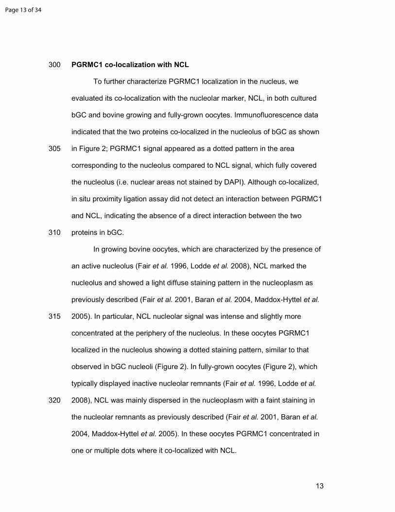
PGRMC1 co-localization with NCL 300
To further characterize PGRMC1 localization in the nucleus, we
evaluated its co-localization with the nucleolar marker, NCL, in both cultured
bGC and bovine growing and fully-grown oocytes. Immunofluorescence data
indicated that the two proteins co-localized in the nucleolus of bGC as shown
in Figure 2; PGRMC1 signal appeared as a dotted pattern in the area 305
corresponding to the nucleolus compared to NCL signal, which fully covered
the nucleolus (i.e. nuclear areas not stained by DAPI). Although co-localized,
in situ proximity ligation assay did not detect an interaction between PGRMC1
and NCL, indicating the absence of a direct interaction between the two
proteins in bGC. 310
In growing bovine oocytes, which are characterized by the presence of
an active nucleolus (Fair et al. 1996, Lodde et al. 2008), NCL marked the
nucleolus and showed a light diffuse staining pattern in the nucleoplasm as
previously described (Fair et al. 2001, Baran et al. 2004, Maddox-Hyttel et al.
2005). In particular, NCL nucleolar signal was intense and slightly more 315
concentrated at the periphery of the nucleolus. In these oocytes PGRMC1
localized in the nucleolus showing a dotted staining pattern, similar to that
observed in bGC nucleoli (Figure 2). In fully-grown oocytes (Figure 2), which
typically displayed inactive nucleolar remnants (Fair et al. 1996, Lodde et al.
2008), NCL was mainly dispersed in the nucleoplasm with a faint staining in 320
the nucleolar remnants as previously described (Fair et al. 2001, Baran et al.
2004, Maddox-Hyttel et al. 2005). In these oocytes PGRMC1 concentrated in
one or multiple dots where it co-localized with NCL.
Page 13 of 34
14
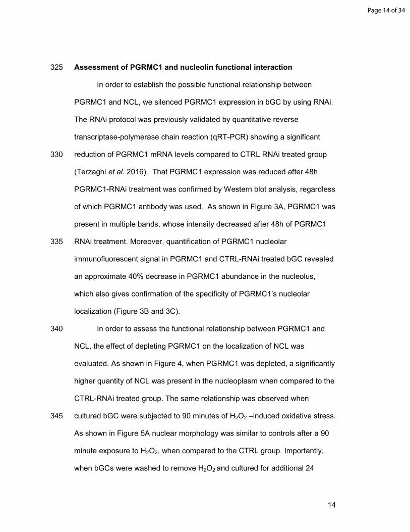
Assessment of PGRMC1 and nucleolin functional interaction 325
In order to establish the possible functional relationship between
PGRMC1 and NCL, we silenced PGRMC1 expression in bGC by using RNAi.
The RNAi protocol was previously validated by quantitative reverse
transcriptase-polymerase chain reaction (qRT-PCR) showing a significant
reduction of PGRMC1 mRNA levels compared to CTRL RNAi treated group 330
(Terzaghi et al. 2016). That PGRMC1 expression was reduced after 48h
PGRMC1-RNAi treatment was confirmed by Western blot analysis, regardless
of which PGRMC1 antibody was used. As shown in Figure 3A, PGRMC1 was
present in multiple bands, whose intensity decreased after 48h of PGRMC1
RNAi treatment. Moreover, quantification of PGRMC1 nucleolar 335
immunofluorescent signal in PGRMC1 and CTRL-RNAi treated bGC revealed
an approximate 40% decrease in PGRMC1 abundance in the nucleolus,
which also gives confirmation of the specificity of PGRMC1’s nucleolar
localization (Figure 3B and 3C).
In order to assess the functional relationship between PGRMC1 and 340
NCL, the effect of depleting PGRMC1 on the localization of NCL was
evaluated. As shown in Figure 4, when PGRMC1 was depleted, a significantly
higher quantity of NCL was present in the nucleoplasm when compared to the
CTRL-RNAi treated group. The same relationship was observed when
cultured bGC were subjected to 90 minutes of H2O2 –induced oxidative stress. 345
As shown in Figure 5A nuclear morphology was similar to controls after a 90
minute exposure to H2O2, when compared to the CTRL group. Importantly,
when bGCs were washed to remove H2O2 and cultured for additional 24
Page 14 of 34
15
hours, virtually all the bGCs appears to be apoptotic as judged by nuclear
morphology after DNA staining, while non-treated cells did not show any sign 350
of nuclear damage. Moreover, when compared to non-treated cells, PGRMC1
nucleolar signal decreased (Figure 5B) while NCL nucleoplasmic signal
increased in H2O2 treated cells. (Figure 5C).
Discussion 355
The present findings demonstrate that PGRMC1 localizes to the
nucleolus of both bovine granulosa cells and oocytes, suggesting that
PGRMC1 has a role in regulating the function of the nucleolus of these two
cell types. The prominent nucleolar localization of PGRMC1 as revealed
using the Protein Tech antibody is consistent with investigations of non-360
ovarian cells that detect PGRMC1 within the nucleolus by either
immunohistochemistry or mass spectrometric analysis (Ahmad et al. 2009,
Luciano et al. 2010, Boisvert et al. 2012, Thul et al. 2017) (see also
http://www.proteinatlas.org). However, the rabbit polyclonal antibody to
PGRMC1 provided by Sigma Prestige detects PGRMC1 not only within the 365
nucleolus but also in other interchromatin regions that resemble the nuclear
speckles (Spector & Lamond 2011). The reason for this discord likely relates
to the two antibodies detecting different molecular weight forms of PGRMC1.
Western blots using either the Proteintech or the Sigma Prestige antibody
detect PGRMC1 as bands at ≈ 25 and ≈ 55 kDa, while the Sigma antibody 370
also detects an additional band at 37 kDA and two bands greater than 55
kDA. All the bands detected by either antibody are specific since their
intensity is decreased in PGRMC1 RNAi-treated cells. The different size
Page 15 of 34
16
forms of PGRMC1 are due to dimerization and post-translational modifications
such as phosphorylation and sumoylation (Neubauer et al. 2008, Peluso et al. 375
2010b, Peluso et al. 2012, Kabe et al. 2016). Therefore, it is not surprising
that polyclonal antibodies obtained using different immunogens may
preferentially recognize one or multiple forms of PGRMC1, which in turn might
preferentially localize in different subcellular compartment.
Because the Proteintech antibody precisely localizes PGRMC1 to the 380
nucleolus, it was used to determine whether PGRMC1 co-localizes with the
nucleolar protein, NCL. This approach reveals that PGRMC1 and NCL co-
localizes to the nucleolus in bGC. Moreover, depletion of PGRMC1 results in
NCL within the nucleolus redistributing to nucleoplasm in these cells. Thus,
localization of NCL is likely dependent in part on PGRMC1. This observation 385
is important since NCL mobilization from the nucleolus into the nucleoplasm is
induced by different types of cellular stress. For example, heat shock,
ionizing radiation, and hypoxia all promote the translocation of NCL into the
nucleoplasm (Daniely & Borowiec 2000, Daniely et al. 2002). In particular,
NCL redistribution is induced by heat stress in HeLa cells and accompanied 390
by an increase in the formation of a complex between NCL and Replication
Protein A (RPA) (Daniely & Borowiec 2000), which exerts important functions
during DNA replication (Iftode et al. 1999). NCL-RPA interaction in turn
strongly inhibits DNA replication, likely by sequestering RPA away from sites
of ongoing DNA synthesis (Daniely & Borowiec 2000). Other studies in U2-OS 395
and U2-OS p53-depleted cells demonstrate that NCL redistribution occurs
when stress is induced by γ-irradiation and treatment with the radiomimetic
Page 16 of 34
17
agent, camptothecin. Under these stress conditions, NCL binds p53, which
facilitates its transit into the nucleoplasm (Daniely et al. 2002).
The stress-induced changes in NCL’s localization suggest that various 400
stressors alter PGRMC1’s ability to retain NCL, which would allow NCL to
transit to the nucleoplasm. Our study demonstrates that H2O2-induced
oxidative stress decreases the PGRMC1 signal in the nucleolus and
increases the nucleplasmic NCL signal, further reinforcing this concept. The
H2O2-induced oxidative stress model is biologically relevant in the ovary, 405
especially during luteolysis. Reactive oxygen species (ROS) are indeed
released locally by leucocyte invading the corpus luteum, which induce
apoptosis of ovarian luteal cells. (Vega et al. 1995, Davis & Rueda 2002, Del
Canto et al. 2007, Will et al. 2017). Moreover, it has been recently shown that
PGRMC1 participates in this process since inhibition of PGRMC1 using an 410
antagonist (AG205), eliminates P4’s ability to prevent H2O2-induced apoptosis
in human granulosa/luteal cells (Will et al. 2017). Interestingly it has been
proposed that AG205 may act by promoting PGRMC1’s translocation from the
nucleus to the cytoplasm and by regulating the expression of Harakiri (Hkr),
which is a BH-3 only member of the B-cell lymphoma 2 (BCL2) family that 415
promotes apoptosis by binding to and antagonizing the antiapoptotic action of
BCL2- and BCL2-like proteins (Will et al. 2017). These observations are
consistent with the NCL’s role in regulating H2O2 -induced apoptosis in
HUVEC (Zhang et al. 2010) and cardiomyocytes (Jiang et al. 2010).
The precise mechanism by which PGRMC1 controls NCL localization 420
and function remains to be further explored. To start to assess this issue, we
have focused on the nature of PGRMC1/NCL association in the nucleolus.
Page 17 of 34

18
Although PGRMC1 and NCL often co-localize to the same sub-region of the
nucleolus of bGC, they do not seem to directly interact, since we were not
able to demonstrate a direct interaction using the PLA assay. This might 425
suggest that their functional interaction could involve the participation of other
yet to be identified protein. Interestingly a known PGRMC1 binding protein,
Plasminogen Activator Inhibitor 1 RNA-Binding Protein (PAIRBP1) (Peluso et
al. 2006, Peluso et al. 2008, Peluso et al. 2013) (also known as SERPINE1
mRNA Binding Protein 1), which is typically found in the cytoplasm, localizes 430
to the nucleolus under specific experimental stress induced conditions in Hela
cells (Lee et al. 2014). Therefore, it is possible that under stress conditions
PAIRBP1 translocates to the nucleolus and competes with this putative
intermediary protein for binding to PGRMC1. This would potentially interfere
with PGRMC1’ ability to retain NCL within the nucleolus and account for the 435
translocation of nucleolin from the nucleolus into the nucleoplasm under
stress condition.
Finally, the present study reveals the relationship between PGRMC1
and NCL in bovine oocytes. PGRMC1 is present in the nucleolus of growing
oocytes and the signal is retained to some extent in the nucleolar remnants of 440
fully-grown bovine oocytes. During growth, the oocyte’s nucleus is
characterized by the presence of a diffuse filamentous transcriptionally active
chromatin and by a functional fibrillogranular nucleolus, which is gradually
disassembled forming the so called ‘nucleolar remnants’, along with the
progressive inactivation of rRNA synthesis that occurs at the end of oocyte 445
growth (Fair et al. 1996, Lodde et al. 2008). Ultrastructurally, the nucleolar
remnants appear as electron dense spheres often showing a semilunar
Page 18 of 34
19
fibrillar center-like structures attached (Fair et al. 1996, Lodde et al. 2008). In
bovine oocytes, proteins such as RNA polymerase I and Upstream Binding
Factor (UBF) remain associated to the inactive nucleolar remnants, while 450
others, such as NCL and nucleophosmin mostly disperse in the nucleoplasm
(Fair et al. 2001, Baran et al. 2004, Maddox-Hyttel et al. 2005). Upon meiotic
resumption and during oocyte maturation the nucleolar remnant further
disassembles and nucleolar proteins are probably dispersed in the cytoplasm.
After fertilization the so called ‘nucleolar precursor bodies’ (NPBs) appear as 455
electron dense compact spheres in the male and female pronuclei reviewed in
(Maddox-Hyttel et al. 2005). The NPBs serve for the re-establishment of a
functional fibrillogranular nucleolus, which in bovine occurs at the time of
major embryonic genome activation (at the 8-16 cell stage). It has been
proposed that proteins engaged in late rRNA processing of maternal origin, 460
including NCL, are to some extent re-used for nucleologenesis in the embryo
while others need to be de-novo transcribed before being incorporated in the
nucleolus (reviewed in (Maddox-Hyttel et al. 2005)). In this scenario,
PGRMC1 localization in growing and fully-grown oocytes and in the NPBs of
bovine zygotes (Luciano et al. 2010) 465
suggests a role in both the disassembly and the reassembly of the nucleolus
during meiosis and early embryogenesis. Interestingly, in growing oocytes, as
well as in bGC, PGRMC1 and NCL show a different localization pattern, with
PGRMC1 present in a dotted pattern. A similar pattern in growing bovine
oocytes has been reported for the RNA polymerase I-specific transcription 470
initiation factor, UBF (Baran et al. 2004). In future studies, it will be important
to assess whether a specific functional interaction between NCL or other
Page 19 of 34
20
nucleolar proteins and PGRMC1 exists during early embryonic development
and thereby influences the embryogenesis.
Page 20 of 34
21
References 475
Ahmad Y, Boisvert FM, Gregor P, Cobley A & Lamond AI 2009 NOPdb:
Nucleolar Proteome Database--2008 update. Nucleic Acids Res 37 D181-
184. 480
Ahmed IS, Rohe HJ, Twist KE & Craven RJ 2010 Pgrmc1 (progesterone
receptor membrane component 1) associates with epidermal growth
factor receptor and regulates erlotinib sensitivity. The Journal of biological
chemistry 285 24775-24782.
Aparicio IM, Garcia-Herreros M, O'Shea LC, Hensey C, Lonergan P & Fair T 485 2011 Expression, regulation, and function of progesterone receptors in
bovine cumulus oocyte complexes during in vitro maturation. Biol Reprod
84 910-921.
Bali N, Arimoto JM, Morgan TE & Finch CE 2013a Progesterone antagonism of
neurite outgrowth depends on microglial activation via Pgrmc1/S2R. 490 Endocrinology 154 2468-2480.
Bali N, Morgan TE & Finch CE 2013b Pgrmc1: new roles in the microglial
mediation of progesterone-antagonism of estradiol-dependent neurite
sprouting and in microglial activation. Front Neurosci 7 157.
Baran V, Pavlok A, Bjerregaard B, Wrenzycki C, Hermann D, Philimonenko 495 VV, Lapathitis G, Hozak P, Niemann H & Motlik J 2004
Immunolocalization of upstream binding factor and pocket protein p130
during final stages of bovine oocyte growth. Biol Reprod 70 877-886.
Beausoleil SA, Jedrychowski M, Schwartz D, Elias JE, Villen J, Li J, Cohn MA,
Cantley LC & Gygi SP 2004 Large-scale characterization of HeLa cell 500 nuclear phosphoproteins. Proc Natl Acad Sci U S A 101 12130-12135.
Boisvert FM, Ahmad Y, Gierlinski M, Charriere F, Lamont D, Scott M, Barton
G & Lamond AI 2012 A quantitative spatial proteomics analysis of
proteome turnover in human cells. Mol Cell Proteomics 11 M111 011429.
Boisvert FM, van Koningsbruggen S, Navascues J & Lamond AI 2007 The 505
multifunctional nucleolus. Nat Rev Mol Cell Biol 8 574-585.
Boulon S, Westman BJ, Hutten S, Boisvert FM & Lamond AI 2010 The
nucleolus under stress. Mol Cell 40 216-227.
Bramley TA, Menzies GS, Rae MT & Scobie G 2002 Non-genomic steroid
receptors in the bovine ovary. Domest Anim Endocrinol 23 3-12. 510 Brinton RD, Thompson RF, Foy MR, Baudry M, Wang J, Finch CE, Morgan TE,
Pike CJ, Mack WJ, Stanczyk FZ & Nilsen J 2008 Progesterone receptors:
form and function in brain. Front Neuroendocrinol 29 313-339.
Cahill MA 2007 Progesterone receptor membrane component 1: an integrative
review. J Steroid Biochem Mol Biol 105 16-36. 515 Cahill MA, Jazayeri JA, Catalano SM, Toyokuni S, Kovacevic Z & Richardson
DR 2016 The emerging role of progesterone receptor membrane
component 1 (PGRMC1) in cancer biology. Biochim Biophys Acta 1866
339-349.
Page 21 of 34

22
Daniely Y & Borowiec JA 2000 Formation of a complex between nucleolin and 520 replication protein A after cell stress prevents initiation of DNA
replication. J Cell Biol 149 799-810.
Daniely Y, Dimitrova DD & Borowiec JA 2002 Stress-dependent nucleolin
mobilization mediated by p53-nucleolin complex formation. Mol Cell Biol
22 6014-6022. 525 Davis JS & Rueda BR 2002 The corpus luteum: an ovarian structure with
maternal instincts and suicidal tendencies. Front Biosci 7 d1949-1978.
Del Canto F, Sierralta W, Kohen P, Munoz A, Strauss JF, 3rd & Devoto L 2007
Features of natural and gonadotropin-releasing hormone antagonist-
induced corpus luteum regression and effects of in vivo human chorionic 530
gonadotropin. J Clin Endocrinol Metab 92 4436-4443.
Engmann L, Losel R, Wehling M & Peluso JJ 2006 Progesterone regulation of
human granulosa/luteal cell viability by an RU486-independent
mechanism. J Clin Endocrinol Metab 91 4962-4968.
Fair T, Hyttel P, Greve T & Boland M 1996 Nucleus structure and 535 transcriptional activity in relation to oocyte diameter in cattle. Mol Reprod
Dev 43 503-512.
Fair T, Hyttel P, Lonergan P & Boland MP 2001 Immunolocalization of
nucleolar proteins during bovine oocyte growth, meiotic maturation, and
fertilization. Biol Reprod 64 1516-1525. 540 Ginisty H, Sicard H, Roger B & Bouvet P 1999 Structure and functions of
nucleolin. J Cell Sci 112 ( Pt 6) 761-772.
Griffin D, Liu X, Pru C, Pru JK & Peluso JJ 2014 Expression of progesterone
receptor membrane component-2 within the immature rat ovary and its
role in regulating mitosis and apoptosis of spontaneously immortalized 545 granulosa cells. Biol Reprod 91 36.
Hand RA & Craven RJ 2003 Hpr6.6 protein mediates cell death from oxidative
damage in MCF-7 human breast cancer cells. Journal of cellular
biochemistry 90 534-547.
Iftode C, Daniely Y & Borowiec JA 1999 Replication protein A (RPA): the 550 eukaryotic SSB. Crit Rev Biochem Mol Biol 34 141-180.
Jia W, Yao Z, Zhao J, Guan Q & Gao L 2017 New perspectives of physiological
and pathological functions of nucleolin (NCL). Life Sci 186 1-10.
Jiang B, Zhang B, Liang P, Song J, Deng H, Tu Z, Deng G & Xiao X 2010
Nucleolin/C23 mediates the antiapoptotic effect of heat shock protein 70 555
during oxidative stress. FEBS J 277 642-652.
Kabe Y, Nakane T, Koike I, Yamamoto T, Sugiura Y, Harada E, Sugase K,
Shimamura T, Ohmura M, Muraoka K, Yamamoto A, Uchida T, Iwata
S, Yamaguchi Y, Krayukhina E, Noda M, Handa H, Ishimori K,
Uchiyama S, Kobayashi T & Suematsu M 2016 Haem-dependent 560
dimerization of PGRMC1/Sigma-2 receptor facilitates cancer proliferation
and chemoresistance. Nat Commun 7 11030.
Keator CS, Mah K & Slayden OD 2012 Alterations in progesterone receptor
membrane component 2 (PGRMC2) in the endometrium of macaques
afflicted with advanced endometriosis. Mol Hum Reprod 18 308-319. 565 Kowalik MK, Martyniak M, Rekawiecki R & Kotwica J 2016 Expression and
immunolocalization of membrane progesterone receptors in the bovine
oviduct. Domest Anim Endocrinol 55 83-96.
Page 22 of 34
23
Lee YJ, Wei HM, Chen LY & Li C 2014 Localization of SERBP1 in stress granules
and nucleoli. FEBS J 281 352-364. 570 Lodde V, Modina S, Maddox-Hyttel P, Franciosi F, Lauria A & Luciano AM
2008 Oocyte morphology and transcriptional silencing in relation to
chromatin remodeling during the final phases of bovine oocyte growth.
Mol Reprod Dev 75 915-924.
Lodde V & Peluso JJ 2011 A novel role for progesterone and progesterone 575
receptor membrane component 1 in regulating spindle microtubule
stability during rat and human ovarian cell mitosis. Biol Reprod 84 715-
722.
Luciano AM, Corbani D, Lodde V, Tessaro I, Franciosi F, Peluso JJ & Modina S
2011 Expression of progesterone receptor membrane component-1 in 580 bovine reproductive system during estrous cycle. Eur J Histochem 55 e27.
Luciano AM, Franciosi F, Lodde V, Tessaro I, Corbani D, Modina SC & Peluso
JJ 2013 Oocytes Isolated from Dairy Cows with Reduced Ovarian Reserve
Have a High Frequency of Aneuploidy and Alterations in the Localization
of Progesterone Receptor Membrane Component 1 and Aurora Kinase B. 585 Biology of Reproduction 88.
Luciano AM, Lodde V, Franciosi F, Ceciliani F & Peluso JJ 2010 Progesterone
receptor membrane component 1 expression and putative function in
bovine oocyte maturation, fertilization, and early embryonic
development. Reproduction 140 663-672. 590 Maddox-Hyttel P, Bjerregaard B & Laurincik J 2005 Meiosis and embryo
technology: renaissance of the nucleolus. Reprod Fertil Dev 17 3-14.
Meyer C, Schmid R, Scriba PC & Wehling M 1996 Purification and partial
sequencing of high-affinity progesterone-binding site(s) from porcine
liver membranes. Eur J Biochem 239 726-731. 595 Miguel F, Augusto AC & Gurgueira SA 2009 Effect of acute vs chronic H2O2-
induced oxidative stress on antioxidant enzyme activities. Free Radic Res
43 340-347.
Min L, Strushkevich NV, Harnastai IN, Iwamoto H, Gilep AA, Takemori H,
Usanov SA, Nonaka Y, Hori H, Vinson GP & Okamoto M 2005 Molecular 600
identification of adrenal inner zone antigen as a heme-binding protein.
The FEBS journal 272 5832-5843.
Mir SU, Ahmed IS, Arnold S & Craven RJ 2012 Elevated progesterone receptor
membrane component 1/sigma-2 receptor levels in lung tumors and
plasma from lung cancer patients. International journal of cancer. Journal 605 international du cancer 131 E1-9.
Mir SU, Schwarze SR, Jin L, Zhang J, Friend W, Miriyala S, St Clair D & Craven
RJ 2013 Progesterone receptor membrane component 1/Sigma-2
receptor associates with MAP1LC3B and promotes autophagy. Autophagy
9 1566-1578. 610 Neubauer H, Adam G, Seeger H, Mueck AO, Solomayer E, Wallwiener D,
Cahill MA & Fehm T 2009 Membrane-initiated effects of progesterone on
proliferation and activation of VEGF in breast cancer cells. Climacteric :
the journal of the International Menopause Society 12 230-239.
Neubauer H, Clare SE, Wozny W, Schwall GP, Poznanovic S, Stegmann W, 615 Vogel U, Sotlar K, Wallwiener D, Kurek R, Fehm T & Cahill MA 2008
Breast cancer proteomics reveals correlation between estrogen receptor
Page 23 of 34
24
status and differential phosphorylation of PGRMC1. Breast cancer
research : BCR 10 R85.
Neubauer H, Ma Q, Zhou J, Yu Q, Ruan X, Seeger H, Fehm T & Mueck AO 2013 620
Possible role of PGRMC1 in breast cancer development. Climacteric 16
509-513.
Peluso JJ 2006 Multiplicity of progesterone's actions and receptors in the
mammalian ovary. Biol Reprod 75 2-8.
Peluso JJ, Gawkowska A, Liu X, Shioda T & Pru JK 2009 Progesterone receptor 625 membrane component-1 regulates the development and Cisplatin
sensitivity of human ovarian tumors in athymic nude mice. Endocrinology
150 4846-4854.
Peluso JJ, Liu X, Gawkowska A, Lodde V & Wu CA 2010a Progesterone inhibits
apoptosis in part by PGRMC1-regulated gene expression. Molecular and 630 cellular endocrinology 320 153-161.
Peluso JJ, Liu X, Gawkowska A, Lodde V & Wu CA 2010b Progesterone inhibits
apoptosis in part by PGRMC1-regulated gene expression. Mol Cell
Endocrinol 320 153-161.
Peluso JJ, Liu X, Saunders MM, Claffey KP & Phoenix K 2008 Regulation of 635 ovarian cancer cell viability and sensitivity to cisplatin by progesterone
receptor membrane component-1. The Journal of clinical endocrinology
and metabolism 93 1592-1599.
Peluso JJ, Lodde V & Liu X 2012 Progesterone regulation of progesterone
receptor membrane component 1 (PGRMC1) sumoylation and 640 transcriptional activity in spontaneously immortalized granulosa cells.
Endocrinology 153 3929-3939.
Peluso JJ, Pappalardo A, Losel R & Wehling M 2006 Progesterone membrane
receptor component 1 expression in the immature rat ovary and its role
in mediating progesterone's antiapoptotic action. Endocrinology 147 645
3133-3140.
Peluso JJ & Pru JK 2014 Non-canonical progesterone signaling in granulosa cell
function. Reproduction 147 R169-178.
Peluso JJ, Yuan A, Liu X & Lodde V 2013 Plasminogen activator inhibitor 1
RNA-binding protein interacts with progesterone receptor membrane 650 component 1 to regulate progesterone's ability to maintain the viability of
spontaneously immortalized granulosa cells and rat granulosa cells. Biol
Reprod 88 20.
Raza FS, Takemori H, Tojo H, Okamoto M & Vinson GP 2001 Identification of
the rat adrenal zona fasciculata/reticularis specific protein, inner zone 655 antigen (IZAg), as the putative membrane progesterone receptor.
European journal of biochemistry / FEBS 268 2141-2147.
Roy L, Laboissiere S, Abdou E, Thibault G, Hamel N, Taheri M, Boismenu D,
Lanoix J, Kearney RE & Paiement J 2010 Proteomic analysis of the
transitional endoplasmic reticulum in hepatocellular carcinoma: an 660 organelle perspective on cancer. Biochimica et biophysica acta 1804
1869-1881.
Runko E, Wideman C & Kaprielian Z 1999 Cloning and expression of VEMA: a
novel ventral midline antigen in the rat CNS. Mol Cell Neurosci 14 428-
443. 665
Page 24 of 34
25
Ryu CS, Klein K & Zanger UM 2017 Membrane Associated Progesterone
Receptors: Promiscuous Proteins with Pleiotropic Functions - Focus on
Interactions with Cytochromes P450. Front Pharmacol 8 159.
Saint-Dizier M, Sandra O, Ployart S, Chebrout M & Constant F 2012
Expression of nuclear progesterone receptor and progesterone receptor 670 membrane components 1 and 2 in the oviduct of cyclic and pregnant cows
during the post-ovulation period. Reprod Biol Endocrinol 10 76.
Sakamoto H, Ukena K, Takemori H, Okamoto M, Kawata M & Tsutsui K 2004
Expression and localization of 25-Dx, a membrane-associated putative
progesterone-binding protein, in the developing Purkinje cell. 675 Neuroscience 126 325-334.
Shin BK, Wang H, Yim AM, Le Naour F, Brichory F, Jang JH, Zhao R, Puravs E,
Tra J, Michael CW, Misek DE & Hanash SM 2003 Global profiling of the
cell surface proteome of cancer cells uncovers an abundance of proteins
with chaperone function. J Biol Chem 278 7607-7616. 680
Spector DL & Lamond AI 2011 Nuclear speckles. Cold Spring Harb Perspect Biol
3.
Tahir MZ, Reynaud K, Grimard B, Thoumire S, Chastant-Maillard S & Saint-
Dizier M 2013 Expression of nuclear and membrane progesterone
receptors in the canine oviduct during the periovulatory period. Reprod 685
Fertil Dev 25 1065-1076.
Tajrishi MM, Tuteja R & Tuteja N 2011 Nucleolin: The most abundant
multifunctional phosphoprotein of nucleolus. Commun Integr Biol 4 267-
275.
Terzaghi L, Tessaro I, Raucci F, Merico V, Mazzini G, Garagna S, Zuccotti M, 690 Franciosi F & Lodde V 2016 PGRMC1 participates in late events of
bovine granulosa cells mitosis and oocyte meiosis. Cell Cycle 15 2019-
2032.
Thomas P, Pang Y & Dong J 2014 Enhancement of cell surface expression and
receptor functions of membrane progestin receptor alpha (mPRalpha) by 695 progesterone receptor membrane component 1 (PGRMC1): evidence for a
role of PGRMC1 as an adaptor protein for steroid receptors. Endocrinology
155 1107-1119.
Thul PJ, Akesson L, Wiking M, Mahdessian D, Geladaki A, Ait Blal H, Alm T,
Asplund A, Bjork L, Breckels LM, Backstrom A, Danielsson F, 700 Fagerberg L, Fall J, Gatto L, Gnann C, Hober S, Hjelmare M, Johansson
F, Lee S, Lindskog C, Mulder J, Mulvey CM, Nilsson P, Oksvold P,
Rockberg J, Schutten R, Schwenk JM, Sivertsson A, Sjostedt E, Skogs
M, Stadler C, Sullivan DP, Tegel H, Winsnes C, Zhang C, Zwahlen M,
Mardinoglu A, Ponten F, von Feilitzen K, Lilley KS, Uhlen M & 705 Lundberg E 2017 A subcellular map of the human proteome. Science 356.
Vega M, Carrasco I, Castillo T, Troncoso JL, Videla LA & Devoto L 1995
Functional luteolysis in response to hydrogen peroxide in human luteal
cells. J Endocrinol 147 177-182.
Will EA, Liu X & Peluso JJ 2017 AG 205, a progesterone receptor membrane 710
component 1 antagonist, ablates progesterone's ability to block oxidative
stress-induced apoptosis of human granulosa/luteal cellsdagger. Biol
Reprod 96 843-854.
Page 25 of 34
26
Wu W, Shi SQ, Huang HJ, Balducci J & Garfield RE 2011 Changes in PGRMC1, a
potential progesterone receptor, in human myometrium during 715 pregnancy and labour at term and preterm. Molecular human
reproduction 17 233-242.
Xu J, Zeng C, Chu W, Pan F, Rothfuss JM, Zhang F, Tu Z, Zhou D, Zeng D,
Vangveravong S, Johnston F, Spitzer D, Chang KC, Hotchkiss RS,
Hawkins WG, Wheeler KT & Mach RH 2011 Identification of the 720
PGRMC1 protein complex as the putative sigma-2 receptor binding site.
Nature communications 2 380.
Zhang B, Wang H, Jiang B, Liang P, Liu M, Deng G & Xiao X 2010
Nucleolin/C23 is a negative regulator of hydrogen peroxide-induced
apoptosis in HUVECs. Cell Stress Chaperones 15 249-257. 725 Zhang L, Kanda Y, Roberts DJ, Ecker JL, Losel R, Wehling M, Peluso JJ & Pru
JK 2008 Expression of progesterone receptor membrane component 1
and its partner serpine 1 mRNA binding protein in uterine and placental
tissues of the mouse and human. Mol Cell Endocrinol 287 81-89. 730
Page 26 of 34
1
Figure caption
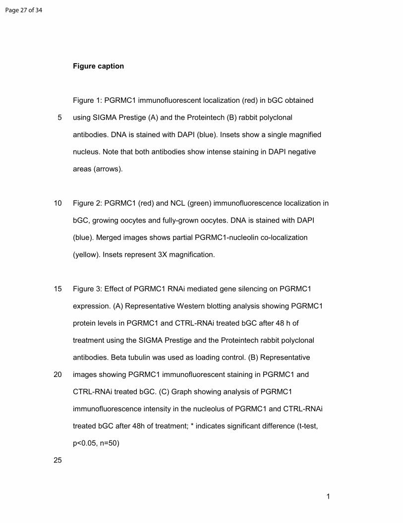
Figure 1: PGRMC1 immunofluorescent localization (red) in bGC obtained
using SIGMA Prestige (A) and the Proteintech (B) rabbit polyclonal 5
antibodies. DNA is stained with DAPI (blue). Insets show a single magnified
nucleus. Note that both antibodies show intense staining in DAPI negative
areas (arrows).
Figure 2: PGRMC1 (red) and NCL (green) immunofluorescence localization in 10
bGC, growing oocytes and fully-grown oocytes. DNA is stained with DAPI
(blue). Merged images shows partial PGRMC1-nucleolin co-localization
(yellow). Insets represent 3X magnification.
Figure 3: Effect of PGRMC1 RNAi mediated gene silencing on PGRMC1 15
expression. (A) Representative Western blotting analysis showing PGRMC1
protein levels in PGRMC1 and CTRL-RNAi treated bGC after 48 h of
treatment using the SIGMA Prestige and the Proteintech rabbit polyclonal
antibodies. Beta tubulin was used as loading control. (B) Representative
images showing PGRMC1 immunofluorescent staining in PGRMC1 and 20
CTRL-RNAi treated bGC. (C) Graph showing analysis of PGRMC1
immunofluorescence intensity in the nucleolus of PGRMC1 and CTRL-RNAi
treated bGC after 48h of treatment; * indicates significant difference (t-test,
p<0.05, n=50)
25
Page 27 of 34
2
Figure 4: Effect of PGRMC1 RNAi mediated gene silencing on NCL
localization. (A) Representative images showing NCL immunofluorescent
staining in PGRMC1 and CTRL-RNAi treated bGC; note the increased
nucleoplasmic signal in PGRMC1 RNAi treated cell. (C) Graph showing
analysis of PGRMC1 immunofluorescence intensity in the nucleoplasm of 30
PGRMC1 and CTRL-RNAi treated bGC after 48h of treatment; * indicates
significant difference (t-test, p<0.05, n=50)
Figure 5: Effect of H2O2 treatment on nuclear morphology, and PGRMC1 and
NCL localization. (A) Pictures in the left panel show the nuclear morphology 35
(as assessed by DAPI staining) in CTRL group (not treated) and H2O2-treated
bGC for 90 minutes. Pictures in the right panel show effect on nuclear
morphology after washing and culturing the cells for additional 24 hours in
both CTRL and H2O2 –treated groups. (B, C) Graphs show analysis of
PGRMC1 and NCL immunofluorescence intensity in the nucleolus and 40
nucleoplasm, respectively, in non-treated bGC (CTRL) and H2O2 treatment
after 90 min of culture * indicates significant difference (t-test, p<0.05, n=75)
Page 28 of 34
1
Tables
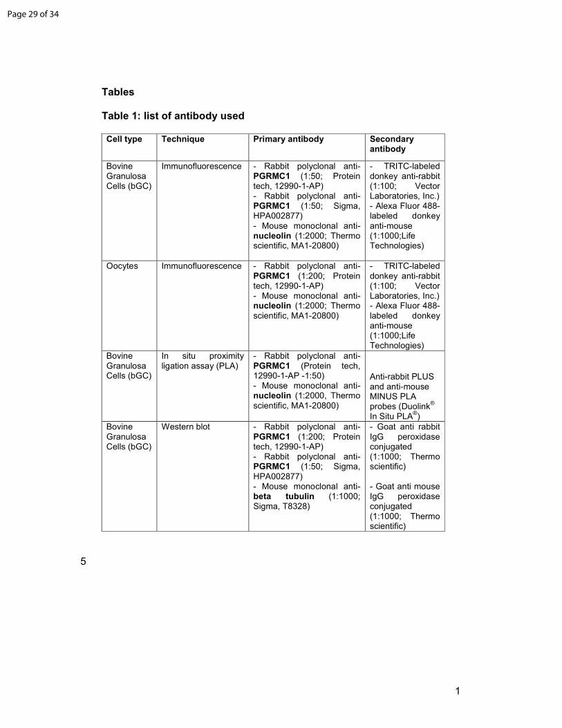
Table 1: list of antibody used
Cell type Technique Primary antibody Secondary antibody
Bovine Granulosa Cells (bGC)
Immunofluorescence - Rabbit polyclonal anti-PGRMC1 (1:50; Protein tech, 12990-1-AP) - Rabbit polyclonal anti-PGRMC1 (1:50; Sigma, HPA002877) - Mouse monoclonal anti-nucleolin (1:2000; Thermo scientific, MA1-20800)
- TRITC-labeled donkey anti-rabbit (1:100; Vector Laboratories, Inc.) - Alexa Fluor 488-labeled donkey anti-mouse (1:1000;Life Technologies)
Oocytes Immunofluorescence - Rabbit polyclonal anti-PGRMC1 (1:200; Protein tech, 12990-1-AP) - Mouse monoclonal anti-nucleolin (1:2000; Thermo scientific, MA1-20800)
- TRITC-labeled donkey anti-rabbit (1:100; Vector Laboratories, Inc.) - Alexa Fluor 488-labeled donkey anti-mouse (1:1000;Life Technologies)
Bovine Granulosa Cells (bGC)
In situ proximity ligation assay (PLA)
- Rabbit polyclonal anti-PGRMC1 (Protein tech, 12990-1-AP -1:50) - Mouse monoclonal anti-nucleolin (1:2000, Thermo scientific, MA1-20800)
Anti-rabbit PLUS and anti-mouse MINUS PLA probes (Duolink
®
In Situ PLA®)
Bovine Granulosa Cells (bGC)
Western blot - Rabbit polyclonal anti-PGRMC1 (1:200; Protein tech, 12990-1-AP) - Rabbit polyclonal anti-PGRMC1 (1:50; Sigma, HPA002877) - Mouse monoclonal anti- beta tubulin (1:1000; Sigma, T8328)
- Goat anti rabbit IgG peroxidase conjugated (1:1000; Thermo scientific) - Goat anti mouse IgG peroxidase conjugated (1:1000; Thermo scientific)
5
Page 29 of 34
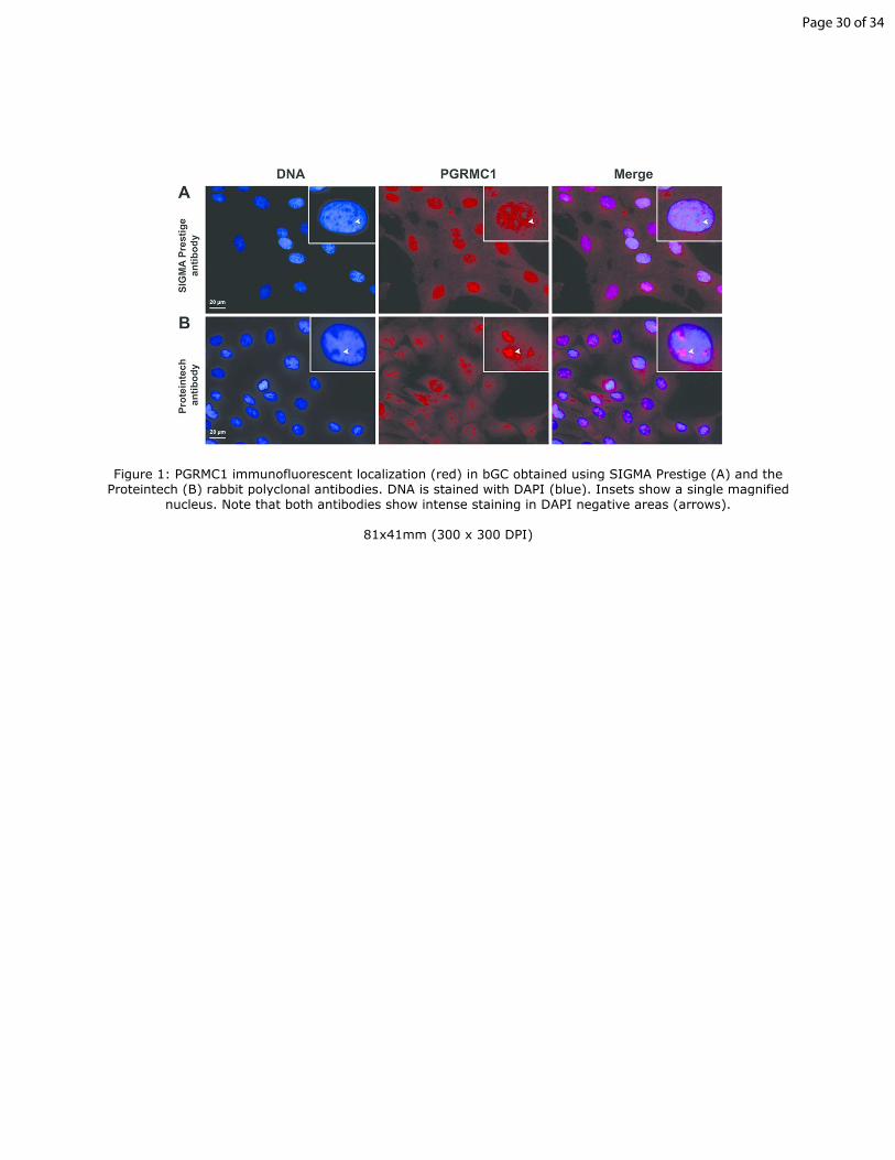
Figure 1: PGRMC1 immunofluorescent localization (red) in bGC obtained using SIGMA Prestige (A) and the Proteintech (B) rabbit polyclonal antibodies. DNA is stained with DAPI (blue). Insets show a single magnified
nucleus. Note that both antibodies show intense staining in DAPI negative areas (arrows).
81x41mm (300 x 300 DPI)
Page 30 of 34
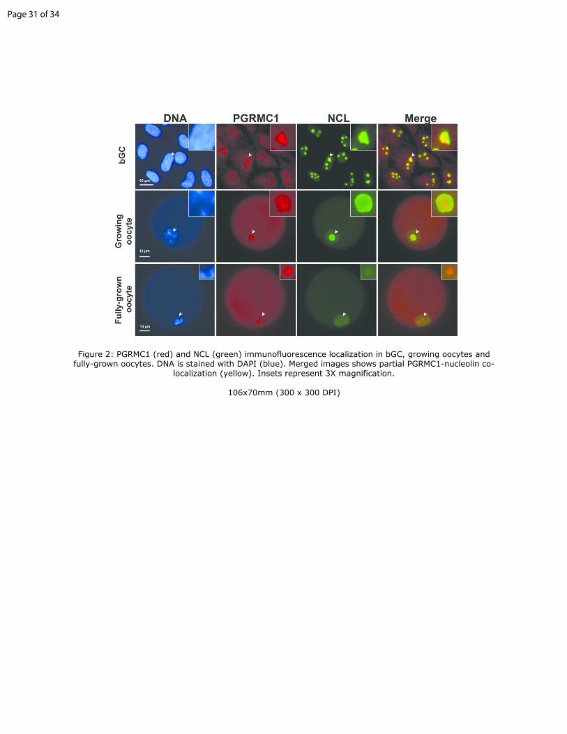
Figure 2: PGRMC1 (red) and NCL (green) immunofluorescence localization in bGC, growing oocytes and fully-grown oocytes. DNA is stained with DAPI (blue). Merged images shows partial PGRMC1-nucleolin co-
localization (yellow). Insets represent 3X magnification.
106x70mm (300 x 300 DPI)
Page 31 of 34
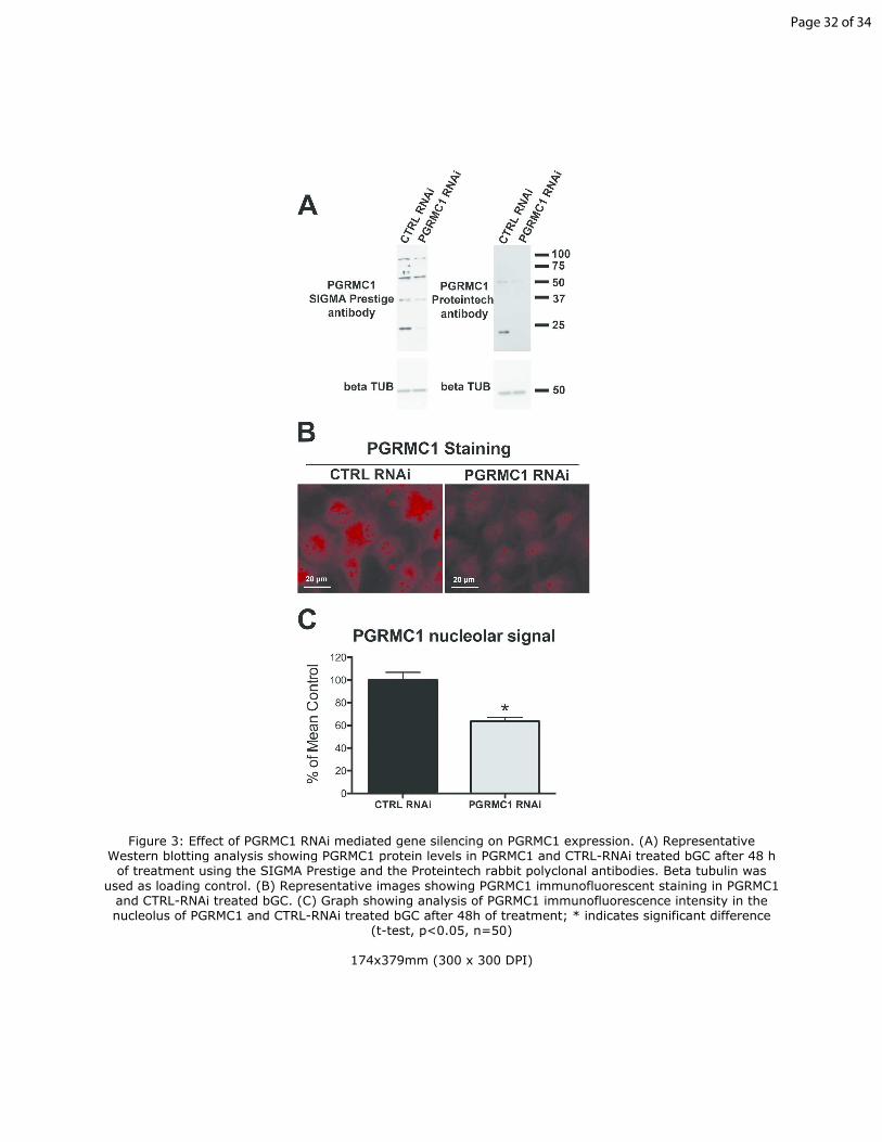
Figure 3: Effect of PGRMC1 RNAi mediated gene silencing on PGRMC1 expression. (A) Representative Western blotting analysis showing PGRMC1 protein levels in PGRMC1 and CTRL-RNAi treated bGC after 48 h of treatment using the SIGMA Prestige and the Proteintech rabbit polyclonal antibodies. Beta tubulin was
used as loading control. (B) Representative images showing PGRMC1 immunofluorescent staining in PGRMC1 and CTRL-RNAi treated bGC. (C) Graph showing analysis of PGRMC1 immunofluorescence intensity in the nucleolus of PGRMC1 and CTRL-RNAi treated bGC after 48h of treatment; * indicates significant difference
(t-test, p<0.05, n=50)
174x379mm (300 x 300 DPI)
Page 32 of 34
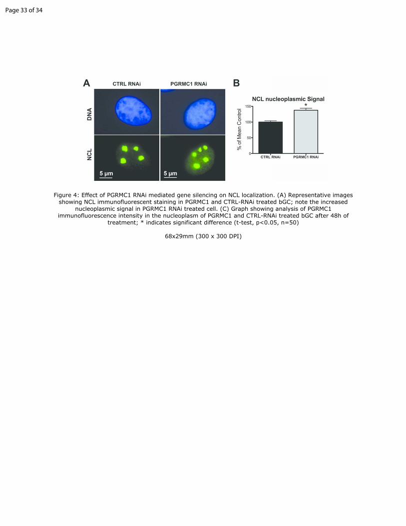
Figure 4: Effect of PGRMC1 RNAi mediated gene silencing on NCL localization. (A) Representative images showing NCL immunofluorescent staining in PGRMC1 and CTRL-RNAi treated bGC; note the increased
nucleoplasmic signal in PGRMC1 RNAi treated cell. (C) Graph showing analysis of PGRMC1 immunofluorescence intensity in the nucleoplasm of PGRMC1 and CTRL-RNAi treated bGC after 48h of
treatment; * indicates significant difference (t-test, p<0.05, n=50)
68x29mm (300 x 300 DPI)
Page 33 of 34
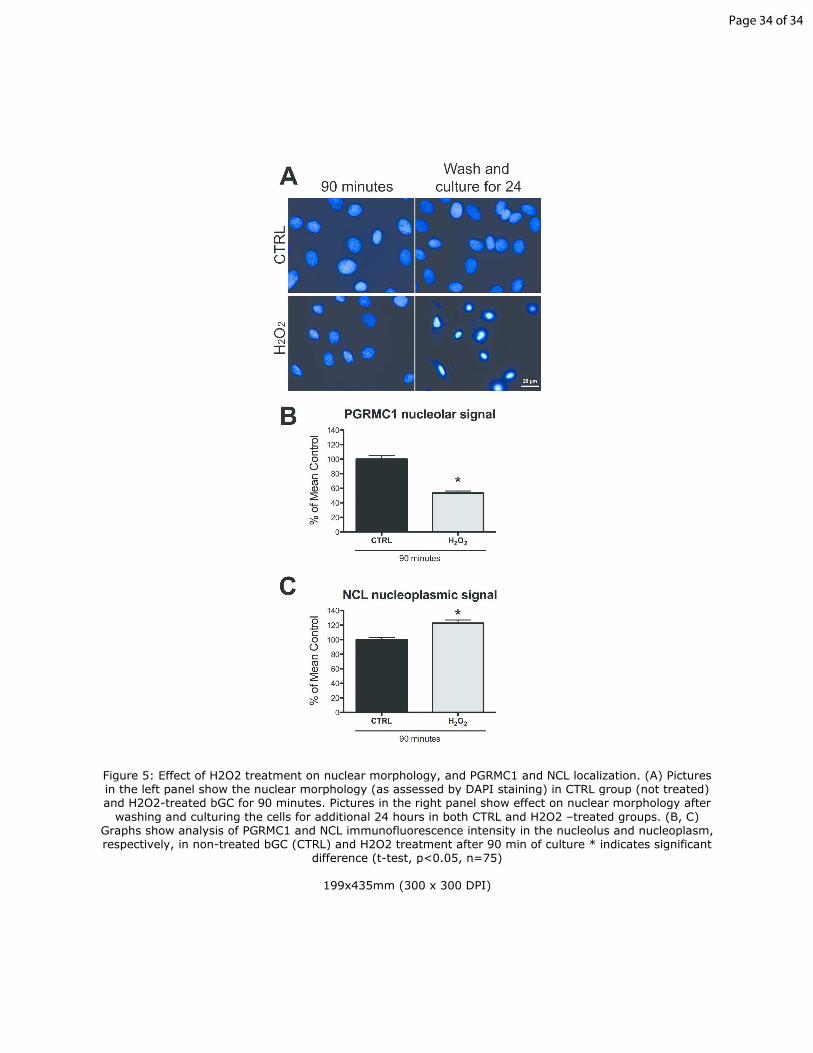
Figure 5: Effect of H2O2 treatment on nuclear morphology, and PGRMC1 and NCL localization. (A) Pictures in the left panel show the nuclear morphology (as assessed by DAPI staining) in CTRL group (not treated) and H2O2-treated bGC for 90 minutes. Pictures in the right panel show effect on nuclear morphology after
washing and culturing the cells for additional 24 hours in both CTRL and H2O2 –treated groups. (B, C) Graphs show analysis of PGRMC1 and NCL immunofluorescence intensity in the nucleolus and nucleoplasm, respectively, in non-treated bGC (CTRL) and H2O2 treatment after 90 min of culture * indicates significant
difference (t-test, p<0.05, n=75)
199x435mm (300 x 300 DPI)
Page 34 of 34