tlr7 ligand augments gm-csf initiated antitumor immunity...
TRANSCRIPT

Research Article
TLR7 Ligand Augments GM-CSF–Initiated AntitumorImmunity through Activation of Plasmacytoid Dendritic Cells
Megumi Narusawa1, Hiroyuki Inoue1,2,3, Chika Sakamoto1, Yumiko Matsumura1, Atsushi Takahashi1,Tomoko Inoue1, Ayumi Watanabe1, Shohei Miyamoto1, Yoshie Miura1, Yasuki Hijikata3,Yoshihiro Tanaka3, Makoto Inoue5, Koichi Takayama2, Toshihiko Okazaki4, Mamoru Hasegawa5,Yoichi Nakanishi2, and Kenzaburo Tani1,3
AbstractVaccination with irradiated granulocyte macrophage colony-stimulating factor (GM-CSF)–transduced autol-
ogous tumor cells (GVAX) has been shown to induce therapeutic antitumor immunity. However, its effectivenessis limited. We therefore attempted to improve the antitumor effect by identifying little-known key pathways inGM-CSF–sensitized dendritic cells (GM-DC) in tumor-draining lymph nodes (TDLN). We initially confirmed thatsyngeneic mice subcutaneously injected with poorly immunogenic Lewis lung carcinoma (LLC) cells transducedwith Sendai virus encoding GM-CSF (LLC/SeV/GM) remarkably rejected the tumor growth. Using cDNAmicroarrays, we found that expression levels of type I interferon (IFN)–related genes, predominantly expressedin plasmacytoid DCs (pDC), were significantly upregulated in TDLN-derived GM-DCs and focused on pDCs.Indeed, mouse experiments demonstrated that the effective induction of GM-CSF–induced antitumor immunityobserved in immunocompetent mice treated with LLC/SeV/GM cells was significantly attenuated when pDC-depleted or IFNa receptor knockout (IFNAR�/�) mice were used. Importantly, in both LLC and CT26 coloncancer–bearing mice, the combinational use of imiquimod with autologous GVAX therapy overcame therefractoriness to GVAX monotherapy accompanied by tolerability. Mechanistically, mice treated with thecombined vaccination displayed increased expression levels of CD86, CD9, and Siglec-H, which correlate withan antitumor phenotype, in pDCs, but decreased the ratio of CD4þCD25þFoxP3þ regulatory T cells in TDLNs.Collectively, these findings indicate that the additional use of imiquimod to activate pDCs with type I IFNproduction, as a positive regulator of T-cell priming, could enhance the immunologic antitumor effects of GVAXtherapy, shedding promising light on the understanding and treatment of GM-CSF–based cancer immunother-apy. Cancer Immunol Res; 2(6); 568–80. �2014 AACR.
IntroductionIn recent clinical trials of patients with diverse solid can-
cers, cancer immunotherapy such as therapeutic vaccinationwith granulocyte macrophage colony-stimulating factor(GM-CSF) gene-transduced tumor vaccines (GVAX), as wellas sipuleucel-T (Provenge; Dendreon), the first FDA-approvedGM-CSF–based therapeutic dendritic cell (DC) vaccine forprostate cancer, induced antitumor immune responses withtolerability (1–3). However, the efficacy of this therapy alone
is not satisfactory, raising an urgent need to improve theantitumor effect of GVAX. Although GM-CSF signaling isessential in conventional DC (cDC) maturation, which leadsto effective generation of tumor-associated antigen (TAA)-specific T cells and differentiation, the underlying molecularmechanism of how GM-CSF sensitizes and matures DCs (GM-DC, i.e., GM-CSF–sensitized DCs) to trigger host antitumorimmunity remains unclear.
Therefore, in this study, we attempted to improve theantitumor effects of GVAX therapy through identification ofthe key cluster genes upregulated in GM-DCs that operate T-cell priming in tumor-draining lymph nodes (TDLN) by con-ducting a cDNA microarray analysis. We used a syngeneicLewis lung carcinoma (LLC)–bearing mouse, which exhibitedremarkable tumor regression following subcutaneous admin-istration of fusion (F) gene-deleted nontransmissible Sendaivirus vector–mediated GM-CSF gene-transduced LLC (LLC/SeV/GM) cells (4). Using this experimental system, the expres-sion microarray analysis elucidated that pathways involvingToll-like receptor 7 (TLR7) and interferon regulatory factor 7(IRF7), which induce type I interferon (IFN) production inplasmacytoid DCs (pDC; ref. 5), were upregulated in GM-CSF–activatedmature DCs. Further activation of this pathway using
Authors' Affiliations: 1Department of Molecular Genetics, Medical Insti-tute of Bioregulation; 2Research Institute for Diseases of the Chest, Grad-uate School of Medical Sciences; 3Department of Advanced Cell andMolecular Therapy and 4Center for Clinical and Translational Research,Kyushu University Hospital, Kyushu University, Fukuoka; and 5DNAVECCorporation, Tsukuba, Japan
Note: Supplementary data for this article are available at Cancer Immu-nology Research Online (http://cancerimmunolres.aacrjournals.org/).
Corresponding Author:Kenzaburo Tani, Department of Molecular Genet-ics, Medical Institute of Bioregulation, Kyushu University, 3-1-1 Maidashi,Higashi-ku, Fukuoka 812-8582, Japan. Phone: 81-92-642-6449; Fax: 81-92-642-6444; E-mail: [email protected]
doi: 10.1158/2326-6066.CIR-13-0143
�2014 American Association for Cancer Research.
CancerImmunology
Research
Cancer Immunol Res; 2(6) June 2014568
on December 30, 2019. © 2014 American Association for Cancer Research. cancerimmunolres.aacrjournals.org Downloaded from
Published OnlineFirst April 10, 2014; DOI: 10.1158/2326-6066.CIR-13-0143

B
D
MOI =
0
MOI =
3
MOI =
10
MOI =
100
0
2×10³
4×10³
6×10³
8×10³
1×10⁴
ng/1
06 c
ells
/48 h
MOI = 0
MOI = 3
MOI = 10
MOI = 100
A
C
0 20 40 60 800
20
40
60
80
100
Days after tumor challenge
Su
rviv
al (%
)
LLC
LLC/SeV/GFP (MOI = 100)
LLC/SeV/GM (MOI = 100)
0 5 10 15 200
2,000
4,000
6,000
8,000
Tum
or
volu
me (
mm
3)
Days after tumor challenge
B16
B16/SeV/GFP (MOI = 30)
B16/SeV/GM (MOI = 30)
*
*
0 10 20 30 400
20
40
60
80
100
Days after tumor challenge
Su
rviv
al (%
)
B16
B16/SeV/GFP (MOI = 30)
B16/SeV/GM (MOI = 30)
0 10 200
500
1,000
1,500
2,000
Days after tumor challenge
Tu
mo
r vo
lum
e (
mm
3)
LLC
LLC/SeV/GM (MOI = 1)
LLC/SeV/GM (MOI = 10)
LLC/SeV/GM (MOI = 100)
*
*****
0 10 20 300
500
1,000
1,500
2,000
2,500
Tu
mo
r vo
lum
e (
mm
3)
Days after tumor challenge
LLC
LLC/SeV/GFP (MOI = 100)
LLC/SeV/GM (MOI = 100)
***
Figure 1. Tumor development of poorly immunogenic LLC andB16F10 cells modified to produceGM-CSFwasmarkedly inhibited. A, dose-escalation studiesto assess GM-CSF production from LLC/SeV/GM cells (MOI ¼ 0, 3, 10, and 100). GM-CSF production levels in the supernatants from the 48-hourculture were measured by ELISA. B and C, tumorigenicity assays using LLC cells. B, a total of 3.0 � 105 LLC and LLC/SeV/GM (MOI of 1, 10, or 100)cellswere subcutaneously inoculated into the right flankofC57/BL6Nmice (n¼3).C, a total of 2.0�105LLC, LLC/SeV/GFP, or LLC/SeV/GM (MOI¼100) cellswere inoculated into the right flank of C57/BL6N mice (n ¼ 6). Significant tumor regression (left) and prolonged survival (right) was shown in mice treatedwith LLC/SeV/GM cells. D, tumorigenicity assays using B16F10 cells. In total, 1.0 � 105 B16F10, B16/SeV/GFP, or B16/SeV/GM (MOI ¼ 30) cells wereinoculated into the right flanks of C57/BL6N mice (n¼ 6). Significant tumor regression (left) and prolonged survival (right) were observed in mice treated withB16/SeV/GM cells. The asterisks indicate statistically significant differences (�,P < 0.05; ��,P < 0.01; ���,P < 0.001). Kaplan–Meier survival curves are shown,and mortality was determined by the log-rank test (LLC vs. LLC/SeV/GM and LLC/SeV/GFP vs. LLC/SeV/GM; P < 0.001, LLC vs. LLC/SeV/GFP; P ¼ 0.67,B16 vs. B16/SeV/GM and B16/SeV/GFP vs. B16/SeV/GM; P < 0.05).
pDCs as Positive Regulator in GM-CSF–Based Antitumor Effect
www.aacrjournals.org Cancer Immunol Res; 2(6) June 2014 569
on December 30, 2019. © 2014 American Association for Cancer Research. cancerimmunolres.aacrjournals.org Downloaded from
Published OnlineFirst April 10, 2014; DOI: 10.1158/2326-6066.CIR-13-0143
A
B
LLC
LLC/S
eV/G
FP
LLC/S
eV/G
M
0
20
40
60
80
CF
SE
low
ce
lls (%
)
CD4+ T cell (day 2)
CD8+ T cell (day 2)
CD4+ T cell (day 4)
CD8+ T cell (day 4)
**
100
101
102
103
104
100
101
102
103
104
100
101
102
103
104
100
101
102
103
104
CFSE
Ce
ll c
ou
nts
Day 2 Day 4
CD8+
T cell
CD4+
T cell
Unstimulated
LLC/SeV/GM
LLC/SeV/GFP
LLC
Day
2
Day
40
50
100
150
200
250
MF
I o
f C
D8
0 g
ate
d o
n C
D1
1c
+ D
Cs
LLC
LLC/SeV/GFP
LLC/SeV/GM
*
*
Fre
qu
en
cy (
%)
Day 2
Day 4
Log fluorescence
CD80 CD86
Isotype
LLC/SeV/GM
LLC/SeV/GFP
LLC
100
101
102
103
104
0
20
40
60
80
100
LLC LLC/SeV/GFP LLC/SeV/GM
100
101
102
103
104
100
101
102
103
104
0.0732
100
101
102
103
104
100
101
102
103
104
0.0747
100
101
102
103
104
100
101
102
103
104
0.396
100
101
102
103
104
100
101
102
103
104
0.111
100
101
102
103
104
100
101
102
103
104
0.103
100
101
102
103
104
100
101
102
103
104
0.126
CD11c
PK
H2
6
TDLNs
CLNs
TDLN
CLN
0.0
0.1
0.2
0.3
0.4
PK
H26
+ D
Cs (%
)
LLC
LLC/SeV/GFP
LLC/SeV/GM
*
C
D
MF
I of C
D86
gate
d o
n
PK
H26
+C
D11c
+ c
ells
TDLN
CLN
0
2,000
4,000
6,000
LLC
LLC/SeV/GFP
LLC/SeV/GM
******
Narusawa et al.
Cancer Immunol Res; 2(6) June 2014 Cancer Immunology Research570
on December 30, 2019. © 2014 American Association for Cancer Research. cancerimmunolres.aacrjournals.org Downloaded from
Published OnlineFirst April 10, 2014; DOI: 10.1158/2326-6066.CIR-13-0143

TLR7 agonist enhanced the therapeutic antitumor effects ofGVAX therapy using irradiated autologous GM-CSF gene-transduced vaccine cells in both LLC and CT26 tumor-bearingmouse models with augmented pDC activation. These resultsshowed that the combination of GVAX and imiquimod is aneffective therapeutic strategy for cancer immunotherapy, andindicate that activated pDCs have a critical role in the GM-CSF–induced induction of antitumor immunity.
Materials and MethodsMiceFive- to 10-week-old female immunocompetent C57/BL6N
and BALB/cN mice were purchased from Charles River Lab-oratories Japan and housed in the animal maintenance facilityat Kyushu University (Fukuoka, Japan). Type I IFN receptorknockout (IFNAR�/�) mice were purchased from The JacksonLaboratory. All animal experiments were approved by theCommittee of the Ethics on Animal Experiments in the Facultyof Medicine, Kyushu University. Mouse experiments werecarried out at least twice to confirm results.
Tumor cell linesLLC andCT26 cells were purchased from the American Type
Culture Collection (ATCC) and passaged for 3 to 4months after
resuscitation. The mouse melanoma cell line (B16F10) was akind gift from Dr. Shinji Okano (Kyushu University) and wasvalidated as free from Mycoplasma infection; no other valida-tions were performed. Both LLC and CT26 cells were validatedas free from Mycoplasma infection. No other validations wereperformed; besides, the former were found as free from ectro-melia virus. LLC and B16F10 cells were maintained in Dulbec-co's Modified Eagle Medium (DMEM; Nakalai Tesque) supple-mented with 10% heat-inactivated fetal bovine serum (FBS)and 1% antibiotic mixture (Nakalai Tesque). CT26 was main-tained in RPMI-1640 (Nakalai Tesque) supplemented with 10%FBS and 1% antibiotic mixture.
Gene transduction with nontransmissible recombinantSendai virus vectors
LLC, B16F10, or CT26 cells were infected with nontransmis-sible Sendai virus vectors encoding green fluorescence protein(GFP) or mouse GM-CSF (SeV/GFP or SeV/GM, respectively),which were prepared by DNAVEC Corp. (6), at the indicatedmultiplicity of infection (MOI) for 90 minutes (termed as LLC/SeV/GFP, LLC/SeV/GM, B16/SeV/GFP, B16/SeV/GM, orCT26/SeV/GM cells, respectively). They were cultured for 48hours after viral gene transduction and used for followingmouse studies.
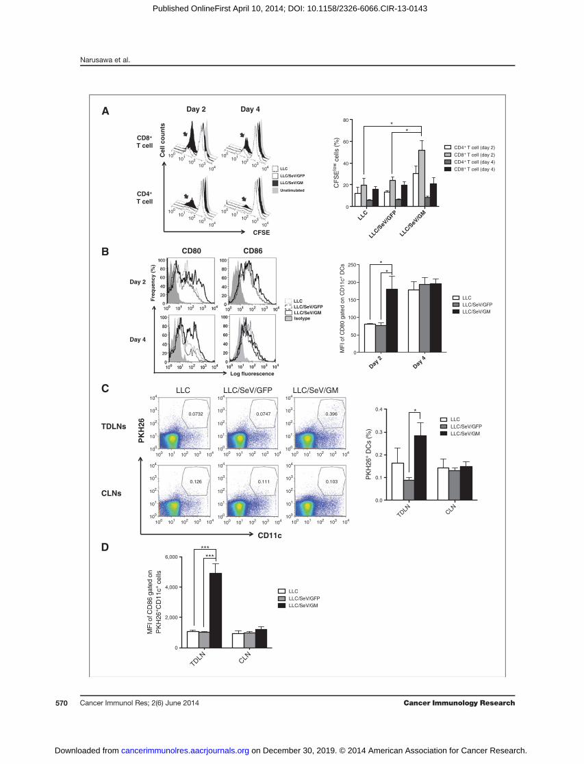
Figure 2. GM-CSF–sensitized DCs elicited superior capacities to stimulate T-cell proliferation and to mobilize TAA-phagocytosed mature DCs into TDLNs. A,CFSE-labeled allogeneic MLR assay. Irradiated CD11cþDCs from mice treated with indicated tumor challenge were mixed with CFSE-labeled allogeneicT cells. After 3 days of coculture, the proliferation rates of T cells were assessed by flow cytometric analysis. Representative histograms depict CFSEexpression of allogeneic CD4þCD3þ or CD8þCD3þ T cells (left). Bar graphs, mean þ SEM percentage of CFSE-diluted cells/total indicated T cells (right). B,representative histograms depict frequency distributions of MFI of CD80 or CD86 expression in CD11cþ DCs from indicated mouse groups on day 2 or4 after the tumor challenge (left). Bar graphs, mean þ SEM of MFI of CD80 on DCs in TDLNs (right). C, representative dot plots show PKH26þCD11cþ
cells gated by their FSC/SSCprofiles in TDLNs orCLNs (left). Bar graphs,meanþSEMof percentage ofCD11cþPKH26þ cells in TDLNs orCLNs (right). D, bargraphs, mean þ SEM of MFI of CD86 expression levels in PKH26þCD11cþ cells (�, P < 0.05; ���, P < 0.001).
Table 1. Canonical pathways identified by IPA
Pathways �log (P value) Molecules
Role of pattern recognition receptors inrecognition of bacteria and viruses
7.42Eþ00 OAS1, C3, OAS2, IL6, CCL5, Oas1f, OAS3, IFNA1/IFNA13,TLR2, IFIH1, IRF7, DDX58, TLR7, PIK3R6, EIF2AK2
Pathogenesis of multiple sclerosis 5.33Eþ00 CXCL10, CXCL9, CCL4, CCL5, CXCL11Activation of IRF by cytosolic patternrecognition receptors
4.38Eþ00 DHX58, IFIH1, IRF7, DDX58, ZBP1, STAT2, IL6, IFIT2,IFNA1/IFNA13, ISG15
IFN signaling 3.96Eþ00 IFIT3, IFIT1, OAS1, MX1, IFI35, STAT2, IFNA1/IFNA13DC maturation 3.01Eþ00 FCGR2A, HLA-DMB, IL6, MAPK13, FCGR2B, TREM2,
IFNA1/IFNA13, FCGR1A, TLR2, COL1A2, IL1RN,FSCN1, PIK3R6, STAT2
Hepatic fibrosis/hepatic stellate cell activation 2.58Eþ00 COL1A2, CXCL3, FN1, CXCL9, IGF1, PDGFA, CCL21,CD14, MMP13, CCL5, IL6, IFNA1/IFNA13
Role of hypercytokinemia/hyperchemokinemiain the pathogenesis of influenza
2.49Eþ00 CXCL10, CCL4, IL1RN, CCL5, IL6, IFNA1/IFNA13
Communication between innate andadaptive immune cells
2.47Eþ00 CXCL10, TLR2, CCL4, IL1RN, TLR7, CCL5, IL6,IFNA1/IFNA13, Ccl9
Role of tissue factor in cancer 2.45Eþ00 F10, PDIA2, PIK3R6, HCK, MMP13, F7, LIMK2,MAPK13, FGR, F2
LXR/RXR activation 2.26Eþ00 APOE, SCD, C3, MSR1 (includes EG:20288), IL1RN, LPL,CLU, CD14, IL6, GC
pDCs as Positive Regulator in GM-CSF–Based Antitumor Effect
www.aacrjournals.org Cancer Immunol Res; 2(6) June 2014 571
on December 30, 2019. © 2014 American Association for Cancer Research. cancerimmunolres.aacrjournals.org Downloaded from
Published OnlineFirst April 10, 2014; DOI: 10.1158/2326-6066.CIR-13-0143

A
B
LL
C
LL
C/S
eV
/GF
P
LL
C/S
eV
/GMC
Day 2
pDC
CD8+ cD
C
CD11
b+ c
DC
0
5
10
15
20
25
Ce
ll num
ber (1
× 1
04)
LLC/SeV/GFP
LLC/SeV/GM
*
Day 4
pDC
CD8+ cD
C
CD11
b+ c
DC
0
5
10
15
20
25
Cell n
um
ber (1
× 1
04)
LLC/SeV/GFP
LLC/SeV/GM
**
*
**
D
Ro
le o
f p
att
ern
re
co
gn
itio
n
rec
ep
tors
in
re
co
gn
itio
n
of
ba
cte
ria
an
d v
iru
se
s
Pa
tho
ge
ne
sis
of
mu
ltip
le s
cle
ros
is
Ac
tiv
ati
on
of
IRF
by
cy
tos
oli
c
pa
tte
rn r
ec
og
nit
ion
re
ce
pto
rs
IFN
sig
na
lin
g
DC
ma
tura
tio
n
He
pa
tic
fib
ros
is /
h
ep
ati
c s
tell
ate
ce
ll a
cti
va
tio
n
Ro
le o
f h
yp
erc
yto
kin
em
ia/
hy
pe
rch
em
ok
ine
mia
in
th
e p
ath
og
en
es
is o
f in
flu
en
za
Co
mm
un
ica
tio
n b
etw
ee
n i
nn
ate
an
d a
da
pti
ve
im
mu
ne
ce
lls
Ro
le o
f ti
ss
ue
fa
cto
r in
ca
nc
er
LX
R/R
XR
ac
tiv
ati
on
0
2
4
6
8
−log (
P v
alu
e)
E
0 5 10 150
200
400
600
800
1,000
Days after tumor challenge
Tu
mo
r volu
me (
mm
3)
WT
IFNAR–/–
**
*
F
0 5 10 15 200
500
1,000
1,500
Days after tumor challenge
Tu
mo
r vo
lum
e (
mm
3)
Isotype
pDC depletion
**
Narusawa et al.
Cancer Immunol Res; 2(6) June 2014 Cancer Immunology Research572
on December 30, 2019. © 2014 American Association for Cancer Research. cancerimmunolres.aacrjournals.org Downloaded from
Published OnlineFirst April 10, 2014; DOI: 10.1158/2326-6066.CIR-13-0143

In vivo experimentsFor tumorigenicity assays, syngeneic C57/BL6N mice were
subcutaneously challenged with 2.0� 105 LLC, LLC/SeV/GFP,or LLC/SeV/GM cells with or without imiquimod (R-837; 50mg/mouse; Invivogen) or lipopolysaccharide (LPS; 5 mg/mouse;Sigma-Aldrich) resuspended in 100-mL Hanks' Balanced SaltSolution (HBSS; Life Technologies) in the right or left flank. Todissect the role of type I IFN and pDCs in the tumorigenicityassays, IFNAR�/� or pDC-depleted mice were subcutaneouslychallenged with 2.0� 105 LLC/SeV/GM cells in the right flank.For therapeutic tumor vaccination assays, LLC/SeV/GFP, LLC/SeV/GM, and CT26/SeV/GM cells were irradiated at 50 Gy andwere designated as irLLC/SeV/GFP, irLLC/SeV/GM, andirCT26/SeV/GM cells, respectively. On days 2 and 9 after tumorchallenge with parental LLC or CT26 cells, C57/BL6N or BALB/cN mice were subcutaneously vaccinated with the indicatedtumor vaccine cells in the opposite flank. Tumor volume wasmeasured every 2 to 4 days and calculated with the followingformula: 0.4 � (largest diameter) � (smallest diameter)2.
ELISA assayIn vitro expression levels of mouse GM-CSF produced from
LLC, LLC/SeV/GFP, or LLC/SeV/GM cells at the MOI and timepoints were measured using mouse GM-CSF enzyme-linkedimmunosorbent assay (ELISA) kits (R&D Systems).
Flow cytometric analysisTDLNs, spleen, and tumor vaccine sites (TVS) harvested
from the indicated groups ofmice (n¼ 3–5)were homogenizedand filtered through a 100-mm cell strainer (BD Biosciences).For splenocyte preparation, smashed spleenswere treatedwithammonium chloride to lyse red blood. For T-cell detection inmixed lymphocyte reaction (MLR) assays, cells were stainedwith anti-CD4 (RM4.5)–PE (eBioscience), anti-CD3e–APC(145-2C11), and anti-CD8a–PerCP (53-6.7; BioLegend). Forphenotypic analyses of DCs in TDLNs, cells were stainedwith an anti-mouse CD11c Ab [anti-CD11c–APC (N418);BioLegend] in combination with anti-mouse Abs, includinganti-B220–PE (RA3-6B2), anti-CD317 (PDCA-1, BST2)–PE(eBio129c; all eBioscience), anti-CD80–PE (16-10A1), anti-CD8a–PerCP, anti-CD86–FITC (GL-1), or anti-CD11b–FITC(M1/70; all BioLegend). For phenotypic analyses of pDCs inTDLNs, cells were stained with either anti-CD317 (PDCA-1,BST2)–PE, anti-PDCA-1–APC (JF05-1C2.4.1; Miltenyi Biotec),or anti-CD11c–PerCPCy5.5 (N418; eBioscience) in combina-tion with anti-mouse Abs, including anti-CD86–FITC, anti-CD9–FITC (MZ3; BioLegend), and anti-Siglec-H–FITC
(551.3D3; Miltenyi Biotec). For regulatory T-cell (Treg) detec-tion in TDLNs, cells were permeabilized with Cytofix/Cyto-perm kit (BD Biosciences), washed with BD Perm/Wash buffer(BD Biosciences), and stained with anti-CD4, anti-CD25–FITC(PC61.5), and anti-FoxP3–APC (FJK-16s; all eBioscience). Cellswere incubated with Abs and analyzed with BD FACSCaliburflow cytometer, CellQuest software (BD Biosciences), andFlowJo software (TreeStar).
Allogeneic MLR assaysTo prepare CD11cþ DCs as stimulators, on day 2 of the
tumorigenicity assay, CD11cþ DCs were purified from TDLNsin mice treated with LLC, LLC/SeV/GFP, or LLC/SeV/GMcells using CD11c MicroBeads (Miltenyi Biotec). To preparethe pDC subset as stimulators, total bone marrow cellsharvested from na€�ve C57/BL6N mice were cultured inRPMI-1640 supplemented with 50 ng/mL murine Fms-relatedtyrosine kinase 3 ligand (Flt3L; PeproTech) for 8 days andSiglec-H–positive cells (pDCs) were purified using anti-Siglec-H–FITC Ab and anti-FITC MicroBeads (Miltenyi Biotec).Sorted pDCs were then incubated overnight with or without2.5 mg/mL of imiquimod or 10 ng/mL of murine recombinantGM-CSF (PeproTech). To prepare allogeneic T cells as respon-ders, T cells were sorted from splenocytes harvested fromna€�ve BALB/cN mice using a Pan T-cell isolation kit II(Miltenyi Biotec). A total of 5.0 � 104 responder T cellslabeled with 1.0 mmol/L CFSE [5(6)-carboxyfluorescein dia-cetate N-succinimidylester; Sigma-Aldrich] were coculturedwith an equal number of 30 Gy–irradiated CD11cþ DCs. Atotal of 2.0 � 105 T cells labeled with 2.5 mmol/L of CFSE werecocultured with 4.0� 104 of pDCs for 5 days. The proliferationrate of the gated CD3þ T-cell fraction was visualized by CFSEdilution.
Detection of DCs that engulfed TAAsLLC, LLC/SeV/GFP, and LLC/SeV/GM cells were labeled
with the PKH26 Red Fluorescent Cell Linker Mini Kit (Sig-ma-Aldrich), respectively, according to the manufacturer'sinstruction. On day 2 after they were subcutaneouslyinjected into the right flanks of mice, axillary lymph nodesin both TDLNs and CLNs were harvested, incubated withanti-CD86–FITC and anti-CD11c–APC Abs, and subjected toflow cytometric analysis.
cDNA microarrayDead cells were excluded from CD86þCD11cþ DCs using 7-
AAD viability dye (Beckman Coulter), which were sorted by
Figure 3. Transcriptome analysis suggested the involvement of type I IFN–related pathways in GM-DCs during GM-CSF–induced antitumor immunity. A, totalRNA was isolated from CD86þ DCs in TDLNs from mice inoculated with LLC, LLC/SeV/GFP, or LLC/SeV/GM cells 2 days after the tumor challengeand subjected to cDNA microarray. The top 10 canonical pathways significantly upregulated in GM-DCs, in comparison with those in GFP-DCs, bywhicha right-tailed Fisher exact testwascalculatedusing the entire dataset.B, IPAwasperformedusing the type I IFNpathway–relatedgenes from theoriginalcommonly regulated probes differentially expressed between GFP-DCs and GM-DCs. Differentially expressed genes are indicated in red and green,representing up- anddownregulation inducedbyGM-CSFactivation, respectively. A highdegree of gene regulation is indicatedbybold-coloredgenes. Director indirect associations with the differentially expressed genes indicated by no color were not found to be significantly different in this assessment.Positive regulatory interactions are depicted by solid arrows (direct interactions) or dashed arrows (indirect interactions). C, heatmap based on type I IFNpathway–related genes that were differentially expressed in CD86þ DCs in TDLNs from indicated mouse groups. D, cell numbers of DC subsets (pDC, CD8þ
cDCs, and CD11bþ cDC) in TDLNs at days 2 (top) and 4 (bottom) after the respective tumor challenge were comparatively quantified (�,P < 0.05; ��, P < 0.01).E and F, representative tumor growth curves observed in IFNAR�/� (E) or pDC-depleted (F) mice (��, P < 0.01).
pDCs as Positive Regulator in GM-CSF–Based Antitumor Effect
www.aacrjournals.org Cancer Immunol Res; 2(6) June 2014 573
on December 30, 2019. © 2014 American Association for Cancer Research. cancerimmunolres.aacrjournals.org Downloaded from
Published OnlineFirst April 10, 2014; DOI: 10.1158/2326-6066.CIR-13-0143

FACSAria (BD Biosciences) from TDLNs of mice on day 2during the tumorigenicity assay. Cells were transferred to RNAlater (Life Technologies) to stabilize and protect intact cellularRNA. RNA isolation was performed according to the TRIzolReagent technical manual (Life Technologies). Total RNA (50ng) was amplified and labeled using the Agilent Low-InputQuickAmp Labeling Kit, one color (Agilent Technologies).Labeled cRNA was hybridized to Agilent Whole MouseGenome Oligo DNA microarray (4 � 44 K) v2 (Agilent Tech-nologies). All gene transcription products were hybridized tomicroarray slides and were scanned by an Agilent scanner.Relative hybridization intensities and background hybridiza-tion values were calculated using the Agilent Feature Extrac-tion Software (v9.5.1.1; Agilent Technologies). The raw signalintensities of two samples were log2-transformed and normal-ized by a quantile algorithm with the "preprocessCore" librarypackage on Bioconductor software. We used Z-scores tocompare significant changes in gene expression in each of thethree groups (DCs frommice treated with LLC, LLC/SeV/GFP,and LLC/SeV/GM cells). Lists of genes with statistically sig-nificant expression in GM-DCs in comparison with GFP-DCswere submitted to Ingenuity Pathway Analysis (IPA; IngenuitySystems) and analyzed for overrepresented general functionsand the resulting networks. Microarray data were deposited inGene Expression Omnibus (GEO; http://www.ncbi.nlm.nih.gov/geo/; accession number GSE43169).
In vivo depletion experimentsTo deplete pDCs, mice were injected intraperitoneally with
100 mg of anti-PDCA-1 mAb (JF05-1C2.4.1; Miltenyi Biotec) orcontrol Ab (rat IgG; Jackson Immunoresearch), as previouslydescribed (7). Effective depletion of PDCA-1þ cells was con-firmed by flow cytometric analysis (Supplementary Fig. S1).CD4þ T or CD8þ T cells were depleted by using GK1.5 or 2.43mAbs, as previously described (8). Briefly, mice received intra-peritoneal injections of anti-mouse GK1.5 mAb, anti-mouse2.43 mAb (50 mg/mouse), or control Ab 6, 4, and 2 days beforetumor challenge, and once every 3 days thereafter. Effectivedepletion of CD4þ and CD8þ T cells was confirmed by flowcytometric analysis (data not shown).
ResultsProduction of GM-CSF from LLC and B16F10 cellsremarkably impaired the tumorigenicity
To test the possibility that substantial secretion of GM-CSFfrom syngeneic mouse cancer cells facilitates the developmentof antitumor immune responses, we used recombinant non-transmissible Sendai virus vectors expressing GM-CSF (SeV/GM) at various MOI. Abundant GM-CSF production from theinfected LLC (LLC/SeV/GM) cells was observed and was MOIdependent (Fig. 1A). The proliferation rate of LLC cells was notaffected by transduction with SeV/GM, as previously described(6). We next performed tumorigenicity assays in which eachLLC and LLC/SeV/GM cells (MOI ¼ 1, 10, and 100) weresubcutaneously injected into the left flank of syngeneic mice.All mice treated with LLC/SeV/GM cells exhibited significantsuppression of the tumor outgrowth in an MOI-dependentmanner (Fig. 1B). We thus determined MOI ¼ 100 for gene
transduction as an optimized infection dose. Notably, micetreated with LLC/SeV/GM cells showed significantly suppres-sed tumor growth and prolonged survival of mice, comparedwith control groups (P < 0.001; Fig. 1C). Similar suppressionof tumor growth and prolongation of mouse survival wereobserved when SeV/GM-infected B16F10 melanoma cellswere injected to C57BL/6N mice (Fig. 1D).
Increased ability of GM-CSF–sensitized DCs to stimulateT-cell proliferation, accelerate their maturation, anddeliver phagocytosed TAAs in TDLNs
To determine a putative phase when GM-CSF–sensitizedDCs fromTDLNs of mice treated with LLC/SeV/GM cells (GM-DCs) effectively prime na€�ve T cells, we performed allogeneicMLR assays. GM-DCs exhibited a significantly markedresponse on day 2 compared with DCs from mice treatedwith LLC/SeV/GFP cells (GFP-DCs), and stimulated the pro-liferation of allogeneic CD3þCD4þ T and CD3þCD8þ T cells(Fig. 2A). Furthermore, GM-DCs harvested on day 2 elicitedhigher expression levels of costimulatory maturation markersCD80 and CD86 than those from control mice (Fig. 2B),suggesting that day 2 could be the putative phase to mountoptimum immunologic responses by GM-DCs. To explore themigratory capacity of GM-DCs that phagocytosed TAAs at thetumor injection site, we inoculated PKH26-labeled LLC, LLC/SeV/GFP, or LLC/SeV/GMcells into the rightflank ofmice, andevaluated PKH26þ DC numbers in both TDLNs and contra-lateral LNs (CLN). The frequencies of PKH26þ DCs in TDLNs,but not CLNs, harvested frommice treated with LLC/SeV/GMcells were significantly increased, indicating that GM-CSFproduction potentiated the migration of PKH26-labeled LLCcells (TAA)–phagocytosed DCs from the tumor injection siteinto TDLNs (P < 0.05; Fig. 2C). PKH26þ GM-DCs derived fromTDLNs, but not from CLNs, showed significantly higher CD86expression than controls (P < 0.001; Fig. 2D).
cDNA microarray analysis revealed the involvement oftype I IFN–related pathways in GM-CSF–inducedantitumor immunity
On the basis of the aforementioned results, we determinedday 2 to be an adequate time point for the peak in T-cell primingby TAA-phagocytosed CD86þ DCs. To address the importantfactor of DC/T-cell priming, we isolated CD86þ DCs from micetreated with LLC/SeV/GM cells and control groups, and com-pared the comprehensive gene expression patterns of isolatedCD86þ DCs in TDLNs. After normalization of microarray dataand statistical analysis, 1,318 genes were found to be differen-tially expressed between GM-DCs and GFP-DCs with statisticalsignificance (upregulated genes; Z-score �2 and ratio >1.5,downregulated genes; Z-score ��2 and ratio <0.66; data notshown). A list of the genes significantly upregulated in the top 10canonical pathways in CD86þ GM-DCs in comparison withCD86þ GFP-DCs is shown in Table 1. As expected, these genescomposed immunologic response–related pathways (Fig. 3A).Among the activatedpathways triggeredbyGM-CSF,we focusedon the following representative molecules: IRF7, OAS3 (20-50-oligoadenylate synthetase 3), and TLR7, which constitute thetype I IFN (IFN-a/IFN-b)–associated pathways (Fig. 3B and C;
Narusawa et al.
Cancer Immunol Res; 2(6) June 2014 Cancer Immunology Research574
on December 30, 2019. © 2014 American Association for Cancer Research. cancerimmunolres.aacrjournals.org Downloaded from
Published OnlineFirst April 10, 2014; DOI: 10.1158/2326-6066.CIR-13-0143

A
B
0 5 10 15 20 250
200
400
600
800
1,000
Days after tumor challenge
Tum
or
volu
me (
mm
3)
LLC
LLC/SeV/GFP
LLC/SeV/GM
LLC/SeV/GM+imiquimod
LLC/SeV/GM+LPS
*
C
0 5 10 15 200
200
400
600
Days after tumor challenge
Tu
mo
r vo
lum
e (
mm
3)
Untreated
irLLC/SeV/GFP
irLLC/SeV/GM
irLLC/SeV/GM+imiquimod
Imiquimod
*
Day
0
Tumor
challenge
s.c.
2
Tumor
vaccine
or
imiquimod
s.c.
9
Tumor vaccine
or
imiquimod
s.c.
Imiquimod
50 µg/mouse
s.c.
4
Imiquimod
50 µg/mouse
s.c.
6
0 5 10 15 200
300
600
900
1,200
Days after tumor challenge
Tu
mo
r vo
lum
e (
mm
3)
Untreated
irCT26/SeV/GM
irCT26/SeV/GM+imiquimod
Imiquimod
**
D
Figure 4. Combined imiquimod andirLLC/SeV/GM cells exertsignificant therapeutic antitumoreffects compared with irLLC/SeV/GM cells alone. A, a total of 2.0 �105 LLC and LLC/SeV/GM cellswith or without LPS or imiquimodwere subcutaneously inoculatedinto the right flanks of C57/BL6Nmice. Bar graphs, mean þ SEM oftumor volumes. Combined datafrom two independent experimentswith similar results are shown(�, P < 0.05). B, schematic diagramof the experimental protocol oftherapeutic GM-CSF–based tumorvaccination. Briefly, 2.0 � 105 LLCcells or 3.0 � 105 CT26 cells wereinoculated subcutaneously toC57/BL6N or BALB/cN mice.LLC-bearing mice were dividedinto the following groups:untreated, imiquimod alone,irLLC/SeV/GFP, irLLC/SeV/GM, orirLLC/SeV/GM cells plusimiquimod. CT26-bearing micewere divided into the followinggroups: untreated, imiquimodalone, irCT26/SeV/GM orirCT26/SeV/GM cells plusimiquimod. On days 2 and 9, micewere inoculated subcutaneouslywith the indicated vaccine cells.For imiquimod administration,mice were subcutaneouslyinoculated with imiquimod on days2, 4, 6, and 9. Represented aretumor growth curves observed ineither LLC- (C) or CT26-bearing (D)mice (�, P < 0.05; ��, P < 0.01).
www.aacrjournals.org Cancer Immunol Res; 2(6) June 2014 575
pDCs as Positive Regulator in GM-CSF–Based Antitumor Effect
on December 30, 2019. © 2014 American Association for Cancer Research. cancerimmunolres.aacrjournals.org Downloaded from
Published OnlineFirst April 10, 2014; DOI: 10.1158/2326-6066.CIR-13-0143

irLLC
/SeV
/GM
irLLC
/SeV
/GM
+im
iquim
od
0
20
40
60
80
100
PD
CA
-1+ c
ells
in T
VS
(%
)
*
A
C
B
D
PD
CA
-1+ c
ells
in
TD
LN
s (%
)irL
LC/S
eV/G
M
irLLC
/SeV
/GM
+imiquim
od
0
5
10
15
20**
Siglec-H
(%)
irLLC/SeV/GM
irLLC/SeV/GM +imiquimod
CD86
(%)
CD
9+P
DC
A-1
+ D
Cs (%
)
irLLC
/SeV
/GFP
irLLC
/SeV
/GM
irLLC
/SeV
/GM
+imiqui
mod
0
20
40
60
80
*
*
PD
CA
-1
CD9
irLLC/SeV/GFP irLLC/SeV/GM
irLLC/SeV/GM +Imiquimod
100
101
102
103
104
100
101
102
103
104
12.2 50.8
9.9827.1
100
101
102
103
104
100
101
102
103
104
15.5 49.4
6.8528.3
100
101
102
103
104
100
101
102
103
104
5.76 74.4
11.58.34
100
101
102
103
104
0
20
40
60
80
100
(%)
CD9
irLLC/SeV/GFP irLLC/SeV/GM
irLLC/SeV/GM +imiquimod
CD25
Fo
xP
3
100
101
102
103
104
100
101
102
103
104
2.55
7.56
100
101
102
103
104
100
101
102
103
104
1.37
9.39
irLLC/SeV/GM
irLLC/SeV/GM +imiquimod
E
100
101
102
103
104
0
20
40
60
80
100
100
101
102
103
104
0
20
40
60
80
100
100
101
102
103
104
0
20
40
60
80
100
100
101
102
103
104
0
20
40
60
80
100
No cytokine GM-CSF Imiquimod
GM-CSF +imiquimod
100
101
102
103
104
0
20
40
60
80
100
22.5
100
101
102
103
104
0
20
40
60
80
100
18.2
100
101
102
103
104
0
20
40
60
80
100
15
100
101
102
103
104
0
20
40
60
80
100
6.98
CFSE
(%)
CD4+
T cell
CD8+
T cell
F
0 5 10 150
200
400
600
Tu
mo
r vo
lum
e (
mm
3)
irLLC/SeV/GM+imiquimod+Isotype
irLLC/SeV/GM+imiquimod+αCD4
*
*
**
0 5 10 150
100
200
300
400
500
Tu
mo
r vo
lum
e (
mm
3)
irLLC/SeV/GM+imiquimod+Isotype
irLLC/SeV/GM+imiquimod+αCD8
*
*
G
Cancer Immunol Res; 2(6) June 2014 Cancer Immunology Research576
Narusawa et al.
on December 30, 2019. © 2014 American Association for Cancer Research. cancerimmunolres.aacrjournals.org Downloaded from
Published OnlineFirst April 10, 2014; DOI: 10.1158/2326-6066.CIR-13-0143

refs. 5, 9). Microarray results for the expression levels of Irf7 andOas3 were validated by performing qRT-PCR (SupplementaryFig. S2). As pDCs provoke initial defensive antiviral responses bytype I IFN production and are the main producers of type I IFNs(10), we speculated that pDCs could be positively involved in theinduction of effective GM-CSF–sensitized DC/T-cell priming(11). Indeed, the numbers of pDCs, CD11bþ cDCs, and CD8þ
cDCs subsets fromtotalGM-DCs fromTDLNsharvestedondays2 and 4were greater than the equivalent subsets from total GFP-DCs (Fig. 3D). Furthermore, the results of in vivo experimentsusing IFNa receptor knockout (IFNAR�/�) mice demonstratedthat IFNAR�/� mice inoculated with LLC/SeV/GM cells signif-icantly abrogated the impairment of tumorigenicity seen in thecorresponding wild-type (WT) mice (Fig. 3E). Importantly,similar results were also obtained when pDC-depleted micewere used (Fig. 3F). These results collectively demonstrate thepositive role of type I IFN–producing pDCs in the induction ofGM-CSF–mediated antitumor immunity.
Combination of TLR7 ligand and GM-CSF–secreting LLCcells enhanced the induction of antitumor immunity inboth tumorigenicity and therapeutic vaccinationmodelsTLR7-dependent type I IFN pathways are activated by
binding with their corresponding ligand, imiquimod (12). Toexamine the impact of the TLR7-mediated activation of type IIFN–related pathways primarily in pDCs on GM-CSF–inducedantitumor immunity, we performed a gain-of-function assayby evaluating the tumorigenicity of LLC/SeV/GM cells with orwithout imiquimod or TLR4 ligand, LPS, as an irrelevantcontrol. Mice treated with LLC/SeV/GM cells combined withimiquimod showed significantly suppressed tumor develop-ment accompanied with complete tumor regression (P < 0.05).Conversely, treatment with LLC/SeV/GM cells combined withLPS attenuated the GM-CSF–induced antitumor effects(Fig. 4A), and these mice exhibited no significant changes inbody weight (Supplementary Fig. S3). We next attempted totranslate these findings into a tumor vaccination therapy byadding imiquimod to the subcutaneous administration ofirradiated LLC/SeV/GM (irLLC/SeV/GM) cells to investigatethe synergistic effect. Notably, mice treated with combinedimiquimod and irLLC/SeV/GM cells elicited a significantlymarked suppression of tumor growth of preestablished LLCcells, whereas control mice treated with irLLC/SeV/GMcells or imiquimod alone manifested negligible antitumoreffects (P < 0.05; Fig. 4B and C). Similarly, mice vaccinated
with irradiated GM-CSF gene-transduced (MOI ¼ 100)CT26 colon cancer cells in combination with imiquimodshowed significantly suppressed tumor development (P <0.01; Fig. 4D).
Admixed use of TLR7 ligand in combination with GVAXtherapy induced pDC activation leading to generation ofT-cell–mediated antitumor immunity
To elucidate the effect of imiquimod on GM-CSF–inducedinitial immune responses, we performed phenotypic immu-noanalyses. Six hours after the first tumor vaccination, micetreated with irLLC/SeV/GM cells plus imiquimod showed asignificantly higher frequency and number of cells expressingPDCA-1, a pDC-specific marker, than control mice in bothTVSs and TDLNs (Fig. 5A and Supplementary Fig. S4). Fur-thermore, pDCs (CD11cþPDCA-1þ cells) derived from micetreated with irLLC/SeV/GM cells plus imiquimod expressedincreased levels of CD86 and sialic acid binding Ig-like lectin(Siglec)-H, a functional pDC-specific receptor (Fig. 5B; ref. 13),accompanied with significantly higher levels of serum IFNa(Supplementary Fig. S5). Because CD9þ pDCs stimulated withTLR agonists induced higher amounts of IFNa and provokedprotective T-cell–mediated antitumor immunity (14), we com-pared CD9 expression levels on pDC subsets. Mice treated withirLLC/SeV/GM cells plus imiquimod had significantly increas-ed frequency and mean fluorescence intensity (MFI) ofCD9þPDCA1þCD11cþ pDCs in TDLNs (Fig. 5C and D). How-ever, the frequency of CD4þCD25þFoxP3þ Tregs wasdecreased in TDLNs from mice treated with irLLC/SeV/GMcells and imiquimod, whereas the frequency of CD4þ
CD25þFoxP3� T cells was increased in mice treated withcombined therapy (Fig. 5E). To investigate the effect of imi-quimod and GM-CSF on the T-cell proliferation capacity ofpDCs, we performed an allogeneicMLRassay. pDCs stimulatedwith GM-CSF and imiquimod elicited the most pronouncedproliferative activity of CD8þ T cells, but not CD4þ T cells,when compared with controls (Fig. 5F). Moreover, the syner-gistic therapeutic efficacy of irLLC/SeV/GM cells and imiqui-mod was significantly inhibited when the corresponding micewere depleted of CD4þ or CD8þ T cells (Fig. 5G).
DiscussionThis study demonstrates that SeV/dF-mediated exogenous
expression of GM-CSF caused poor growth of cancer cells in
Figure 5. Mice vaccinated with combined irLLC/SeV/GM cells and imiquimod augmented the recruitment of activated pDCs in TDLNs. A, 6 hours after thefirst tumor vaccination, infiltrating lymphocytes in TVSs or TDLNs were harvested from indicated mouse groups. Bar graphs, mean þ SEM of frequencyof PDCA-1þ cells gated on FSC/SSC profiles. B, histograms represent expression levels of Siglec-H or CD86 expression on CD11cþPDCA-1þ cells (pDCs) inTDLNs from indicated mouse groups (tinted light gray, isotype control; bold line, anti-CD86 or anti-Siglec-H Ab. C, on day 12, TDLNs were harvestedfrom mice treated with irLLC/SeV/GFP, irLLC/SeV/GM, or irLLC/SeV/GM cells plus imiquimod (n ¼ 3). Representative dot plots depict CD9 and PDCA-1expression gated on CD11cþ cells in TDLNs (left). Bar graphs, mean þ SEM of frequency of CD9þPDCA-1þ cells on DCs (�, P < 0.05; right). D,histograms depict MFI representing CD9 expression levels on PDCA-1þCD11cþ subpopulations in TDLNs (tinted light gray, isotype control; bold line, anti-CD9 Ab). E, representative dot plots illustrate CD25 and FoxP3 expression gated on CD4þ T cells in TDLNs from indicated mouse groups. F, CFSE-labeledallogeneic MLR assay. Bone marrow–derived pDCs treated with GM-CSF or imiquimod or in combination with GM-CSF plus imiquimod weremixed with CFSE-labeled T cells. Representative histograms show CFSE expression of allogeneic CD4þCD3þ or CD8þCD3þ T cells stimulated by theindicated pDCs. G, tumor growth curves in CD4þ T-cell (left)– or CD8þ T-cell (right)–depleted mice treated with irLLC/SeV/GM cells plus imiquimod(�, P < 0.05; ��, P < 0.01).
www.aacrjournals.org Cancer Immunol Res; 2(6) June 2014 577
pDCs as Positive Regulator in GM-CSF–Based Antitumor Effect
on December 30, 2019. © 2014 American Association for Cancer Research. cancerimmunolres.aacrjournals.org Downloaded from
Published OnlineFirst April 10, 2014; DOI: 10.1158/2326-6066.CIR-13-0143

syngeneic mice, concomitant with an early appearance ofmature DCs in TDLNs. We used SeV/dF vectors for the genetransduction of vaccine cells because they have relativelyhigher capacities in terms of gene transduction, induction ofantitumor immunity, and safety (6, 15). Expression microarrayanalyses of the GM-CSF–sensitized CD86þ DCs revealedincreased expression of the TLR7–IRF7 pathway components,which induce type I IFN production in pDCs (5). Furthermore,the addition of imiquimod was found to be an effectivepotential approach to improve the antitumor effects of GVAXtherapy (Fig. 4).
As LLC cells have been considered as poorly immuno-genic in lung cancer (16), it was surprising that tumorchallenge with LLC/SeV/GM cells markedly impaired itstumorigenicity with complete tumor disappearance in halfof the mice tested (Fig. 1). In addition, prophylactic vacci-nation with irLLC/SeV/GM cells also significantly inhibitedsubsequent tumor challenge with LLC cells (SupplementaryFig. S6). However, therapeutic vaccination using irLLC/SeV/GM cells alone failed to exert significant antitumor immu-nity (Fig. 4C). We, therefore, attempted to potentiate thetherapeutic antitumor effects of irLLC/SeV/GM cellsthrough scrutinizing the gene expression signature ofGM-CSF–sensitized DCs in TDLNs from mice that stronglyrejected the tumor challenge with LLC/SeV/GM cells. Weconfirmed that GM-CSF facilitated the maturation of DCsinto antigen-presenting cells with enhanced ability to primena€�ve T cells to proliferate, and to increase expression ofCD80, CD86, MHC class I, MHC class II, and CD40 (Fig. 2Band Supplementary Fig. S7), consistent with the previousfinding that GM-CSF promotes DCs maturation and differ-entiation (17). Herein, transcriptome analyses revealed thatGM-CSF also modulated signal transduction in pDCs byupregulation of the TLR7–IRF7 pathway related to type IIFN production (Fig. 3), consistent with a previous reportthat GM-CSF stimulation upregulated TLR7 expression inmouse immune cells (18). Our observation that the pDCsubset was markedly increased in GM-DCs from TDLNs wasunexpected, as the GM-CSF receptor is mainly expressed onCD34þ progenitor cells and myeloid cells (19, 20), and GM-CSF administration preferentially expands CD11bþ cDC (21,22) and inhibits pDC differentiation (23). However, recentstudies showing that pDC precursors differentiated toCD11bþMHC IIhigh cDCs by GM-CSF stimulation (24), andthe identification of GM-CSF as a novel activator of pDCsrevealed by systematic analysis of cytokine receptors (25),may explain the increase of GM-CSF–sensitized pDC sub-sets in TDLNs (Fig. 3D). In the development of activeimmunotherapeutic strategies, much attention has beenfocused on CD11bþ cDC-based vaccines that have failedto induce sufficient clinical efficacy (26), as pDCs areconsidered to be involved in the maintenance of anti-tumor tolerance (27) and to be inversely correlated withprognosis in patients with cancer (28, 29). However, pDCsubsets can be pivotal players in TAA-specific antitumorimmune responses by functioning as antigen-presentingcells (30) that use distinct MHC class II antigen-presentationmolecules (31), leading to the effective priming of na€�ve
CD4þ T cells (32), and cross-present antigens with anefficiency comparable with CD11bþ cDCs (33), implicatingtheir potential as promising antigen-presenting cells forcancer immunotherapy. Indeed, imiquimod or CpG, a TLR9agonist, reverted immunotolerant pDCs to antitumor pDCs(34), resulting in clinical antitumor effects (35, 36). Impor-tantly, results of our in vivo experiments using pDC deple-tion and/or IFNAR�/� mice demonstrated the positiveimpact of the pDC subset and/or type I IFN signaling onthe effective generation of GM-CSF–induced antitumorimmunity (Fig. 3E and F). Thus, there may be a functionaldichotomy in pDC biology between immune tolerance andantitumor phenotype, where their redirection is dependenton the tumor microenvironment.
Imiquimod, a TLR7 ligand, could be regarded as the mosteffective adjuvant among all approved immunomodulatorsbased on the following: (i) topical imiquimod is currentlyFDA approved with a good safety profile; (ii) it potentlyactivates antigen-presenting cells to release type I IFNs andTh1-skewing cytokines; and (iii) imiquimod treatment leadsto CCL2-dependent recruitment of pDCs and their trans-formation into killer DCs (37). The underlying mechanismof substantial antitumor efficacy by the combined vacci-nation may be due to generation of functionally maturepDCs in TVSs and TDLNs (Fig. 5A and Supplementary Fig.S4). IFNa, mainly produced from pDCs upon exposure toviruses via TLR7 or TLR9 (38), acts directly on memory Tcells, which potentiate the antigen presentation and cross-priming capacities of CD11bþ cDCs (39, 40). We detectedCD9þ pDCs, which produce abundant IFNa (14), in TDLNsfrom mice injected with ir.LLC/SeV/GM cells (Fig. 5C andD). Furthermore, GM-CSF–sensitized pDCs expressedhigher CD86 and Siglec-H (Fig. 5B), a regulator of pDCdifferentiation and CD8þ T-cell responses (13, 41). More-over, pDCs activated with GM-CSF plus imiquimod furtherenhanced the proliferation of CD8þ T cells (Fig. 5F), indi-cating that GM-CSF–activated pDCs with or without imi-quimod could serve as functional antigen-presenting cellsto prime the potent generation of TAA-specific adaptiveimmunity. ELISPOT assay demonstrated that the numberof IFNg-producing splenocytes from mice treated withirLLC/SeV/GM cells plus imiquimod was increased com-pared with control mice (data not shown). Indeed, deple-tion assays revealed that CD4þ and CD8þ T cells signifi-cantly contributed to the augmentation of the antitumorefficacy by combination GVAX therapy (Fig. 5G), thusreflecting the imiquimod-driven accelerated TAA-specificTh1 responses.
Although other researchers showed that the addition ofimiquimod negates the antitumor efficacy of a GM-CSF–basedvaccine (42), these conflicting results may stem from thedifference in doses and administration schedule. It is note-worthy that the ability of imiquimod to potentiate the anti-tumor effect of GVAX therapy in two different types of cancersand in two different host strains might confirm the generalityof our findings (Fig. 4C and D).
In conclusion, we, for the first time, elucidated that thebeneficial roles of the pDCs and relevant type I IFN pathway
Cancer Immunol Res; 2(6) June 2014 Cancer Immunology Research578
Narusawa et al.
on December 30, 2019. © 2014 American Association for Cancer Research. cancerimmunolres.aacrjournals.org Downloaded from
Published OnlineFirst April 10, 2014; DOI: 10.1158/2326-6066.CIR-13-0143

in GM-CSF–induced antitumor immunity and that the com-binational use of imiquimod with GVAX therapy producedsynergistic antitumor effects, underscoring its potential as apromising approach for the treatment of cancer.
Disclosure of Potential Conflicts of InterestNo potential conflicts of interest were disclosed.
Authors' ContributionsConception and design: M. Narusawa, H. Inoue, Y. Matsumura, K. TaniDevelopment of methodology: M. Narusawa, H. Inoue, Y. Matsumura,M. Inoue, M. HasegawaAcquisition of data (provided animals, acquired and managed patients,provided facilities, etc.):M. Narusawa, H. Inoue, C. Sakamoto, Y. Matsumura,A. Watanabe, K. TaniAnalysis and interpretation of data (e.g., statistical analysis, biostatistics,computational analysis): M. Narusawa, H. Inoue, Y. Matsumura, T. Inoue,S. MiyamotoWriting, review, and/or revision of themanuscript:M. Narusawa, H. Inoue,A. Takahashi, Y. Tanaka, K. Takayama, T. Okazaki, Y. Nakanishi
Administrative, technical, or material support (i.e., reporting or orga-nizing data, constructing databases): Y. Matsumura, T. Inoue, Y. Miura,M. Hasegawa, K. TaniStudy supervision: H. Inoue, Y. Matsumura, A. Takahashi, Y. Hijikata,T. Okazaki, K. Tani
AcknowledgmentsThe authors thank Michiyo Okada, Michiko Ushijima, Yosuke Yokota, and
Haruka Nabeta for excellent technical assistance, and Kaori Yasuda and AtsushiDoi for performing the cDNA microarray analysis. The authors also thankKatsuaki Sato for valuable discussion.
Grant SupportThis work was supported by JSPS KAKENHI grant number 21790773 and a
grant for students through the Kyushu University Foundation research grantprogram.
The costs of publication of this article were defrayed in part by the payment ofpage charges. This article must therefore be hereby marked advertisement inaccordance with 18 U.S.C. Section 1734 solely to indicate this fact.
Received September 5, 2013; revised February 5, 2014; accepted March 3, 2014;published OnlineFirst April 10, 2014.
References1. Dranoff G, Jaffee E, Lazenby A,GolumbekP, Levitsky H, BroseK, et al.
Vaccination with irradiated tumor cells engineered to secrete murinegranulocyte-macrophage colony-stimulating factor stimulates potent,specific, and long-lasting anti-tumor immunity. Proc Natl Acad Sci U SA 1993;90:3539–43.
2. Tani K, AzumaM,Nakazaki Y, Oyaizu N, HaseH, Ohata J, et al. Phase Istudy of autologous tumor vaccines transduced with the GM-CSFgene in four patients with stage IV renal cell cancer in Japan: clinicaland immunological findings. Mol Ther 2004;10:799–816.
3. Longo DL. New therapies for castration-resistant prostate cancer. NEngl J Med 2010;363:479–81.
4. Li HO, Zhu YF, Asakawa M, Kuma H, Hirata T, Ueda Y, et al. Acytoplasmic RNA vector derived from nontransmissible Sendaivirus with efficient gene transfer and expression. J Virol 2000;74:6564–9.
5. HondaK, Yanai H, Negishi H, Asagiri M, SatoM,Mizutani T, et al. IRF-7is the master regulator of type-I interferon-dependent immuneresponses. Nature 2005;434:772–7.
6. Inoue H, Iga M, Nabeta H, Yokoo T, Suehiro Y, Okano S, et al. Non-transmissible Sendai virus encoding granulocyte macrophagecolony-stimulating factor is a novel and potent vector system forproducing autologous tumor vaccines. Cancer Sci 2008;99:2315–26.
7. Yoneyama H, Matsuno K, Toda E, Nishiwaki T, Matsuo N, Nakano A,et al. Plasmacytoid DCs help lymph node DCs to induce anti-HSVCTLs. J Exp Med 2005;202:425–35.
8. YokotaY, InoueH,MatsumuraY,NabetaH,NarusawaM,WatanabeA,et al. Absence of LTB4/BLT1 axis facilitates generation of mouse GM-CSF–induced long-lastingantitumor immunologicmemorybyenhanc-ing innate and adaptive immune systems. Blood 2012;120:3444–54.
9. Sadler AJ,WilliamsBR. Interferon-inducible antiviral effectors.Nat RevImmunol 2008;8:559–68.
10. Gilliet M, Cao W, Liu YJ. Plasmacytoid dendritic cells: sensing nucleicacids in viral infection and autoimmune diseases. Nat Rev Immunol2008;8:594–606.
11. Colonna M, Trinchieri G, Liu YJ. Plasmacytoid dendritic cells in immu-nity. Nat Immunol 2004;5:1219–26.
12. Akira S, Hemmi H. Recognition of pathogen-associated molecularpatterns by TLR family. Immunol Lett 2003;85:85–95.
13. Takagi H, Fukaya T, Eizumi K, Sato Y, Sato K, Shibazaki A, et al.Plasmacytoid dendritic cells are crucial for the initiation of inflamma-tion and T cell immunity in vivo. Immunity 2011;35:958–71.
14. Bjorck P, LeongHX, Engleman EG. Plasmacytoid dendritic cell dichot-omy: identification of IFN-alpha producing cells as a phenotypicallyand functionally distinct subset. J Immunol 2011;186:1477–85.
15. Shi L, Chen J, Zhong Q, Li M, Geng P, He J, et al. Inactivated Sendaivirus strain Tianjin, a novel genotype of Sendai virus, inhibits growth ofmurine colon carcinoma through inducing immune responses andapoptosis. J Transl Med 2013;11:205.
16. Sumimoto H, Tani K, Nakazaki Y, Tanabe T, Hibino H, HamadaH, et al.GM-CSF and B7-1 (CD80) co-stimulatory signals co-operate in theinduction of effective anti-tumor immunity in syngeneic mice. Int JCancer 1997;73:556–61.
17. Wada H, Noguchi Y, Marino MW, Dunn AR, Old LJ. T cell functions ingranulocyte/macrophage colony-stimulating factor deficient mice.Proc Natl Acad Sci U S A 1997;94:12557–61.
18. YangH,Wei J, ZhangH, Lin L, ZhangW,HeS. Upregulation of Toll-likereceptor (TLR) expression and release of cytokines from P815 mastcells by GM-CSF. BMC Cell Biol 2009;10:37.
19. Barreda DR, Hanington PC, Belosevic M. Regulation of myeloiddevelopment and function by colony stimulating factors. Dev CompImmunol 2004;28:509–54.
20. Kingston D, Schmid MA, Onai N, Obata-Onai A, Baumjohann D, ManzMG. The concerted action of GM-CSF and Flt3-ligand on in vivodendritic cell homeostasis. Blood 2009;114:835–43.
21. Mausberg AK, Jander S, Reichmann G. Intracerebral granulocyte-macrophage colony-stimulating factor induces functionally compe-tent dendritic cells in the mouse brain. Glia 2009;57:1341–50.
22. Daro E, Pulendran B, Brasel K, Teepe M, Pettit D, Lynch DH, et al.Polyethylene glycol-modified GM-CSF expands CD11b(high)CD11c(high) but notCD11b(low)CD11c(high) murine dendritic cells in vivo: acomparative analysis with Flt3 ligand. J Immunol 2000;165:49–58.
23. Esashi E, Wang YH, Perng O, Qin XF, Liu YJ, Watowich SS. The signaltransducer STAT5 inhibits plasmacytoid dendritic cell development bysuppressing transcription factor IRF8. Immunity 2008;28:509–20.
24. Schlitzer A, Loschko J, Mair K, Vogelmann R, Henkel L, Einwachter H,et al. Identification of CCR9�murine plasmacytoid DC precursors withplasticity to differentiate into conventional DCs. Blood 2011;117:6562–70.
25. Ghirelli C, Zollinger R, Soumelis V. Systematic cytokine receptorprofiling reveals GM-CSF as a novel TLR-independent activator ofhuman plasmacytoid predendritic cells. Blood 2010;115:5037–40.
26. Banchereau J, Palucka AK. Dendritic cells as therapeutic vaccinesagainst cancer. Nat Rev Immunol 2005;5:296–306.
27. Munn DH, Sharma MD, Hou D, Baban B, Lee JR, Antonia SJ, et al.Expression of indoleamine 2,3-dioxygenase by plasmacytoid den-dritic cells in tumor-draining lymph nodes. J Clin Invest 2004;114:280–90.
28. Labidi-Galy SI, Sisirak V,MeeusP, GobertM, Treilleux I, Bajard A, et al.Quantitative and functional alterations of plasmacytoid dendritic cells
www.aacrjournals.org Cancer Immunol Res; 2(6) June 2014 579
pDCs as Positive Regulator in GM-CSF–Based Antitumor Effect
on December 30, 2019. © 2014 American Association for Cancer Research. cancerimmunolres.aacrjournals.org Downloaded from
Published OnlineFirst April 10, 2014; DOI: 10.1158/2326-6066.CIR-13-0143

contribute to immune tolerance in ovarian cancer. Cancer Res 2011;71:5423–34.
29. ZouW,MachelonV,Coulomb-L'HerminA,Borvak J,NomeF, IsaevaT,et al. Stromal-derived factor-1 in human tumors recruits and alters thefunction of plasmacytoid precursor dendritic cells. Nat Med 2001;7:1339–46.
30. Kim R, Emi M, Tanabe K, Arihiro K. Potential functional role of plas-macytoid dendritic cells in cancer immunity. Immunology 2007;121:149–57.
31. Villadangos JA, Young L. Antigen-presentation properties of plasma-cytoid dendritic cells. Immunity 2008;29:352–61.
32. Sapoznikov A, Fischer JA, Zaft T, Krauthgamer R, Dzionek A, Jung S.Organ-dependent in vivo priming of naive CD4þ, but not CD8þ, T cellsby plasmacytoid dendritic cells. J Exp Med 2007;204:1923–33.
33. Di Pucchio T, Chatterjee B, Smed-Sorensen A, Clayton S, Palazzo A,Montes M, et al. Direct proteasome-independent cross-presentationof viral antigen by plasmacytoid dendritic cells on major histocom-patibility complex class I. Nat Immunol 2008;9:551–7.
34. Palamara F, Meindl S, Holcmann M, Luhrs P, Stingl G, Sibilia M.Identification and characterization of pDC-like cells in normal mouseskin and melanomas treated with imiquimod. J Immunol 2004;173:3051–61.
35. MolenkampBG, Sluijter BJ, van LeeuwenPA, SantegoetsSJ,Meijer S,Wijnands PG, et al. Local administration of PF-3512676 CpG-B insti-gates tumor-specific CD8þ T-cell reactivity in melanoma patients. ClinCancer Res 2008;14:4532–42.
36. Aspord C, Charles J, Leccia MT, Laurin D, Richard MJ, Chaperot L,et al. A novel cancer vaccine strategy based on HLA-A�0201matched allogeneic plasmacytoid dendritic cells. PLoS ONE 2010;5:e10458.
37. Drobits B, Holcmann M, Amberg N, Swiecki M, Grundtner R, HammerM, et al. Imiquimod clears tumors in mice independent of adaptiveimmunity by converting pDCs into tumor-killing effector cells. J ClinInvest 2012;122:575–85.
38. Fonteneau JF, Gilliet M, Larsson M, Dasilva I, Munz C, Liu YJ, et al.Activation of influenza virus-specific CD4þ and CD8þ T cells: a newrole for plasmacytoid dendritic cells in adaptive immunity. Blood 2003;101:3520–6.
39. Le Bon A, Etchart N, Rossmann C, Ashton M, Hou S, Gewert D, et al.Cross-priming of CD8þ T cells stimulated by virus-induced type Iinterferon. Nat Immunol 2003;4:1009–15.
40. Honda K, Sakaguchi S, NakajimaC,Watanabe A, Yanai H,MatsumotoM, et al. Selective contribution of IFN-alpha/beta signaling to thematuration of dendritic cells induced by double-stranded RNA or viralinfection. Proc Natl Acad Sci U S A 2003;100:10872–7.
41. Blasius AL, Cella M, Maldonado J, Takai T, Colonna M. Siglec-H is anIPC-specific receptor that modulates type I IFN secretion throughDAP12. Blood 2006;107:2474–6.
42. Dang Y, Wagner WM, Gad E, Rastetter L, Berger CM, Holt GE, et al.Dendritic cell-activating vaccine adjuvants differ in the ability to elicitantitumor immunity due to an adjuvant-specific induction of immuno-suppressive cells. Clin Cancer Res 2012;18:3122–31.
Cancer Immunol Res; 2(6) June 2014 Cancer Immunology Research580
Narusawa et al.
on December 30, 2019. © 2014 American Association for Cancer Research. cancerimmunolres.aacrjournals.org Downloaded from
Published OnlineFirst April 10, 2014; DOI: 10.1158/2326-6066.CIR-13-0143
2014;2:568-580. Published OnlineFirst April 10, 2014.Cancer Immunol Res Megumi Narusawa, Hiroyuki Inoue, Chika Sakamoto, et al. through Activation of Plasmacytoid Dendritic Cells
Initiated Antitumor Immunity−TLR7 Ligand Augments GM-CSF
Updated version
10.1158/2326-6066.CIR-13-0143doi:
Access the most recent version of this article at:
Material
Supplementary
http://cancerimmunolres.aacrjournals.org/content/suppl/2014/04/17/2326-6066.CIR-13-0143.DC1
Access the most recent supplemental material at:
Cited articles
http://cancerimmunolres.aacrjournals.org/content/2/6/568.full#ref-list-1
This article cites 42 articles, 17 of which you can access for free at:
E-mail alerts related to this article or journal.Sign up to receive free email-alerts
Subscriptions
Reprints and
To order reprints of this article or to subscribe to the journal, contact the AACR Publications Department
Permissions
Rightslink site. Click on "Request Permissions" which will take you to the Copyright Clearance Center's (CCC)
.http://cancerimmunolres.aacrjournals.org/content/2/6/568To request permission to re-use all or part of this article, use this link
on December 30, 2019. © 2014 American Association for Cancer Research. cancerimmunolres.aacrjournals.org Downloaded from
Published OnlineFirst April 10, 2014; DOI: 10.1158/2326-6066.CIR-13-0143