tlr9 activation induces normal neutrophil responses in a ... · the pi3k pathway has variously been...
TRANSCRIPT

TLR9 activation induces normal neutrophil responses in
a child with IRAK-4 deficiency: involvement of the
direct PI3K pathway.
Cyrille Hoarau, Benedicte Gerard, Emmanuel Lescanne, Dominique Henry,
Stephanie Francois, Jean-Jacques Lacapere, Jamel El Benna, Pham My-Chan
Dang, Bernard Grandchamp, Yvon Lebranchu, et al.
To cite this version:
Cyrille Hoarau, Benedicte Gerard, Emmanuel Lescanne, Dominique Henry, Stephanie Francois,et al.. TLR9 activation induces normal neutrophil responses in a child with IRAK-4 deficiency:involvement of the direct PI3K pathway.. Journal of Immunology, American Association ofImmunologists, 2007, 179 (7), pp.4754-65. <inserm-00179394>
HAL Id: inserm-00179394
http://www.hal.inserm.fr/inserm-00179394
Submitted on 28 Oct 2008

HAL is a multi-disciplinary open accessarchive for the deposit and dissemination of sci-entific research documents, whether they are pub-lished or not. The documents may come fromteaching and research institutions in France orabroad, or from public or private research centers.
L’archive ouverte pluridisciplinaire HAL, estdestinee au depot et a la diffusion de documentsscientifiques de niveau recherche, publies ou non,emanant des etablissements d’enseignement et derecherche francais ou etrangers, des laboratoirespublics ou prives.

1
TLR9 activation induces normal neutrophil responses in a child with
IRAK-4 deficiency: involvement of the direct PI3K pathway
Cyrille Hoarau,* Bénédicte Gérard,† Emmanuel Lescanne,* Dominique Henry,†
Stéphanie François,† Jean-Jacques Lacapère,† Jamel El Benna,†
Pham My-Chan Dang,† Bernard Grandchamp,† Yvon Lebranchu,*
Marie-Anne Gougerot-Pocidalo,† and Carole Elbim1†
* Université François Rabelais, UFR de Médecine, Tours, JE 2448 « Cellules Dendritiques &
Greffes » F-37032, France ; Centre Hospitalier Universitaire de Tours, Unité Transversale
d’Allergologie, Néphrologie et Immunologie Clinique et Service d’ORL, Tours, F-37044,
France.
† Université Paris 7 Denis Diderot, Faculté de Médecine, site Bichat, Paris, F-75018, France ;
AP-HP, Centre Hospitalier Universitaire Xavier Bichat, Service d’Immunologie et
d’Hématologie et Service de Biochimie Hormonale et Génétique, CIB Phenogen, Paris, F-
75018, France ; INSERM, U773, Paris, F-75018, France.
Running title: IRAK-4 deficiency and normal PMN TLR9 responses
Key words: Neutrophils, IRAK-4, transduction pathways, PI3K
HA
L author manuscript inserm
-00179394, version 1
HAL author manuscriptJournal of Immunology 2007;179(7):4754-65
2
Abstract
Polymorphonuclear neutrophils (PMN) play a key role in innate immunity. Their activation
and survival are tightly regulated by microbial products via pattern-recognition receptors such
as TLRs, which mediate recruitment of the IRAK complex. We describe a new inherited
IRAK-4 deficiency in a child with recurrent pyogenic bacterial infections. Analysis of the
IRAK4 gene showed compound heterozygosity with two mutations: a missense mutation in
the death domain of the protein (p.Arg12Cys) associated in cis with a predicted benign variant
(p.Arg391His) and a splice-site mutation in intron 7 that led to the skipping of exon 7. A non-
truncated IRAK-4 protein was detected by western blotting. The patient’s functional
deficiency of IRAK-4 protein was confirmed by the absence of IRAK-1 phosphorylation after
stimulation with all TLR agonists tested. The patient’s PMN showed strongly impaired
responses (L-selectin and CD11b expression, oxidative burst, cytokine production, cell
survival) to TLR agonists which engage TLR1/2, TLR2/6, TLR4, and TLR7/8; in contrast,
the patient’s PMN responses to CpG-DNA (TLR9) were normal, except for cytokine
production. The surprisingly normal effect of CpG-DNA on PMN functions and apoptosis
disappeared after pretreatment with PI3K inhibitors. Together, these results suggest the
existence of an IRAK-4-independent TLR9-induced transduction pathway leading to PI3K
activation. This alternative pathway may play a key role in PMN control of infections by
microorganisms other than pyogenic bacteria in inherited IRAK-4 deficiency.
HA
L author manuscript inserm
-00179394, version 1

3
Introduction
Polymorphonuclear neutrophils (PMN) play a key role in host defense against bacterial and
fungal pathogens (1). They contribute to early innate response by rapidly migrating into
inflamed tissues, where their activation triggers microbicidal mechanisms such as release of
proteolytic enzymes and antimicrobial peptides, and rapid production of reactive oxygen
species (ROS), in the so-called oxidative burst. PMN die spontaneously by apoptosis and are
then recognized and phagocytosed by macrophages (2).
PMN directly recognize microbial products via pattern-recognition receptors such as
TLRs. Human PMN have been reported to express all TLRs except TLR3 (3); TLR5 and
TLR7 are weakly expressed (4). TLRs are members of the IL-1R superfamily, characterized
by an intracytoplasmic Toll-IL-1 receptor (TIR) domain which mediates recruitment of the
interleukin-1 receptor-associated kinase (IRAK) complex via TIR-containing adapter
molecules such as MyD88. During formation of this complex, IRAK-4 is activated, leading to
hyperphosphorylation of IRAK-1, which in turn induces the interaction of tumor necrosis
factor receptor-associated factor 6 (TRAF6) with the complex. TRAF6 then triggers
downstream signalling, and this results in NF-κB activation (5, 6). In addition, TLR
engagement activates stress kinases such as MAP kinases, c-Jun NH2-terminal kinase (JNK),
and phosphatidylinositol 3,4,5-triphosphate kinase (PI3K) in most cells, including PMN (7,
8). The PI3K pathway has variously been shown to regulate TLR-mediated inflammatory
responses, through negative feedback functions (9, 10), or to enhance NF-κB nuclear
translocation (11, 12). In particular, it was recently suggested that the PI3K signaling cascade
occupies a central role in TLR2-induced activation of PMN (13).
PMN stimulation through TLRs causes an immediate defensive response, including
modulation of adhesion molecule expression (L-selectin shedding and β2-integrin
HA
L author manuscript inserm
-00179394, version 1

4
upregulation), production of an array of antimicrobial molecules (ROS and cytokines) (3, 13),
and inhibition of apoptosis (4). The major role of the TIR-IRAK signaling pathway in
immunity to infections by pyogenic bacteria is illustrated by the recent descriptions of
children with inherited IRAK-4 deficiency associated with recurrent infections (14-22). The
cells of these patients fail to respond to IL-1 and IL-18, and to the stimulation of at least five
TLRs (TLR2, TLR3, TLR4, TLR5, TLR9).
Here we describe a case of inherited IRAK-4 deficiency related to new double
heterozygous mutations generating a non functional IRAK-4 protein. We show that some
PMN functions (adhesion molecule expression, ROS production, survival) which are critical
for antimicrobial defenses, occur normally in response to CpG-DNA (TLR9), despite an
impaired response to the other TLR agonists, suggesting the existence of a distinct TLR9-
induced transduction pathway.
HA
L author manuscript inserm
-00179394, version 1

5
Materials and Methods
Case report
We investigated a 14-year-old boy with recurrent infections, osteomyelitis and cellulitis. He
was born in July 1991 and was the second child of healthy unrelated parents. There was no
family history of recurrent or severe infections, autoimmune disease, or lymphoma. His
brother and sister were healthy. At age 15 days he developed a severe necrotic infection of his
palate, due to Pseudomonas aeruginosa. Despite several surgical procedures he had
velopharyngeal insufficiency and recurrent otitis media. Vaccines were normally tolerated.
From the age of 5 years he had severe chronic otitis media, arthritis, and impetigenous
infections of the face and limbs, usually after skin trauma. Local and systemic antibiotics
were usually necessary to eradicate the infections. At age 9 years he underwent
tympanoplasty for tympanic membrane perforation, which was complicated by retroauricular
cellulitis due to Staphylococcus aureus, and severe impetigo of the face and hands, leading to
graft loss. The procedure failed to close the tympanic perforation. At age 10 years he was
hospitalized for cervical adenitis associated with fatigue and weight loss. The CRP was
elevated, at 24 mg/ml. The PMN count was reduced at 920/mm3 but chest radiograph and
tuberculin test were normal. Serological tests for Bartonella, Borrelia and Lyme’s disease
were negative. Surgical biopsy showed non specific subacute lymphadenitis. The adenitis
regressed on amoxicillin + clavulanic acid. At age 14 years he developed asthma and common
verrucas. Chest radiography and computed tomography were normal. Inhaled steroid therapy
and smoking cessation improved his bronchospasm. Prophylactic antibiotic therapy with
sulfamethoxazole + trimethoprim was started and he underwent a second tympanic repair,
without infectious complications.
HA
L author manuscript inserm
-00179394, version 1

6
His growth and development were normal. He had no severe viral or fungal infections or
infections due to intracellular bacteria.
Immunological studies gave normal results (lymphocyte counts: CD3+=2037/mm3,
CD4+=1328/mm3, CD8+=798/mm3, CD19+=798/mm3, CD16+=321/mm3; IgA=1.27g/l;
IgG=12.08g/l (IgG1=10g/l, IgG2=2.52g/l, IgG3=0.72g/l, IgG4=0.05g/l); IgM=1.38g/l)
excepted for a still low number of PMN. The complement system was normal.
PMN migration was normal when tested with the under-agarose method, with and without
fMLP (10-7 M) and activated serum (23), ruling out leucocyte adhesion deficiency (LAD).
PMN phagocytosis of Staphylococcus epidermidis was normal, as was PMN
chemiluminescence after stimulation with PMA (100 ng/ml), ruling out a chronic
granulomatous disease.
Reagents
The reagents and sources were as follows:
Ultrapurified LPS from E. coli serotype R515 (LPS) and synthetic macrophage-activating
lipopeptide-2 (MALP-2) (Alexis, Lausen, Switzerland); R-848 and a synthetic palmitoylated
mimic of bacterial lipopeptides (Pam3CSK4) (Invivogen, San Diego, CA); unmethylated
CpG-DNA (HyCult Biotechnology, Lausen, Switzerland); hydroethidine (HE, Fluka, Buchs,
Switzerland); N-formyl-methionyl-leucyl-phenylalanine (fMLP), phorbol myristate acetate
(PMA) and ionomycin (Sigma Chemical CO., St Louis, MO); SN50, SB203580, PD98059,
genistein, wortmannin, rottlerin, and GF109203X (Calbiochem, La Jolla, CA);
allophycocyanin (APC)-conjugated annexin V, 7-amino-actinomycin D (7-AAD), fluorescein
(FITC)-anti-CD15, purified anti-L-selectin and FITC-conjugated goat anti-rabbit antibodies
(Abs), phycoerythrin (PE)-conjugated anti-phosphorylated p38MAPK and ERK1/2 Abs, and
cytometric bead array (CBA) kit (Pharmingen, Becton Dickinson, San Jose, CA); PE-
HA
L author manuscript inserm
-00179394, version 1

7
conjugated anti-CD45 Ab (Immunotech, Marseille, France); PE-conjugated anti-CD11b Ab
(Dakopatts, Glostrup, Denmark); FITC-conjugated goat anti-mouse Ab (Nordic Immunology,
Tilburg, The Netherlands); anti-IRAK-4, anti-phospho-IRAK-1 and anti-phospho-Bad (S136)
Abs (Cell Signaling Technology, Beverly, MA); anti-Mcl-1 Ab (Santa Cruz Biotechnology,
Santa Cruz, CA); TNFα and GM-CSF (R&D, Abington, UK); IL-18 (MBL, Tokyo, Japan).
Incubation of whole blood with TLR agonists
One-milliliter aliquots of fresh blood, collected on lithium heparinate (10 U/ml), were
incubated at 37°C for various times with phosphate-buffered saline (PBS), IL-18 (500 ng/ml),
or the following TLR agonists (reported to stimulate PMN functions) (4): LPS (10 ng/ml)
(TLR4), MALP-2 (10 ng/ml) (TLR2/6), Pam3CSK4 (500 ng/ml) (TLR1/2), R-848 (10 µg/ml)
(TLR7/8), and CpG-DNA (100 µg/ml) (TLR9). These optimal concentrations were
determined in preliminary concentration-response experiments (personal data).
In some experiments samples were pretreated with the NF-κB inhibitor SN50 (100 µg/ml) or
kinase inhibitors at optimal concentrations previously determined in whole blood
(wortmannin, 2500 nM; LY2940002, 25 µM; GF109203X, 5µM; genistein, 100 µM;
PD98059, 50 µM; SB203580, 25 µM; rottlerin, 10 µM) (4).
Determination of adhesion molecule expression at the PMN and monocyte surface
Whole-blood samples were either kept on ice or incubated at 37°C for 1 hour with PBS, IL-18
or TLR agonists as described above; TNFα (100 U/ml) was used as control. Samples (100 µl)
were then stained at 4°C for 30 minutes with PE-anti-human CD11b or purified anti-L-
selectin Abs. To study L-selectin expression, samples were then washed with ice-cold PBS
and incubated at 4°C for 30 minutes with FITC-goat anti-mouse Ab. Red blood cells were
lysed with FACS lysing solution (Becton Dickinson, Mountain View, CA) and white blood
HA
L author manuscript inserm
-00179394, version 1

8
cells were resuspended in 1% paraformaldehyde-PBS. Nonspecific Ab binding was
determined on cells incubated with the same concentration of an irrelevant Ab of the same
isotype.
NADPH oxidase activity in priming conditions
Superoxide anion (O2-°) production was measured with a flow cytometric assay derived from
the HE oxidation technique (24): Whole-blood samples (500 µl) were loaded for 15 minutes
with HE (1500 ng/ml) at 37°C and then incubated with PBS, IL-18 or TLR agonists as
described above; TNFα (100 U/ml) was used as positive control; samples were then treated
with PBS or 10-6 M fMLP for 5 minutes. Red cells were lysed as described above and white
cells were resuspended in 1% paraformaldehyde-PBS.
Measurement of PMN apoptosis
Apoptosis of PMN in whole blood was quantified by using annexin V and 7-AAD (an
impermeant nuclear dye) as previously described (4, 25). Samples were incubated in 24-well
tissue cultures plates at 37°C with 5% CO2 for 8 hours with PBS, IL-18 or TLR agonists as
described above; GM-CSF (1000 pg/ml) was used as anti-apoptotic control. Samples (100 µl)
were washed twice in PBS, incubated on ice with FITC-anti-CD15 and PE-anti-CD45 Abs for
15 minutes, and then with APC-annexin V for 15 minutes. After dilution in PBS (500 µl),
samples were incubated with 7-AAD at room temperature for 15 minutes and analyzed
immediately by flow cytometry. PMN were identified as CD15high cells. Use of the
combination of APC-annexin V and 7-AAD distinguishes between early apoptotic PMN
(annexin V+, 7-AAD-) and late apoptotic PMN (annexin V+, 7-AAD+).
HA
L author manuscript inserm
-00179394, version 1

9
Study of intracellular phospho-IRAK-1, phosho-p38MAPK, phospho-ERK1/2 and Bcl-2
family protein content by flow cytometry
After incubation of whole blood with TLR agonists or PBS for various times at 37°C,
leukocytes were permeabilized in 90% methanol as previously reported (4, 26). Cells were
then stained with anti-IRAK-1 phosphospecific, anti-Mcl-1 or anti-Bad phosphospecific Abs
for 1 hour at room temperature and washed once in PBS-2% HSA. Samples were then
incubated for 30 minutes with FITC-goat anti-mouse or anti-rabbit Ab. Phospho-p38MAPK
and phospho-ERK1/2 contents were studied by staining with PE-conjugated anti-phospho-
p38MAPK and phospho-ERK1/2 Abs. After one wash, leukocytes were resuspended in 1%
paraformaldehyde-PBS.
Cytokine production by blood cells
Blood PMN were isolated in LPS-free conditions in medium containing 9% Dextran T-500
(Pharmacia, Uppsala, Sweden) and 38% Radioselectan (Schering, Lys-Lez-Lannoy, France);
the leukocyte suspension was then centrifuged on Ficoll-Paque medium (Pharmacia). The cell
pellet was washed with PBS, and erythrocytes were removed by hypotonic lysis; PMN were
further purified by negative selection with pan anti-human HLA class II-coated magnetic
beads (Miltenyi Biotec) to deplete B lymphocytes, activated T lymphocytes and monocytes as
previously described (27). Less than 0.5% of cells were positive by nonspecific esterase
staining, and flow cytometry showed the absence of CD45+/CD14high, CD45+/CD3+, and
CD45+/CD19+ cells; this showed that the PMN were highly purified, without contaminating
monocytes. In parallel, the mononuclear cell ring obtained after Ficoll-Paque centrifugation
was treated with anti-CD14-coated magnetic beads (Miltenyi Biotec) to positively select
monocytes.
HA
L author manuscript inserm
-00179394, version 1

10
Whole blood, pure PMN (5x10⁶/ml) or pure monocytes (5x10⁵/ml) were cultured for 18 hours
at 37°C with 5% CO2 in 24-well tissue culture plates (Costar, Cambridge, MA) in RPMI 1640
culture medium (Sigma, St Louis, MO). TLR agonists, IL-1β and IL-18 were added to the
culture medium. PMA (100 ng/ml) and PMA (100 ng/ml) + ionomycin (10-5 M) were used as
positive controls. Supernatants were stored at -70°C for no longer than 15 days before assay.
IL-8, IL-6, IL-1β and TNFα were detected simultaneously in supernatants by using the
human inflammatory cytokine cytometric bead array (CBA) kit (BD Pharmingen, San Diego,
CA). The CBA working range was 20-5000 pg/ml for each cytokine.
Flow cytometry
We used a Becton Dickinson FACScalibur (Immunocytometry Systems, San Jose, CA) with a
15-mW, 488-nm argon laser and a 635-nm diode laser. PMN functions were analysed using
CellQuest software. To measure apoptosis in whole blood, PMN were identified on the
CD15/SSC dot plot and 2x10⁵ events were counted per sample. In other experiments, forward
and side scatter were used to identify the PMN population and to gate out other cells and
debris; 104 events were counted per sample. Plasma cytokine levels were analyzed with CBA
software (BD Pharmingen).
HA
L author manuscript inserm
-00179394, version 1

11
Blot analysis of IRAK-4
PMN were isolated and highly purified as described above. Suspensions of 40 x 106 PMN/ml
in PBS buffer were incubated with PBS or TLR agonists for 5 minutes and treated with 2.7
mM diisopropylfluorophosphate for 20 minutes at 4°C then pelleted at 400 g for 8 minutes at
4°C (28). The pellet was resuspended in Chaps solubilization buffer containing 50 mM Tris
pH 7.5, 15 mM Chaps, 1 mM EDTA and antiproteases. The cells were incubated on ice and
the suspension was then centrifuged at 1500 g for 5 minutes. Following SDS-PAGE on 10%
acrylamide gels, the proteins were transferred to nitrocellulose filters. The filters were
incubated for 1 hour at room temperature in 50 mM Tris, 150 mM NaCl, 0.1% Tween 20
(TBST) containing 5% (w/v) fat-free dried milk. The nitrocellulose membranes were
incubated overnight with anti-IRAK-4 Ab at 1/500 dilution. Following 5 washes with TBST,
the membranes were incubated with goat anti-mouse or goat antirabbit Abs conjugated to
horseradish peroxidase. After 5 washes with TBST, the blots were revealed with a
chemiluminescence method (ECL; Amersham Life Sciences, Arlington Heights, IL)
following the manufacturer's instructions.
Genetic analysis
The propositus and his parents underwent genetic analysis with their written informed
consent. DNA and RNA were extracted from whole blood with Qiagen extraction kits
following the manufacturer's instructions. The IRAK4 coding sequence and intron-exon
junctions were sequenced in the patient and his parents (PCR conditions and primers are
available on request), using an ABI sequencing kit (Applera, Foster City, CA) and a 3130xl
DNA sequencer (Applera). cDNA was analyzed after reverse transcription of the patient’s
RNA by PCR using sets of primers located in various exons (Ex6F cDNA: CTA CTG AAG
AAC TGA AAC AGC AGT TTG A; Ex7F cDNA: GTT TAC ATG CCT AAT GGT TCA
HA
L author manuscript inserm
-00179394, version 1
12
TTG C, Ex11R cDNA: CGA CAT TGG CTA GCA CCA GAG TA). Forward primers were
6-FAM-labelled. Allele-specific amplification of cDNA was performed using modified
oligonucleotides (the 3’ end nucleotide is one LNA molecule from Proligo) (Ex10R LNA_G:
TAT CTA GCA ATA ACT GAG GTT CAC; Ex10R LNA_A: TAT CTA GCA ATA ACT
GAG GTT CAT ). Analysis of the fluorescent PCR products was done with a 310 DNA
sequencer (Applera).
Statistical analysis
Data are reported as means ± SEM. Comparisons were based on ANOVA and Tukey’s
posthoc test, using Prism 3.0 software (Graph Pad Software).
HA
L author manuscript inserm
-00179394, version 1

13
Results
IRAK4 mutations
The patient’s disease was characterized by recurrent infections due to extracellular pyogenic
bacteria. Standard immunological studies gave normal results and major PMN defects (i.e.
chronic granulomatous disease) were ruled out. As inherited IRAK-4 deficiency has been
associated with recurrent infections (14-22), we analyzed the IRAK4 gene in our patient.
DNA sequencing revealed three mutations: one missense mutation resulting in the
substitution of arginine by cystein at position 12 (c. 34 C>T; p.Arg12Cys), a second missense
mutation at position 391 (c. 1170 G>A; p.Arg391His), and an intronic mutation at position +
5 of intron 7 (G>T) (designated c.831 +5 G>T) (Figure 1, A and B). Analysis of DNA from
the two parents showed that c. 34 C>T; p.Arg12Cys was inherited from the father, along with
p.Arg391His, whereas c.831 +5 G>T was inherited from the mother (Figure 1A). Polyphen
software (http://tux.embl-heidelberg.de/ramensky/polyphen.cgi) and SIFT software
(http://blocks.fhcrc.org/sift/SIFT.html) both predicted a benign effect of the p.Arg391His
substitution and detrimental effect of the p.Arg12Cys mutation. Indeed, the p.Arg12Cys
mutation involves a highly conserved residue that is located in the external region of the death
domain of the protein (Figure 1C). P.Arg391His affects a non conserved amino acid residue
of the IRAK-4 kinase domain and is located near a previously described polymorphism
(rs4251583, p. His390Arg). In addition, the p.Arg391His mutation does not alter mRNA
splicing (data not shown). Although we cannot rule out the possibility that the presence of the
two mutations on the same paternal allele has a detrimental effect on the protein function, our
findings make it more likely that only p.Arg12Cys is deleterious and that p.Arg391His is a
rare neutral variant.
HA
L author manuscript inserm
-00179394, version 1

14
RNA from the patient was further studied to assess the potential consequence of the c.831 +5
G>T mutation. For this purpose, primers were designed in exons 6 and 11 and PCR products
from the patient’s cDNA were analyzed onto ABI310 (Figure 2A). The presence of an
abnormal band revealed that the intronic mutation resulted in a splice defect leading to the
skipping of exon 7 and a predicted stop codon at position 249. Sequencing of the shortened
PCR band confirmed the abnormal exon 6-8 junction (Figure 2B). The observation of a
relative abundance of a shortened mRNA without exon 7 as compared with full-length mRNA
(see figure 2) argued against marked mRNA nonsense-mediated decay and could thus
theoretically lead to the production of a truncated protein ending at position 248
(p.Cys240MetfsX8).
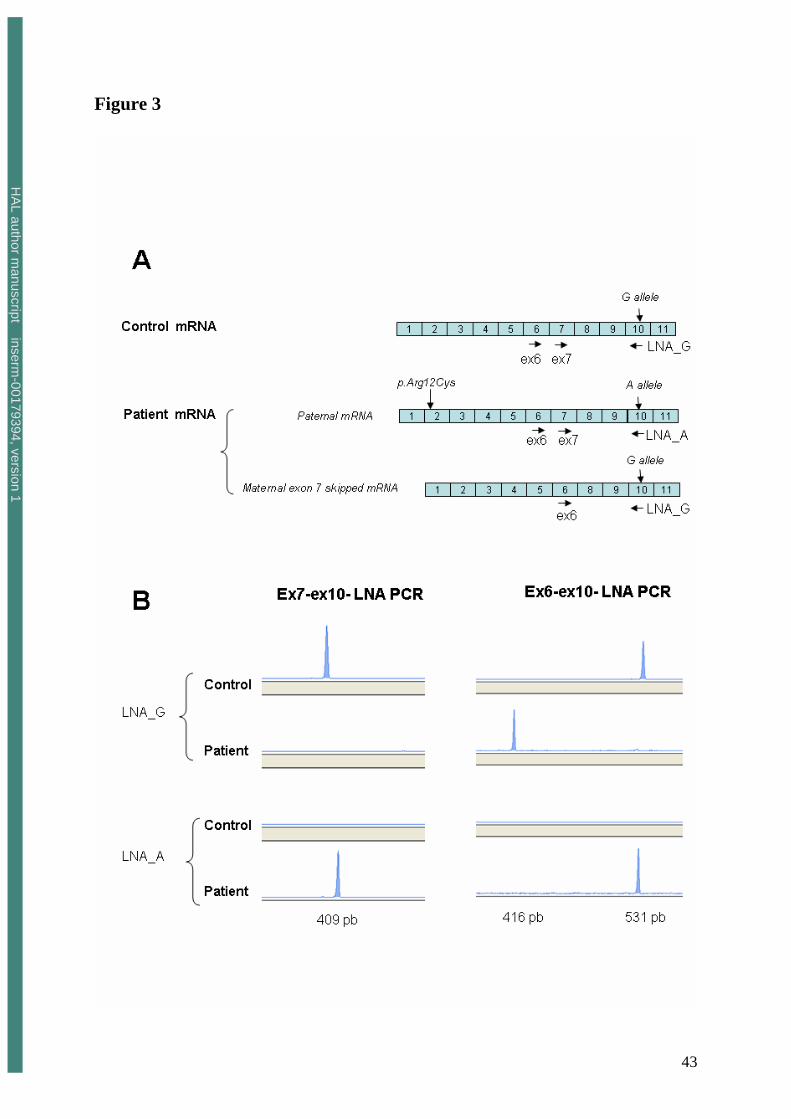
The possibility that residual full-length RNA molecules were produced from the maternal
allele was excluded by taking advantage of the patient’s heterozygosity for the p.Arg391His
variant in exon 10. cDNA from the patient was amplified using LNA modified primers with a
C or a T at the 3’ end (thus specific for the wild-type allele G, ex10R LNA_G, or for the
paternal allele A, ex10R LNA_A, figure 3A). Maternal mRNA molecules were theoretically
specifically amplified using ex10R LNA_G and paternally derived mRNA, using ex10R
LNA_A. The specificity of the LNA primers was confirmed using the ex7F and ex10 LNA
primers as shown in figure 3B. PCR amplification was only observed with the ex7F and ex10
LNA_G primers in the control (homozygous for the wild-type allele, c.1170 G/G), whereas
PCR amplification was positive in the patient using primers ex7F and ex10 LNA_A and
negative using primers ex7F and ex10 LNA_G. These results confirmed that full-length
mRNA molecules (containing exon 7) were exclusively produced from the paternal allele.
They were confirmed by using Ex6F – ex10R LNA_G, that amplified only exon 7-truncated
mRNA, and full-length mRNA molecules using Ex6F – ex10R LNA_A (Figure 3B). Together
HA
L author manuscript inserm
-00179394, version 1

15
these data confirmed that full-length mRNA molecules were exclusively produced from the
paternal allele and thus carried the [p.Arg12Cys and p.Arg391His] mutations.
Presence of a non functional IRAK-4 protein
As expected, a band corresponding to an IRAK-4 protein of apparently normal molecular
weight was detected by western blotting of the patient’s PMN; no shortened protein was
observed (Figure 1D). As the first step of IRAK-4 activity is the phosphorylation of IRAK-1
(29), we studied the functionality of IRAK-4 protein by analyzing the phospho-IRAK-1
content of intact PMN treated with TLR agonists in whole blood, by means of flow cytometry
with a mouse anti-human phospho-IRAK-1 Ab. Incubation of whole blood from healthy
controls with TLR agonists for 5 minutes significantly increased IRAK-1 phosphorylation as
compared to PBS (Table I). In contrast, pretreatment of the patient’s PMN with all the TLR
agonists, including the TLR9 agonist, did not modify IRAK-1 phosphorylation as compared
to PBS. This result suggested that IRAK-4 was non functional.
Impaired PMN adhesion molecule expression and ROS production in response to IL-18 and
TLR agonists, except for TLR9
PMN dysfunctions have been reported in IRAK-4 deficiencies (15), especially in response to
LPS (TLR4), contrasting with normal responses to TNF. We therefore analyzed the effect of a
broad range of TLR agonists on adhesion molecule expression and ROS production by PMN.
CD11b expression by resting patient’s PMN was normal, in keeping with normal chemotaxis
and with the absence of LAD (not shown). In controls, incubation of whole-blood samples
with TNFα, GM-CSF, IL-18 and the following TLR agonists: LPS (TLR4), MALP-2
(TLR2/6), Pam3CSK4 (TLR1/2), R-848 (TLR7/8) and CpG-DNA (TLR9) induced a
significant increase in CD11b expression (Figure 4A) and a significant decrease in L-selectin
HA
L author manuscript inserm
-00179394, version 1

16
expression (Figure 4B) related to activation-induced shedding of this molecule (30). In the
patient, TNFα and GM-CSF stimulation induced normal CD11b expression and normal L-
selectin shedding at the PMN surface as compared to PBS-treated samples. In contrast, after
treatment of patient’s samples with IL-18 and the following TLR agonists: LPS (TLR4),
MALP-2 (TLR2/6), Pam3CSK4 (TLR1/2) and R-848 (TLR7/8), no significant increase in
CD11b expression was observed as compared with the sample incubated with PBS; In
addition, L-selectin was still detectable at the PMN surface, reflecting a defect in the shedding
of this molecule. Surprisingly, however, the response to CpG-DNA (TLR9) was conserved
(Figure 4, A and B).
In controls, pretreatment of whole blood with TNFα, GM-CSF, IL-18 or with the
various TLR agonists, followed by stimulation with fMLP, a structural analog of bacterial
metabolic products, strongly increased ROS production (Figure 4C). A similar increase in
ROS production was also observed in the patient’s PMN after pretreatment with TNFα or
GM-CSF and stimulation with fMLP, ruling out defective priming of the phagocyte oxidative
burst (31). This priming effect on the fMLP-stimulated PMN oxidative burst, which was also
observed after treatment with CpG-DNA, was no longer detectable after incubation with the
other TLR agonists or IL-18 (Figure 4C).
Although the effect of TLR agonists on adhesion molecule expression and ROS
production by monocytes is far lower than with PMN, the patient’s monocytes showed a
pattern of responses similar to that of his PMN, with altered responses to TLR agonists except
for CpG-DNA (not shown).
HA
L author manuscript inserm
-00179394, version 1

17
Impaired prolongation of PMN survival by IL-18 and TLR agonists, except for TLR9
As PMN are usually very short-lived immune cells, prolongation of their lifespan by
proinflammatory mediators is critical for their efficacy against pathogens (32). In keeping
with previous reports (4, 33, 34), treatment of control PMN for 8 hours with GM-CSF, IL-18
and TLR agonists induced ~ 50 to 90% inhibition of PMN apoptosis in whole blood (total
annexin V+ cells); similar levels of inhibition were found in the early (annexin V+/7AAD-
cells) and late stage (annexin V+/7AAD+ cells) of PMN apoptosis (not shown). In contrast,
neither IL-18 nor LPS (TLR4), MALP-2 (TLR2/6), Pam3CSK4 (TLR1/2) and R-848
(TLR7/8) were able to inhibit the patient’s PMN apoptosis (percentage inhibition of PMN
apoptosis ~ 0%), while GM-CSF induced a normal prolongation of PMN survival. In keeping
with the results for adhesion molecule expression and ROS production, the effect of CpG-
DNA (TLR9) on the patient’s PMN apoptosis was conserved (Figure 4D).
We recently reported that the TLR-induced delay in PMN apoptosis was associated
with modulation of Bcl-2 family members (4), with an increased level of the anti-apoptotic
protein Mcl-1 and increased phosphorylation of the proapoptotic protein Bad, which have
been reported to inhibit apoptosis (35). In our patient, while CpG-DNA (TLR9) induced a
normal increase in Mcl-1 and phospho-Bad content, no modulation of either of these two
proteins was observed after stimulation with the other TLR agonists, as compared to samples
incubated with PBS (Table II).
Impaired cytokine production by PMN and monocytes in response to IL-18, IL-1 and all TLR
agonists.
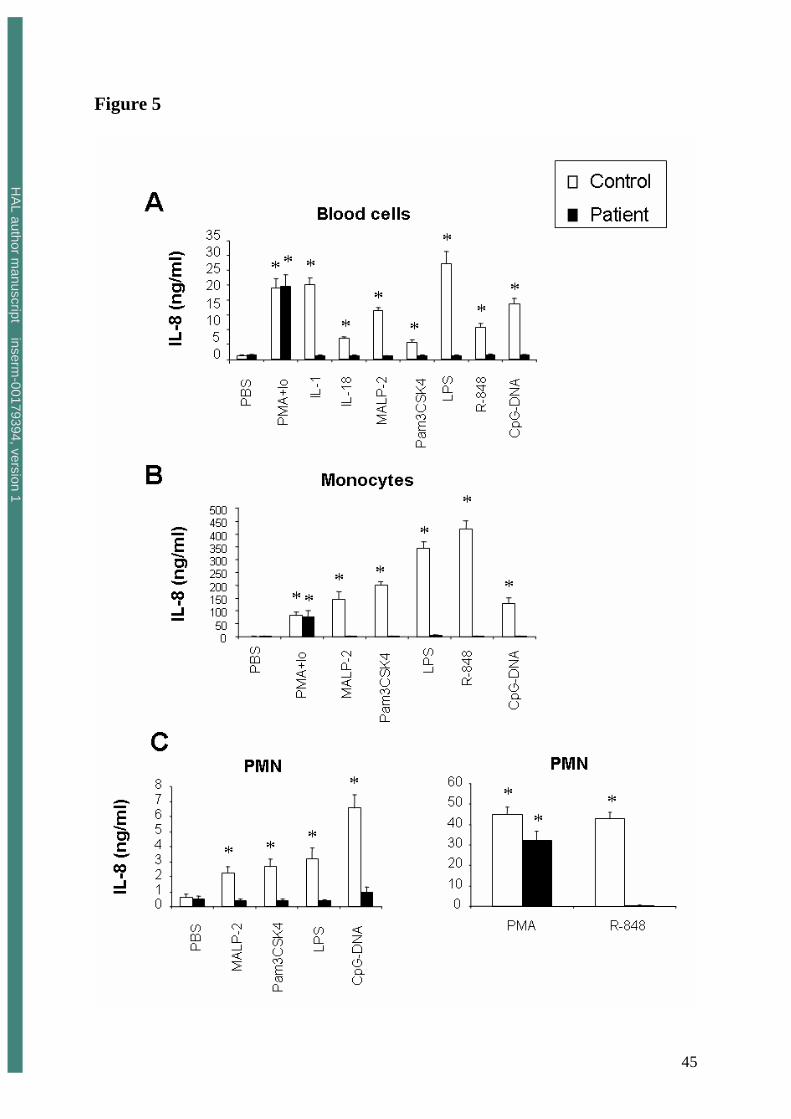
In keeping with previous data on patients with IRAK-4 deficiency (15, 16), we found strongly
impaired pro-inflammatory cytokine (IL-8) production in the supernatant of the patient’s
HA
L author manuscript inserm
-00179394, version 1

18
whole-blood samples cultured with all TLR agonists, including CpG-DNA (Figure 5A).
Similar results were observed for IL-6, IL-1β and TNFα production (not shown).
As cytokine production by whole blood cells mainly reflects synthesis by monocytes,
the patient’s monocytes and PMN were isolated and highly purified in order to analyze the
individual response of the two cellular subpopulations to TLR agonists, and especially to
CpG-DNA (TLR9). As expected, IL-8 production by control monocytes incubated with TLR
agonists was far higher than that of control PMN. Neither monocytes (Figure 5B) nor PMN
(Figure 5C) were able to produce significant amounts of IL-8 in response to LPS (TLR4),
MALP-2 (TLR2/6), Pam3CSK4 (TLR1/2) nor R-848 (TLR7/8). IL-8 production by the
patient’s PMN and monocytes in response to CpG-DNA was also strongly diminished as
compared to that of cells from a healthy control.
Finally, the parents’ PMN exhibited normal responses (adhesion molecule expression,
ROS production, delayed apoptosis, cytokine production) to all TLR agonists (not shown).
Involvement of direct stimulation of the PI3K pathway in the preserved PMN responses to
TLR9
CpG-DNA induced normal responses by the patient’s PMN in terms of survival, adhesion
molecule expression and ROS production, despite the lack of functional IRAK-4. This
strongly suggested that an alternative pathway, independent of the classic TLR9/IRAK-4
pathway, was involved in TLR9 signaling. We therefore examined the effects of various
kinase inhibitors on L-selectin and CD11b expression at the PMN surface, as well as on PMN
apoptosis in whole blood incubated with CpG-DNA. These inhibitors were used at optimal
concentrations previously determined in whole blood (4). Pretreatment with inhibitors of
conventional protein kinase C (GF109203X: 5 µM), PKCδ kinase (rottlerin: 10 µM) and
tyrosine kinase (genistein: 100 µM) had no effect on CpG-DNA-induced responses (not
HA
L author manuscript inserm
-00179394, version 1

19
shown). In contrast, pretreatment of the patient’s whole blood with PI3K inhibitors
(wortmannin and LY2940002) suppressed the effect of CpG-DNA on PMN responses. CpG-
DNA-induced shedding of L-selectin led to a significant decrease in L-selectin expression at
the PMN surface as compared to PBS-treated samples (Figure 6A); this decrease was totally
abolished after preincubation of the patient’s PMN with PI3K inhibitors (the MFI of the
sample incubated with PI3K inhibitors + CpG-DNA was similar to that observed after
treatment with PBS alone) (Figure 6A). CpG-DNA-induced modulation of L-selectin
expression was also significantly reduced after preincubation with PD98059 (a ERK1/2
kinase inhibitor) or SB203580 (a p38MAPK inhibitor). Similarly, the CpG-DNA-induced
increase in CD11b expression (reflected by the increase in stimulation index) (Figure 6B) was
significantly reduced after pre-incubation with PI3K inhibitors as well as with ERK1/2 kinase
and p38MAPK inhibitors.
Finally, in keeping with previous data (4), the CpG-DNA-induced increase in the percentage
of cell survival (annexin V-/7-AAD- cells) was significantly reduced after preincubation of
control samples with PI3K inhibitors and the NF-κB inhibitor SN50, while MAPKinase
inhibitors did not affect PMN apoptosis (Figure 6C). In the patient’s PMN, the inhibitory
effect of PI3K inhibitors was conserved while the NF-κB inhibitor SN50, which strongly
suppressed CpG-DNA-induced survival of control PMN, had no effect on TLR9-induced
survival of the patient’s PMN.
The PI3K inhibitor-induced reduction in PMN responses was also observed in healthy
controls but at a lower level than in the patient (Figure 6, A, B and C). This result suggested a
critical role of the direct TLR9/PI3K pathway in the patient’s PMN responses.
The effect of ERK1/2 kinase and p38MAPK inhibitors on CpG-DNA-induced modulation of
adhesion molecule expression observed in control and the patient’s PMN strongly suggested
the involvement of these kinases downstream of TLR9 activation. We therefore studied the
HA
L author manuscript inserm
-00179394, version 1

20
phospho-ERKK1/2 and phospho-p38MAPK contents of intact PMN treated in whole blood,
by means of flow cytometry. As shown in Figure 7, incubation of whole blood from controls
and the patient with CpG-DNA significantly increased ERK1/2 and p38MAPK
phosphorylation after 10 min as compared to PBS. This effect was significantly reduced by
preincubation with PI3K inhibitors. Total ERK1/2 and p38MAPK content, measured in the
same conditions, was not modified by treatment with CpG-DNA (data not shown).
HA
L author manuscript inserm
-00179394, version 1

21
Discussion
We describe an inherited IRAK-4 deficiency in a patient with compound heterozygosity
generating a non functional IRAK-4 protein. PMN functional responses (adhesion molecule
expression, ROS production, survival and pro-inflammatory cytokine production) to MALP-
2, Pam3CSK4, LPS, and R-848, which engage TLR2/6, TLR1/2, TLR4 and TLR7/8,
respectively, were strongly impaired, as were PMN responses to IL-18 (whose receptor shares
the same intracytoplasmic TIR domain). In contrast, the patient’s PMN responses to CpG-
DNA (TLR9) were normal, except for cytokine production, suggesting the existence, in
parallel to the MyD88/IRAK-4-dependent pathway, of a distinct TLR9-induced transduction
pathway regulating adhesion molecule expression, ROS production and survival.
The patient’s PMN exhibited an impaired response to several agonists of the IL-1R
family, and especially TLRs, while a normal response to other stimuli, including TNF, was
observed. These results suggest that the patient has a defect in the common TIR signalling
pathway, upstream of TRAF-6 and downstream of individual TIR membrane receptors.
IRAK4 gene analysis showed two compound heterozygous mutations in our patient. The
maternally inherited mutation at position + 5 of intron 7 (G>T) was predicted to result in a
protein of 248 amino acids, truncated of a large part of the kinase domain. However, no
shortened band was observed on western blots with a polyclonal Ab directed against the
whole IRAK-4 protein. These results suggest that the truncated protein resulting from the
maternal mutation is degraded as exon 7 skipped mRNA molecules were not subject to drastic
nonsense mediated decay. The two paternally inherited missense mutations, located in the
death domain (p.Arg12Cys) and in the kinase domain of IRAK-4 (p.Arg391His), did not
interfere with IRAK-4 synthesis: IRAK-4 protein was detected in the patient’s PMN by
western blot. However, none of the TLR agonists increased IRAK-1 phosphorylation, further
demonstrating the non function of IRAK-4 protein. These results suggest that the p.Arg12Cys
HA
L author manuscript inserm
-00179394, version 1

22
missense mutation of the paternal allele may hamper protein-protein interactions via the death
domain in the TIR pathway. Indeed, the p.arg12Cys mutation involves a residue that is
located at the surface of the protein; this domain is thought to interact with the different
ligands of IRAK-4 such as MyD88. This residue is highly conserved through evolution in
IRAK-4 orthologous proteins including human, Mus musculus, Bos taurus, Gallus gallus,
Xenopus tropicalis, Danio rerio and Euprymna scolopes (Supplementary data available on
request) but is not conserved in paralogues of the IRAK family, suggesting that it participates
to the specificity of interaction. It is therefore most likely that the substitution of this
positively charged residue (arginine) by a neutral one (cystein) interferes either with the
conformation of the IRAK-4 death domain or with the interaction of IRAK-4 with its
partners, preventing the assembly of an active signaling complex following TLR activation.
P.Arg391His, located in cis of the p.Arg12Cys substitution, was predicted to be benign with
two software programs based on structural and amino acid conservation, and had no effect on
mRNA splicing. A deleterious effect is thus unlikely, but a synergistic deleterious effect of
the two missense mutations could not be excluded. The non function of IRAK-4 protein in our
patient was also confirmed by the absence of significant IKK phosphorylation, which results
from TLR pathway activation (not shown).
TLR9-induced responses, i.e. adhesion molecule expression, ROS production and
survival, were normal in the patient’s PMN, while cytokine production was lower than with
control PMN. This suggests that different pathways may be involved in these functions; in
particular, the IRAK-4 dependent pathway is necessary for TLR9-induced cytokine
production, while other functions might use independent pathways. PI3Ks have been reported
to enhance nuclear translocation of NF-κB through phosphorylation and activation of IκB-
kinase and activation of MAPK, especially in TLR2-stimulated PMN (13); nevertheless, it is
not known whether MyD88/IRAK-4 complexes are required for this pathway. We clearly
HA
L author manuscript inserm
-00179394, version 1
23
found that PI3K inhibitors (wortmannin and LY2940002) totally suppressed the effect of
CpG-DNA on adhesion molecule expression and survival of our patient’s PMN. These results
suggest that TLR9 may be directly linked to PI3K, while the MyD88/IRAK-4 dependent
pathway may be required for PI3K activation through the other TLRs. Such direct PI3K
activation has been described for TLR2 in a human monocytic cell line (36).
The lipid products of PI3K – mainly phosphatidylinositol 3,4,5-triphosphate – induce
translocation of Akt/PKB to the plasma membrane, where it is phosphorylated and activated
by phosphatidyl-inositol 3,4,5-phosphate-dependent kinase (PDK1) (37). This pathway has
been forwarded as a major mediator downstream of PI3K (38). In particular, Akt activation
induces modulation of Bcl-2 family proteins such as Mcl-1 and phospho-Bad (39, 40), and
could therefore be involved in the inhibitory effect of CpG-DNA on our patient’s PMN
apoptosis. In addition, it has been reported that class I PI3K catalytic subunits can lead to
phosphorylation of ERK1/2 and p38MAPK (9, 10, 41, 42); activation of these signaling
pathways has been implicated in the upregulation of CD11b expression (43, 44) and L-
selectin shedding (45, 46) after PMN treatment with various inflammatory stimuli. In keeping
with these data, we demonstrated that CpG-DNA-induced modulation of CD11b and L-
selectin on the surface of our patient’s PMN is partially inhibited by pharmacological
inhibitors of ERK1/2 and p38MAPK; furthermore, we found a CpG-DNA-induced increase in
phosphorylation of ERK1/2 and p38MAPK in the patient’s PMN, which was reduced by
PI3K inhibitors. These findings strongly suggest that IRAK-4-independent TLR9-induced
PI3K activation leads to MAPK recruitment. As CpG-DNA has no direct effect on ROS
production, the use of kinase inhibitors did not allow us to analyse the involvement of MAPK
in the priming effect of CpG-DNA on the PMN oxidative burst in response to fMLP.
Nevertheless, PI3K products have also been reported to exert their effects on the PMN
HA
L author manuscript inserm
-00179394, version 1

24
oxidative burst by activating downstream protein kinases such as Akt, which may directly
phosphorylate components of the oxidase complex (47).
Taken together, our results suggest that activation of class I PI3K (PI3K (I) –
PDK1/Akt/PKB) through TLR9, and subsequent recruitment of MAPK, could be an
alternative pathway to the IRAK/IKK/NF-κB pathway involved in PMN adhesion, oxidative
burst, and prolonged survival, which are major components of PMN functional activity. A
schematic representation of the different pathways involved in PMN functions is proposed in
Figure 8. Nevertheless, we cannot formally exclude the involvement of other, unidentified
signalling pathways leading to CpG-DNA-induced PMN responses. In particular, there is
evidence of a TLR9-independent pathway leading to downstream PI3K activation and CD11b
upregulation in response to bacterial CpG-containing DNA in murine neutrophils. (44, 48).
This pathway described in human PMN by Alvarez et al is MyD88-dependent and leads to
IRAK-1 phosphorylation, suggesting the involvement of IRAK-4 in subsequent PI3K
activation (44). However, we cannot formally exclude the possibility that the IRAK-4-
independent activation of PI3K observed in our patient after CpG-DNA stimulation may be
related to the existence of TLR9-independent mechanisms, thus implicating non-CpG
molecular motifs in synthetic oligonucleotides.
IRAK-4-deficient patients suffer from pyogenic infections but are resistant to viruses,
fungi and parasites, as well as many other bacteria. It has been speculated that cell-surface
TLRs rapidly sense bacterial infections by recognizing bacterial cell wall constituents in the
extracellular medium. In contrast, several lines of evidence suggest that molecular recognition
of CpG-DNA occurs inside the cells (49). TLR9 might enter the phagosome from the
endoplasmic reticulum (50) and bind bacterial DNA released into the phagosome following
bactericidal processes. In addition, TLR9 was recently implicated in host defenses against
intracellular pathogens (51, 52). Further studies are necessary to elucidate the role of direct
HA
L author manuscript inserm
-00179394, version 1

25
PI3K activation by TLR9 in the phagosome, relative to cell surface activation of the
TLRs/IRAK-4-dependent pathway in defenses against microorganisms, and especially
intracellular pathogens. Nevertheless, we clearly observed that CpG-DNA induced normal
PMN functions in terms of adhesion molecule expression and survival in our IRAK4-deficient
patient, suggesting that the IRAK-4 dependent pathway may be compensated for by the
TLR9-dependent IRAK-4-independent pathway. This may account, at least in part, for the
observed clinical improvement with age.
In conclusion, this study provides the first description of persistent TLR9-induced
responses, critically involved in anti-microbial defenses, by PMN from a patient with
inherited IRAK-4 deficiency. These results strongly suggest the existence of a TLR9
alternative pathway leading to PI3K activation independently of the classical MyD88/IRAK-4
pathway. This may explain the control of infections due to microorganisms other than
pyogenic bacteria by PMN in patients with inherited IRAK-4 deficiency. Finally, our study
emphasizes the importance of “lessons of nature” in understanding the role of the TLR in
human defenses.
Acknowledgments
We thank Steven and his family.
HA
L author manuscript inserm
-00179394, version 1

26
References
1. Babior, B. M. 1984. Oxidants from phagocytes: agents of defense and destruction. Blood.
64: 959-966.
2. Savill, J., I. Dransfield, C. Gregory, and C. Haslett. 2002. A blast from the past: clearance
of apoptotic cells regulates immune responses. Nat. Rev. Immunol. 2: 965-975.
3. Hayashi, F., T. K. Means, and A. D. Luster. 2003. Toll-like receptors stimulate human
neutrophil function. Blood. 102: 2660-2669.
4. François, S., J. El Benna, P. M. C. Dang, M. A. Gougerot-Pocidalo, and C. Elbim. 2005.
Inhibition of neutrophil apoptosis by Toll-like receptor agonists in whole blood:
involvement of the phosphoinositide 3-kinase/Akt and NF-κB signaling pathways leading
to increased levels of Mcl-1, A1 and phosphorylated Bad. J. Immunol. 174: 3633-3642.
5. Beutler, B. 2000. TLR4: central component of the sole mammalian LPS sensor. Curr.
Opin. Immunol. 12: 20-26.
6. Akira, S. 2003. Toll-like Receptor Signaling. J. Biol. Chem. 278: 38105-38108.
7. Yum, H. K., J. Arcaroli, J. Kupfner, R. Shenkar, J. M. Penninger, T. Sasaki, K. Y Yang, J.
S. Park, and E. Abraham. 2001. Involvement of phosphoinositide 3-kinases in neutrophil
activation and the development of acute lung injury. J. Immunol. 167: 6601-6608.
8. Zhu, D., H. Hattori, H. Jo, Y. Jia, K. K. Subramanian, F. Loison, J. You, Y. Le, M.
Honczarenko, L. Silberstein, and H. R. Luo. 2006. Deactivation of phosphatidylinositol
3,4,5-triphosphate/Akt signaling mediates neutrophil spontaneous death. Proc. Natl. Acad.
Sci. USA. 103: 14836-14841.
9. Guha, M., and N. Mackman. 2002. The phosphatidylinositol 3-kinase-Akt pathway limits
lipopolysaccharide activation of signaling pathways and expression of inflammatory
mediators in human monocytic cells. J. Biol. Chem. 277: 32124-32132.
HA
L author manuscript inserm
-00179394, version 1
27
10. Fukao, T., and S. Koyasu. 2003. PI3K and negative regulation of TLR signaling. TRENDS
in Immunology. 24: 358-363.
11. Madrid, L. V., C. Y. Wang, D. C. Guttridge, A. J. Schottelius, A. S. Baldwin, and M. W.
Mayo. 2000. Akt suppresses apoptosis by stimulating the transactivation potential of the
RelA/p65 subunit of NF-κB. Mol. Cell. Biol. 20: 1626-1638.
12. Madrid, L. V., M. W. Mayo, J. Y. Reuther, and A. S. Baldwin. 2001. Akt stimulates the
transactivation potential of the RelA/p65 subunit of NF-κB through utilization of the IκB
kinase and activation of the mitogen-activated protein kinase p38. J. Biol. Chem. 276:
18934-18940.
13. Strassheim, D., K. Asehnoune, J. S. Park, J. Y. Kim, Q. He, D. Richter, K. Kuhn, S. Mitra,
and E. Abraham. 2004. Phosphoinositide 3-kinase and Akt occupy central roles in
inflammatory responses of Toll-like receptor 2-stimulated neutrophils. J. Immunol. 172:
5727-5733.
14. Medvedev, A. E., A. Lentschat, D. B. Kuhns, J. C. G. Blanco, C. Salkowski, S. Zhang, M.
Arditi, J. I. Gallin, and S. N. Vogel. 2003. Distinct mutations in IRAK-4 confer
hyporesponsiveness to lipopolysaccharide and interleukin-1 in a patient with recurrent
bacterial infections. J. Exp. Med. 198: 521-531.
15. Picard, C., A. Puel, M. Bonnet, C. L. Ku, J. Bustamante, K. Yang, C. Soudais, S. Dupuis,
J. Feinberg, C. Fieschi, C. Elbim, R. Hitchcock, D. Lammas, G. Davies, A. Al-Ghonaium,
H. Al-Rayes, S. Al-Jumaah, S. Al Hajjar, I. Zaid Al-Mohsen, H. H. Frayha, R. Rucker, T.
R. Hawn, A. Aderem, H. Tufenkeji, S. Haraguchi, N. K. Day, R. A. Good, M. A.
Gougerot-Pocidalo, A. Ozinsky, and J. L. Casanova. 2003. Pyogenic bacterial infections in
humans with IRAK-4 deficiency. Science. 299: 2076-2079.
HA
L author manuscript inserm
-00179394, version 1

28
16. Currie, A. J., D. J. Davidson, G. S. D. Reid, S. Bharya, K. L. MacDonald, R. Devon, and
D. P. Speert. 2004. Primary immunodeficiency to pneumococcal infection due to a defect
in Toll-like receptor signaling. J. Pediatr. 144: 512-518.
17. Day, N., N. Tangsinmankong, H. Ochs, R. Rucker, C. Picard, J. L. Casanova, S.
Haraguchi, and R. Good. 2004. Interleukin receptor-associated kinase (IRAK-4) deficiency
associated with bacterial infections and failure to sustain antibody responses. J. Pediatr.
144: 524-526.
18. Enders, A., U. Pannicke, R. Berner, P. Henneke, K. Radlinger, K. Schwarz, and S. Ehl.
2004. Two siblings with lethal pneumococcal meningitis in a family with a mutation in
Interleukin-1 receptor-associated kinase 4. J. Pediatr. 145: 698-700.
19. Yang, K., A. Puel, S. Zhang, C. Eidenschenk, C. L. Ku, A. Casrouge, C. Picard, H. von
Bernuth, B. Senechal, S. Plancoulaine, S. Al-Hajjar, A. Al-Ghonaium, L. Marodi, D.
Davidson, D. Speert, C. Roifman, B. Z. Garty, A. Ozinsky, F. J. Barrat, R. L. Coffman, R.
L. Miller, X. Li, P. Lebon, C. Rodriguez-Gallego, H. Chapel, F. Geissmann, E. Jouanguy,
and J. L. Casanova. 2005. Human TLR7-, -8-, and -9-mediated induction of IFN-alpha/beta
and -lambda is IRAK-4 dependent and redundant for protective immunity to viruses.
Immunity. 235: 465-78.
20. Cardenes, M., H. von Bernuth, A. Garcia-Saavedra, E. Santiago, A. Puel, C.L. Ku, J. F.,
C. Picard, J. L. Casanova, E. Colino, A. Bordes, A. Garfia, and C. Rodriguez-Gallego.
2006. Autosomal recessive interleukin-1 receptor-associated kinase 4 deficiency in fourth-
degree relatives. J. Pediatr. 148: 549-51.
HA
L author manuscript inserm
-00179394, version 1
29
21. Davidson, D. J., A. J. Currie, D. M. Bowdish, K. L. Brown, C. M. Rosenberger, R. C. Ma,
J. Bylund, P. A. Campsall, A. Puel, C. Picard, J. L. Casanova, S. E. Turvey, R. E.
Hancock, R. S. Devon, and D. P. Speert. 2006. IRAK-4 mutation (Q293X): rapid detection
and characterization of defective post-transcriptional TLR/IL-1R responses in human
myeloid and non-myeloid cells. J. Immunol. 1;177: 8202-11.
22. Ku, C. L., C. Picard, M. Erdos, A. Jeurissen, J. Bustamante, A. Puel, H. von Bernuth, O.
Filipe-Santos, H. H. Chang, T. Lawrence, M. Raes, L. Marodi, X. Bossuyt, and J. L
Casanova. 2007. IRAK4 and NEMO mutations in otherwise healthy children with
recurrent invasive pneumococcal disease. J. Med. Genet. 44: 16-23.
23. Amar, M., N. Amit, T. Pham Huu, S. Chollet-Martin, M. T. Labro, M. A. Gougerot-
Pocidalo, and J. Hakim. 1990. Production by K562 cells of an inhibitor of adherence-
related functions of human neutrophils. J. Immunol. 144: 4749-4756.
24. Rothe, G., and G. Valet. 1990. Flow cytometric analysis of respiratory burst activity in
phagocytes with hydroethidine and 2',7'-dichlorofluorescin. J. Leukocyte Biol. 47: 440-448.
25. Herault, O., P. Colombat, J. Domenech, M. Degenne, J. L. Bremond, L. Sensebe, M. C.
Bernard, and C. Binet. 1999. A rapid single-laser flow cytometric method for
discrimination of early apoptotic cells in a heterogenous cell population. Br. J. Haematol.
104: 530-537.
26. Elbim, C., H. Reglier, M. Fay, C. Delarche, V. Andrieu, J. El Benna, and M. A. Gougerot-
Pocidalo. 2001. Intracellular pool of IL-10 receptors in specific granules of human
neutrophils: differential mobilization by proinflammatory mediators. J. Immunol. 166:
5201-5207.
27. Grenier, A., M. Dehoux, A. Boutten, M. Arce-Vicioso, G. Durand, M. A. Gougerot-
Pocidalo, and S. Chollet-Martin. 1999. Oncostatin M production and regulation by human
polymorphonuclear neutrophils. Blood. 93: 1413-1421.
HA
L author manuscript inserm
-00179394, version 1

30
28. Dang, P. M. C., C. Elbim, J. C. Marie, M. Chiandotto, M. A. Gougerot-Pocidalo, and J.
El-Benna. 2006. Anti-inflammatory effect of interleukin-10 on human neutrophils involves
inhibition of GM-CSF-induced p47phox phosphorylation through a decrease in ERK1/2
activity. FASEB J. 20: 1504-1516.
29. Suzuki, N., S. Suzuki, and W. C. Yeh. 2002. IRAK-4 as the central TIR signaling
mediator in innate immunity. Trends Immunol. 23: 503-506.
30. Bevilacqua, M. P. 1993. Selectins. J. Clin. Invest. 91: 379-387.
31. Elbim, C., P. Rajagopalan-Levasseur, S. Chollet-Martin, J. L. Gaillard, M. Fay, J. Hakim,
A. Fischer, J. L. Casanova, and M. A. Gougerot-Pocidalo. 1999. Defective priming of the
phagocyte oxidative burst in a child with recurrent intracellular infections. Microbes and
infection. 1: 581-587.
32. Haslett, C., J. S. Savill, M. K. Whyte, M. Stern, I. Dransfield, and L. C. Meagher. 1994.
Granulocyte apoptosis and the control of inflammation. Philos. Trans. R. Soc. Lond. Biol.
Sci. 345: 327-333.
33. Derouet, M., L. Thomas, A. Cross, R. J. Moots, and S. W. Edwards. 2004. Granulocyte
macrophage colony-stimulating factor signaling and proteasome inhibition delay
neutrophil apoptosis by increasing the stability of Mcl-1. J. Biol. Chem. 279: 26915-26921.
34. Jablonska, E., M. Marcinczyk, and J. Jablonski. 2006. Toll-like receptors types 2 and 6
and the apoptotic process in human neutrophils. Arch. Immunol. Ther. Exp. (Warsz). 54:
137-142.
35. Perianayagam, M. C., V. S. Balakrishnan, B. J. Pereira, and B. L. Jaber. 2004. C5a delays
apoptosis of human neutrophils via an extracellular signal-regulated kinase and Bad-
mediated signaling pathways. Eur. J. Clin. Invest. 34: 50-56.
HA
L author manuscript inserm
-00179394, version 1

31
36. Arbibe, L., J. P. Mira, N. Teusch, L. Kline, M. Guha, N. Mackman, P. J. Godowski, R. J.
Ulevitch, and U. G. Knaus. 2000. Toll-like receptor 2-mediated NF-κB activation requires
a Rac1-dependent pathway. Nat. Immunol. 1: 533-540.
37. Cantley, L. C. 2002. The phosphoinositide 3-kinase pathway. Science. 296: 1655-1657.
38. Toker, A, and L. C. Cantley. 1997. Signaling through the lipid products of
phosphoinositide-3-OH kinase. Nature. 387: 673-676.
39. del Peso, L., M. Gonzalez-Garcia, C. Page, R. Herrera, and G. Nunez. 1997. Interleukin-
3-induced phosphorylation of BAD through the protein kinase Akt. Science. 278: 687-689.
40. Schubert, K. M., and V. Duronio. 2001. Distinct roles for extracellular-signal-regulated
protein kinase (ERK) mitogen-activated protein kinases and phosphatidylinositol 3-kinase
in the regulation of Mcl-1 synthesis. Biochem. J. 356: 473-480.
41. Coxon, P. Y., M. J. Rane, D. W. Powell, J. B. Klein, and K. R. McLeisch. 2000.
Differential mitogen-activated protein kinase stimulation by Fcγ receptor IIa and Fcγ
receptor IIIb determines the activation phenotype of human neutrophils. J. Immunol. 164:
6530-6537.
42. Rane, M. J., P. Y. Coxon, D. W. Powell, R. Webster, J. B. Klein, W. Pierce, P. Ping, and
K. R. McLeisch. 2001. P38 Kinase-dependent MAPKAPK-2 activation functions as 3-
phosphoinositide-dependent kinase-2 for Akt in human neutrophils. J. Biol. Chem. 276:
3517-3523.
43. Mocsai, A., Z. Jakus, T. Vantus, G. Berton, C. A. Lowell, and E. Ligeti. 2000. Kinase
pathways in chemoattractant-induced degranulation of neutrophils: the role of p38
mitogen-activated protein kinase activated by Src family kinase. J. Immunol. 164: 4321-
4331.
HA
L author manuscript inserm
-00179394, version 1

32
44. Alvarez, M. E., J. I. Bass, J. R. Geffner, P. X. Calotti, M. Costas, O. A. Coso, R.
Gamberale, M. E. Vermeulen, G. Salamone, D. Martinez, T. Tanos, and A. S. Trevani.
2006. Neutrophil signaling pathways activated by bacterial DNA stimulation. J. Immunol.
177: 4037-4046.
45. Fan, H., and R. Derynck. 1999. Ectodomain shedding of TGF-alpha and other
transmembrane proteins is induced by receptor tyrosine kinase activation and MAP kinase
signaling cascades. EMBO J. 18: 6962-6972.
46. Smolen, J. E., T. K. Petersen, C. Koch, S. J. O’Keefe, W. A. Hanlon, S. Seo, D. Pearson,
M. C. Fossett, and S. I. Simon. 2000. L-selectin signaling of neutrophil adhesion and
degranulation involves p38 mitogen-activated protein kinase. J. Biol. Chem. 26: 15876-
15884.
47. Chen, Q., D. W. Powell, M. J. Rane, S. Singh, W. Butt, J. B. Klein, and K. R. McLeish.
2003. Akt phosphorylates p47phox and mediates respiratory burst activity in human
neutrophils. J. Immunol. 170: 5302-5308.
48. Trevani, A. S., A. Chorny, G. Salamone, M. Vermeulen, R. Gamberale, J. Schettini, S.
Raiden, and J. Geffner. 2003. Bacterial DNA activates human neutrophils by a CpG-
independent pathway. Eur. J. Immunol. 33:3164-3174.
49. Wagner, H. 2004. The immunobiology of the TLR9 subfamily. Trends Immunol. 25: 381-
386.
50. Leifer, C. A., M. N. Kennedy, A. Mazzoni, C. W. Lee, M. J. Kruhlak, and D. M. Segal.
2004. TLR9 is localized in the endoplasmic reticulum prior to stimulation. J. Immunol.
173: 1179-1183.
51. Khan, I. A. 2007. Toll road for Toxoplasma gondii: the mystery continues. Trends
Parasitol. 23: 1-3.
HA
L author manuscript inserm
-00179394, version 1

33
52. von Meyenn, F., M. Schaefer, H. Weighardt, S. Bauer, C. J. Kirschning, H. Wagner, and
T. Sparwasser. 2006. Toll-like receptor 9 contributes to recognition of Mycobacterium
bovis Bacillus Calmette-Guerin by Flt3-ligand generated dendritic cells. Immunobiology.
211: 557-565.
HA
L author manuscript inserm
-00179394, version 1

34
Footnotes
1 Adress correspondence and reprint requests to:
Carole Elbim, INSERM U773, Faculté Xavier Bichat, 16 rue Henri Huchard, 75877 Paris
Cedex 18, France.
Phone: 33 1 44 85 62 06; Fax: 33 1 44 85 62 07. E-mail: [email protected]
2 Non standard abbreviations: IRAK: interleukin-1 receptor associated kinase; PMN:
polymorphonuclear neutrophil; TIR: Toll-IL-1 receptor; TRAF6: tumor necrosis factor
receptor-associated factor 6; LAD: leucocyte adhesion deficiency; HE: hydroethidin; fMLP:
N-formyl-methionyl-leucyl-phenylalanine; APC: allophycocyanin; 7-AAD: 7-amino-
actinomycin D;
HA
L author manuscript inserm
-00179394, version 1

35
Figure legends
FIGURE 1. Genetic analysis of IRAK4 gene and protein expression in the patient
A, Schematic representation of the IRAK4 gene and alleles in the patient. Sequencing
chromatograms obtained in our patient are shown.
B, Schematic representation of IRAK-4 protein, with the death domain and kinase domain.
The positions of the mutations found in the patient are indicated.
C, Schematic representation of the IRAK-4 death domain (Protein Data Bank accession code
2A9I). This ribbon diagram was generated with PyMOL (DeLano Scientific,
www.pymol.org). The Arg12 is shown as full surface amino-acid residue.
D, Expression of IRAK-4 by Western blotting
A total of 2.5x10⁶ cell equivalents were loaded in each well. Following SDS-PAGE, the
proteins were transferred to nitrocellulose membranes and incubated with anti-human IRAK-4
Ab at 1/500 dilution overnight. The Western blots were revealed as described in Materials and
Methods.
FIGURE 2. Mutation + 5 G>T in intron 7 induces exon 7 skipping
A, PCR amplification products using ex6F cDNA and ex11R cDNA primers and cDNA from
a control and the patient were analysed onto ABI310 DNA sequencer. A normal PCR product
size (647 pb) and a truncated one (532 pb) was observed in our patient.
B, Sequencing of the shortened band was performed using ex6F cDNA and ex11R cDNA non
fluorescent primers.
HA
L author manuscript inserm
-00179394, version 1

36
FIGURE 3. Full length mRNA molecules are exclusively produced from the paternal allele.
Allele specific PCR was performed using two different reverse LNA primers. Ex10R LNA_G
primer matches the wild type allele (c.1170 G allele) whereas Ex10R LNA_A primer is
specific of the variant allele (c.1170 A allele). The paternal mRNA contained the A allele and
the maternal mRNA the G allele. The control used in this experiment was homozygous for the
G allele. Fluorescent products obtained after PCR amplification of cDNA from the patient or
the control were then analysed on a ABI310 DNA sequencer. Expected PCR product sizes are
indicated. The 416-bp peak observed with the Ex6F-Ex10R LNA_G set of primers
corresponds to the exon 7-skipped form of mRNA.
HA
L author manuscript inserm
-00179394, version 1

37
FIGURE 4. Impaired PMN functions in response to IL-18 and TLR agonists, except for
TLR9
A and B, Adhesion molecule expression at the PMN surface: whole-blood samples were
incubated at 37°C for 1 hour with PBS, TNFα (100 U/ml), GM-CSF (1000 pg/ml), IL-18
(500 ng/ml) or with the following TLR agonists: LPS (10 ng/ml) (TLR4), MALP-2 (10
ng/ml) (TLR2/6), Pam3CSK4 (500 ng/ml) (TLR1/2), R-848 (10 µg/ml) (TLR7/8), or CpG-
DNA (100 µg/ml) (TLR9). Samples were then stained with PE-anti-CD11b and purified anti-
L-selectin antibodies at 4°C for 30 minutes.
Results are expressed as mean fluorescence intensity (MFI).
* Significantly different from sample incubated with PBS (p<0.05).
C, PMN oxidative burst: whole-blood samples were pretreated with HE for 15 minutes at
37°C and then incubated with TNFα, GM-CSF, IL-18 or TLR agonists as described above,
followed by fMLP stimulation (10-6 M, 5 minutes).
Results are expressed as a stimulation index (ratio of the mean fluorescence intensity of
stimulated cells to that of unstimulated cells).
* Significantly different from the sample incubated with PBS (stimulation index=1) (p<0.05).
D, PMN apoptosis: whole-blood samples were incubated in 24-well tissue cultures plates at
37°C with 5% CO2 for 8 hours with PBS, GM-CSF, IL-18 or TLR agonists as described
above. PMN were identified by using a FITC-anti-CD15 Ab. Apoptosis was quantified by
staining with APC-annexin V and 7-AAD as described in Materials and Methods.
Results are expressed as the percentage inhibition of PMN apoptosis [1 – (% of total annexin
V+ PMNs in stimulated sample/% of total annexin V+ PMN in PBS-treated sample)] x 100.
* Significantly different from the sample incubated with PBS (percentage inhibition of
apoptosis=0) (p<0.05).
Panel A to D: three independent experiments, each with a different healthy control.
HA
L author manuscript inserm
-00179394, version 1

38
FIGURE 5. Impaired cytokine production by blood cells in response to TLR agonists, IL-1β
and IL-18.
Whole-blood samples (Panel A), isolated monocytes (5x10⁵/ml) (Panel B), and highly
purified PMN (5x10⁶/ml) (Panel C) were incubated for 18 hours with PMA-ionomycin (10-7M
and 10-5M), PMA (10-7M), IL-1β (50 ng/ml), IL-18 (500 ng/ml) or TLR agonists as described
in the legend of figure 4.
IL-8 production was measured by using the human inflammatory cytokine cytometric bead
array (CBA) kit.
* Significantly different from the sample incubated with PBS (p<0.05) (n=3, each experiment
performed with a different healthy control).
HA
L author manuscript inserm
-00179394, version 1

39
FIGURE 6. Involvement of the putative IRAK-4-independent PI3K signalling pathway in the
persistent PMN response to TLR9.
A and B, Effect of kinase inhibitors on CpG-induced modulation of adhesion molecule
expression at the PMN surface: whole-blood samples were pretreated at 37°C with PBS, PI3K
inhibitors (wortmannin: 2500 nM; LY2940002: 25 µM), MEK1/2 inhibitor (PD98059: 50
µM) or p38MAPK inhibitor (SB203580: 25 µM) for 15 minutes and then with PBS or CpG-
DNA for 1 hour. L-selectin and CD11b expression at the PMN surface were then studied as
described in the legend of figure 4. Results are expressed in MFI (L-selectin expression) and
as a stimulation index (CD11b expression: ratio of the MFI of CpG-DNA-stimulated cells to
that of unstimulated cells).
C, Effect of an NF-κB inhibitor (SN50) and kinase inhibitors on CpG-DNA-induced PMN
survival.
Whole-blood samples were pretreated in 24-well tissue culture plates at 37°C in 5% CO2/air
with PBS, SN50 (100 µg/ml), PI3K inhibitors (wortmannin: 2500 nM; LY2940002: 25 µM),
MEK1/2 inhibitor (PD98059: 50 µM) or p38MAPK inhibitor (SB203580: 25 µM) for 1 hour.
Samples were then incubated with PBS or CpG-DNA for 8 hours. Survival was quantified as
described in the legend of figure 4. Results are expressed as the percentage of viable cells
(annexin V-/7-AAD- cells).
* Significantly different from the CpG-DNA-stimulated sample incubated with PBS alone
instead of kinase inhibitors (p<0.05) (n=3, each experiment performed with a different healthy
control).
HA
L author manuscript inserm
-00179394, version 1

40
FIGURE 7. Effect of CpG-DNA on intracellular ERK1/2 and p38MAPK phosphorylation.
Whole-blood samples were preincubated at 37°C in a water bath with PBS or PI3K inhibitors
(wortmannin: 2500 nM; LY2940002: 25 µM) for 15 minutes and then with PBS or CpG-
DNA (100 µg/ml) for 10 min. Phospho-p38MAPK and phospho-ERK1/2 contents were then
measured by flow cytometry on methanol-permeabilized cells as described in Materials and
Methods.
Results are expressed as the mean fluorescence intensity (MFI). Values obtained with an
irrelevant Ab of the same isotype were subtracted.
Values are means ± SEM (n=3, each experiment was performed with a different healthy
control).
* Significantly different from samples incubated with PBS alone (p<0.05).
FIGURE 8. Scheme of CpG-DNA/TLR9-mediated cellular signalling in PMN.
IRAK-4 dependent pathway. Recruitment of the TIR domain activates IRAK-4-TRAF6-
TAK1 complex formation. This leads to the activation of both MAPKs and IKK complexes,
culminating in upregulation of transcription factors, including NF-κB. NF-κB activation leads
to pro-inflammatory cytokine production and delays apoptosis. MAPK activation may be
involved in the modulation of adhesion molecule expression at the PMN surface and in
increased ROS production by primed PMN.
Alternative IRAK-4-independent pathway. Activation of class I PI3K (PI3K (I) – PD-
K1/Akt/PKB) through TLR9 could be an alternative to the IRAK/IKK/NF-κB pathway. Its
activation could lead to 1) delayed apoptosis through independent modulation of Bcl-2 family
proteins, and 2) recruitment of MAPKs involved in PMN adhesion and the oxidative burst.
HA
L author manuscript inserm
-00179394, version 1

41
Figure 1
HA
L author manuscript inserm
-00179394, version 1

42
Figure 2
HA
L author manuscript inserm
-00179394, version 1
43
Figure 3
HA
L author manuscript inserm
-00179394, version 1

44
Figure 4
HA
L author manuscript inserm
-00179394, version 1
45
Figure 5
HA
L author manuscript inserm
-00179394, version 1

46
Figure 6
HA
L author manuscript inserm
-00179394, version 1

47
Figure 7
HA
L author manuscript inserm
-00179394, version 1

48
Figure 8
HA
L author manuscript inserm
-00179394, version 1