to - defense technical information center station alexandria, virginia unclassified. notice: when...
TRANSCRIPT

UNCLASSIFIED
AD NUMBER
AD462433
NEW LIMITATION CHANGE
TOApproved for public release, distributionunlimited
FROMDistribution authorized to U.S. Gov't.agencies only; Administrative/OperationalUse; APR 1965. Other requests shall bereferred to Aeromedical Research Lab.[6571ST], Holloman AFB, NM.
AUTHORITY
ARL ltr dtd 1 Dec 1969
THIS PAGE IS UNCLASSIFIED

UNCLASSIFIED
AD4 6243 3
DEFENSE DOCUMENTATION CENTERFOR
SCIENTIFIC AND TECHNICAL INFORMATION
CAMERON STATION ALEXANDRIA, VIRGINIA
UNCLASSIFIED
NOTICE: When government o other drawings, speci-fications or other data aie used for any purposeother than in connection with a definitely relatedgovernment procurement operation, the U. S.Government thereby incurs no responsibility, nor any
obligation whatsoever; and the fact that the Govern-ment may have formulated, furnished, or in any waysupplied the said drawings, specifications, or otherdata is not to be regarded by implication or other-wise as in any manner licensing the holder or anyother person or corporation, or conveying any rightsor permission to manufacture, use or sell anypatented invention that my in any way be relatedthereto.

C RL-TR65-3
CTUDY OF MONKEY, APE. AND HUMAN MORPHOLOGY AND PHYSIOLOGY-RELATING TO STRENGTH AND ENDUVANCE'
PHASE IV
THE MUSCULOSKELETAL ANATOMY OF THE THORAX AND BRACHIUM OF ANADULT FEMALE CHIMPANZEE
LLJCDWilliam E. Edwards
~462433 jApril 1965
CDD
MAY 194 1965
DDC-IRA E
6571st Aeromedical Research LaboratoryAerospace Medical Division
Air Force Systems CommandHolloman Air Force Base, New Mexico

The animals used in this study were handled in accordance with the"Principles of Laboratory Animal Care" established by the NationalSociety for Medical Research.
This report may be reproduced to satisfy needs of U. S.- Governmentagencies. No other reproduction is authorised except. with permissionof the 6571stAeromedical Research Laboratory, Holloman ATF, NMex.
This report is made available for study with the understanding thatproprietary interests in and relating thereto will not be impaired. r Incase of apparent conflict or any other questions between the Govern-ment's rights and those of others, notify the Judge Advocate, AirForce Systems Command, Andrews Air-Force Base, Washington, D.C.Z0331.
Certain of the animals used for this research were provided by theNational Aeronautics and Space Administration under Order R-ZS.
Do not return this copy. Retain.or destroy.

STUDY OF MONKEY, APE, AND HUMAN MORPHOLOGY AND PHYSIOLOGYRELATING TO STRENGTH AND ENDURANCE
PHASE IV
THE MUSCULOSKELETAL ANATOMY OF THE THORAX AND BRACHIUM OF ANADULT FEMALE CHIMPANZEE
William E. Edwards

FOREWORD
This is the first in a series of four papers concerned with themusculoskeletal system of the thorax and upper extremities of thechimpanzee and the squirrel monkey. This study was conducted duringthe period 1961-1963 by William E. Edwards, under Contract AF 29(600)-3466, Project 6892, Task 689201. The program was monitored byMajor James E. Cook, Veterinary Services Division, ARRV.
The author wishes to acknowledge the assistance of Mr. T. ErskineClarke in the phase of dissection and of Mr. Robert Halferty for helpwith the photographic work. The writer is also indebted to Dr. EdwardM. Burn of the South Carolina State Hospital, Dr. Daris R. Swindlerof the South Carolina Medical College, Lt. Col. Hamilton H. Blackshear,USAF, MC, Major Robert H. Edwards, USAF, MC, Major James E. Cook,USAF, VC, and Major Clyde H. Kratochvil, USAF, MC, of the 6571stAerome,lical Research Laboratory for their helpful cooperation and en-couragement in this study.
Publication of this report does not constitute Air Force approvalof the report's findings or conclusions. It is published only for the ex-change and stimulation of ideas.
C.H. KRAT VLMajor, USAF, MCCommander
ii

ABSTRACT
The left thoracic and brachial musculature of a 'young-adultfemale chimpanzee is described and illustrated with the accuracy ofdetail plus clarity made possible by the photo-etching process. Otherdata are depicted graphically by section drawings. Comparisons withdata from the literature on other chimpanzees, apes, humans, andnon-hominoid primates are also provided, with emphasis on quantitativeaspects.
,ii

TABLE OF CONTENTS
Page
1. INTRODUCTION.....................................
2. HISTORY OF RESEARCH PRESENTED HERE ... .. ........ 2
3. DESCRIPTION OF SUBJECT. .. ....... ....... ... 2
4. PROCEDURE OF STUDY. .. .. ...... ........ .... 4
5. Description of Muscle Morphology .. .... ........ ... 7
A. Muscles of the, Shoulder Girdle. .. ..... .......... 7
B. Muscles of the Shoulder. .. .... ....... ....... 11
C. Muscles of the Arm. .. ..... ....... ........ 13
6. EVALUATION .... ....... ....... .......... 19
REFERENCES. ..... ....... ........ ....... 2
APPENDIX - Figures 1 through 19. ... ........ ...... 5

THE MUSCULO-SKELETAL ANATOMY OF THE THORAX AND BRACHIUM
OF AN ADULT FEMALE CHIMPANZEE
1. INTRODUCTION
Before the Christian era, non-human primates were long employed assubstitute subjects for anatomical studies because of the taboo againstdissection of cadavers -- a taboo not significantly violated until thestudies of the Alexandrian Herophilus, approximately 300 B.C. By thetime of Galen (c. 130-200 A.D.), some of the anatomical subjects wereapparently anthropoid apes (which likely ranged at least somewhat morewidely at that time) but most were catarrhine monkeys, such as the tail-less "Barbary ape" (Macaca sylvana) of North Africa (Yerkes and Yerkes,1929, pp. 3-7). Thus Ve-a ulearned thirteen centuries later thatGalen's errors for man tended to be correct for "apes."
Through the Middle Ages and into the Renaissance - when the humanstudies of da Vinci (1452-1519) and Vesalius (1514-1564) were conducted-- general prohibition of human dissection, despite some permitted excep-tions, encouraged some of that minority of physicians unwilling to relyupon the work of Galen to use other primates as substitutes.
The first identifiable ape to be dissected with detailed anatomicaldescriptions still extant was the chimpanzee ("a Pygmie") reported uponby Tyson (1699). Since the work of Tyson, scores of primatologists havepublished studies of ape anatomy. The chimpanzee has been the objc-t ofa plurality of these studies because of its relative accessibility ind itssimilarity to man morphologically and in other respects (Schultz, 1936;Yerkes, 1943, pp. 3-8; Hooton, 1946, pp. 39-44).
It might seem, therefore, that no further anatomical studies of thechimpanzee are needed. However, many studies are limited to brief -- andthus necessarily incpmplete -- verbal descriptions, while with only partialexceptions the papers which have presented drawings have shown the voluntarymusculeture only diagrammatically or vaguely and imprecisely. "Dependablesources of anatomical information.. .on the chimpanzee are few and whollyinadequate. There is no convenient atlas and no text which is completeand reliable. Sperino's Anatomia del Cimpanza (1897) includes an accountof the muscular, respiratory, gastrointestinal, and nervous systems, but itis based almost entirely on the dissection of a single female whose age wasestimated as two years" (Yerkes, 1943, p. 282).

2, HISTORY OF RESEARCH PRESENTED HERE
On June 14, 1961, an Aeromedical Research Laboratory adult femalechimpanzee died, and the writer decided upon his arrival a month later
that the formalin-fixed cadaver would be used for detailed, quantitative
studies of the musculature of the upper extremities -- during extrahours, since this was not part of the strength-testing project for
which he had been employed, The anatomical study seemed significant,
however, because the factors determining any differences in AStrengthbetween chimpanzee and man would almost surely include anatomicaldifferences.
Studies of the thoracic and brachial musculature were initiated
August 4, 1961, and continued intermittently until September 10.
Various factors necessitated postponement of the resumption of intensive
study until Marcih, 1963, and its completion until June, 1963.
3. DESCRIPTION OF THE SUBJECT
The subject, ARL Chimpanzee #133, was a young adult female chim-panzee of unidentified race (for geographic distribution of species,
see Coolidge, 1933, pp. 34-35), with an estimated age of 15 years at
the time of death. Extensive gangrene following an accident to her
right forearm necessitated euthanasia under anesthesia. Her last re-
corded body-weight, on May 19, 1961, was 89.5 pounds.
Immediately after death, the chimpanzee was injected with a 10 per
cent formalin solution through the arterial system, but without individ-ual treatment of the left upper extremity. In the subsequent autopsy,
the gangrenous right arm and all viscera were removed, with transection
of the rib-cage and such ascles as pectorilis major (Fig. 1). Much of
the head was removed with the brain, with resulting loss of the cephalad
portion of muscles traversing the cervical region. From the time ofthe autopsy, June 14, to the initiation of dissection, August 4, the
cadaver was kept in a refrigerated locker at approximately 2 C., ascontinued to be the case between intervals of dissection and study untilmid-September, 1961.
Panometric data procured immediately prior to dissection include
the following, in millimeters:
Cephalad surface of acromion tip (13 mm. medial to
lateral border of brachium) to distal tip of lateralepicondyle 308
Cephalad surface of acromion tip to distal tip ofmedial epicondyle 326
2

Distal tip of medial epicondyle to distal tip ofulnar styloid process 254
Distal tip of lateral epicondyle to distal tip ofradial styloid process 268-15
Medial surface of epidermis at tip of medialepicondyle to opposite lateral surface at elbow 96
Medial surface of epidermis at ulnar styloidprocess to lateral surface at radial styloid process 56
Distal tip of radial styloid process to distal tip +of digit I 103-5
Distal tip of ulnar styloid process to distal tip +of digit V 161-4
Length of the fleshy portion of the digits from thebase of the cleft between the digits1
I (from I-Il) 42II (from I-I) 121II (from 1I-Il) 80
III (from I-Il) 95III (from III-IV) 98IV (from Ill-IV) 82IV (from IV-V) 90V (from IV-V) 68
Circumferences
Through axilla and over shoulder 63 mm.medial to lateral surface of brachium 425
Brachium at inferior (caudal) border ofaxilla on dorsal surface of body (30 mm.inferior to inferior border of axilla onventral surface) 265
Middle of brachium 263
Brachium 63 mm. proximal to distal tip ofmedial epicondyle 238
Elbow across lateral and medial epicondyles 261
1These measurements of the length of digits are not precise (-3 or
4 mm.) because of the impossibility of extending fingers fully withoutdamaging the specimen.
3

Antebrachium 50 mm. distal to distal tip of medialepicondyle 256
Middle of antebrachium 229
Distal end of antebrachium across styloid processes 172
2Circumferences at the middle of the secodd phalanges
I (proximal phalanx) 42II 55
III 64IV 54V 42
Measurements of the cranium
Bi-orbital breadth 84
Middle of superior border of supraorbitaltorus to prosthion 97.5
Upper facial height (nasion-prosthion) 68
Bi-zygomatic diameter (10 mm. posterior toanterior border of orbits) 106
Prosthion-gonion 128
Some active decomposition of the specimen began soon after death,and grossly apparent autolysis continued until the study was completed.Ev'an during the first month of study in 1961, the muscular, tendinous,and fascial tissues and even the arteries, veins, and nerves of this sub-ject developed a progressively uniform gray coloration.
Formalin was reapplied in South Carolina and the specimen was keptrefrigerated at a temperature slightly above freezing, but the process ofdeterioration was never entirely arrested. In the spring of 1963, duringwhich the remainder and vast majority of the study was carried out, to theproblem of uniform coloration was added that of some softening of muscletisues.
4. PROCEDURE OF STUDY
As the dissection of the chimpanzee (preceded by that of the squirrel
2These five measures of the fleshy portions of each digit were muchmore significantly affected by dessication and other changes than the othermeasures in the 50 days between death and study.
4

monkey) proceeded in August, 1961, the writer prepared fairly detailed
verbal descriptions, including extensive measurements. It was soonrecognixed that written descriptions and measurements were entirelyinadequate, as has been confirmed by a subsequent survey of pertinentliterature, which reveals that much undoubtedly meticulous study isrelatively worthless because of the limitations of verbal descriptionsand the validity, at least in anatomical studies, of the adage concern-ing a thousand words versus one picture. Therefore, tne writer pre-pared diagrammatic sketches and then careful drawings to clarify photo-graphs, but he decided that even the latter were quantitatively too
imprecise.
It was finally decided that only very precise drawings could showthe form, size, and relationships of the musculoskeletal system entirelysatisfactorily, and the most feasible way to produce these was by theprocess of photo-etching, in which an India ink drawing is made directlyupon a photographic print, which is then bleached out. A brief surveyof the literature reveals that a somewhat comparable technique has beenutilized in the preparation of Grant's An Atlas of Anatomy (1947), forwhich muscle outlines were traced by theuse of photographs and a viewingbox.
Several variants of the photo-etching process were tested. The oneselected involves the use of "Kodak Illustrators' Special E" double-weight enlarging paper, with the print fixed in plain sodium thiosulphate
(100 gm. to 320 gm. of water). In drying, no ferrotyping is used; thesurface of the print can be made more receptive to the ink by rubbingwith talcum powder. After the drawing is completed, the picture is
immersed for 90 seconds in a solution of 10 gm. of potassium permanganateand 10 gm. of sulphuric acid in 2250 gm. of water. The resultingbronzed print is then washed gently for 5 minutes and "cleared" in asolution of 40 gm. of sodium thiosulphate plus 10 gm. of glacial aceticacid in 160 gm. of water. Although more experimentation may effectfurther improvement, the writer still finds that there is generally somerunning of the ink and smudging, requiring laborious application of whitepaint and India ink after the bleaching.
At.the Aeromedical Field Laboratory, by the time the photographswere printed, parts of the specimen, had in some cases been removed, andvital anatomical landmarks and lines of separation between parallelmuscles were often confusingly absent in the photographs. Thus a moreprecise and complete photographic record was sought. Photographs in
color as well as black-and-white were helpful but far from entirelyadequate, and by 1963 such photographs would have been virtually uselessfor analysis of tissue boundaries because of the remarkably uniformcoloration previously noted. After some experimentation, it was foundthat application of a purple ("Venus Copying") indelible pencil directlyon the wetted tissue yields just the proper amount of pigment absorptionby the tissues for clear markings and photographic reproduction oftendon boundaries and fiber alignments (although diffusion of the pigmentleads to gradual blurring of the marked lines over a period of hours).
5
It might seem that this combination of techniques would make thepreparation of excellent, accurate photo-etchings both very simple andquite rapid; unfortunately# such is not the case. Not only do manyof the essential features fail to show up with sufficient clarity onthe photographs, but, especially after imperfect preservation, it isextremely difficult to d-istinguish types of tissue as well as tissueboundaries and fiber directions for a large proportion of.the features,
Typically in the current study (1963), after dissection and arrange-ment of all structures in their proper form for the illustration andafter subsequent meticulous drawing on the tissues by indelible pencil,12 toc. 25 photographs were taken for a single figure, From enlargementsof these photographs, the one best showing the features and theirrelationships was chosen and printed in multiple copies, whereupon thespecimen was further studied and discussed by the writer and his assistant.With frequent comparison with the specimen, the artist-assistant thenprepared a rough draft, which was checked and errors noted by the writer.A second draft was next prepared by the assistant and checked and cor-rected by the writer to provide the basis for the final draft by theassistant and the writer. Even when this final draft was, as in somecases, the fourth version, and had required most of'the time of thewriter and the assistant for some four days from the start of the dis-section for the single illustration, it generally manifested a few flawsrequiring correction. Subsequent rechecking with the specimen andthorough comparison with associated drawings has been found to be worthwhile, for errors have thereby been'discovered long after a drawing hadbeen considered complete. But such errors have been relatively few and,with only one or two exceptions, minor, so it is hoped that any publishedmistakes, which have escaped detection, are at least rare and insignificant.
Some improvements in procedure were made during the course of thework to date, as noted. Another involves greater reliance upon drawingpens with retractable "needles" which make lines of constant width easierto draw instead of exclusively upon ctow-quill pens -- st-ill best forthe finest lines. These drawing pens facilitated transfer in style ofrepresentation from dashed lines for both muscle fiber and tendon(earliest drawings of squirrel monkey) to tendon only (Figs. 1-4) tomedium solid line for muscle fiber and fine solid line for tendon(Figs. 5-12). A further improvement in the process of preparation wasprovided by spraying (with precautions to avoid damage to the technician'slungs) with an acrylic resin ("Krylon Crystal Clear Spray Coating") afterthe drawing was bleached, to protect the softened ink from smudging andpeeling.. Identifications are typed directly on the plastic-protecteddrawing, which after application of another plastic film has label linesapplied in ink or pencil (or cut by scalpel where solid black is crossed)and is given a final layer of plastic spray.
in the future, selective staining of anatomic components will betested when tissue differentiation is poor. After further experiment-ation, the writer also plans to use pencils for the drawings on the
6

photographs; development of a suitable procedure for use of pencilshould result in faster and more readily modifiable drawings. Whetherpen or pencil is used, it is hoped that the bleaching process can beimproved to obviate the presently necessary painting and drawing overthe damaged lines.
5. DESCRIPTION OF MUSCLE MORPHOLOGY
A. Muscles of the Shoulder Girdle
Pectoralis major. This most superficial muscle of the chest --although covered by platysma in much of its most cephalad portion -- islarge and flat (Figs. 1, 2, 7, and 8). It takes origin from the medialtwo-thirds of the anteroinferior (ventroposterior) border of the clavicle,from the sternum and first six costal cartilages, and from the aponeurosisof the external oblique muscle (Sonntag, 1924, p. 181), As Sonntag noted,differing from the other apes and man, the clavicular portion seems ent-irely continuous with the costo-sternal part. From this broad fan-shaped origin, it converges to insert upon the crest of the greater tuber-osity of the humerus and along the lateral lip of the very deep bicipitalgroove. Most distally, its heavy tendon is shared with deltoid (Figs. 7and 10); perhaps this is the condition referred to by Fick (1925, p. 121),who observed that much of the clavicular portion inserted not on thehumerus but on the tendon of the deltoid. The horizontal sectioning ofthis muscle made prior to this study may be seen in Figure 1 and isdepicted in greater detail in Figure 16, where the maximum thicknessshown is 22 mm.
Because of the earlier damage to pectoralis major, detailed compar-isons with the human condition are not possible. Yet, since the actionof this muscld upon the humerus is a major component of muscular activityinvolved in arboreal climbing and brachiating and is thus more crucial tothe chimpanzee and requires the application of more force than in man,obvious adaptations might be anticipated. At least two such adaptationsare evident. First, the muscle is proportionately much thicker in thechimpanzee; all else equal, muscle force is proportionate to musclethickness. Second, the area of insertion, 37 mm. to 118 mm. below theproximal end of the humerus, extends appreciably further down the324,5-mm. humerus (measured radiographically) from the shoulder-jointthan in man; all else equal, exertable force is proportionate to thedistance of the point of application of force (muscle insertion) fromthe fulcrum (joint). For comparison, Stewart's chimpanzee manifestedan insertion 23 mm. to 67 mm. below the proximal end of the humerus (1936,p. 161).
Pectoralis minor. This flat, roughly triangular muscle of moderatesize lies deep to pectoralis major and -- though its origin was damaged
7
before the specimen was studied -- likely arises from the sternal endsof the second or third to fifth ribs, as in man. The tendon of inser-tion of pectoralis minor crosses the tip of the coracoid process super-ficial and slightly cephalad to it, with the tendon's center-linepassing within 12.5 mm. of the distalmost tip (Figs. 8-12). It insertsinto the heavy capsule over the anterosuperior aspect of the head of thehumerus, broadening to some 12 mm. just before fusing into the capsule.The insertion is located directly beneath the most lateral tip of theacromion.; a third of the insertion's breadth extends laterally beyond
the acromiono A more superficial layer of tendinous capsule -- entirelycontinuous with the tendinous slip accessory to the common tendon oforigin of biceps and coracobrachialis, to be described, and as noted byFick (1925, p. 122) -- is penetrated by the tendon of pectoralis minorimmediately lateral to the coracoid process. This superficial sheathfuses with the deeper one a short distance lateroinferior to the tendonof pectoralis minor, approximately 6 mm. from the nearer border of thetendon in the middle third of :the distance from the center Of the cora-coid tip to the pectoralis minor tendon's insertion 42 mm. laterally.The tendon's smallest transverse section (4.8 x 2.2 mm.) occurs 23 mm.from its insertion.
Pick's (1925, p. 121) chimpanzee specimens manifested origins fromthe second and mainly the third and fourth ribs, similar to Stewart's(1936, p. 163), but others varied in origin considerably (Chapman, 1879,p. 54; Sonntag, 1924, p. 182). Insertion of pectoralis minor in thechimpanzee is not usually upon the coracoid process, as in man, althoughsuch insertions have been observed. In rare examples in the chimpanzee,insertion is both on the coracoid process and into the capsule of theshoulder-joint; but a capsular insertion alone, as in the present sub-ject, is most common (Sonntag, 1924, p. 182). In the lower primates,the insertion is similarly into the capsule or restricted to the lesseror greater tuberosity (Hill, 1953, p. 63); among the platyrrhine generaCebus and Callicebus it is also inserted into the capsule (Hill, 1960,T-3), as it largely is in Ateles, where its insertion is fused to thatof pectoralis abdominis (Hill, 1962, p. 393).
In the gorilla, pertoralis minor and pectoralis quartus both takecostal origins and coracoid insertions (Raven, 1950, p. 41 and Pl. 29).A similar condition has been reported as an anomaly in the chimpanzee(Sonntag, 1924, p. 182).
Trapezius. On the dorsal surface of the thorax, this large, flatmusclebilaterally of trapezium outline, extends superficially acrossmost of the upper back (Figs. 3 and 4). From its extensive, partiallyaponeurotic origin along the midline of the back, the muscle convergesto insert upon the spine of the scapula, the acromion, and the lateralthird of the clavicle (Figs. 1-5, 8, and 10). As shown in cross-section in Figure 13, it manifests the surprising thickness of 26 mm.
(slightly more than an inch) where originally transected in the mid-to-lower cervical region.
8

Despite its greater mass in the chimpanzee, it is very similar tothe human trapezius (Vrolik, 1841, pp. 18 and 27; Champneys, 1871,p. 178). Sonntag (1924, p. 174) contends that in the chimpanzee it mayfuse with latissimus dorsi caudally, as in the specimen of Schuck (1913,p. 375), but at least in the present specimen only the aponeurotictendons were very firmly adherent in the small area of overlap, withtrapezius more superficial (for comparable observations, see Fick, 1925,p. 119).
Latissimus dorsi. Most of the remainder of the back is covered bythis muscle, even more extensive than trapezius and more caudally locatedexcept for the small area where it is deep to trapezius near the middleof the back's midline (Figs. 1, 4, and 5). It arises from the posteriorlayer of the lumbodorsal fascia, which is attached to the spinous pro-cesses of the lowest thoracic and the lumbar vertebrae, the posteriorsurface of the sacrum, and the crest of the ilium. Accessory digitationswere observed on the eleventh, twelfth, and thirteenth (last) ribs,although some studies have reported costal origins not quite so caudal(Sonntag, 1924, pp. 174-175; Fick, 1925, p. 119). As in all primates,there is no scapular origin (Hill, 1953, p. 63; Hill, 1957, p. 31).
Triangular in form, it donverges as it crosses the posterior wall of theaxilla behind the brachial plexus. It converges further to pass beneathcoracobrachialis as a flat and relatively narrow tendon of insertion --26 mm. wide and 0.6 - 1.1 mm. thick, 35 mm. from the center of the inser-tion, and 27 mm. wide at the insertion, measured perpendicular to thetendon borders. This tendon attaches to the medial lip of the deepbicipital groove of the humerus, slightly medial to the tendon ofinsertion of coracobrachialis and immediately lateral to that of teresmajor (Figs; 6, 0, and 10-12). As shown in cross-section in Figure 14,latissimus dorsi has a maximum thickness of at least 20 mm.
Levator scapulae. Under trapezius, arising from the transverseprocesses of the cervical vertebrae, this broad and faitly thick muscleinserts fairly extensively upon the border of the scapula from 13 mm6lateral to the medial angle (this point here defined as that with thesmallest radius of curvature) to a point 71 mm. distant measured alongthe vertebral border; the inferior edge is 17 mm. cephalad from thecenter of the scapular spine's junction with the vertebral border.Rhomboideus overlaps upon the caudal end of the deeper levator scapulae(f6r 26 mm. The superior border of levator scapulae inserts at the samepoint and immediately superficial to the superior border of serratusanterior's insertion on the cranial border of the scapula.
In different chimpanzees studied, the origin of levator scapulaevaries widely from the atlas and axis (Champneys, 1871) to the first
five cervical vertebrae (Sutton, 1884, p. 76; Virchow, 1909, p. 144),with two (Gratiolet and Alix, 1866, p. 139) to five slips of origin,which in some cases may "remain separate to near the insertion" (Sonntag,1924, pp. 175-176).
9

The writer would offer the rather obvious suggestion that in thechimpanzee the levator scapulae functions primarily to elevate the neckand head rather than to raise the scapula, as in man, so the terms
"origin" and "insertion" -- terms presently employed very little by manyanatomists anyway -- might better be reversed from their usage for man.Consideration of relatively greater gravitational resistance in thegenerally more nearly pronograde ape would also account for themassiveness of the muscle relative to that of man, especially in the
great apes because of geometrical similitude (Edwards, 1963a).
In many of the haplorhines (tarsier and higher primates), levatorscapulae is represented by the anterior and posterior atlanto-scapulares(Hill, 1955, p. 37); this separation, noted for other chimpanzees bySonntag, is likely reflected in the partial differentiation of the musclein the present specimen (Fig. 9).
Rhomboideus. This muscle, with the important function in arboreallocomotion of fixing the vertebral border of the scapula, is very thickbut otherwise shows an origin and insertion quite similar to rhomboideusmajor and minor in man, except for the previously noted overlap withlevator scapulae. From the superficial surface, the muscle is readilyseparable into approximately equal halves (Fig. 5), but deeper thanapproximately half-way through the muscle's thickness, this cleavagebecomes no more marked than that between other bundles of muscle fibers.The ventral surface of rhomboideus reveals a line of change in fiberdirection fairly near the cranial border of the muscle (Fig. 9), suggest-ing a dichotomy more like the general condition in man.
In most lower primates (Hill, 1953, p. 62) and in many othercatarrhines -- especially the apes, including the chimpanzee (Schuck,1913, p. 390; Fick, 1925, p. 119), though Champneys (1871) described anexample of marked division in a chimpanzee -- there is no clearlydistinct rhomboideus minor (hill, 1957, p. 31), nor even in man in somecases (Hollinshead, 1951, p. 88).
Serratus anterior (serratus ventralis), This muscle is primarilyinferior to the scapula and in all but its scapular portion almost com-pletely covered by latissimus dorsio Because of its significant climbingand brachiating functions, including pronating and depressing the scapula,this mostly lateral and dorsal chimpanzee muscle is broader and thickerthan in man and arises by digitations from the lateral costal surfacesnot only as far caudally as the eighth or ninth rib, as in man, but tothe penultimate twelfth rib (Figs. 5, 9, and 11).
Sonntag (1924, p. 184) reports the origin of serratus anteriorcaudally as far as the eleventh rib and Miller (1952, p. 199) only tothe tenth. But Stewart (1936, p. 189) found the origin extending tothe twelfth rib on one side and inserting on the entire vertebral border
10

of the scapula, with the heaviest insertion marked by fairly thick ten-dinous attachment to the inferior angle. Reportedly, all primatesmanifest cervical as well as costal origins, with the exception of thetarsier, apes, and man (Miller, 1932, p. 11).
Serratus posterior. Limited observations of this muscle revealed
no particularly notable features (Fig,. 5).
Cleidomastoid and omocervicalis. Lacking their superior portions,these muscles are shown in Figures 5 and 10 arising and inserting, res-pectively, on the clavicle, consistent with their description by Stewart
(1936, pp. 146-149) for the chimpanzee and other apel, although not withMiller's (1952) description. Omocervicalis in man is "usually completelyabsorbed by the trapezius and [as lost its identity as a separatest-ructure" (Miller, 1932, p. 9), but it occurs more fully developed as arare anomaly (Chapman, 1879, p. 54).
B. Muscles of the Shoulder
Deltoideus. Very similar to the human muscle, this triangularmuscle, the largest of the shoulder muscles, weighing 210 gm. during thefirst interval of disseation (1961) in this probably slightly dehydratedsoecimen, covers the pointi of the shoulder and portions of deeper muscles,including much of infraspinatus and all of teres minor. It arises inan essentially contiguous manner from the spine of the scapula, theacromion, and the clavicle. As shown in Figures 3 and 4, there is alsoa relatively small but heavy accessory slip which takes its origin fromthe heavy fascia covering infraspinatus, as has also been observed bySonntag (1924, p. 182) and Fick (1925, p. 121), the latter having des-cribed it as "a strong distal bundle from the infraspinatus attached tothe upper edge of the teres major." The deltoid converges to insert bymeans of heavy tendons upon the anterolateral surface of the humerus inan area approximately one-fourth to one-half of the way from the proximalto the distal end of the humerus, between the origin of the lateral headof triceps and brachialis posteriorly and the more distal portion of the
insertion of pectoralis major anteriorly (Figs. 1-5, 7, 8, and 10).The horizontal section of this muscle approximately half-way between theorigin and insertion reveals its great thickness, to 2.7 cm. in thissubject (Fig. 15).
Supraspinatus. Immediately underlying most of the more lateralportion of trapezius and occupying the superomedial half of the scapula,above the spine, this roughly triangular muscle is covered by fairlythick fascia with varying fiber alignment (Fig. 5). Supraspinatustakes origin from the supraspinous fossa, which it alone occupies, and
11
inserts upon the uppermost facet of the greater tuberosity of thehumerus by a relatively narrow tendon which extends laterally beneaththe acromion.
Infraspinatus. This muscle, much larger than supraspinatus, arisesfrom much of the surface of the infraspinous fossa which it occupies, andinserts upon the greater tuberosity in a relatively small area immediatelybetween the insertions of supraspinatus and teres minor. Despite closesimilarity to the human condition in all the features noted above, the
chimpanzee infraspinatus differs by being divided in its inferior(caudal) half into nearly equal halves by a fairly marked central sulcusalmost parallel to the scapular spine, resulting in essentially two headsof origin, medial and lateral. This sulcus is marked by a moderatelythick tendinous sheet in a plane which at the dorsal surface of the
muscle is fairly perpendicular to the dermis. Heaviest near the infer-ior (posterior) angle of the scapula, the tendinous sheet gradually thinsuntil it is no longer visible on the muscle surface some 78 mm. above theangle; some.7 mm. above the end of this septum (and some 95 mm. from the
insertion of infraspinatus upon the humerus), the sulcus virtually dis-appears as well, with clear representation above this point only by adifference in muscle fiber alignment. This inferior septum is undoubt-edly a tendon of origin, associated with the concentration of origin along
the spine., axillary border, and inferior portion of the fossa of thescapula (Beddard, 1893, p. 187). The lateral portion of the muscle
manifests muscle fiber alignment markedly oblique to the sulcus and tothe fibers of the medial portion (Figs. 3-5, and 7). The muscle iscovered by a moderately heavy fascial sheath, half of which is illustratedin Figure 4.
Teres minor. This "small round" muscle arises from the middle andupper portions of the axillary border of the scapula and inserts into thelowermost facet of the greater tuberosity of the humerus, immediatelydistal to the insertion of infraspinatus. The muscle is heavier than
in man, has a markedly broader insertion, and is divided into two portionswith an obliqueness of fiber direction rather similar to that of infra-
spinatus (Figs. 5 and 7).
In strepsirhines (lower primates), teres minor is incompletely dif-ferentiated from infraspinatus (Hill, 1953, p. 63), while in the Cebidae,
"no teres minor is differentiated" (Hill, 1960, p. 33). Others haveregarded teres minor as having evolved as a gradually differentiating
scapular slip of the deltoid (Parsons, 1898, p. 723).
Teres maLo. This very thick muscle arises from the inferior angleby a moderately heavy tendon as well as from the inferior portion of theaxillary border of the scapula and is separated from teres minor by the
long head of triceps as teres major passes across the posterior wall of
12

the axilla to the anterior surface of the humerus. There it inserts bya broad tendon of irregular length upon the medial lip of the bicipitalgroove, in its proximal half immediately medial to and beneath the tendonof latissimus dorsi -- from which it is largely separated by a bursa --and, in its more distal portion, immediately medial to and beneath thetendon of insertion of the short head of coracobrachialis (Figs. 3-5, 11,and 12).
Subscapularis. Arising across the ventral surface of the scapula,this thick muscle inserts upon the lesser tuberosity of the humerus.Quite similar to its form in man, subscapularis is divided into some sixmajor segments; as shown in Figure 11, the muscle fibers within eachsegment are aligned obliquely to the sulci between segments. Thesesulci are only rather faintly developed, but, as indicated by somewhatbroader lines in the drawing, are more marked than the correspondingfurrows and planes of cleavage between the fiber bundles, which are 1.5-3.0 mm. in diameter in this muscle. Most of the fascia covering theventral surface of subscapularis is quite tendinous in appearance, butthe fascia is nevertheless quite thin, with fibers aligned in as manyas three directions at a single locus, as evident in the inferomedialportion of the fascial sheath illustrated in Figure 11. For a somewhatdiffering description, see Fick (1925, p. 121).
C. Muscles of the Arm
Biceps brachii. This large, hemicylindrical .&scle occupiesapproximately the anterolateral quadrant of the brachium (Figs. 1-11).The long head arises from the supraglenoid tuberosity of the scapula bya long and relatively slender tendon, which traverses the capsule of theshoulder joint and extends down the bicipital groove, which is verydeep and relatively broad, consistent with the role of biceps as thechiqf flexor of the antebrachium at the elbow in climbing and brachiating.In this subject, neither the one accessory head noted by Howell and Straus(1931, pp. 2-3) on one side nor the two accessory contralateral heads oftheir specimen were evident. The short head arises from the coracoidprocess by a broad, heavy common tendon with coracobrachialis and alsomore distally by muscle fibers tightly adherent to the tendon sheetseparating biceps from coracobrachialis (Fig. 10). This tendon sheetmight also be considered a common tendon, but it is properly assignableto coracobrachialis, primarily because it extends distally on the super-ficial surface of coracobraclialis 24 mm. beyond to its area of fusionwith biceps.
Separation between the two heads continues distally far beyond thepoint where the two come into intimate contact with one another approx-imately 100 mm. from the coracoid process, pnly a third the length ofthe humerus (Fig. 8); complete fusion is not achieved on the deep
13

surface before 68 per cent or on the superior surface until 80 per centof the total length of the fleshy portion of the muscle, some 220 mm.,has been traversed.
Distal to its belly mid-point, tendons begin to develop within thefleshy portion of biceps, mostly on or near the deep surface, and becomeprogressively heavier and more numerous toward the insertion (Fig. 17),which is primarily upon the tuberosity of the radius and very second-arily by the weakly developed lacertus fibrosus to the fascia over theflexor surface of the foreatm. Although Howell and Straus -(1931, p. 2)report that the lacertus fibrosus was "quite . . . well-marked," Pira(1913, p. 325) reported that among various studies of the closelyrelated gorilla only hboand Sommer had observed a lacertus fibrosus, andSonntag (1924, p. 184) generalized that it is "reduced in the apes"relative to man, which accords with present observations.
More detailed quantitative data on general dimensions and locationof tendons is provided by Figure 17.
Although this subject had only two biceps heads, as in Homo, Macaca,and Tarsius, a third head (accessory slip of origin) from the humerushas been frequently reported for chimpanzees as well as other apes, whilegibbons may even manifest a fourth head (Hill, 1955, p. 38). The areaof fusion of the two heads in this subject, much more distal than in man,is fairly typical of other chimpanzees and many other haplorines, butmore proximal than in some of the other catarrhines, such as Macaca andCercopithecus, in which it is not achieved until near the insertTn(Hill, 1955, p. 38).
Coracobrachialis. This flat muscle of irregular shape is almostentirely covered by biceps and pectoralis major. As noted, it arisesby a broad tendon in common with the short head of biceps, but the muchthinner medial third of this -endon is more properly assignable tocoracobrachialis alone (Figs 1, 6, and 8-12). Distally, the commontendon of origin gradually ecomes broader and, more rapidly, thinner.Extending from the medial third of the tendon at the coracoid pr6cessis a thinner tendon paralleling the common tendon, medial to it, andalso continuous with a tendon sheet separating the fleshy portion ofbiceps from coracobrachialis; it also extends laterally beyond thecommon tendon, with which it is distally contiguous but not fused, andin all three parallel and continuous areas is assignable to coracO-brachialis (Figs. 10 and 19). There is also illustrated (Figs. 9-12)a tendinous extension from the lateral side of the coracoid process to-ward the acromion which might be considered an accessory common tendon oforigin of coracobrachialis and biceps. In the specimen as drawn, thistendon is quite thick and is 6mm. wide, but this is primartly an arti-fact of dissection, largely through expansion of the foramen penetratedby the tendon of pectoralis minor for a better view of this tendon. Sothis reinforcing tendon of origin is in actuality only a portion of the
14

locally two-layered capsule of the shoulder-joint, and it has been morefully described in the section on pectoralis minor.
The fleshy portion of this flat and broad but only moderately thickmuscle extends as high as the distal end of the prominent coracoid pro-cess and rapidly broadens to insert proximally along the mid-line of thebottom of the deep, broad bicipital groove, and even somewhat morelaterally as the insertion proceeds distally and the groove becomesshallow and terminates. This insertion extends in the same line to apoint approximately 60 per cent of the distance down the total length ofthe humerus and is above and immediately lateral to the tendons of latis-simus dorsi and teres major (Figs. 10-12). The musculocutaneous nerve
pierces the muscle, but unlike the human pattern, the nerve traverses acleft which largely divides the muscle into a large, more lateral, prox-
imal portion and a longer but more attenuated distal portion. These twoportions represent the "profundus (brevis)" and "medius" heads describedand figured by Howell and Straus (1931, pp. 2-3 and Pl. 1). But the
two intimately conjoined portions of the muscle in the writer's subjectmanifest nothing comparable to the 18-mm. hiatus between insertions intheir specimen, while the right arm of their subject completely lackedthe shorter head. Sonntag (1924, p. 184) states that the short head isabsent in apes and man, which is confirmed by Hill (1957, p. 31), whoasserts that the musculocutaneous nerve simply pierces the medial head
in apes and man but passes between this head and profundus in monkeys andstrepsirhines. It appears likely that the absence of the more proximalportion on one side and the marked sepairation at the nerve's penetrationon the other side of the Howell and Straus specimen both represent fairlyrare anomalies. But despite interpretations in part based upon anatypical subject by Howell and Straus while that of Sonntag and Hill waspresumably based upon a larger number of subjects, it-seems to the
writer more probable that the short (brevis) head has not been eliminatedin man and the apes but has shifted distalward and broadened, as impliedin the identifications of Howell and Straus.
The right arm of the Howell and Straus (1931, p. 2) subject revealedan "anomalous superficial portion (of coracobrachialis which) took originin common with, and upon the medial side of, the short biceps origin fromthe coracoid. It continued entirely superficial to all nerves andmuscles of the brachium, except the dorsoepitrochlearis, to its insertionupon the medial epicondyle. It was slender but sharply defined, thedistal half being nonmuscular and weakly tendinous." Such extremedevelopment of this part of coracobrachialis is also apparently veryrare, although others have noted comparable but less well-developed headsin the chimpanzee, despite its general rareness among platyrrhines andcatarrhines (Hill, 1957, p. 31). Sonntag (1924, p. 184) contends that
this superficial portion, which he terms the "long part," is among apesand man "only present in the Orang and Chimpanzee," where it "runs to
the internal intermuscular septum and dorso-epitrochlearis." Thereseems little doubt that the small slip of coracobrachialis in the speci-men here reported is homologous to this superficial portion, despite its
15

more distal origin from the medial superficial tendon of the main bodyof coracobrachialis and its insertion by very thin and only slightlytendinous fascia -- precluding any significant contractile function --
upon the fascia of dorsoepithrochlearis and the fascia on both sides ofthe sulcus between the medial and long heads of triceps (Fig. 9). Itseems to conform closely to the l-cm.-wide coracobrachialis slip reportedby Fick (1925, p. 122).
From the foregoing discussion, it seems evident that coracobrachialisis extremely variable between different primate species, individuals ofthe same species, and even sides of the same individual primate (Wood,1867, pp. 45-55). Despite such apparent inconsistencies as its "power-ful" development in the gorilla (Sommer, 1907, p. 197), this variabilityquite surely reflects, the writer would suggest, the relative insignifi-cance of the coracobrachialis -- especially of its accessory superficialportion -- among the higher primates, as is confirmed by the moderatesize of the muscle and by the generally small size of the superficialpart when present.
Some of the dimensions and relationships of coracobrachialis toneighboring features are clarified in Figure 19.
Brachialis. This broad, flat muscle arises from the anterolateralsurface of the distal 65 per cent of the humerus and extends over mostof the corresponding anterolateral portion of the arm, with its medialhalf largely covered by biceps. The medial intermuscular septum, whichin the chimpanzee is less marked than in man and near its superficialborder much more nearly parallels the dermis, provides some additionalarea of origin for brachialis muscle fibers, the most superficial ofwhich are indicated by the few slightly overlapping muscle lines inFigures 10 and 12. Brachialis inserts upon the coronoid process andtuberosity of the ulna (Figs. 1-5, 7, 10, and 12). As might be antici-pated because of its exclusive function of flexing the forearm in thislargely arboreal animal, brachialis is a very thick muscle which issimilar to the human homologous organ except for its lesser developmentmedially and greater thickness laterally, consistent with its lack ofan origin on the medial side of the deltoid in this specimen -- althoughat least one exception to this condition was discovered in a chimpanzeedissected by Sonntag (1924, p. 184) and another partial exception in thespecimen of Howell and Straus (1931, p. 2). Unlike a presumably largeproportion of chimpanzee brachialis muscles (Howell and Straus, 1931,pp. 2 and 24) and those of some other iaplorhines (Hill, 1955, p. 39),in this specimen brachialis was not partly separable longitudinally intoa pars medialis and a pars lateralis.
In many other catarrhines, such as the rhesus monkey (Hartman andStraus, 1933, p. 132), the origin of brachialis extends appreciablyhigher on the humerus than in the chimpanzee and man. In the lowerprimates and tarsier, it may have an extra head from the surgical neck
16

of the humerus, although usually it is similar to the condition in man(Hill, 1953, p. 64; Hill, 1955, p. 39). Brachialis is relatively moreextensive and complex in most -more generalized placentals, such asinsectivores and rodents (Parsons, 1898, p. 728).
Despite the variations noted in comparisons with other specimens, itmay be observed that interspecifically and probably intraspecificallybrachialis is the most constant of the primate brachial flexors (Howelland Straus, 1931, p. 28).
Dorsoepitrochlearis (latissimo-epicondyloideus). This long,fairly slender, flat-fusiform muscle extends down the arm as the mostsuperficial muscle on the medial aspect of the brachium (Figs. 1, 6,and 8-12). As in most haplorhines (Hill, 1955, p. 37), it arisesfrom the base of the broad band of heavy tendon 6f insertion of latis-simus dorsi where the tendon fuses with the fleshy portion of thatmuscle. In this very unusual muscular relationship, therefore, thereis in a sense a common tendon of insertion of latissimus dorsi and oforigin of dorsoepitrochlearis. The fleshy belly of the muscle isrestricted exclusively to the proximal 60 per cent of its length; aheavy, broad band of tendon comprises the distal 40 per cent and insertsafter some fiber convergence upon the medial aspect of the medial epic-ondyle -- and at least in this specimen apparently only very secondarily
into the distal end of th& medial intermuscular septum, cited as thearea of insertion for all apes by Sonntag (1924, p. 185), although the
insertion in this specimen closely approximates that of the gorillareported by Raven (1950, p. 43). In the present specimen, the lengthof the fleshy portion is 125 mm. and the total length, including thetendon of insertion, some 210 mm. It is 10 mm. thick at its thickestpoint and 35 mm. wide at its widest. Further data on the form anddimensions of dorsoepitrochlearis in this chimpanzee are provided byFigure 18. For comparison, "In Tschika, this muscle is about 4 cm.broad on the left side and 5 cm. broad on the right side; in Tschegothey were only 21 cm." (Fick, 1925, p. 122); both of Fick's subjectswere apparently adults. in an earlier study by Fick (1895, pp. 313-314), no dimensions were given, but the muscle apparently differedsomewhat from the present specimen, for (unlike an orangutan with atendinous origin described in the same paper) the origin was fleshy anda few bundles inserted upon the triceps tendon.
Since dorsoepitrochlearis is a climbing muscle, which functions "inheaving the body upwards to a position previously gained by the hand"(Hill, 1957, p. 31), it is characteristic of all apes and monkeys, andin many, such as Ateles (Hill, 1962, p. 392), it is very similar to Pan.Likewise, its absence in man -- except for homologous fascia or theligament of Struthers and rare, more extensively developed anomalies(Champneys, 1871, pp. 180-181) -- is not surprising, despite its gooddevelopment in the primarily non-arboreal drill (Sonntag, 1924, p. 185).
17

Raven (1950, p. 43) has suggested that dorsoepitrochlearis "may bederived from the long head of triceps. They are parallel throughout,part of the origin is from the scapula, a strong aponeurotic sheet connectstheir tendons of insertion, and the nerve supply is from rami of thesame branch of the radial nerve." The present writer feels that suchsuggestions, however speculative of necessity at first, are urgently neededto change anatomy from that which is still primarily a descriptive scienceto an equally interpretive one, with determination of the events ofmorphological evolution and, at a deeper interpretive level, the factorsaccounting for such events (see also Davis, 1955, pp. 34-35). In thiscase, however, neither the parallelism nor the innervation seems con-vincing. The scapular origin and aponeurotic sheet are characteristicof the gorilla in contrast to other primates as a response to the muchgreateerdifficulty of achieving sufficient strength for climbing,caused by the enormous size of this partially arboreal primate and theresulting demands of the principles of geometrical similitude (Edwards,1963b). The suggested interpretation of Raven therefore appears to beanother example of a fallacious generalization based upon a single --and in this instance atypical -- case. It should be noted, however,that Hartman and Straus (1933, p. 133) had also come to the conclusionthat dorsoepitrochlearis "is really a part of the triceps complex thathas become secondarily connected with the m. latissimus dorsi." Itis also interesting to observe that more than a century ago dorsoep-itrochlearis was widely interpreted as a portion of latissimus dorsi(Champneys, 1871, p. 180). For more adequate phylogenetic inter-pretations, the presence and form of dorsoepitrochlearis in the widestvariety of species should be considered; in the lower primates, forexample, it arises not from latissimus dorsi "but from two heads fromopposite sides of its fascial sheath" (Hill, 1953, p. 63) and, in lemurs,at least, "continues beyond the olecranon far down into the forearmfascia" (Miller, 1932, p. 22). It also appears in a wide variety of
non-primates, including, for example, the dog (Parsons, 1898, p. 723).
Triceps brachii. This powerful extensor of the forearm is thelargest in the arm of this chimpanzee -- although the flexors combinefor greater total mass -- and occupies the entire posterior surface ofthe humerus. The lateral head arises from the posteromedial surface(apparently slightly more medial than in man) of the humerus, extendingproximally as high as the area immediately medial to the insertion ofteres minor; it also arises from the proximal portion of the lateralintermuscular septum, The medial head takes its origin from the medialand anteromedial surfaces of the humerus (extending proximally almost ashigh as the lateral head) and from the entire length of.the medialintermuscular spptum. Except for some rotation about the humeralcircumference, the lateral and medial heads are very similar to those ofman, but the long head id more markedly distinct. It is much heavierthan in man, and originates from a larger portion of the axillary borderof the scapula -- from the border of the glenoid cavity to a distanceof some 55 mm. inferomedially, approximately 40 per cent of the distanceto the inferior angle. Running parallel with the humerus, the three
18

heads undergo incomplete fusion distally and insert broadly upon thedorsal aspect of the olecranon process and on both sides of the olecranonby an aponeurotic extension into the antebrachial fascia, as in thegorilla (Raven, 1950, p. 43).
One difference from man associated with the medial head in thischimpanzee is the relative lack of development of the medial intermuscularseptum, as noted previously; another is that the ulnar nerve does not,as in men, traverse most of the distal half of the medial head approx-
imating the septum, but instead occupies a shallow, two-thirds-enclosinggroove some 1 cm. medial from the septum, approximately as in the gorilla(Raven, 1950, Pl. 36). The present writer would suggest that, despitethe greater development of brachialis in the chimpanzee, the relativelysmaller medial portion of brachialis, noted previously, has less needfor a heavy accessory septum for origin. The ulnar nerve more preciselyparallels the muscle fibers of the medial head in apes than in man, soits groove protection -- in addition to protection by the tendon ofdorsoepitrochlearis -- is provided at little "cost" in semi-arborealforms in which injury to nerves of the upper extremity are both morefrequent and more deleterious to survival than in man. The superficialhead of coracobrachialis, apparently generally only vestigial in chimp-anzees and occurring only as rare anomalies in man, may likewise haveretained in the chimpanzee the function of protecting underlying nervesand blood vessels, just sufficient to justify its survival in greatlyreduced form.
6. EVALUATION
The reason for the lack of any prior study of comparable nature --despite the fact that the chimpanzee, closely related to man, is likelyman's closest extant analog -- has become abundantly evident to thewriter upon completion of the present study. This first of the seriesof four studies has alone required more than a thousand man-hours.
On the other hand, it is felt that much has been accomplished bythis research. During the past few decades, and especially during thelast five years, the study of non-human primates has accelerated rapidly,especially for the apes, with application to problems from the under-standing of man's ancestry of forty million years ago to space-flightof the future. The present paper apparently provides the most detailedand precise gross anatomical study of a t@ajor portion of a non-humanprimate specimen to date.
Nevertheless, even within the present scope of study, many additionalobservations, measurements, and drawings are needed for such a singlespecimen to be studied in a truly definite fashion. Furthermore, markedcontralateral variability has been noted by a number of primatologistsfor the chimpanzee; for example, Miller (1952, p. 230) notes that at
19

least ten of the some two hundred pairs of muscles manifested markedright-versus-left differences in her pygmy chimpanzee. Finally, as withman, chimpanzee inter-individual variability in the musculo-skeletalsystem is high, so the anatomy of this species cannot be at all adequatelydefined from one or two specimens.
So it must regretably be concluded that, significant though it ishoped the present study will prove to be, the "general and inclusivedescription of chimpanzee anatomy . . drawn from competent dissectionof an adequate number of normal mature specimens of each sex &hich i17
urgently needed" (Yerkes, 1943, p. 282) is satisfied only in very smallpart by the present study.
20

REFERENCES
Beddard, F. E. 1893. "Contributions to the Anatomy of the Anthropoid Apes."Transactions of the Zoological Society of London, Vol. 13, pp. 177-218.
Champneys, F. 1871. "On the Muscles and Nerves of a Chimpanzee (TroglodytesNiger) and a Cynocephalus Anubiso" Journal of Anatomy and Physiology,Vol. 6, pp. 176-211.
Chapman, H. C. 1879. "On the Structure of the Chimpanzee." Proceedings ofthe Academy of Natural Sciences of Philadelphia. pp. 52-63.
Coolidge, H. J. 1933. "Pan Paniscus. Pigmy Chimpanzee from South of theCongo River." American Journal of Physical Anthropology, Vol. 18,pp. 1-59.
Davis, D. D. 1955. "Primate Evolution from the Viewpoint of ComparativeAnatomy." The Non-Human Primates and Human Evolution, pp. 33-41. WayneUniversity Press, Detroit.
Edwards, W. E. 1963a. "Factors in the Posture and Grasping Strength of Monkeys,Apes, and Man." Technical Documentary Report, No. ARL-TD-63-21, 6571stAeromedical Research Laboratory, Holloman Air Force Base, New Mexico.
Edwards, W. E. 1963b. "The Relationships of Human Size to Strength." TechnicalDocumentary Reports, No. ARL-TDR-63-19, 6571st Aeromedicol ResearchLaboratory, Holloman Air Force Base, New Mexico.
Fick, R. 1895. "Beobachtungen an einem zweiten ,rwachsenen Orangutang und einemSchimpanseti." Archive fUr Anatomie und Physiologie, pp. 289-318.
Fick, R. 1925. "Beobachtungen an den Muskeln einiger Schimpansen." Zeitschriftfir Anatomie u:nd Entwicklungsgeschichte, Vol. 76, pp. 117-141,
Gratiolet, L.P., and Alix, H.-P. 1866. "Recherches sur l'Anatomie du TroglodytesAubryi, Chimpanze d'une Espace Nouvelle." Nouvelle Archives du Muste d'Histoire NatureIle, Paris, Vol. 2, pp. 1-264.
Hartman, C. G., and Straus, W. L., Jr. 1933. The Anatomy of the Rhesus Monkey.Williams & Wilkins Co., Baltimore.
Hill, W. C. 0. 1953. "Strepsirhini." Primates: Comparative Anatomy and Taxonomy,Vol. 1. Edinburgh University Press, Edinburgh.
Hill, W. C. 0. 1955. "Haplorhini: Tarsioidea." Primates: Comparative Anatomyand- Taxonomy, Vol. 2. Edinburgh University Press, Edinburgh.
21

Hill, W. C. 0. 1957. "Pithecoidea: Platyrrhini: Hapalidae and Callimiconidae."Primates: Comparative Anatomy and Taxonomy, Vol. 3. Edinburgh UniversityPress, Edinburgh.
Hill, W. C. 0. 1960. "Cebidae: Part A." Primates: Comparative Anatomy andTaxonomy, Vol. 4. Edinburgh University Press, Edinburgh.
Hill, W. C. 0. 1962. "Cebidae: Part B." Primates: Comparative Anatomy andTaxonomy, Vol. 5. Edinburgh University Press, Edinburgh.
Hollinshead, W. H. 1951. Functional Anatomy of the Limbs and Back. W. B.Saunders Company, London.
Rooton, E. A. 1946. Up from the Ape. The Macmillan Company, New York.
Howell, A. and Strauss, W. L., Jr. 1931. "The Brachial Flexor Muscles inPrimates." Proceedings of the U. S. National Museum, Vol. 80, No. 2913,pp. 1-31.
Miller, R. A. 1952. "The Musculature of Pan Paniscus." American Journalof Anatomy, Vol. 91, pp. 183-232.
Parsons, F. G. 1897. "The Muscles of Mammals, with Special Relation toHuman Myology: A Course of Lectures Delivered to the Royal College ofSurgeons of England." Journal of Anatomy and Physiology, Vol. 32, pp.721-752.
Pira, A. 1913. "Beitrage zur Anatomie des Gorilla." Morphologie Jahrbuch,Vol. 47, pp. 309-353.
Raven, H. C. 1950. The Anatomy of the Gorilla. Columbia University Press,New York.
Schultz, A. H. 1936. "Characters Common to Higher Primates and CharactersSpecific for Man." Quarterly Review of Biology, Vol. 11, pp. 259-283 and425-455.
Schuck, A. 1913. "Beitrage zur Myologie der Primaten." Morphologie Jahrbuch,Vol. 47, pp. 355-418.
Sommer, A. 1907. "Das Muskelsystem des Gorilla." Jenaische Zeitschrift furNaturwissenschaften, Vol. 42, pp. 181-308.
Sonntag, C. F. 1924. The Morphology of Apes and Man. John Bale Sons &Danielson, Ltd., London.
Stewart, T. D. 1936. "The Musculature of the Anthropoids: I. Neck and Trunk."American Journal of Physical Anthropology, Vol. 21, pp. 141-204.
22

Sutton, J. B. 1884. "On Some Points in the Anatomy of the Chimpanzee." Journalof Anatomy and Physiology, Vol. 18, pp. 66-85.
*Virchow, R. 1909. "UIber die Ruckenmuskeln eines Schimpanse." Archive fMr
Anatomie und Physiologie. pp. 137-174.
Vrolik, W. 1841. Recherches d' anatomie Comparge sur le Chimpanse. Amsterdam.
Wood, J. 1867. "On the Human Muscular Variations and Their Relation to ComparativeAnatomy." Journal of Anatomy and Physiology, Vol. 1, pp. 42-59.
Yerkes, R. M. and Yerkes, A. W. 1929. The Great Apes: A Study of Anthropoid
Life. Yale University Press, New Haven.
Yerkes, R. M. 1943. Chimpanzees: A Laboratory Colony. Yale University Press,New Haven.
23
APPENDIX
Figures 1 through 19
25

Pectoralis major
Anteior(Vetral vi~~ f Let hlf f ThraxandBracium wih Drmiis Rslove
major ~ ~ ~ ~ ~ ~ ~ ~ ~ ~ ~ hr aead ofisiu dobiceeetprasin rmeale atpy
Notetherepesenatin o nuale ell bsoli ie n ednb ahdlns
26rc Baho

VV
Peetorelis majos
Longf head of trcepu .--
Latiesini dorsi 11
Lateral head of triceps-
Long head of biceps-
Short head of biceps
Brachicradiallik
o4 9
Figure 2
Anterolateral View of Left Brachi~um, with Dermis Removed
V7

Mid-line of back
Illk oI head of
,11111 llt \,I% Latiaiusa oa
111111 La1a hea ofW\1 ,\kkl,
fit' InfrasBrachie
Tiarre mao
Superolateral ~ ~ ~ ~ ~ Lon Vied ofToa rcim ihDri eove
Notederisrmovl exend slghtl tothe igh ofthiepdosami-ne
281

0I
homlidsiu -
TerFigureor
Loteror (hera IVewoLetHlofToaardrciuWihesReod
og the accdsr hedo\etiS\ars rmtespefca acaoe
inrahspisaua
tion,~~~~~ coee odl byfaryhikfa!ia
Note the dotted lines,~~~~~t iniaitepprxmt Sodr fte cpl
of hetedonoforginofla iizdrsi.1z:,
Extens9

44
Omocervitu fcais..\ plnu cpis
Seesmnrtsatroxtensr carIradaingtus
liestora rersnttedo ad asis
fringes of oriicepisriosedo n bly
teres~.,re maorojoeruers
muscl fibr algnmet inthismusce asta tere n.
Notethevared fberalinn of thiepfsci oeigspapnts
Not th fisur lagey dvidng homoeraus inotrofr y e.a.prio.
Noe hethc tndn finerio ormuh f h ifeio orio o erratus anterior otheinerir xngenof thcapul~da, where its fused totetiks edno rgno ee
Exeao cjor.dili rei
30i

U 44
4 414
@4'
A414
U
(%4 4
* a,
0.0ax
U 31

CM
Deltoideue
Infraspinatus
Teres minor
Pectoralis minor
Tendon of deltoideus
Long head of triceps
"Deltoideus
Lateral head of
triceps
BracbisliS
Long head of biceps
Short head of biceps
Figure 7
Anterolateral View of Left Shoulder after Removal of Deltoideus
Note that the fleshy portion of deltoideus has been transected near theorigin and near the insertion. The anterior three-fourths of the deltoideustendon of insertion depicted proximal to the distal transection of the fleshymuscle is firmly adherent to the underlying humerus; this tendon is showntransected approximately at the proximal boundary of its extensive area ofadherence to the bone.
32

ATrapezius
gdeltoideus
Pectorli major
commontendon of
Nb.-b. and
i r, l s oas.h.of bleeps
Pector~f is ino
superfc urfa d acmig
L tissimus dors i--
Short head of biceps.
Dorsoepitrochleari-
0 a 4
Figure 8
Anterior View of Deeper Musculature of Left Shoulder Area
Note that the removal of all but the fringing portions of deltoideus and
pectoralis ma jor presents to view pectoralis minor a~nd the more proximal por-
tions of the long head of biceps, coracobrachislis, and the brschial plexus.
The middle portion of sectioned pectoralis major is attached to bone only
by relatively thin connective tissue, which thus does not constitute an effee-
ti~e area of origin or insertion.
Note that onf either side of the broad common tendon of biceps and coraco-
brachislis are markedly thinner layers of tendon attached to the underlying
superficial muscle surface and assignable to coraeobrachislis alone.
33

Rhomboidaus
Common tendon of- 1 .. ~/ /!~ bico s and coraco-
*'I Tendon of coraco-
-Tendon of pectoralis
Coracobrach is i- ' Pectorelis minor
pectoralis
kY Slid of
Dorsoepitiochlenri
Shr head
bicpsandthesepraton f drsopitochears fom ricps o rvea th inerton tri e
suprfcil li o coacbrchals ntoth fsca f orsoepitroclericad oeethr id
natura positoncapthfor the mdri-age, nisorallytintevenin teenaaino the ep surfacs of
pectoralis minor and serratus anterior, has been removed.
34
OwocervLecolsm
Trm az iua
Sternal and (:ledol~to~d - Iltoideus
of clavicle-,PectAL ins
or "Common tendon ofr ~cormaobroobhali
PeeLi 'and short head 'fa no rbiceps
Tendon of coreco-brachislis
Corecobreehialin
l.fibers of i.h.of biceps
Lattssimus dor-- Petorelis major
Slip of coracobrachLalis(ouperttctalts)
Nuculocutaneous nerve
lorsoeptroohlearis CoracobrachLete
Long head uf triceps-
u~nar nerve- ,Arschils
edial head of tricep eHadiel intermaculer septum
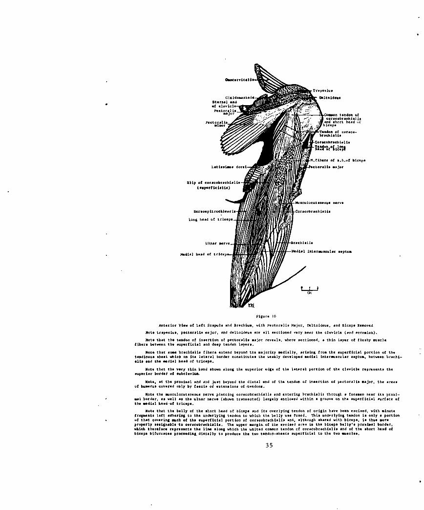
Figure 10
Anterior View of Left Scapula and Brachium, with Pectoralie Hajor, Deltoideus, and Biceps Removed
Note trapesiue, pectoralie major, and deltoideus are all sectioned very near the clavicle (and acromeLon).
Note that the tendon of insertion of pectoralis major reveals, where sectioned, a thin layer of fleshy musclefibers between the superficial end deep tendon layers.
Note that some brachislia fibers extend beyond the majority medially, arising from the superficial portion of thetendinous sheet which on its lateral border constitutes the weakly developed medial intermusculir septum, between brachi-ella and the medial head of triceps.
Note that the very thin band shown along the superior edge of the lateral portion of the clavicle represents thesuperior border of eubelovis,
Note, at the proximal end snd just beyond the distal end of the tendon of insertion of pectoralis major, the areasof humerus covered only by fmaciS or extensions of tendons.
Note the masculocutsneous nerve piercing corecobrachialis and entering brachialis through a foramen near its proxi-mal border, as well ae the ulnar nerve (shown transected) largely enclosed within a groove on the superficial surface ofthe mdial head of triceps.
Note that the belly of the short head of biceps and its overlying tendon of origin have been excised, with minutefragments left odhering to the underlying tendon to which the belly was fused. This underlying tendon is only a portionof that covering meh of the superficial portion of coracobrachislis and, although shared with biceps, t thus moreproperly assigneble to corecobrachiolis. The upper mrgin of the excised aree is the biceps belly's proximal border,which therefore represents the line along which the united common tendon of coraecobrachialis end of the short head ofbiceps bifurcates proceeding, distally to produce the two tendon-sheets superficial to the two mscles.
35
Pct.oralis minor
Supraspinstum CoracobrachialisCommon tendon of corsco-
Sorratus brnehislis and short hoodanterior of biceps
Pectoralhi Imnjor
-ubsespularis
-Tendon of latisbimus dorsi
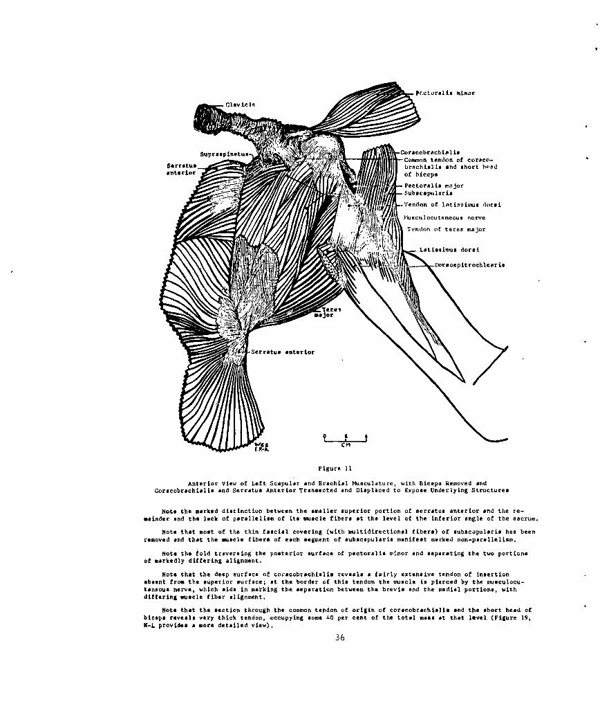
Figure 11Anterior View of Left Scapular and Brachial Msculture, with Biceps Removed and
Coracobrachiahis and Serratus Anterior Trrnsected ad Displaced to Expose Underlying Structures
Note the marked distinction between the smaller superior portion of serratus anterior and the re-mainder and the lack of parallelism of its muscle fibers at the level of the inferior angle of the sacrum.
Note that most of the thin fascial covering (with nltidirectional fibers) of aubac~pularis has beenremoved and that the muscle fibers of each segment of subacapularis manifest marked non-parallelim.
Note the fold traversing the posterior surface of pectoralis minor and separating the two portionsof markedly differing alignment..
Note that the deep surface of co,~'cobrachialis reveals a fairly extensive tendon of insertionasent from the superior surface; at the border of this tendon the muscle is pierced by the musculocu-taneous nerve, which aids in marking the separation between the brevis and the medial portions, withdiffering muscle fiber alignment.
Note that the section through the common tendon of origin of corseobrachaltsi and the short head ofbiceps revea very thick tendon, ocupying some 40 per cent of the total mss at that level (Figure 19,K-L provides a more detailed view).
36

Pectoralis
-Coinfon tendonSubseapulati. of corabrach-
aL81 IN a borthead of biceps
edon of lon~g head of
N iftoracobrachisl is
<" Tendon of lat is-
@IU$s dorsi
Tarespitomajori
Medial hoodsaala setumep
Dorsoepirochlears Trenseted and isplece
superor *d ofthis inrin
to the t~~~~~~~~~~~~~endon rgno h ogha of biceps, neligtebra n ewe h di edo
Cr-&

Figure 13
Section of Trapezius
Total length of section shown is 203 mm. Thickness at A is 26 mm.,at 26 mm. lower is 27 mm., at 17 mm. lower still is 10 mm., and atsuccessive 20 mm. intervals below that 8, 8, 7, 7, 7, 6, and 5 mm. (thelast at 20 mm. above B).
Figure 14
Section of Latissimus Dorsi
Total length of section shown is 186 mm. on the superficial periph-ery, plus an estimated 10 mm. at the anterior end (dotted). Thickness6 mm. below C is 3 mm., and at successive 20 mm. intervals below that 8,13, 19, 20, 18, 21, 16, 10, and 7.5 (the last at the end of the extantsection), measured perpendicular to the superficial surface.
Figure 15
Section of Del oideus
This lateral view of the left deltoideus depicts the muscle withX the anteromedial and Y the posteromedial extreme points of origin, withZ the distalmost tip of insertion. Along their curved borders on thesuperficial surface, X to W is 85 mm.; W to Y, 125 mm.; X to Z, 192 mm.;Y to Z, 206 mm.; W to Z, 197 mm.; Z to E, 119 mm.; Z to V, 109 mm.; andZ to F, 89 mm. Along the deep surface, E to F is 101 mm. At 10 mm. alongthis deep surface from E and perpendicular to it (Fig. 15A), the muscle is15 mm. thick; at 35 mm. (immediately lateral from the anterior surface ofthe shaft of the humerus), 20 mm.; at 65 mm. from E (immediately lateralfrom the posterior surface of the humerus), 27 mm.; and at 85 mm. from E(16 mm. from F), 21 mm.
Figure 16
Section of Pectoralis Major
This is the major section of the muscle resulting from autopsy, asshown in Figure 1; this section is shown as viewed from below, with thedeep surface at the bottom of the outline. With a straight-line lengthof 85 mm., pectoralis major becomes steadily thicker from the medialborder (G) to 13 mm. at 18 mm. distance, 19 mm. at 32 mm., 22 mm. from42 to 61 mm., and, in the final 24 mm. to H, becomes abruptly thinner.
38

Fiue1
figure 1
/GFi ur 15
i u e I BCA iu e 1/7l
for Fig re13 14 IS,1
/ / / ~
''39
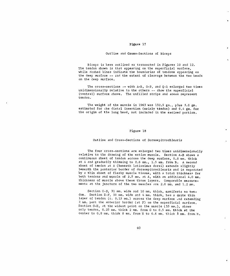
Figure 17
Outline and Cross-Sections of Biceps
Biceps is here outlined as transected in Figures 10 and 12.The tendon shown is that appearing on the superficial surface,while dashed lines indicate the boundaries of tendons appearing onthe deep surface -- and the extent of cleavage between the two headson the deep surface.
The cross-sections -- with A-B, O-P, and Q-R enlarged two timesunidimensionally relative to the others -- show the superficial(ventral) surface above. The unfilled strips and areas representtendon.
The weight of the muscle in 1963 was 132.5 gm., plus 3.0 gm.estimated for che distal insertion (mainly tendon) and 0.4 gm. forthe origin of the long head, not included in the excised portion.
Figure 18
Outline and Cross-Sections of Dorsoepitrochlearis
The four cross-sections are enlarged two times unidimensionallyrelative to the drawing of the entire muscle. Section A-B shows acontinuous sheet of tendon across the deep surface, 0.8 mm. thickat A and gradually thinning to 0.6 mm., 1.5 mm. from B. A secondsheet of tendon at A (beneath latissimus dorsi) extends slightlybeneath the posterior border of dorsoepitrochlearis and is separatedby a thin sheet of fleshy muscle tissue, with a total thickness forboth tendons and muscle of 2.5 mm. at A, with an additional 4.0 mm.thickness of muscle above these three layers. Comparable measure-ments at the juncture of the two muscles are 2.0 mm. and 1.2 mm.
Section C-D, 31 mm. wide and 10 mm. thick, manifests no ten-don. Section E-F, 35 mm. wide and 4 mm. thick, has a quite thinlayer of tendon (c. 0.15 mm.) across the deep surface .,nd extending2 mm. past the anterior border (at F) on the superficial surface.Section G-11, at the widest point on the muscle (35 mm.), showsonly tendon, 0.25 mm. thick 2 mm. from G to 0.5 mm. thick at thecenter to 0.8 mm. thick 8 mm. from H to 0.6 mm. thick 3 mm. from H.
40

Gc K0
cm
-L ~Cm ~1~
cpI
I~ ..
C m
0 .'£6 -= - - ---------------
Figure 17 Fiigure 18
outline and Cross-SectiOfll Of BicePs outline and Croon-Sectionsof Dorsoapitrochlearis
41

Figure 19
Outline and Sections of Coracobrachialis and Adjacent Muscles
In the upper left corner is outlined the 1.11 gm. superficial slip ofcoracobrachialis, with section A-B (8.9 mm. x 1.3 mm. average thickness),C-D (9.2 mm. x 1.4 mm.), and E-F (8.3 mm. x 0.95 mm.).
The outline in the upper right shows the location of section G-H(22.8 mm. wide and tapering from 1.1 mm. thick near the superior border,G, to 0.9 mm. in the middle and 0.6 mm. at I mm. from H) perpendicularlythrough the tendon of insertion of latissimus dorsi, 45 mm. from the hume-rus insertion on the superior and 28 mm. on the inferior border. SectionI-J was taken, as indicated, near the origin of dorsoepitrochlearis. Thecentral outline depicts the location of the four remaining sections:K-L through the common tendon of origin of coracobrachialis and the shorthead of biceps, 4 mm. distal to the tip of the coracoid process; M-Nthrough the common tendon, coracobrachialis, the humerus, and the tendonof insertion of pectoralis major (at N); O-P through coracobrachialis(with the slip removed), the short head of biceps, and the musculocutaneousnerve; and Q-R through teres major and its tendon (at the bottom near Q),latissimus dorsi and dorsoepitrochlearis and their tendons, the slip ofcoracobrachialis, coracobrachialis, the musculocutaneous nerve, the shorthead of biceps, the humerus, and the tendon of pectoralis major envelopingthe attenuated fleshy portion of that muscle.
In all sections, the deeper surfaces are represented at the bottom.
4Z

AA
0 4-
cCm
00
0 1
0~6.
Poo *A
C mT
Figure 19
Outline and Sections of Coracobrachialis and Adjacent Muscles
43

DISTRIBUTION
AFSC (SCTB) 2 Academy Library-DFSLB 2Andrews AFB United States Air Force AcademyWash, DC 20331 Colorado 80840
Hq, USAF, Science Division 1 American Institute of Aero- 1(AFRSTA) nautics and Astronautics
Dir/Science and Technology, 750 Third AvenueDCS/R&D New York, NY 10017
Wash, DC 20330Boeing Airplane Company I
AMD I Aero-Space DivisionATTN: Chief Scientist ATTN: Ruth E. Peerenboon,
Brooks AFB, Texas 78236 Process SupervisorP.O. Box 3707
AMD (AMAP) 10 Seattle, Wash 98124Brooks AFB, Texas 78236
Redstone Scientific Information 5DDC (TIAAS) 20 CenterCameron Station ATTN: Chief, Document SectionAlexandria, Va 22314 U.S. Army Missile Command
Redstone Arsenal, Ala 35809AFETR Tech Library (MU-135) lPatrick AFB, Fla 32922 British Liaison Office 1
Army Missile Test and Evalu-APGC (PGBAP-1) 1 ation DirectorateEglin AFD, Fla 32542 White Sand. Missile Range
New Mexico 88002ESD (ESTI) 2L.G. Hanscom Field Defence Research Member IBedford, Mass 01730 Canadian Joint Staff
Director of Biosciences ResearchAir University Library 1 2450 Massachusetts Ave., N. W.Maxwell AFB, Ala. 36100 Wash, DC 20008
Central Intelligence Agency 2 USAF School of Aerospace 1
ATTN: OCR/DD-Standard Medicine
Distribution ATTN: Aeromedical LibraryWash, DC 20505 Brooks AFB, Texas 78236
Life Sciences Dept, Code N300 1U.S. Naval Missile CenterPoint Mugu, Calif 93041

Aviation Medical Acceleration 2 Medical Records Section
Laboratory Room 325ATTN: AMAL Library Division of Medical SciencesJohnsville, Pa 18974 National Academy of Sciences
National Research CouncilLibrarian 1 2101 Constitution Avenue NW
C.A.R.I. Wash, DC 20418
F.A.A.
P.O. Box 1082 Aviation Safety EngineeringOklahoma City, Okla 73101 Research
A Division of Flight Safety
Director 2 Foundation
U.S. Naval Research Laboratory 2871 Sky Harbor Blvd(Code 5360) Sky Harbor Airport
Wash, DC Z0390 Phoenix, Arizona 85034
Biomedical Library 1 NASA Langley Research Center 3Center for Health Sciences Langley Station
University of California ATTN: Librarian MS185Los Angeles, Calif 90024 Hampton, Va 23365
Technical Reference Library 1 Archive-Library Dept
U.S. Naval Medical Research American Medical Assoc
Institute 535 N. Dearborn St.,
Bethesda, Md 20014 Chicago, Ill 60610
Director 1 Information Officer
Walter Reed Army Institute of USAFE French Liaison OfficeResearch Det 0100, 7260th Support Group
ATTN: Neuropsychiatry Division APO New York, NY 09686Wash, DC 20012
The Decker CorpCommanding Officer 2 45 Monument Road
ATTN: Code 373 Bala-Cynwyd, Pa 19004U. S. Naval School of Aviation
Medicine Douglas Aircraft Co
U.S. Naval Aviation Medical Advance Biotechnology DeptCenter Missile and Space Systems Div
Pensacola, Fla 32512 Santa Monica, Calif 90406
Dr. W.M. Helvey 1 Literature Acquisition Dept0/55-60 B/151 Biological AbstractsLockheed Missiles and Space Co 3815 Walnut St
P.C. Box 504 Philadelphia, Pa 19104Sunnyvale, Calif 94088
The Lovelace Foundation 1 Mr. Elliott H. MorseDept of Aerospace Medicine Librarian
and Bioastronautics Library of the College of5200 Gibson Blvd., S. E. Physicians of PhiladelphiaAlbuquerque, NMex., 87108 19 South 22nd Street
Philadelphia, Pa 19103Dr. William D. Thompson 1Department of Psychology Dr. Leon H. Schmidt, Director IBaylor University National Center for PrimateWaco, Texas Biology
University of CaliforniaDr. Deets Picket I Davis, California 956168505 Lee BlvdLeawood, Kansas 66Z06 Del Electronics Corp I
Research & Development DeptDr. Walter J. Frajola 1 ATTN: Leonard R. Solon, Ph.D.M-351 Starling-Loving Hall 250 E. Sandford Blvd.,Ohio State University Mount Vernon, New YorkColumbus 10, Ohio 43210
Civil Aeromedical Research 1Dr. R.D. Gafford I InstituteProject Manager, Space Federal Aviation Agency
Engineering Aeronautical CenterBeckman Instruments, Inc ATTN: John N. Ice, AM-119Z500 Harbor Blvd Editor, Acceleration ProjectFullerton, Calif 92632 P.O. Box 1082
Oklahoma City, Oklahor- ,101Commanding Officer 1U. S. Naval Medical Field Research Director
Laboratory Flyvemedicinsk InstituteCamp Lejeune, NC Copenhagen, DenmarkATTN: Library
National Aeronautics and Space 2Aerospace Medicine Bibliography 1 Administration
Project Scientific and Technical Infor-Science and Technology Division mation FacilityLibrary of Congress ATTN: NASA RepresentativeWash, DC 20540 Bethesda, Maryland 20014
Dr. F.H. Rohles 1 Human Research Branch 2Institute for Environmental (Code RBH)
Research OART, NASA HeadquartersKansas State University Wash, DC 20546Manhattan, Kansas
Hq, U.S. Army Medical R&D 1 Dean 2Command School of Veterinary Medicine
ATTN: Chief, Behavioral Sciences Oklahoma State UniversityResearch Branch Stillwater, Okla
Room 2608, Main Navy Bldg.,Wash, D.C. 20315 Biosciences Division
Eoar Shell BldgD.L. Patterson 1 47 CantersteenSchool )f Veterinary Medicine Brussels, BelgiumUniversity of PennsylvaniaPhiladelphia, Pa Dr. J.E. Green, Dean 2
School of Veterinary MedicineDr. Clement A. Stone, Director 1 Auburn UniversityMerck Institute of Theraputic Auburn, Ala 36830Research
West Point, Pa 19486 Library, New York State 2Veterinary College at
Dean, College of Veterinary 2 Cornell UniversityMedicine Ithaca, NY 14850
Iowa State UniversityAmes, Iowa 50010 Dean, Ralph L. Ketchell 2
College of Veterinary MedicineDean 2 Kansas State UniversitySchool of Veterinary Medicine Manhattan, Kansas 66504Ohio State University1900 Coffey Road Dean 2Columbus 8, Ohio 43210 College of Veterinary Medicine
Michigan State UniversityC.J. Marshall Memorial Library 2 East Lansing, MichSchool of Veterinary MedicineUniversity of Pennsylvania Dean 23800 Spruce Street School of Veterinary MedicinePhiladelphia, Pa 19104 University of Georgia
Athens, GaDr. W.R. Pritchard 2Dean, School of Veterinary Dr. Robert L. Hamlin
Medicine 1900 Coffey Road
University of California Columbus 10, Ohio 43210Davis, Calif 95616
Librarian IDean C.A. Brandly 2 Department of Health, EducationCollege of Veterinary Medicine and Welfare
University of Illinois Public Health ServiceUrbana, Ill 61803 Occuptational Health Research and
Training Facility
Bureau of State Services1014 B roadwayCincinnati, Ohio 45202
Major James E. Cook 1Department of Veterinary
PathologyKansas State UniversityManhattan, Kansas
Local
MDNH 1NLO 1ARGS 80RRRT 3RRRS 1
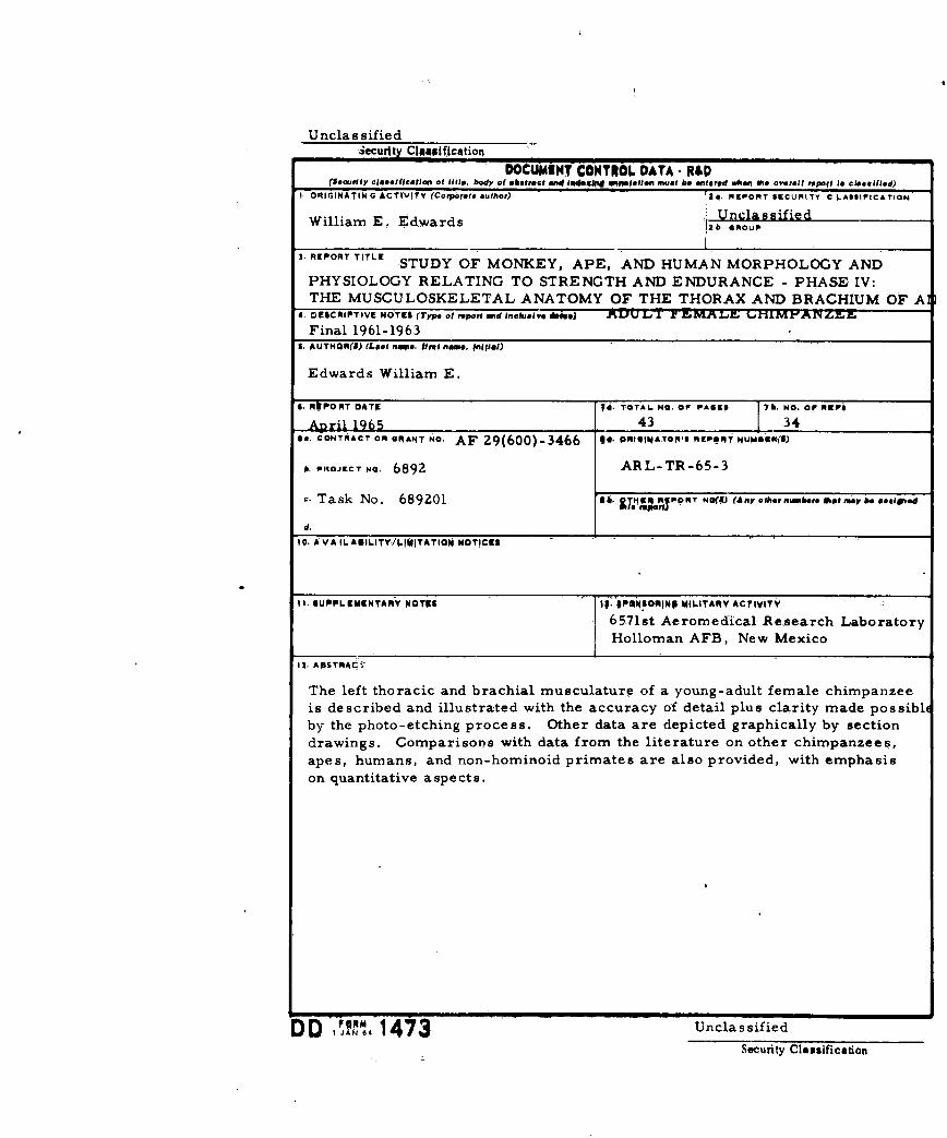
Unclassified-3ecurity C10gp1fication
(Security clossiicoreo of MWl* body of sketrael aRE040# iRE ad lpatiofl must A. etered aA*i, sh overall r,poll Is cleasiid)1, ORIGINATING ACTIVITY (Corporae. author) 31. REPORT SECuRtTY C L.ASSIFtCATION
William E. Edwards Unclassified2b opROUP
3. RlEPORT TITLE STUDY OF MONKEY, APE, AND HUMAN MORPHOLOGY AND
PHYSIOLOGY RELATING TO STRENGTH AND ENDURANCE - PHASE IV:THE MUSGULOSKELETAL ANATOMY OF THE THORAX AND BRACHIUM OF A]
4. DESCRIPTIVE NOTES (Type of report and inclusive d~a) ALU k LMLIAM'XN LZA.
Final 1961-1963S. AUTHOR(S) (Lost no*@. ftra$ name, #nflfal)
Edwards William E.
S. RkPO PT DATE TO. TOTAL Na. OF PAO"I 76. NO. OF REPSl
April 1965 1 43 ______34___
On. CONTRACT ORk GRANT NO.- AF 29(600)-3466 IS. PRONAO' REPORT NUM66008)
A. PROJECT No, 6892 ARL-TR-65-3
cTask No. 689201 sh. &TMF mjpotrT NO(I (Any othoetnabere Ifttmysassinead~
10. AVAIL AUILITY/IIWITATIOW NOTICES .-
11. SUPPLRMENTARY NOTES 1p. 3PSI4SORjN9 MIW-TA RY ACTIVITY
6571st Aeromedical Re,search LaboratoryHolloman AFB, New Mexico
13, ABSTRAci"
The left thoracic and brachial musculature of a young-adult female chimpanzeeis described and illustrated with the accuracy of detail plus clarity made possibioby the photo-etching process. Other data are depicted graphically by sectiondrawings. Comparison~s with data from the literature on other chimpanzees,apes, humans, and non-hominoid primates are also provided, with emphasison quantitative aspects.
DD ~ 1473UnclassifiedSecurity Clmaification
UnclassifiedSecurity, Clssification
14, KLY WORDS INE A LINKS LINK CNOLA WT OLE - oLa wT
ChimpanzeeStrengthEnduranceMusculo skeletal AnatomyThoraxB rachium
INSTRUCTIONsI. ORIGINATING ACTIVITY: Enter the name and address Imposed by security classification, using standard statementsof the contractor, subcontractor, grantee, Department of Do- much so:fanes activity or other organization (corporate euthor) issuingthe report. (1) "Qualified requesters may obtain copies of thisreport from DDC."2a. REPORT SEC.UNTY CLASSIFICATION. Enter the over- ( " announcement and dissmination of thisall security classification of the report. Indicate whether report b nno t ed."Restricted Data" in included. Marking Is to be in accord- report by DDC is not authorized."ance with appropriate security regulations. (3) "U. S. Government agencies may obtain copies of
this report directly from DCC. Other quialified DC2b. GROUP: Automatic downgrading is specified in DD Di- users shall request throughrective 5200. 10 and Armed Forces Industrial ManuaL Fnterthe group number. Also, when applicable, show that optionalmarkings have been used for Group 3 and Group 4 s author- "U. Sized. (4) ".S. military agencies roy obtain copies of th~i s
report directly from DDC. Other qualified users3, REPORT TITLE: Eater the complete report 1o to in all shall request throughcapital letters. Titles in Il cases should be unclassified.If a meaningful title cannot be selected without classifica-tion, show title classification in all capitals in parenthesis (5) "All distribution of. this report is controlled. Qual.immediately following the title. ified DDC users shall request through4. DESCRIPTIVE NOTES: If appropriate, enter the type of __,,
report, e.g., interim, progress, summary, annual, or final. If the report has been furnished to the Office of TechnicalGive the inclusive dates when a specific reporting period is Services, Department of Commerce, for sale to the public, indi-coveed. cate this fact and enter the price, if known.S. AUTHOR(S): Enter the name(*) of author(s) as shown o I1L SUPPI.EMENTARY NOTES: Use for additional explsne-or in the report. Enter last name, first name, middl Initial, tory notes.If military, sho* rank and branch of service. The name ofthe principal ;fthor is an absolute minimum requirement. 12. SPONSORING MILITARY ACTIVITY: Eter the name of
the departmental project office or laboratory sponsoring (pay-6. REPORT DATZ. Eter the date of the report as day, in# for) the research and development. Include idiness.month, year; or month, year If more than one date appearson the report, use date of publication. 13. ABSTRACT: Enter an abstract giving a brief and factual
summery of the document indicative of the report, even though7s. TOTAL NUMBER OF PAGES: The total peo count it may so0 appear elsewhere in the body of the technical re-should follow normal pigintion procedures, i.e., enter the port. If additional space is required, a continuation sheet $hall'number of pages contannng information, be attached,7b. NUMBER OF REFERENCE& Enter the total number of It is highly desirable that the abstract of classified reportsreferences cited in the report, be unclassified. Each paragraph of the abstract shall and withBe. CONTRACT OR GRANT 4UMBER: If appropriate, enter an indication of the military security classifIcsion of the in-the applicable number of the contract or grant under which formation in the paragraph, represented as (,n), (s), (c), or (u),the report was written. There is no limitation on the length of the abstract. How-Sb, 8c, & Sd. PROJECT NUMBER: Enter the appropriate ever, the suggested length is from ISO to 225 words.military deprtment identification, such as project number,subproject number, system numbers, task number, etc. 14. KEY WORDS: Key words are technically meaningful termCor short phrases that cherecterixe a report and nay be used as9s. ORIGINATOR'S REPORT NUMER(S): Ritter the affi- index entries for cataloging the report. Key words must becial report number by %%kich the document will be identified, selected so that no security classification is required. ldenti-and controlled by the orginating activity. This number must fiers, such si equipment model designation, trade name, militarybe unique to this report., project code name, geographic location, may be used s key9b. OTHER REPORT NUMBER(S): If the report has been words but will be followed by an indication of technical con-assignitdany other repcrt numbers (either by the originator text. The sssignfment of links, rules, and weights is optioaal.or bk the "kponsor), also enter this number(s).10. AVAILABILITY/LIMITATION NOTICES; Enter any lim-itations on further dissemination of the report, other then thosel
U ncla ssifiedSecurity Classification
5